1. Introduction
Goose astrovirus (GoAstV) is an emerging avian pathogen in China, having disseminated across major goose-farming regions [
1,
2]. Goslings infected with GoAstV develop visceral and articular gout, accompanied by anorexia, lethargy, and excretion of chalky droppings [
3,
4,
5]. The infection induces rapid mortality in goslings, with case fatality rates of 20–50%, causing substantial economic losses to the poultry industry [
6]. Although the gout-inducing mechanisms have been investigated, effective control strategies remain elusive [
5,
7,
8].
GoAstV is a single-stranded, positive-sense RNA virus within the family Astroviridae and genus Avastrovirus [
9]. Similarly to other astroviruses, GoAstV possesses an approximately 7 kb genome comprising two untranslated regions (the 5′-UTR and 3′-UTR) and three open reading frames (ORF1a, ORF1b, and ORF2) [
1,
10,
11]. ORF1a and ORF1b encode non-structural proteins that mediate viral replication and transcription [
12,
13]. ORF2, encoding the structural protein, constitutes the viral capsid [
14]. During host cell invasion, the ORF2-encoded polyprotein undergoes trypsin-mediated proteolytic cleavage into subunits including VP25, VP27, and VP34; notably, VP27 governs viral infectivity and serves as the dominant antigenic protein [
15].
Vaccine development represents one of the most effective strategies for disease control, wherein neutralizing antibody titers serve as a critical metric for evaluating vaccine-induced immunoprotection. To date, several ELISA-based methods for detecting GoAstV-specific serum antibodies have been established, enabling diagnostic confirmation, seroepidemiological surveillance, and immune monitoring [
16,
17]. Furthermore, methods that could detect neutralizing antibodies should also be established to evaluate vaccine efficacy in future applications.
This study aimed to generate a murine monoclonal antibody (mAb) specific to the GoAstV VP27 protein. The immunoreactivity of the produced mAb was characterized using Western blot and immunofluorescence assay (IFA). Subsequently, a sensitive and specific indirect competitive ELISA (ic-ELISA) was developed and validated for its sensitivity, specificity, and reproducibility. This validated ic-ELISA was applied to conduct a seroepidemiological survey of GoAstV infection using clinical serum samples.
2. Materials and Methods
2.1. Cells, Virus, Sera and Animals
SP2/0 murine myeloma cells were obtained from Procell Life Science & Technology Co., Ltd. (Wuhan, China), and maintained in RPMI-1640 medium supplemented with 20% fetal bovine serum (FBS; Thermo Fisher Scientific, Waltham, MA, USA). LMH cells were preserved in our laboratory and cultured in DMEM containing 10% FBS and used for the propagation of GoAstV. One-day old geese were acquired from Guiliu Livestock Farming Co., Ltd. (Qihe, China). GoAstV-negative sera were obtained from goslings confirmed negative by PCR, while positive sera were collected at 7 days post-infection. BALB/c mice were procured from Shandong Pengyue Experimental Animal Breeding Co., Ltd. (Jinan, China).
2.2. Expression and Purification of VP27 Protein
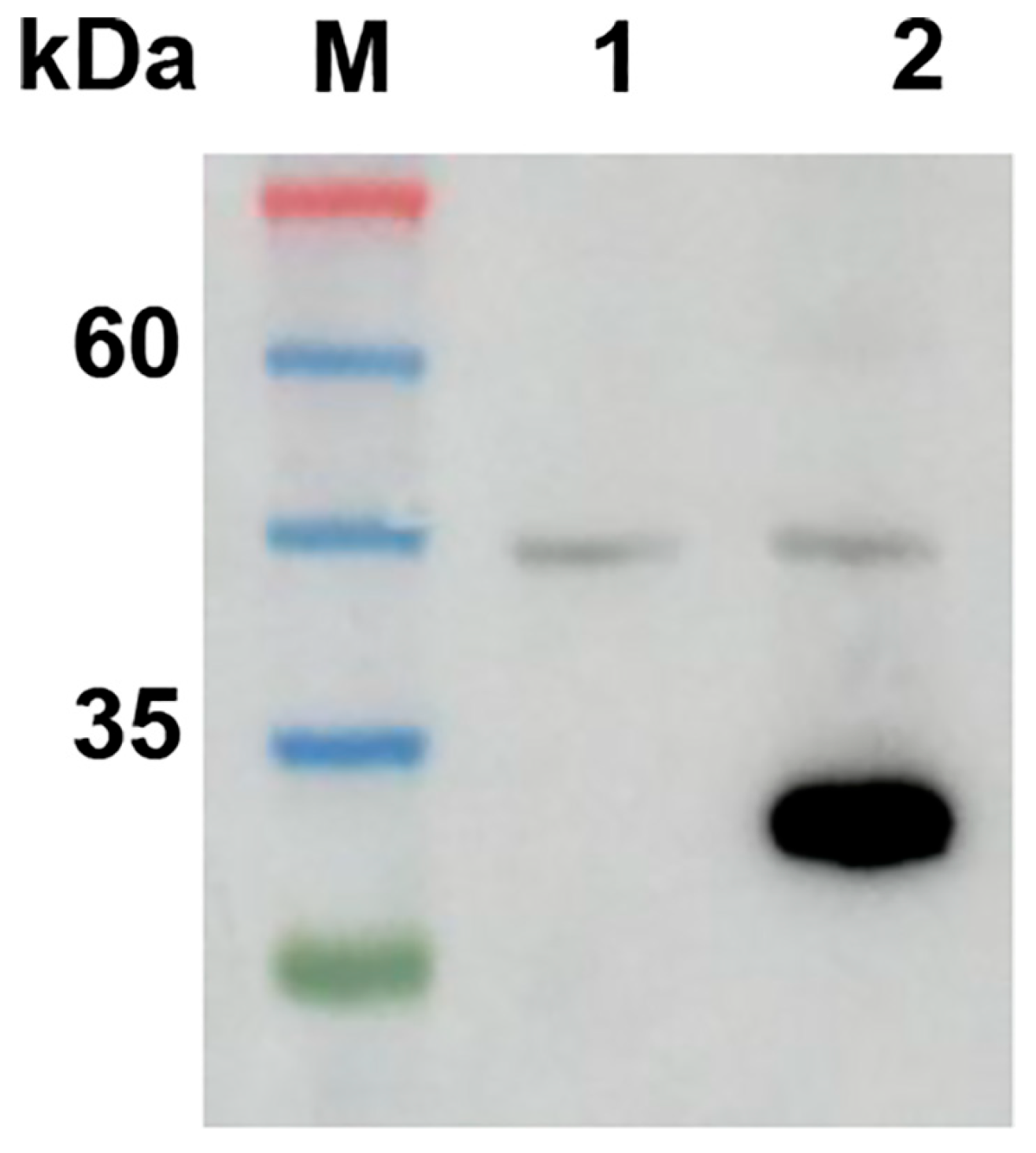
Total RNA of GoAstV was extracted using the Simply P Total RNA Extraction Kit (BioFlux, Hangzhou, China). The VP27 gene segment was amplified by one-step RT-PCR using the AccurSTART One Step RT-PCR Kit (Vazyme, Nanjing, China) with the primer pair 5′-gaattcCAGGTTACTCCCTCGCT-3′ and 5′-gcggccgcAGAGGTCTTGAGCGAGAC-3′, which incorporate EcoRI and NotI restriction sites, respectively. The amplified VP27 sequence was verified by sequencing and subsequently codon-optimized for expression in E. coli by Sangon Biotech Co., Ltd. (Qingdao, China). The optimized VP27 gene was cloned into the pET-28a (+) vector, and the resulting recombinant plasmid, pET-28a-VP27, was confirmed by restriction digestion and DNA sequencing. The validated plasmid was then transformed into E. coli BL21(DE3) competent cells (TransGen, Beijing, China). Recombinant protein expression was induced by the addition of IPTG. VP27 expression was analyzed by SDS-PAGE, and the protein was purified under native conditions using His-Tagged Protein Purification Kit (CWBIO, Beijing, China). The purified VP27 was verified by SDS-PAGE and Western blot analysis using anti-His monoclonal antibody (Biodragon, Beijing, China).
2.3. Development of Anti-VP27 mAbs
The purified VP27 protein was emulsified with an equal volume of incomplete Freund’s adjuvant (Sigma-Aldrich, Shanghai, China). Six-week-old female BALB/c mice were subcutaneously injected in the cervical region with the emulsion containing 50 μg of VP27 protein. Immunizations were administered three times at two-week intervals. Fourteen days following the final immunization, antibody titers in serum were detected by indirect ELISA using VP27 protein as the coating antigen following procedures reported before [
18]. Mice with high serum antibody titers were euthanized by cervical dislocation, and splenocytes were harvested for fusion with SP2/0 murine myeloma cells. The fused cells were cultured in HAT selection medium in 96-well plates. Cell culture supernatants were collected seven days post-fusion and screened for anti-VP27 antibodies by indirect ELISA using GoAstV as coating antigen. Hybridoma clones secreting VP27-specific antibodies were subsequently subjected to three rounds of limiting dilution to establish monoclonal lines. Monoclonal antibodies were produced as ascites by inoculating ten-week-old female BALB/c mice with the selected hybridoma cells.
2.4. Characterization of Anti-VP27 mAbs
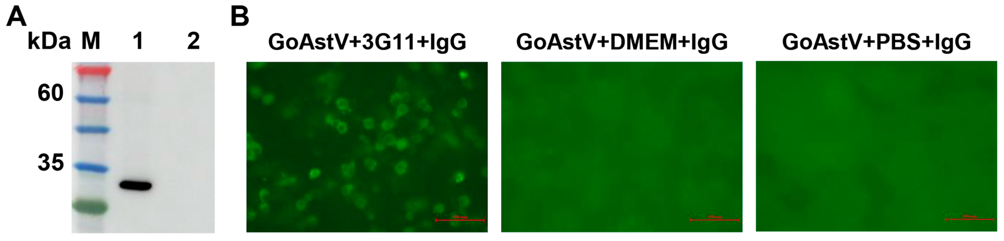
The reactivity of monoclonal antibodies against the VP27 protein was assessed by Western blot analysis. Purified VP27 protein was separated by electrophoresis on a 12.5% SDS-polyacrylamide gel and subsequently transferred onto a nitrocellulose membrane. The membrane was then blocked by incubating with 5% skimmed milk for 2 h at room temperature. Following blocking, the membrane was incubated with anti-VP27 monoclonal antibodies (hybridoma supernatant) for 2 h at room temperature. After three washes with PBST, the membrane was incubated with HRP-conjugated goat anti-mouse IgG (CWBIO, Beijing, China) for 1 h at room temperature. Following a final washing step, protein bands were visualized using Amersha ImageQuant 800 (Cytiva, Shanghai, China).
An IFA was performed on GoAstV-infected LMH cells. LMH cells were seeded in 96-well plates and infected with GoAstV at a multiplicity of infection (MOI) of 0.1. At 48 h post-infection, the cells were fixed with 4% paraformaldehyde in PBS for 30 min at room temperature and stored at 4 °C until use. Fixed cells were incubated with anti-VP27 monoclonal antibodies, followed by incubation with AF488-labeled goat anti-mouse IgG (H+L) (Beyotime, Shanghai, China). DMEM and PBS were used as control for the anti-VP27 mAbs. Immunofluorescence images were acquired using a Nikon ECLIPSE Ts2R fluorescence microscope (Nikon, Tokyo, Japan).
2.5. Serum Sample Detection by Neutralization Test
All negative and positive serum samples used in this study were confirmed with neutralization test. Viral titer was measured with IFA using polyclonal antiserum or mAb against VP27, and calculated and expressed as TCID50. For the neutralization test, serum samples were inactivated at 56 °C for 30 min, after which 100 μL of each serum was mixed with an equal volume of GoAstV suspension containing 105 TCID50. Following incubation at 37 °C for 1 h, the residual viral titer of each mixture was determined. Samples demonstrating a reduction in virus titer to below 104 TCID50 were considered positive for neutralizing antibodies.
2.6. Development of mAb-Based ic-ELISA
GoAstV viral suspensions were diluted and coated onto 96-well plates (100 µL/well, 105 TCID50, 104 TCID50, 103 TCID50) and incubated overnight at 4 °C. Following coating, the plates were washed three times with PBST and blocked with 5% non-fat milk (Beyotime, Shanghai, China) in PBS at 37 °C for 60, 90 and 120 min. After washing, 100 µL of GoAstV positive (P) and negative (N) serum (diluted 1:10 in dilution buffer) were added to the plates and incubated at 37 °C for 45, 60 and 75 min. After washing, 100 µL of anti-VP27 mAb (diluted from 1:1000 to 1:16,000 in PBST) were added to the plates and incubated at 37 °C for 45, 60 and 75 min. Subsequently, 100 µL of HRP-conjugated goat anti-mouse IgG (CWBIO, Beijing, China) was added and incubated for 50 min at 37 °C. After three additional washes with PBST, the plates were developed with TMB substrate for 15 min, and the reaction was terminated using TMB stop solution (Beyotime, Shanghai, China). Absorbance was measured at 450 nm. Optimum reaction parameters and composition were optimized based on the maximum the N/P ratio.
2.7. Determination of the ic-ELISA Cut-Off Percent Inhibition Value
Thirty GoAstV-negative serum samples were analyzed to establish the cut-off percent inhibition (PI) value. The PI for each sample was calculated using the following formula: PI (%) = [1 − (OD
(450) of sample − minimum OD
(450))/(maximum OD
(450) − minimum OD
(450))] × 100% [
19]. The cut-off PI value was defined as the mean PI of these negative samples plus twice the standard deviation (mean + 2 × SD) [
20].
2.8. Sensitivity, Specificity and Reproducibility Assessments of ic-ELISA
The sensitivity of the developed ELISA was evaluated using known positive serum samples subjected to serial dilutions ranging from 1:10, 1:100, 1:1000, and 1:10,000. Specificity was assessed by testing the method against antisera positive for goose Parvovirus (GPV), Tembusu virus (TMUV), avian Reovirus (ARV), avian influenza virus (AIV, H5), and duck Avastrovirus (DAstV), which were generated using vaccine strain or natural infection. To determine reproducibility, five serum samples (comprising three positive and two negative) were analyzed in triplicate, and the intra-assay coefficient of variation (CV) was calculated. The reproducibility was considered satisfactory when the CV values were below the 10% threshold [
18].
2.9. Application of the ic-ELISA
Serum samples were collected by the Avian Pathogens Diagnosis and Monitoring Center of the Poultry Institute, Shandong Academy of Agricultural Sciences, which has obtained CNAS accreditation from commercial and breeding goose farms in Shandong province, including Weifang, Dezhou, Jining, Liaocheng, Linyi, Zaozhuang, Jinan, between October 2024 to May 2025. Samples were categorized into two types: clinical diagnostic specimens from diseased geese and epidemiological surveillance samples from healthy geese. All samples were tested using the newly established serological method.
4. Discussion
GoAstV infection has been identified as a primary causative agent of gout in goslings, posing a significant threat to the Chinese goose industry with substantial economic losses [
21,
22,
23]. Consequently, the development of effective preventive and therapeutic strategies has become a research priority, underpinned by the critical need for rapid and accurate diagnostic methods [
16,
17]. Unlike previously characterized strains, the novel GoAstV isolate used in this study did not induce discernible cytopathic effects in available cell lines. Therefore, viral titers were quantified using an IFA in LMH cells, a system that also facilitated preliminary neutralization assessments.
The ELISA is a cornerstone serological technique in veterinary diagnostics, valued for its operational simplicity, high throughput, and robust sensitivity and specificity [
16,
24,
25,
26]. To develop an ELISA for GoAstV, we targeted the VP27 capsid protein. VP27 constitutes a major immunogenic component of astroviruses, forming the outer spikes of mature virions, mediating host cell entry, and eliciting strong immune responses [
15,
27,
28]. Compared to the precursor VP90, VP27 presents a higher density of immunodominant epitopes [
27,
29]. In this study, recombinant VP27 was expressed in
E. coli and used to generate mAbs in BALB/c mice. The resulting mAb, 3G11, demonstrated specific reactivity against both recombinant VP27 and native GoAstV particles in Western blot and immunofluorescence assays (
Figure 2), confirming its suitability for serological assay development.
In present study, an ic-ELISA based on mAb 3G11 was established, in which GoAstV antibodies present in serum samples compete with mAb 3G11 for binding sites on immobilized particles. Optimal reaction conditions, including antigen concentration and antibody dilution, were determined via checkerboard titration using negative and positive control sera, as previously reported [
18,
24], with selection based on the highest N/P ratio (
Table 1 and
Table 2). A diagnostic cut-off value of 44.52% was calculated from the PI analysis of 30 negative serum samples, a threshold consistent with values reported in prior studies [
16]. Following assay development, key performance parameters—sensitivity, specificity, and repeatability—were rigorously evaluated, as they are critical for reliable serological application [
16,
18]. The results confirmed that the assay meets the requisite standards for clinical and field use (
Table 4,
Table 5 and
Table 6).
The development of effective vaccines is essential for controlling GoAstV infection, although no licensed vaccines are currently available [
30]. Neutralizing antibody titer is a critical correlate of vaccine-induced immune protection [
31]. Therefore, to enhance the probability of detecting such antibodies, intact viral particles were employed both for monoclonal antibody screening and as the coating antigen in our ic-ELISA [
32,
33]. The strong immunoreactivity of mAb 3G11 with viral particles (
Figure 2B) suggests it may target surface epitopes, a premise requiring future validation. Consequently, the particle-based ic-ELISA is postulated to primarily detect serum neutralizing antibodies, a functionality that also necessitates confirmation through future vaccine studies.
5. Conclusions
In conclusion, a murine mAb (designated 3G11) was generated against the GoAstV VP27 protein. Western blot and IFA confirmed its specific immunoreactivity with both recombinant VP27 and native GoAstV particles. Subsequently, an ic-ELISA was developed using mAb 3G11, whose analytical performance in terms of sensitivity, specificity, and reproducibility was validated through comprehensive assessment. Collectively, this study establishes a reliable serological tool for detecting GoAstV-specific antibodies and provides a foundation for evaluating vaccine efficacy in future applications.




 ,
,