
Determining Frequency of Multiple Organ System Involvement and Concurrent Lesions Identified in Feedyard Mortalities and Potential Associations with Cattle Demographics

 ,
,  ,
,  ,
,  and
and
Simple Summary
Abstract
1. Introduction
2. Materials and Methods
3. Results
4. Discussion
5. Conclusions
Supplementary Materials
Author Contributions
Funding
Institutional Review Board Statement
Informed Consent Statement
Data Availability Statement
Conflicts of Interest
Abbreviations
| DOFs | Days on Feed |
| BIP | Bronchopneumonia with Interstitial Pneumonia |
| AIP | Acute Interstitial Pneumonia |
| BP | Bronchopneumonia |
| GI | Gastrointestinal |
| CHF | Congestive Heart Failure |
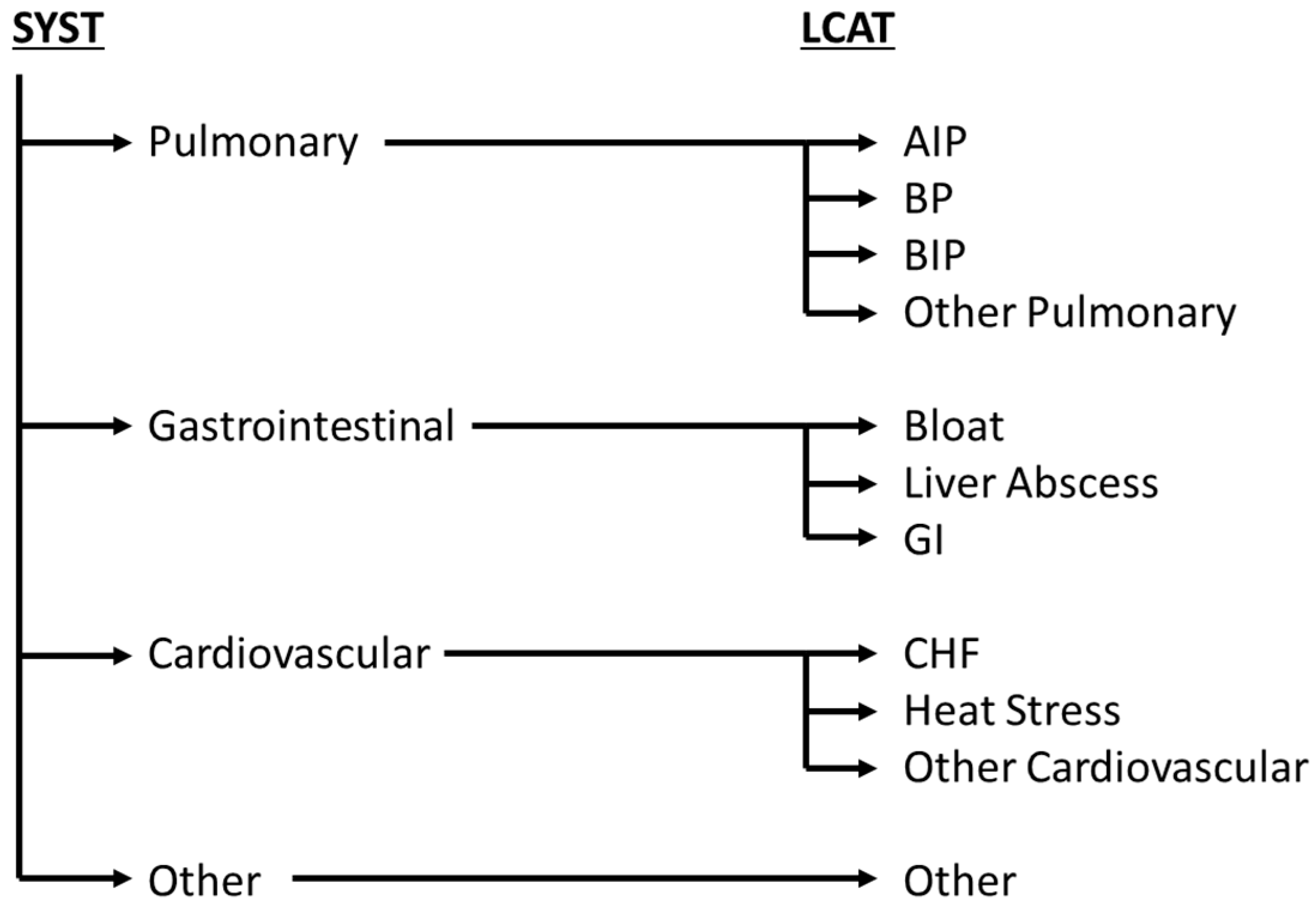
| SYST | Organ System |
| LCAT | Lesion Category |
References
- Loneragan, G.H.; Dargatz, D.A.; Morley, P.S.; Smith, M.A. Trends in mortality ratios among cattle in US feedlots. J. Am. Vet. Med. Assoc. 2001, 219, 1122–1127. [Google Scholar] [CrossRef] [PubMed]
- Ribble, C.S.; Meek, A.H.; Janzen, E.D.; Guichon, P.T.; Jim, G.K. Effect of time of year, weather, and the pattern of auction market sales on fatal fibrinous pneumonia (shipping fever) in calves in a large feedlot in Alberta (1985–1988). Can. J. Vet. Res. Rev. Can. Rech. Vet. 1995, 59, 167–172. [Google Scholar]
- Lechtenberg, K.F.; Smith, R.A.; Stokka, G.L. Feedlot Health and Management. Vet. Clin. N. Am. Food Anim. Pract. 1998, 14, 177–197. [Google Scholar] [CrossRef] [PubMed]
- Galyean, M.L.; Gunter, S.A.; Malcolm-Callis, K.J. Effects of arrival medication with tilmicosin phosphate on health and performance of newly received beef cattle1. J. Anim. Sci. 1995, 73, 1219–1226. [Google Scholar] [CrossRef] [PubMed]
- Kelly, A.P.; Janzen, E.D. A review of morbidity and mortality rates and disease occurrence in north american feedlot cattle. Can. Vet. J. Rev. Vet. Can. 1986, 27, 496–500. [Google Scholar]
- Irsik, M.; Langemeier, M.R. Estimating the impact of animal health and death loss on economic performance of feedlot cattle. Kans. Agric. Exp. Stn. Res. Rep. 2003, 1, 97–100. [Google Scholar] [CrossRef]
- Mason, G.L.; Madden, D.J. Performing the Field Necropsy Examination. Vet. Clin. N. Am. Food Anim. Pract. 2007, 23, 503–526. [Google Scholar] [CrossRef] [PubMed]
- Schneider, M.J.; Tait, R.G.; Busby, W.D.; Reecy, J.M. An evaluation of bovine respiratory disease complex in feedlot cattle: Impact on performance and carcass traits using treatment records and lung lesion scores1,2. J. Anim. Sci. 2009, 87, 1821–1827. [Google Scholar] [CrossRef] [PubMed]
- Hay, K.E.; Barnes, T.S.; Morton, J.M.; Clements, A.C.A.; Mahony, T.J. Risk factors for bovine respiratory disease in Australian feedlot cattle: Use of a causal diagram-informed approach to estimate effects of animal mixing and movements before feedlot entry. Prev. Vet. Med. 2014, 117, 160–169. [Google Scholar] [CrossRef] [PubMed]
- Smith, R.A. Impact of disease on feedlot performance: A review. J. Anim. Sci. 1998, 76, 272–274. [Google Scholar] [CrossRef] [PubMed]
- Belknap, E.B.; Navarre, C.B. Differentiation of Gastrointestinal Diseases in Adult Cattle. Vet. Clin. N. Am. Food Anim. Pract. 2000, 16, 59–86. [Google Scholar] [CrossRef] [PubMed]
- Johnson, B.T.; Amrine, D.E.; Larson, R.L.; Weaber, R.L.; White, B.J. Retrospective analysis of cohort risk factors and feeding phase timing associated with noninfectious heart disease deaths in U.S. feedlot cattle. Transl. Anim. Sci. 2021, 5, txab220. [Google Scholar] [CrossRef] [PubMed]
- Griffin, D. Feedlot Euthanasia and Necropsy. Vet. Clin. N. Am. Food Anim. Pract. 2015, 31, 465–482. [Google Scholar] [CrossRef] [PubMed]
- Heffernan, K.R.; Thomas, M.G.; Enns, R.M.; Holt, T.; Speidel, S.E. Phenotypic relationships between heart score and feed efficiency, carcass, and pulmonary arterial pressure traits. Transl. Anim. Sci. 2020, 4 (Suppl. S1), S103–S107. [Google Scholar] [CrossRef] [PubMed]
- Brown, H.; Bing, R.F.; Grueter, H.P.; McAskill, J.W.; Cooley, C.O.; Rathmacher, R.P. Tylosin and Chlortetracycline for the Prevention of Liver Abscesses, Improved Weight Gains and Feed Efficiency in Feedlot Cattle. J. Anim. Sci. 1975, 40, 207–213. [Google Scholar] [CrossRef] [PubMed]
- Haydock, L.A.J.; Fenton, R.K.; Smerek, D.; Renaud, D.L.; Caswell, J.L. Bronchopneumonia with interstitial pneumonia in feedlot cattle: Epidemiologic characteristics of affected animals. Vet. Pathol. 2023, 60, 226–234. [Google Scholar] [CrossRef] [PubMed]
- Haydock, L.A.J.; Fenton, R.K.; Sergejewich, L.; Veldhuizen, R.A.W.; Smerek, D.; Ojkic, D.; Caswell, J.L. Bronchopneumonia with interstitial pneumonia in beef feedlot cattle: Characterization and laboratory investigation. Vet. Pathol. 2023, 60, 214–225. [Google Scholar] [CrossRef] [PubMed]
- Baptista, A.L.; Rezende, A.L.; Fonseca, P.d.A.; Massi, R.P.; Nogueira, G.M.; Magalhães, L.Q.; Headley, S.A.; Menezes, G.L.; Alfieri, A.A.; Saut, J.P.E. Bovine respiratory disease complex associated mortality and morbidity rates in feedlot cattle from southeastern Brazil. J. Infect. Dev. Ctries. 2017, 11, 791–799. [Google Scholar] [CrossRef] [PubMed]
- Jensen, R.; Pierson, R.E.; Braddy, P.M.; Saari, D.A.; Benitez, A.; Horton, D.P.; Lauerman, L.H.; McChesney, A.E.; Alexander, A.F.; Will, D.H. Brisket disease in yearling feedlot cattle. J. Am. Vet. Med. Assoc. 1976, 169, 515–517. [Google Scholar] [CrossRef] [PubMed]
- Neary, J.M.; Booker, C.W.; Wildman, B.K.; Morely, P.S. Right-Sided Congestive Heart Failure in North American Feedlot Cattle. J. Veter. Intern. Med. 2015, 30, 326–334. [Google Scholar] [CrossRef] [PubMed]
- Heaton, M.P.; Bassett, A.S.; Whitman, K.J.; Krafsur, G.M.; Lee, S.I.; Carlson, J.M.; Clark, H.J.; Smith, H.R.; Pelster, M.C.; Basnayake, V.; et al. Evaluation of EPAS1 variants for association with bovine congestive heart failure. F1000Research 2019, 8, 1189. [Google Scholar] [CrossRef] [PubMed]
- Vogel, G.L.; Parrott, J.C. Mortality survey in feedyards: The incidence of death from digestive, respiratory and other causes in feedyards of the Great Plains. Compend. Contin. Educ. Pract. Vet. 1994, 16, 227–234. [Google Scholar]
- Glock, R.D.; DeGroot, B.D. Sudden death of feedlot cattle. J. Anim. Sci. 1998, 76, 315. [Google Scholar] [CrossRef] [PubMed]
- Edwards, A.J. Respiratory diseases of feedlot cattle in the central USA. Bov. Pract. 1996, 30, 5–7. [Google Scholar] [CrossRef]
- Neal, K.B.; White, B.J.; Amrine, D.E.; Lubbers, B.V.; Tessman, R.K.; Larson, R.L. Risk factors associated with case fatality and treatment success following initial bovine respiratory disease treatment in feedyard cattle. Bov. Pract. 2024, 58, 1–8. [Google Scholar] [CrossRef]


| Area of Examination: | Options, if Not Within Normal Limits: | |
|---|---|---|
| External: | Sex | Steer, Heifer |
| Breed | Native, Holstein, Mexican, Beef × Dairy | |
| Body condition | Thin, Moderate, Fat | |
| Lactation status | Lactating, Not Lactating | |
| Mastitis status | Mastitis, No Mastitis | |
| Integumentary: | Oral cavity | Ulcers, Blunted Papillae, Petechia |
| Esophagus, Trachea, Heart: | Esophagus | Ulcers, Hemorrhage |
| Larynx/Pharynx | Necrosis, Infection, Ulcers, Hemorrhage, Fibrinous | |
| Tracheal contents/Mucosa | Froth, Reflux, Hemorrhage, Fibrinonecrotic, Ulcers, Edema, Infection | |
| Pericardial effusion/Sac | Fibrinous, Mucoid, Purulent, Sanguineous, Serous, Serosanguineous, Thickened, Adhesions | |
| Heart score | 1, 2, 3, 4, 5 | |
| Heart circumference (cm) | ||
| Heart weight (g) | ||
| Myocardium/Endocardium | Necrosis, Petechia, Ecchymosis, Dilated Ventricle, Myocarditis, Endocarditis | |
| Lungs: | Pleural effusion | Fibrinous, Mucoid, Purulent, Sanguineous, Serous, Serosanguineous |
| Pleural effusion volume | Mild, Moderate, Severe | |
| Pulmonary pathology | Bronchopneumonia, Fibrinous, Pleuritis, Acute Interstitial Pneumonia, Bronchopneumonia with Interstitial Pneumonia, Granulomatous, Embolic Pneumonia, Pulmonary Hemorrhage/Petechia, Pulmonary Edema | |
| Abscesses | Small, Medium, Large/Few, Moderate, Many | |
| Adhesions | Present, Absent | |
| GI Tract: | Small intestine (SI) serosa | Red, Black |
| SI mucosa | Ulcer, Hemorrhage, Thickened | |
| SI content | Gas, Fluid | |
| SI lesion | Obstructed, Inflamed, Parasites, Neoplasia | |
| Large intestine (LI) serosa | Red, Black | |
| LI mucosa | Ulcer, Hemorrhage, Thickened | |
| LI content | Gas, Fluid | |
| LI lesion | Obstructed, Inflamed, Parasites, Neoplasia | |
| Rumen score | 1, 2, 3, 4, 5 | |
| Rumen contents | Froth, Full, Empty, Bloated, Fluid | |
| Rumen mucosa | Ulcer, Hemorrhage, Thickened, Parakeratosis | |
| Abomasal mucosa | Ulcer, Hemorrhage, Thickened | |
| Abdomen: | Mesenteric lymph nodes | Enlarged, Hemorrhage |
| Peritoneal fluid | Fibrinous, Mucoid, Purulent, Sanguineous, Serous, Serosanguineous | |
| Adhesions | Liver, SI, LI, Forestomach, Other | |
| Liver | Nutmeg, Congested, Jaundiced, Pale, Scars, Flukes, Enlarged | |
| Liver abscess grade | O, A, A+ | |
| Kidney | Enlarged, Contracted, Infarcts, Petechia, Abscess, Infection | |
| Bladder/urine | Ecchymosis, Calculi, Cystitis, Pale, Dark Brown, Hemorrhage, Exudate, Ruptured, Stones | |
| Reproductive | Infected, Adhesions, Pregnant | |
| Spleen | Swollen, Contracted, Infarcted, Enlarged | |
| Musculoskeletal: | Lesion | Swollen, Trauma, Muscle Necrosis, Ecchymosis |
| Final Diagnosis: | ||
| SYST: | LCAT: | Frequency of Lesion (n) | Percent of Cases (n/889 × 100) |
|---|---|---|---|
| Pulmonary | BP | 354 | 38.8% |
| BIP | 281 | 31.6% | |
| AIP | 76 | 8.6% | |
| Other Pulmonary | 122 | 13.7% | |
| Total Pulmonary | 824 | 92.7% | |
| GI | Bloat | 83 | 9.3% |
| Liver Abscess | 47 | 5.3% | |
| Other GI | 625 | 70.3% | |
| Total GI | 755 | 84.9% | |
| Cardiovascular | CHF | 136 | 15.3% |
| Heat Stress | 40 | 4.5% | |
| Other Cardiovascular | 141 | 15.9% | |
| Total Cardiovascular | 317 | 35.7% | |
| Other | Other | 144 | 16.2% |
| Total other | 144 | 16.2% | |
| Total lesions | 2040 | 230% | |
| Top 5 Concurrent Systems (SYST) | |||||
|---|---|---|---|---|---|
| GI and Pulmonary | Cardiovascular, GI, and Pulmonary | Cardiovascular and Pulmonary | GI1, GI2, and Pulmonary | GI, Other, and Pulmonary | |
| (n = 170) | (n = 57) | (n = 48) | (n = 49) | (n = 23) | |
| Sex (%) | |||||
| Steer | 31% | 28% | 33% | 29% | 26% |
| Heifer | 69% | 72% | 67% | 71% | 74% |
| Arrival Weight (kg; avg) | 324.6 | 331.4 | 324.4 | 323.1 | 353.8 |
| Days on Feed (avg) | 90 | 113 | 97 | 101 | 57 |
| Number of Treatments (avg) | 2 | 1 | 2 | 1 | 2 |
| Top 5 Concurrent Lesion Categories (LCATs) | |||||
|---|---|---|---|---|---|
| BIP and Other GI | BP and Other GI | AIP and Other GI | Other GI and Other | BIP, CHF, and Other GI | |
| (n = 72) | (n = 59) | (n = 23) | (n = 21) | (n = 16) | |
| Sex (%) | |||||
| Steer | 25% | 31% | 35% | 40% | 13% |
| Heifer | 75% | 69% | 65% | 60% | 87% |
| Arrival Weight (kg; avg) | 326.2 | 324.5 | 321.3 | 329.5 | 325.3 |
| Days on Feed (avg) | 94 | 70 | 106 | 72 | 120 |
| Number of Treatments (avg) | 2 | 2 | 2 | 2 | 1 |
Disclaimer/Publisher’s Note: The statements, opinions and data contained in all publications are solely those of the individual author(s) and contributor(s) and not of MDPI and/or the editor(s). MDPI and/or the editor(s) disclaim responsibility for any injury to people or property resulting from any ideas, methods, instructions or products referred to in the content. |
© 2025 by the authors. Licensee MDPI, Basel, Switzerland. This article is an open access article distributed under the terms and conditions of the Creative Commons Attribution (CC BY) license (https://creativecommons.org/licenses/by/4.0/).
Share and Cite
Mancke, M.R.; White, B.J.; Bortoluzzi, E.M.; Depenbusch, B.E.; Schmidt, P.H.; Champagne, R.E.; Jensen, M.; Lancaster, P.A.; Larson, R.L. Determining Frequency of Multiple Organ System Involvement and Concurrent Lesions Identified in Feedyard Mortalities and Potential Associations with Cattle Demographics. Vet. Sci. 2025, 12, 666. https://doi.org/10.3390/vetsci12070666
Mancke MR, White BJ, Bortoluzzi EM, Depenbusch BE, Schmidt PH, Champagne RE, Jensen M, Lancaster PA, Larson RL. Determining Frequency of Multiple Organ System Involvement and Concurrent Lesions Identified in Feedyard Mortalities and Potential Associations with Cattle Demographics. Veterinary Sciences. 2025; 12(7):666. https://doi.org/10.3390/vetsci12070666
Chicago/Turabian StyleMancke, Madeline R., Brad J. White, Eduarda M. Bortoluzzi, Brandon E. Depenbusch, Paige H. Schmidt, Rachel E. Champagne, Makenna Jensen, Phillip A. Lancaster, and Robert L. Larson. 2025. "Determining Frequency of Multiple Organ System Involvement and Concurrent Lesions Identified in Feedyard Mortalities and Potential Associations with Cattle Demographics" Veterinary Sciences 12, no. 7: 666. https://doi.org/10.3390/vetsci12070666
APA StyleMancke, M. R., White, B. J., Bortoluzzi, E. M., Depenbusch, B. E., Schmidt, P. H., Champagne, R. E., Jensen, M., Lancaster, P. A., & Larson, R. L. (2025). Determining Frequency of Multiple Organ System Involvement and Concurrent Lesions Identified in Feedyard Mortalities and Potential Associations with Cattle Demographics. Veterinary Sciences, 12(7), 666. https://doi.org/10.3390/vetsci12070666