Article

Annual and Seasonal Trends in Mastitis Pathogens Isolated from Milk Samples from Dairy Cows of California’s San Joaquin Valley Dairies Between January 2009 and December 2023

by
Daniela R. Bruno
1,*,
Karen H. Tonooka
2,
Terry W. Lehenbauer
2,3,
Sharif S. Aly
2,3 and
Wagdy R. ElAshmawy
2,4,*
1
Cooperative Extension, University of California Agriculture and Natural Resources, Fresno, CA 93710, USA
2
Veterinary Medicine Teaching and Research Center, School of Veterinary Medicine, University of California Davis, Tulare, CA 93274, USA
3
Department of Population Health & Reproduction, School of Veterinary Medicine, University of California Davis, Davis, CA 95616, USA
4
Department of Internal Medicine and Infectious Diseases, Faculty of Veterinary Medicine, Cairo University, Giza 12613, Egypt
*
Authors to whom correspondence should be addressed.
Vet. Sci. 2025, 12(7), 609; https://doi.org/10.3390/vetsci12070609
Submission received: 23 May 2025 / Revised: 10 June 2025 / Accepted: 19 June 2025 / Published: 21 June 2025

Simple Summary

This study examines the microbiological profiles of bovine milk samples submitted to the Milk Quality Laboratory at UC Davis between 2009 and 2023, analyzing over 319,000 samples. The research identifies long-term trends in mastitis-causing pathogens, seasonal variations, and shifts in contamination rates, providing crucial insights into dairy herd health. Findings reveal that environmental pathogens, particularly non-aureus staphylococci and coliforms, dominate mastitis cases, while contagious pathogens such as Staphylococcus aureus, Streptococcus agalactiae, and Mycoplasma spp. appear less frequently. Seasonal effects show higher contamination rates in Winter and increased no-growth samples in Summer, indicating environmental influences on pathogen prevalence. The study highlights a notable decline in sample submissions in recent years, possibly linked to evolving dairy practices and the rise in on-farm culturing techniques. These findings contribute valuable knowledge on pathogen dynamics, mastitis management, and milk quality control strategies in California’s Central Valley. By providing a 14-year perspective on bovine milk microbiology, the study supports dairy producers, veterinarians, and researchers in optimizing herd health and refining disease prevention approaches tailored to regional conditions.

Abstract

Bovine mastitis is a significant disease affecting dairy cattle worldwide, impacting milk quality and farm profitability. Understanding pathogen distribution is crucial for effective disease management. This study analyzed 319,634 individual cow milk samples submitted to the UC Davis Milk Quality Laboratory between 2009 and 2023 to assess pathogen prevalence, seasonal variations, and long-term trends. Routine microbiological cultures identified major and minor mastitis pathogens, with additional testing for Mycoplasma spp. Statistical analyses evaluated annual and seasonal trends in bacterial isolation rates. Results indicated that environmental pathogens, particularly non-aureus staphylococci and coliforms, were most frequently isolated, while contagious pathogens (Staphylococcus aureus, Streptococcus agalactiae, and Mycoplasma spp.) were less prevalent. Seasonal trends revealed higher contamination rates in Winter and increased no-growth samples in Summer. The study also observed a decline in sample submissions in recent years, possibly reflecting evolving dairy management practices. These findings provide a comprehensive perspective on mastitis pathogen dynamics in California’s Central Valley, supporting improved milk quality control measures and tailored mastitis prevention strategies.

1. Introduction

Mastitis is an economically significant disease affecting dairy cattle worldwide, impacting animal health, welfare, and productivity [1,2,3]. Bovine mastitis is most commonly caused by pathogens, typically classified as contagious or environmental based on their primary reservoir and transmission route [4]. Contagious mastitis pathogens reside in the cow’s udder and on teat skin, colonizing and growing in the teat canal. They are primarily transmitted among cows through contact with infected milk during milking [4]. Among these pathogens, Staphylococcus aureus, Streptococcus agalactiae, and Mycoplasma bovis are the most significant, with S. aureus considered to be the most common in North America [4,5]. On the other hand, environmental pathogens live in the cow’s environment, such as in the bedding and housing, and cause an infection when given the opportunity [4]. A wide range of bacterial species cause environmental mastitis, with the most common pathogens including coliforms (Escherichia coli, Klebsiella spp., Enterobacter spp.), Streptococcus species such as Streptococcus uberis and Streptococcus dysgalactiae, and Pseudomonas spp. [4]. Non-aureus staphylococci (NAS) are opportunistic bacteria that can cause intramammary infections [6]. Pathogens can also be categorized as major and minor based on their prevalence and the severity of the symptoms they cause [4].
Understanding the prevalence and distribution of mastitis-causing bacteria is crucial for controlling and preventing bovine mastitis [7]. Milk samples are often submitted for bacteriological examination to identify the causative agent as a part of mastitis control programs. However, limited information is available on the distribution of mastitis-causing organisms in milk from individual dairy cows in North America [8]. In California, one of the largest dairy-producing states, mastitis management is critical for maintaining productivity and profitability. California is the leading milk-producing state in the nation, housing 1.69 million dairy cows and accounting for 18% of the total milk produced in the United States. Over 90% of the cows are housed in dairies in the San Joaquin Valley [9]. Moreover, the California dairy industry has undergone significant structural transformations over the past two decades [10], including changes in environmental regulations and water usage, leading to shifts in bedding management and housing systems. Technological innovations, such as advances in milking technology and hygiene practices, alongside the adoption of genomic testing and improved diagnostic tools, have enhanced the ability to detect and manage mastitis. These advancements have led to better control of both environmental and contagious pathogens, likely influencing the health and management of dairy cows, as well as the profile of udder pathogens affecting them.
Therefore, the objective of this retrospective study was to describe the microbiologic culture results of dairy cows’ milk samples submitted from San Joaquin Valley dairies for routine microbiological testing to the Milk Quality Laboratory at the Veterinary Medicine Teaching and Research Center, UC Davis, Tulare, and to identify the prevalence, seasonal distribution, and annual trends of the most common mastitis pathogens isolated between January 2009 and December 2023.

2. Materials and Methods

The current retrospective study reviewed records of quarter milk samples submitted by dairy farms and veterinarians to the Milk Quality Laboratory at the Veterinary Medicine Teaching and Research Center, UC Davis. It examined data from samples submitted between January 2009 and December 2023, focusing on routine microbiological testing and Mycoplasma spp. detection. The data represented cows from dairy farms located in California’s San Joaquin Valley that submitted samples to the Milk Quality Laboratory at the Veterinary Medicine Teaching and Research Center, UC Davis. However, information on the cows’ clinical conditions, treatment histories, and specific dairy management practices was unavailable. Any samples submitted for research purposes were excluded. The researchers did not contribute to the collection or submission of the milk samples. The study was conducted on laboratory records, and therefore, approval from the Institutional Animal Care and Use Committee was unnecessary.

2.1. Microbiological Evaluation

Milk samples were cultured according to the National Mastitis Council guidelines [11]. Briefly, milk samples were plated on quarter plates of bovine blood agar [Biological Media Services, Davis, CA, USA] using sterile cotton swabs. Plates were incubated at 37 °C, read at 18–24 h, and then again at 48 h after culture. Results were classified as no bacterial growth (NG) or positive for bacterial growth with one colony type (pure culture), two different colony types (mixed infection), or more than two different colony types (contaminated samples). The growing bacteria were identified by colony morphology, catalase test (3% Hydrogen Peroxide), KOH reactions(3% solution – Potassium Hydroxide pellets P250-500 g - Fisher Scientific, Waltham, MA, USA), and Gram stain (BD BBL gram stain kit - Fisher Scientific, Waltham, MA). Staphylococcus aureus and S. agalactiae were confirmed using rabbit plasma coagulase (BD BBK Coagulase Plasma- Fisher Scientific, Waltham, MA) and CAMP tests (Biological Media Services, UC Davis, Davis, CA, USA), respectively. Mycoplasma spp. culture was also performed according to NMC guidelines with modifications. Briefly, the mycoplasma culture from milk samples was carried out by plating a cotton swab of milk on a mycoplasma agar plate (Myco-D, Biological Media Services, UC Davis, Davis, CA). Swabs used for streaking were placed into the enrichment mycoplasma broth (3 mL aliquot from Biological Media Services, UC Davis, Davis, CA). Plates were incubated at 37 °C with 4% CO2 for up to 7 days. Enrichment mycoplasma broths were incubated in an aerobic chamber for 48 h, streaked onto Myco-D plates, and then placed in the incubator under the same conditions mentioned above for mycoplasma growth for up to 7 days. Plates were read twice, on days 4 and 7 of the incubation. Results were recorded as NG if, after 7 days, no colonies were visualized, as contaminated if colonies other than Mycoplasma spp. were visualized, and as positive if they showed any number of small translucent domes with dense centers (fried egg appearance). Fluorescent Antibody Staining (Biological Media Services, UC Davis, Davis, CA) was performed on a subset of samples for Mycoplasma spp. identification.

2.2. Data Management and Statistical Analysis

Records of the milk culture results from the UC Davis-VMTRC Milk Quality Laboratory were compiled using a spreadsheet (Microsoft® Excel® for Microsoft 365 MSO, Version 2308, Redmond, WA, USA). Each entry in the dataset represented the milk culture results for each sample collected from individual cows. The study represents milk samples of mastitis cases in dairy cows housed in the San Joaquin Valley in California and submitted to the UC Davis-VMTRC Milk Quality Laboratory.
Culture results were categorized based on colony growth as follows: pure culture (a single colony type), mixed culture (two different colony types), contaminated (at least three different colony types), or NG (no bacterial growth). The frequencies of these culture outcomes—pure, mixed, contaminated, and NG—were calculated over various years (2009–2023) and seasons (Winter, Spring, Summer, Fall). The bacterial growth of the pure culture was further classified into environmental mastitis pathogens (Gram-positive, Gram-negative, Fungi, Algae, and Yeast) and contagious mastitis pathogens (S. aureus, S. agalactiae, and Mycoplasma spp.). Further classification of pure cultures into major and minor mastitis pathogens was conducted according to a previously published study [4]. Colony growth types were compared across years of isolation and seasons (Winter, Spring, Summer, Fall) using a univariable logistic regression model that adjusted for multiple comparisons. Specifically, pairwise comparisons using the Bonferroni adjustment were employed to compare the differences in colony growth type (pure culture, mixed infections, contaminated, NG), Gram stain (Gram-positive, Gram-negative), major versus minor mastitis pathogens, and contagious versus environmental mastitis pathogens across years and seasons.
All statistical analyses were conducted with Stata statistical software (Stata Corp. 2023, Release 18, College Station, TX, USA). Graphs were created using Microsoft Excel 2010 (Microsoft® Excel® for Microsoft 365 MSO, Version 2308, Redmond, WA, USA).

3. Results

A total of 319,634 milk culture records from dairy cows’ milk samples submitted for routine culture to the University of California Milk Quality Laboratory between January 2009 and December 2023 were evaluated. Of these, 299,152 were also cultured for Mycoplasma spp. The number of samples submitted by year significantly decreased over time, with 2022 and 2023 being the years with the fewest submissions (Table 1).
Overall, samples submitted for routine culture were categorized as NG (27.44%), contaminated (5.96%), or yielding pure (47.74%) and mixed (18.86%) culture results (Table 1). The proportion of samples with no bacterial growth decreased over time, with the highest proportion in 2012 (34.37%) and the lowest in 2021 (19.41%). The proportion of contaminated samples slightly increased over time, peaking in 2020 (8.67%) and reaching its lowest in 2012 and 2017 (4.28 and 4.24%).

Overall Pathogen Prevalence

Microorganisms isolated as pure cultures were categorized by Gram staining (Gram-positive, Gram-negative, or other), origin (contagious or environmental), and infection type (major or minor pathogen). The study found that environmental pathogens were the most frequently isolated in all years compared to contagious mastitis pathogens (Table 2).
Among environmental pathogens, Gram-positive bacteria were the most commonly isolated pathogens (75.37%), while Gram-negative bacteria and other groups (Fungi, Algae, and Yeast) were isolated from 18.24% and 1.63% of the samples, respectively (Table 2). Most isolates (64.63%) were classified as minor pathogens, and the prevalence of major pathogens decreased from 43.57% to 32.39% from 2009 to 2023 (Table 3).
Streptococcus spp. was the most commonly isolated genus (Table 4), with its prevalence increasing from 26.07% in 2009 to 44.52% in 2023, peaking in 2022 at 49.47%. Coliforms were the second most prevalent major pathogens, with their occurrence decreasing from 57.08% in 2009 to 41.35% in 2023, reaching the highest level in 2010 and the lowest in 2022 (Table 4).
Among major pathogens, S. aureus was the most commonly detected, identified as a pure isolate in 11.06% of samples. Its prevalence varied over the years, peaking in 2015 at 20.29% and reaching its lowest level in 2013 at 3.54%. A similar pattern was observed for Mycoplasma spp., which peaked in 2017 at 3.36% and was lowest in 2020, with an overall prevalence of less than 2.0%. Only 12% of samples underwent further Mycoplasma speciation, revealing Mycoplasma bovis as the most commonly isolated species at 43.52%. (Table 4).
Among Streptococcus species, Streptococcus uberis was identified in a small proportion of samples, with the highest prevalence occurring in 2015 (7.20%). Other Streptococcus species were isolated in a larger percentage of samples. Streptococcus agalactiae accounted for 2.60% of the Streptococcus species and had the highest prevalence in 2016 (8.04%). (Table 5).
NAS were the most frequently isolated among minor pathogens, accounting for 97.14% of cases. Their prevalence increased from 96.76% in 2009 to 99.67% in 2023, reaching the highest proportion recorded that year. Meanwhile, the prevalence of other minor pathogens declined over the same period (Table 6).
Seasonal trends significantly influenced results. The percentage of contaminated samples was higher in Winter (7.72%) and lower in Summer (4.12%), Spring (4.43%), and Fall (5.92%) (p < 0.05). Milk samples were more likely to be characterized as NG in Summer (30.33%) than in Spring (28.04%), Fall (28.63%), and Winter (25.78%) (p < 0.05). (Table 7).
The proportion of pathogens also varied by month and season. Contagious and environmental Gram-negative pathogens were more likely to be isolated in Winter and less likely to be isolated in Summer. In contrast, environmental Gram-positive pathogens were more frequently isolated in Summer and less so in Winter. Other microorganisms (Fungi, Algae, and Yeast) were more commonly detected in Summer but were less likely to be found in Fall (Table 8).
Streptococcus agalactiae and Staphylococcus aureus were more frequently isolated in February, whereas Mycoplasma spp. showed minimal variation (Figure 1).
The highest percentage of contaminated samples occurred in the Winter months (December to January) (Figure 2).

4. Discussion

This is the first comprehensive study describing microbiological culture results from milk samples collected from dairy cows housed in California’s Central Valley. Understanding the profile of udder pathogens and monitoring trends over time is crucial for implementing effective mastitis control strategies. Milk samples are often collected from individual quarters or as composite milk samples (all quarters) to identify the pathogen causing an intramammary infection, or are taken from bulk tanks and string samples for herd surveillance. The current study evaluated records of milk samples from mastitis cases submitted to the Milk Quality Laboratory at the Veterinary Medicine Teaching and Research Center—the University of California, Davis, in Tulare, California, for routine milk culture between 2009 and 2023. The selection of the study laboratory was based on access to dairy cattle milk sample submissions. The study aimed to uncover patterns in the results of microbiological tests on milk samples originating from individual cows housed in California dairies. Notably, the results may underrepresent the prevalence of mastitis pathogens in California, as many dairy farms submit their milk samples to veterinary clinics or perform their own on-farm culturing.
Dairy farms rely on milk microbiological culturing to evaluate udder health status, which can be conducted using an on-farm culture (OFC) system or submitted to a diagnostic laboratory [12]. The popularity of performing an OFC has increased in recent years because it provides results within 24–48 h of sample collection. In contrast, laboratory-submitted samples may take several days from submission to the delivery of results to producers. The growing use of OFCs could partially explain the decline in the number of submissions from 2009 to 2023, which was similarly reported in a study evaluating trends of pathogen isolation in milk samples collected from cows in Canada [7]. Another factor that may have affected the number of submissions is a decrease in the number of dairies and animals in California over the years [9].
Conventional bacteriological culturing remains the gold standard for evaluating milk samples and identifying mastitis-causing pathogens. However, many samples yield no bacterial growth, complicating identification [13]. In this study, 27.44% of samples were NG, a result that is similar to findings from routine milk culture studies in Wisconsin [14] but lower than what was reported in a Canadian study [7]. A scoping review [15] analyzing MALDI-TOF results from 50,429 healthy cows and 43,924 clinical mastitis cases in Canada, the U.S., and Brazil found NG in 68.2% of healthy cow samples, but only 39.6% of clinical mastitis cases, with the latter aligning with our results. A recent California study on Gram-negative mastitis treatment in three large dairies reported NG rates ranging from 20.1% to 52.0% [2], suggesting variability based on farm conditions and sampling methods.
Several factors contribute to NG results in routine cultures, including infection clearance before sampling, prior antimicrobial use, improper sample handling, low bacterial loads, post-milking sampling, or cows not shedding the pathogen at the time of collection [13,14]. In this study, 98.68% of milk samples tested negative for Mycoplasma spp., consistent with a Cornell study [16] that found 98.03% of mastitic quarter samples showed no Mycoplasma growth. The low recovery of Mycoplasma spp. may be due to pathogen absence, intermittent shedding, or sample contamination, as Mycoplasma can be easily overgrown by other bacteria [17]. Seasonal trends also influenced NG results. Our study found a higher NG occurrence in Summer, aligning with a Canadian study [7], while a Wisconsin study [14] reported more NG cases in Winter. Regional climate differences may explain these variations—California’s Central Valley experiences hot, dry Summers exceeding 38 °C, whereas Wisconsin’s Summers are warm and wet (21–27 °C), and Canada’s range from 10 to 30 °C with high humidity.
Contamination of milk samples can occur at any point between collection and laboratory culturing. Common sources include the barn environment (e.g., feces, feed, bedding, air), teat skin, and the teat canal [18]. To minimize contamination, aseptic techniques should be followed during sample collection. Contaminated samples complicate pathogen identification, potentially delaying or misguiding treatment and masking the presence of major contagious pathogens. A recent study [15] reported contamination rates in U.S. dairies ranging from 2.5% to 39.7% for healthy cows and 2.4% for clinical mastitis cases. In our study, 5.63% of samples were contaminated, which was lower than in a similar Wisconsin study (15.3%) [14] but higher than findings from a Canadian study [7]. Contamination was more frequent in Winter, likely due to California’s wetter conditions. Since recycled manure is a common bedding material, increased organic matter on teats may contribute to higher contamination if teats are not properly cleaned. Interestingly, contamination rates were notably higher in 2020 and 2023. The COVID-19 pandemic in 2020 caused labor shortages and high turnover in the dairy industry [19,20], potentially impacting sample quality due to reduced training and workforce availability. Post-pandemic challenges continue to affect the agriculture sector [21], possibly influencing sample handling. A contaminated sample is typically identified when a culture yields three or more dissimilar colony types. Contamination can result from dirty teat ends, improper handling (e.g., milk touching hands before entering the tube), nonsterile equipment, contaminated media, excess alcohol on teat ends, and poorly sealed containers leading to alcohol evaporation. In this study, contamination remained below 10%, with most cases occurring in Winter, reinforcing the impact of seasonal conditions on sample quality.
Our findings showed that environmental pathogens were the most frequently isolated bacteria, which agrees with similar studies [7,14,15]. The environment plays a vital role in the growth and survival of environmental bacteria. Environmental mastitis pathogens are inherently present in the cows’ environment and often cause intramammary infections. Common environmental pathogens include major pathogens such as coliforms (Escherichia coli, Klebsiella spp., and Enterobacter spp.) and Streptococcus spp., as well as minor pathogens like NAS and Corynebacterium spp. The type of bedding, bedding management, and climate greatly influence the prevalence of specific bacterial populations. This significantly impacts udder health and mastitis incidence [22]. Organic materials such as composted manure bedding promote the rapid growth of environmental pathogens. Conversely, inorganic bedding, particularly sand, does not facilitate bacterial growth [23]. Composted manure bedding, also referred to as recycled manure bedding, is the most commonly used bedding in California dairies [24] and, if improperly managed, can be a substantial source of coliforms and other environmental pathogens.
Several Gram-negative pathogens can cause mastitis, with Escherichia coli and Klebsiella spp. being the most common, as they belong to a group commonly referred to as coliforms [4]. Many intramammary infections caused by Gram-negative bacteria develop into clinical mastitis [2]. Coliforms are the most common major environmental pathogens isolated in a pure culture. They naturally inhabit the soil and intestinal tract of animals, accumulating and multiplying in manure, as well as in contaminated bedding and water. E. coli is one of the leading causes of bovine mastitis and is found in the cow’s environment, including the bedding material, flies, alleys, and even the bovine gastrointestinal tract, which is a common reservoir for many environmental pathogens [25]. A recent study [4] evaluating the efficacy of intramammary therapy against Gram-negative bacteria reported that only 9.1% of the cases were attributed to Gram-negative bacteria, with over 90% of the isolates being E. coli. Wood-based bedding products are considered the primary source of Klebsiella spp. on dairy farms, although these bacteria can also be present in herds that use recycled manure or sand for bedding [26]. Any bedding contaminated with manure may contain Klebsiella spp., and the nutrients and moisture in bedding enhance the growth of coliforms. Research [26] has shown that healthy adult cows can shed Klebsiella organisms in their feces. The prevalence of Klebsiella spp. varies geographically due to differences in climate and management practices [27].
Staphylococcus spp. are differentiated in the lab into S. aureus and NAS for mastitis management. While S. aureus is considered a major contagious pathogen that causes a significant increase in somatic cell count and production losses, NAS are recognized as minor environmental mastitis pathogens, opportunists, and common skin inhabitants. They can be easily found in milking liners, the milker’s hands, bedding, floors, and air samples [6]. Although their importance in intramammary infections has not been clearly delineated, NAS have been associated with mild clinical and subclinical mastitis and a high elevation of quarter somatic cell count compared to uninfected quarters [6,28]. NAS were the most prevalent among all pathogens found in the present study, with an increase of 1.3-fold from 2009 to 2023. A Canadian study [7] reported a 17-fold increase in the prevalence of NAS from Canadian dairies between 2008 and 2017. A recent study also showed NAS as the most common pathogens isolated from clinical and subclinical cases of mastitis in Germany [29]. It appears that the prevalence of NAS has been increasing over the years; understanding the prevalence of this minor pathogen is therefore important for implementing prevention and control protocols.
Streptococcus spp. was the most frequently isolated Gram-positive species among major mastitis pathogens. It is commonly present on the mucosal surfaces and skin of animals and humans [30,31]. Specific tests are required to differentiate environmental Streptococcus spp., such as S. uberis, S. dysgalactiae, and other Streptococcus spp., from the contagious S. agalactiae. Several Streptococci species can cause bovine mastitis. However, there have been some instances where S. uberis acted as a contagious pathogen [30]. Streptococcus uberis and S. agalactiae can induce chronic mastitis [31].
The pathogens Pseudomonas aeruginosa, Trueperella pyogenes, Nocardia spp., Mycobacterium, Serratia spp., Bacillus spp., Fungi, Algae (Prototheca spp.), and Yeast are considered uncommon causes of mastitis, typically leading to sporadic infections that affect only a few cows within a herd. These opportunistic pathogens often exploit compromised udder health, such as teat injuries or suboptimal milking practices, to establish infections. Our study identified these pathogens in less than 2% of samples, indicating their relatively low significance as mastitis agents in the dairies. This observation aligns with findings from a survey of mastitis pathogens in Australia [32], which reported that the most prevalent isolates were Streptococcus uberis, Staphylococcus aureus, and Escherichia coli, with other pathogens constituting a minor fraction of cases. Additionally, Prototheca zopfii is recognized as an uncommon cause of bovine mastitis, typically leading to sporadic infections within herds. These findings underscore the importance of maintaining proper milking hygiene and udder health to prevent opportunistic infections by these less common mastitis pathogens.
The primary reservoir of contagious pathogens is the udders of infected cows, and transmission occurs during milking through the milkers’ hands, the liners of the milking unit, and cloths. This study found a low prevalence of contagious pathogens, which could be associated with effective mastitis control programs [33]. Staphylococcus aureus, a major mastitis pathogen, can cause a substantial economic loss once introduced into a herd [4,6]. It can colonize the scabs and damaged skin of cows, other animals, and humans and has been isolated from flies and environmental sites [34]. Moreover, S. aureus can be transmitted to heifers before calving by horn flies, and these infections can serve as a source of re-infection for cows in the herd [34]. Although it had low prevalence compared to environmental microorganisms, S. aureus was found to be the most common contagious mastitis pathogen isolated from cows in the study. We found a significant increase in the percentage of samples classified as positive for S. aureus from 2013 to 2015, decreasing thereafter. We suspect that some herds had outbreaks of this pathogen during this period.
Streptococcus agalactiae has been considered a very contagious obligatory intramammary pathogen primarily transmitted from cow to cow during milking, infecting many cows in the herd. However, recent reports suggest that it can be found in extramammary sources [30,31]. In our study, S. agalactiae was identified in a lower percentage (0.4%) of samples. Streptococcus agalactiae is often shed in high numbers in milk, leading to elevated bacterial counts in bulk tank milk [31].
Mycoplasma bovis and other Mycoplasma species have been reported as important contagious mastitis pathogens, with M. bovis being the most common species and likely causing the most severe mastitis problems [35]. Mycoplasma spp. have been detected in California dairies since the 1970s [35]. Mycoplasma spp. were also isolated in a small proportion of samples [1.02%], with the majority classified as Mycoplasma bovis. The low recovery of mycoplasma from the samples could be due to the methodology used (mycoplasma culture), as this method is relatively slow, often taking one to two weeks, with potential non-growth of these bacteria due to their fastidious culture requirements. However, mycoplasma mastitis should be suspected when milk samples from cows with clinical mastitis routinely test negative for pathogens by standard routine culture methods and when multiple quarters, often all four, are affected in individual cows. Other signs of Mycoplasma mastitis include sudden onset, rapid spread within the herd, a marked reduction in milk production, and resistance to treatment [15,16].
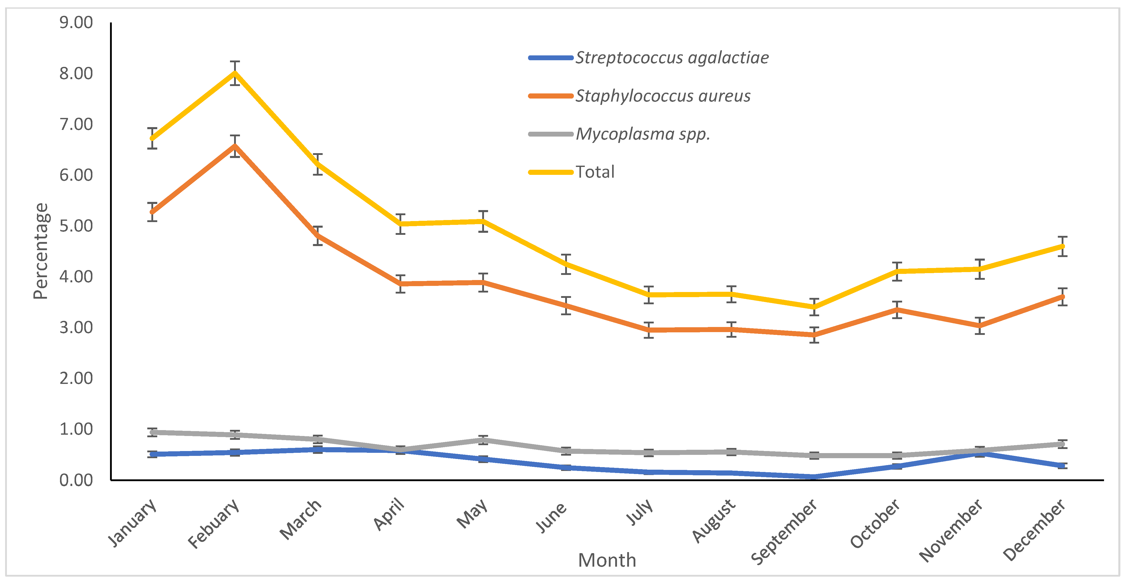
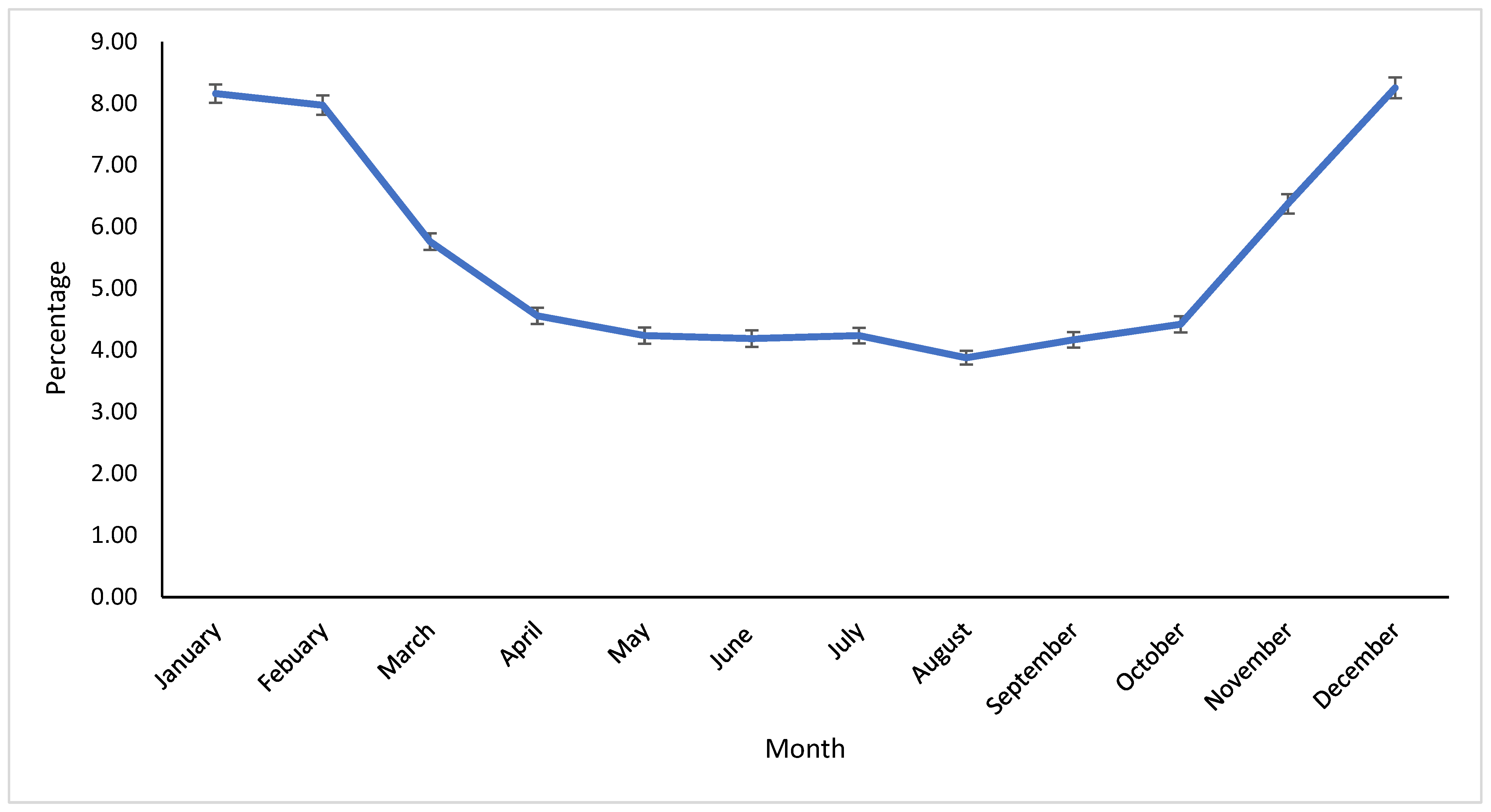
The incidence of clinical mastitis is greatly influenced by weather factors, which affect the seasonal isolation of mastitis-causing pathogens [36]. In our study, the season significantly impacted the proportion of pathogens. Most pathogens were isolated in Winter, except for NAS, which were primarily found in Summer. Reports suggest that the prevalence and distribution of pathogens vary greatly depending on the region [7,15,27]. Differences in the distribution of seasonal pathogens are likely associated with climatological variations. The Midwest and East Coast regions typically experience increased humidity and temperature in Summer, possibly leading to higher bacterial counts in bedding material. Conversely, Winter typically brings freezing conditions, reducing the bacterial population in the environment. California’s Winter tends to be humid, with precipitation levels that are higher than in other seasons, while Summer is usually dry and hot. Furthermore, recycled manure bedding on most dairy farms, combined with increased humidity during Winter, may elevate the risk of coliform exposure. It has been noted that the proportion of contagious pathogens worldwide has been decreasing, likely due to effective contagious mastitis control programs [33]. This suggests that the use of pre- and post-milking teat disinfectants, good milking hygiene, antimicrobial treatments, and dry cow therapy implemented in recent years could contribute to changes in the prevalence and distribution of contagious mastitis pathogens [37]. In our study, contagious pathogens (S. aureus, S. agalactiae, and Mycoplasma spp.), although isolated in low proportions, were more likely to be found in Winter. Infections acquired in Winter may persist into Spring, potentially increasing the likelihood of isolating contagious pathogens, as observed in the current study. The results of this study offer valuable insights into the prevalence and distribution of mastitis-causing pathogens in dairies within the Central Valley of California. The data indicate that both contagious and environmental pathogens are present, with significant variation in prevalence by season. These findings emphasize the importance of season-specific mastitis management strategies that address local pathogen profiles, particularly during Winter.

5. Conclusions

In conclusion, this study provides a comprehensive examination of the microbiological profiles of milk samples from dairy cows in California’s Central Valley over 14 years. The findings highlight the diverse range of pathogens responsible for mastitis in the region, with environmental pathogens being the most frequently isolated. While contagious pathogens were less prevalent, their presence underscores the importance of robust mastitis control programs. Seasonal variations in pathogen prevalence suggest that targeted, season-specific strategies are essential for managing udder health and effectively controlling mastitis. Overall, this study emphasizes the need for the continued monitoring of mastitis-causing pathogens to optimize prevention and treatment strategies tailored to the unique conditions of California dairies.

Author Contributions

Conceptualization, D.R.B. and W.R.E.; methodology, D.R.B. and W.R.E.; formal analysis, W.R.E.; data curation, D.R.B., K.H.T. and W.R.E.; writing—D.R.B., writing—review and editing, D.R.B., K.H.T., T.W.L., S.S.A. and W.R.E. All authors have read and agreed to the published version of the manuscript.

Funding

This research received no external funding.

Institutional Review Board Statement

Not applicable.

Informed Consent Statement

Not applicable.

Data Availability Statement

The data are available from the corresponding author.

Acknowledgments

The authors thank the UC Davis Milk Quality Laboratory staff and John Leach for culturing milk samples and assisting with data collection and analysis.

Conflicts of Interest

The authors declare no conflicts of interest.

Abbreviations

The following abbreviations are used in this manuscript:
UC DavisUniversity of California Davis
VMTRCVeterinary Medicine Teaching and Research Center
NMCNational Mastitis Council
NGNo growth
NASNon-aureus staphylococci
OFCOn-farm culture
SEStandard Error
CIConfidence Interval
USDUnited States Dollar
MALDI-TOFMatrix-Assisted Laser Desorption/Ionization Time-of-Flight

References

  1. Rollin, E.; Dhuyvetter, K.; Overton, M. The cost of clinical mastitis in the first 30 days of lactation: An economic modeling tool. Prev. Vet. Med. 2015, 122, 257–264. [Google Scholar] [CrossRef] [PubMed]
  2. Bruno, D.R.; Cleale, R.M.; Overton, M.W.; Short, T.; Pedraza, J.R.; Wallace, R. Financial implications of treating nonsevere gram-negative clinical mastitis in 3 California dairies. JDS Commun. 2024, 5, 659–663. [Google Scholar] [CrossRef] [PubMed]
  3. Leite de Campos, J.; Gonçalves, J.L.; Kates, A.; Steinberger, A.; Sethi, A.; Suen, G.; Shutske, J.; Safdar, N.; Goldberg, T.; Ruegg, P.L. Variation in partial direct costs of treating clinical mastitis among 37 Wisconsin dairy farms. J. Dairy Sci. 2023, 106, 9276–9286. [Google Scholar] [CrossRef]
  4. Cobirka, M.; Tancin, V.; Slama, P. Epidemiology and Classification of Mastitis. Animals 2020, 10, 2212. [Google Scholar] [CrossRef]
  5. da Costa, L.B.; Rajala-Schultz, P.J.; Schuenemann, G.M. Management Practices Associated with Presence of Staphylococcus aureus in Bulk Tank Milk from Ohio Dairy Herds. J. Dairy Sci. 2016, 99, 1364–1373. [Google Scholar] [CrossRef]
  6. Dego, O.K.; Vidlund, J. Staphylococcal Mastitis in Dairy Cows. Front. Vet. Sci. 2024, 11, 1356259. [Google Scholar] [CrossRef]
  7. Acharya, K.R.; Brankston, G.; Slavic, D.; Greer, A.L. Spatio-Temporal Variation in the Prevalence of Major Mastitis Pathogens Isolated from Bovine Milk Samples Between 2008 and 2017 in Ontario, Canada. Front. Vet. Sci. 2021, 8, 742696. [Google Scholar] [CrossRef]
  8. USDA Dairy 2014, Milk Quality, Milking Procedures, and Mastitis on U.S. Dairies, 2014. U.S. Department of Agriculture. 2016. Available online: https://www.aphis.usda.gov/sites/default/files/dairy14_dr_partiii.pdf (accessed on 22 January 2025).
  9. USDA Census of Agriculture, 2022. National Agricultural Statistics Service. Available online: https://www.nass.usda.gov/Publications/Highlights/2024/Census22_HL_Dairy.pdf (accessed on 12 February 2025).
  10. ERA Economics. Economic Analysis of California Dairy Consolidation, Attrition, and Policy Leakage. California Cattle Council, 2024. Available online: https://calcattlecouncil.org/wp-content/uploads/2024/10/2.-ERA_CCC_FinalReport_Sept2024.pdf (accessed on 12 February 2025).
  11. National Mastitis Council. Laboratory Handbook on Bovine Mastitis; National Mastitis Council Inc.: Madison, WI, USA, 2017. [Google Scholar]
  12. Lago, A.; Godden, S.M.; Bey, R.; Ruegg, P.L.; Leslie, K. The selective treatment of clinical mastitis based on on-farm culture results: I. Effects on antibiotic use, milk withholding time, and short-term clinical and bacteriological outcomes. J. Dairy Sci. 2011, 94, 4441–4456. [Google Scholar] [CrossRef]
  13. Schwaiger, K.; Wimmer, M.; Huber-Schlenstedt, R.; Fehlings, K.; Hölzel, C.S.; Bauer, J. Hot topic: Bovine milk samples yielding negative or nonspecific results in bacterial culturing—The possible role of PCR-single strand conformation polymorphism in mastitis diagnosis. J. Dairy Sci. 2012, 95, 98–101. [Google Scholar] [CrossRef]
  14. Makovec, J.A.; Ruegg, P.L. Results of milk samples submitted for microbiological examination in Wisconsin from 1994 to 2001. J. Dairy Sci. 2003, 86, 3466–3472. [Google Scholar] [CrossRef]
  15. Kurban, D.; Roy, J.P.; Kabera, F.; Fréchette, A.; Um, M.M.; Albaaj, A.; Rowe, S.; Godden, S.; Adkins, P.R.F.; Middleton, J.R.; et al. Diagnosing intramammary infection: Meta-analysis and mapping review on frequency and udder health relevance of microorganism species isolated from bovine milk samples. Animals 2022, 12, 3288. [Google Scholar] [CrossRef] [PubMed]
  16. Gioia, G.; Addis, M.F.; Santisteban, C.; Gross, B.; Nydam, D.V.; Sipka, A.S.; Virkler, P.D.; Watters, R.D.; Wieland, M.; Zurakowski, M.J.; et al. Mycoplasma species isolated from bovine milk collected from U.S. dairy herds between 2016 and 2019. J. Dairy Sci. 2021, 104, 4813–4821. [Google Scholar] [CrossRef] [PubMed]
  17. Dudek, K.; Nicholas, R.A.J.; Szacawa, E.; Bednarek, D. Mycoplasma bovis infections—Occurrence, diagnosis, and control. Pathogens 2020, 9, 640. [Google Scholar] [CrossRef]
  18. Dean, C.J.; Deng, Y.; Wehri, T.C.; Pena-Mosca, F.; Ray, T.; Crooker, B.A.; Godden, S.M.; Caixeta, L.S.; Noyes, N.R. The impact of kit, environment, and sampling contamination on the observed microbiome of bovine milk. mSystems 2024, 9, e0115823. [Google Scholar] [CrossRef]
  19. Acosta, A.; McCorriston, S.; Nicolli, F.; Venturelli, E.; Wickramasinghe, U.; ArceDiaz, E.; Scudiero, L.; Sammartino, A.; Schneider, F.; Steinfeld, H. Immediate effects of COVID-19 on the global dairy sector. Agric. Syst. 2021, 192, 103177. [Google Scholar] [CrossRef]
  20. Valldecabres, A.; Wenz, J.; Ferreira, F.C.; Chahine, M.; Dalton, J.; Marti, M.; Rovai, M.; Silva-del-Rio, N. Perspective of dairy producers from California, Idaho, South Dakota, and Washington: Health and business implications of the COVID-19 pandemic during the second wave. J. Dairy Sci. 2021, 105, 1788–1796. [Google Scholar] [CrossRef]
  21. Mahmood, H.; Furqan, M.; Meraj, G.; Hassan, M.S. The effects of COVID-19 on agriculture supply chain, food security, and environment: A review. PeerJ 2024, 12, e17281. [Google Scholar] [CrossRef]
  22. Freu, G.; Garcia, B.L.N.; Tomazi, T.; Di Leo, G.S.; Gheller, L.S.; Bronzo, V.; Moroni, P.; Dos Santos, M.V. Association between mastitis occurrence in dairy cows and bedding characteristics of compost-bedded pack barns. Pathogens 2023, 12, 583. [Google Scholar] [CrossRef]
  23. Ferraz, P.F.P.; Ferraz, G.A.E.S.; Leso, L.; Klopčič, M.; Barbari, M.; Rossi, G. Properties of conventional and alternative bedding materials for dairy cattle. J. Dairy Sci. 2020, 103, 8661–8674. [Google Scholar] [CrossRef]
  24. Meyer, D.; Price, P.L.; Rossow, H.A.; Silva-del-Rio, N.; Karle, B.M.; Robinson, P.H.; DePeters, E.J.; Fadel, J.G. Survey of dairy housing and manure management practices in California. J. Dairy Sci. 2011, 94, 4744–4750. [Google Scholar] [CrossRef]
  25. Goulart, D.B.; Mellata, M. Escherichia coli mastitis in dairy cattle: Etiology, diagnosis, and treatment challenges. Front. Microbiol. 2022, 13, 928346. [Google Scholar] [CrossRef] [PubMed]
  26. Munoz, M.A.; Ahlström, C.; Rauch, B.J.; Zadoks, R.N. Fecal shedding of Klebsiella pneumoniae by dairy cows. J. Dairy Sci. 2006, 89, 3425–3430. [Google Scholar] [CrossRef]
  27. Song, J.; Xiang, W.; Wang, Q.; Yin, J.; Tian, T.; Yang, Q.; Zhang, M.; Ge, G.; Li, J.; Diao, N.; et al. Prevalence and risk factors of Klebsiella spp. in milk samples from dairy cows with mastitis—A global systematic review. Front. Vet. Sci. 2023, 10, 1143257. [Google Scholar] [CrossRef] [PubMed]
  28. Valckenier, D.; Piepers, S.; Schukken, Y.H.; De Visscher, A.; Boyen, F.; Haesebrouck, F.; De Vliegher, S. Longitudinal study on the effects of intramammary infection with non-aureus Staphylococci on udder health and milk production in dairy heifers. J. Dairy Sci. 2021, 104, 899–914. [Google Scholar] [CrossRef]
  29. Bechtold, V.; Petzl, W.; Huber-Schlenstedt, R.; Sorge, U.S. Distribution of bovine mastitis pathogens in quarter milk samples from Bavaria, Southern Germany, between 2014 and 2023—A retrospective study. Animals 2024, 14, 2504. [Google Scholar] [CrossRef]
  30. Woudstra, S.; Wente, N.; Zhang, Y.; Leimbach, S.; Kirkeby, C.; Gussmann, M.K.; Krömker, V. Reservoirs of Staphylococcus spp. and Streptococcus spp. associated with intramammary infections of dairy cows. Pathogens 2023, 12, 699. [Google Scholar] [CrossRef]
  31. Kabelitz, T.; Aubry, E.; van Vorst, K.; Amon, T.; Fulde, M. The role of Streptococcus spp. in bovine mastitis. Microorganisms 2021, 9, 1497. [Google Scholar] [CrossRef]
  32. Dyson, R.; Charman, N.; Hodge, A.; Rowe, S.M.; Taylor, L.F. A survey of mastitis pathogens including antimicrobial susceptibility in southeastern Australian dairy herds. J. Dairy Sci. 2022, 105, 1504–1518. [Google Scholar] [CrossRef]
  33. LeBlanc, S.J.; Lissemore, K.D.; Kelton, D.F.; Duffield, T.F.; Leslie, K.E. Major advances in disease prevention in dairy cattle. J. Dairy Sci. 2006, 89, 1267–1279. [Google Scholar] [CrossRef]
  34. Gioia, G.; Freeman, J.; Sipka, A.; Santisteban, C.; Wieland, M.; Gallardo, V.A.; Monistero, V.; Scott, J.G.; Moroni, P. Pathogens associated with houseflies from different areas within a New York State dairy. JDS Commun. 2022, 3, 285–290. [Google Scholar] [CrossRef]
  35. Jasper, D.E.; Dellinger, J.D.; Rollins, M.H.; Hakanson, H.D. Prevalence of mycoplasmal bovine mastitis in California. Am. J. Vet. Res. 1979, 40, 1043–1047. [Google Scholar] [CrossRef] [PubMed]
  36. Gao, J.; Barkema, H.W.; Zhang, L.; Liu, G.; Deng, Z.; Cai, L.; Shan, R.; Zhang, S.; Zou, L.; Kastelic, J.P.; et al. Incidence of clinical mastitis and distribution of pathogens on large Chinese dairy farms. J. Dairy Sci. 2017, 100, 4797–4806. [Google Scholar] [CrossRef] [PubMed]
  37. Fitzpatrick, S.R.; Garvey, M.; Flynn, J.; O’Brien, B.; Gleeson, D. The effect of disinfectant ingredients on teat skin bacteria associated with mastitis in Irish dairy herds. Ir. Vet. J. 2021, 74, 1–12. [Google Scholar] [CrossRef] [PubMed]
Figure 1. Monthly percentage of contagious mastitis pathogens (Staphylococcus aureus, Streptococcus agalactiae, and Mycoplasma spp.) from milk samples sent to the Milk Quality Laboratory at the Veterinary Medicine Teaching and Research Center, UC Davis, Tulare, California, between 2009 and 2023. Error bars represent one standard error.
Vetsci 12 00609 g001
Figure 2. Monthly percentage of contaminated milk samples sent to the Milk Quality Laboratory of the Veterinary Medicine Teaching and Research Center, UC Davis, Tulare, California, from 2009 to 2023. Error bars indicate one standard error.
Vetsci 12 00609 g002
Table 1. Annual percentage of no bacterial growth, pure cultures, mixed infections, and contaminated milk samples submitted to the Milk Quality Laboratory at the Veterinary Medicine Teaching and Research Center, UC Davis, Tulare, California.
YearRoutine Culture *No GrowthPure CultureMixed CultureContaminated
%SE%SE%SE%SE
200933,90726.68 ef0.2448.04 ef0.2718.63 d0.216.65 c0.14
201027,65932.55 h0.2846.83 bcde0.3015.93 a0.224.69 ab0.13
201125,82231.70 h0.2946.96 cdef0.3116.46 ab0.234.88 b0.13
201221,67534.37 i0.3245.77 abc0.3415.58 a0.254.28 ab0.14
201314,72927.49 f0.3747.04 bcdef0.4119.53 de0.335.94 c0.19
201417,32633.20 hi0.3646.18 abcd0.3816.15 ab0.284.46 ab0.16
201519,40429.53 g0.3345.59 abc0.3618.45 d0.286.43 c0.18
201622,55926.50 ef0.2947.78 def0.3319.39 de0.266.33 c0.16
201734,34230.07 g0.2549.45 g0.2716.24 ab0.204.24 a0.11
201827,79527.35 f0.2751.10 h0.3017.14 bc0.234.41 ab0.12
201922,39425.38 de0.2948.43 fg0.3320.19 e0.276.00 c0.16
202013,09022.50 bc0.3644.99 ab0.4323.84 f0.378.67 d0.25
202120,62219.41 a0.2856.19 ah0.3518.16 cd0.276.25 c0.17
2022922520.89 ab0.4247.61 cdefg0.5223.49 f0.448.01 d0.28
2023908524.02 cd0.4544.15 a0.5223.74 f0.458.09 d0.29
Total319,63427.440.3047.740.4018.860.305.960.20
* Total number of individual cow milk samples submitted to the laboratory for routine culture. Cells with the same letter within the same column mean that there is no significant difference, and cells with different letters mean that there is a significant difference.
Table 2. Annual percentage of environmental and contagious mastitis pathogens from milk samples sent to the Milk Quality Laboratory of the Veterinary Medicine Teaching and Research Center, UC Davis, Tulare, California, between 2009 and 2023.
YearN *Environmental Mastitis PathogensContagious Mastitis Pathogens **
Gram-PositiveGram-NegativeFungi, Algae, and Yeast
%SE95%CI%SE95%CI%SE95%CI%SE95%CI
200916,28865.39 e0.3764.6565.3926.12 hi0.3425.4526.801.68 bc0.101.501.896.81 e0.206.437.21
201012,95268.11 e0.4167.3068.1127.04 i0.3926.2827.811.44 bc0.101.241.663.42 bc0.163.123.75
201112,12671.30 a0.4170.4971.3024.53 gh0.3923.7825.311.97 cd0.131.742.232.19 a0.131.952.47
2012992072.56 ab0.4571.6772.5623.78 g0.4322.9524.631.45 bc0.121.231.712.21 a0.151.942.52
2013692971.79 a0.5470.7171.7924.85 ghi0.5223.8525.881.17 b0.130.941.452.19 a0.181.872.57
2014800276.18 c0.4875.2476.1817.21 f0.4216.4018.051.51 bc0.141.271.805.10 d0.254.645.60
2015884676.00 c0.4575.1076.0014.88 cde0.3814.1515.631.45 bc0.131.221.727.68 ef0.287.148.25
201610,77874.74 c0.4273.9174.7414.98 cde0.3414.3215.672.75 ef0.162.453.077.53 ef0.257.058.05
201716,98374.33 bc0.3473.6674.3315.29 cde0.2814.7615.842.35 de0.122.132.598.03 f0.217.638.45
201814,20474.98 c0.3674.2674.9816.40 ef0.3115.8017.013.44 fg0.153.163.765.18 d0.194.835.56
201910,84575.77 c0.4174.9575.7716.27 def0.3515.5916.984.14 g0.193.784.533.82 bc0.183.474.19
2020588982.65 d0.4981.6682.6511.56 a0.4210.7712.410.44 a0.090.300.655.35 d0.294.805.95
202111,58782.01 d0.3681.3082.0114.59 c0.3313.9515.240.25 a0.050.170.363.15 b0.162.853.48
2022439283.17 d0.5682.0483.1712.02 ab0.4911.0913.020.23 a0.070.120.424.58 cd0.324.005.24
2023401181.60 d0.6180.3781.6014.09 bcd0.5513.0415.200.17 a0.070.080.374.14 bcd0.313.564.80
* N: Total number of samples with positive growth (pure colonies only). ** Contagious mastitis pathogens: only Staphylococcus aureus, Streptococcus agalactiae, and Mycoplasma spp. Cells with the same letter within the same column mean that there is no significant difference, and cells with different letters mean that there is a significant difference.
Table 3. Annual percentage of major and minor mastitis pathogens from milk samples sent to the Milk Quality Laboratory of the Veterinary Medicine Teaching and Research Center at UC Davis, Tulare, California, between 2009 and 2023.
YearN *MajorMinor
%SE95% CI%SE95% CI
200916,28843.57 h0.3942.8144.3356.43 a0.3955.6757.19
201012,95243.20 gh0.4442.3544.0556.80 ab0.4455.9557.65
201112,12640.92 f0.4540.0541.8059.08 c0.4558.2059.95
2012992038.43 de0.4937.4739.3961.57 de0.4960.6162.53
2013692941.02 efg0.5939.8642.1858.98 bcd0.5957.8260.14
2014800232.17 abc0.5231.1533.2067.83 fgh0.5266.8068.85
2015884634.03 bc0.5033.0535.0265.97 fg0.5064.9866.95
201610,77834.32 c0.4633.4335.2265.68 f0.4664.7866.57
201716,98336.64 d0.3735.9237.3663.36 e0.3762.6464.08
201814,20433.68 bc0.4032.9134.4666.32 fg0.4065.5467.09
201910,84531.68 ab0.4530.8132.5668.32 gh0.4567.4469.19
2020588926.10 i0.5724.9927.2473.90 i0.5772.7675.01
202111,58730.48 a0.4329.6531.3369.52 h0.4368.6770.35
2022439231.81 abc0.7030.4533.2068.19 fgh0.7066.8069.55
2023401132.39 abc0.7430.9533.8567.61 fgh0.7466.1569.05
* N: total number of samples with positive growth (pure colonies only). Cells with the same letter within the same column mean that there is no significant difference, and cells with different letters mean that there is a significant difference. Major pathogens: Staphylococcus aureus, Streptococcus agalactiae, Streptococcus dysgalactiae, Mycoplasma bovis, Streptococcus uberis, Enterococcus spp., Proteus spp., Serratia spp., Yersinia spp., Pseudomonas aeruginosa, Truerperella pyogenes, and Coliforms (Escherichia coli, Klebsiella spp., Enterobacter spp.). Minor pathogens: non-aureus staphylococci, Corynebacterium spp., Fungi, Algae, Yeast.
Table 4. Annual percentage of selected major mastitis pathogens from milk samples sent to the Milk Quality Laboratory at the Veterinary Medicine Teaching and Research Center, UC Davis, Tulare, California, between 2009 and 2023.
YearN *Streptococcus spp.Staphylococcus AureusColiformsMycoplasma spp.Others
%SE95% CI%SE95% CI%SE95% CI%SE95% CI%SE95% CI
2009721626.07 h0.5225.0727.0912.76 de0.3912.0113.5557.08 f0.5855.9458.222.43 c0.182.092.811.66 bc0.151.391.99
2010567031.22 a0.6230.0232.444.30 a0.273.814.8660.37 g0.6559.0961.642.79 c0.222.393.251.32 abc0.151.061.66
2011502035.52 bc0.6834.2136.853.63 a0.263.144.1858.15 fg0.7056.7859.511.55 ab0.171.251.941.16 ab0.150.891.49
2012386833.04 ab0.7631.5834.544.24 a0.323.654.9260.13 fg0.7958.5861.671.14 ab0.170.851.531.45 abc0.191.121.88
2013287935.19 abc0.8933.4636.953.54 a0.342.934.2859.29 fg0.9257.4961.070.69 a0.150.451.071.29 abc0.210.931.77
2014260732.83 ab0.9231.0634.6614.58 def0.6913.2715.9850.56 e0.9848.6452.470.77 a0.170.501.191.27 abc0.220.901.78
2015305534.08 ab0.8632.4235.7820.29 g0.7318.9121.7642.26 bc0.8940.5244.021.90 cd0.251.472.451.47 aabc0.221.101.97
2016376838.61 cde0.7937.0740.1816.59 fg0.6115.4317.8141.11 b0.8039.5542.691.86 de0.221.472.341.83 bc0.221.452.31
2017630240.19 def0.6238.9941.4115.49 f0.4614.6116.4039.69 b0.6238.4840.903.36 ef0.232.953.841.27 abc0.141.021.58
2018489139.01 de0.7037.6540.3913.06 de0.4812.1514.0444.71 cd0.7143.3346.111.02 a0.140.781.352.19 c0.211.812.64
2019351038.95 cd0.8237.3540.5710.11 bc0.519.1611.1647.49 de0.8445.8449.151.34 bc0.191.011.782.11 bc0.241.682.64
2020156540.13 cdef1.2437.7342.5816.23 efg0.9314.4818.1441.66 bc1.2539.2444.120.19 a0.110.060.591.79 abc0.341.242.58
2021356343.70 f0.8342.0845.337.80 b0.456.978.7346.20 cde0.8444.5647.841.43 cd0.201.091.880.87 a0.160.611.23
2022141349.47 g1.3346.8752.0811.32 bcd0.849.7713.0835.39 a1.2732.9337.922.69 f0.431.963.671.13 abc0.280.691.84
2023132344.52 efg1.3741.8647.2111.94 cde0.8910.3013.8041.35 abc1.3538.7244.020.38 abc0.170.160.901.81 abc0.371.222.69
* N: total number of samples submitted to the laboratory with major mastitis pathogens (pure colonies only). Cells with the same letter within the same column mean that there is no significant difference, and cells with different letters mean that there is a significant difference. Classification of major mastitis pathogens was carried out according to [4].
Table 5. Annual percentage of various Streptococcus spp. from milk samples sent to the Milk Quality Laboratory of the Veterinary Medicine Teaching and Research Center at UC Davis, Tulare, California, between 2009 and 2023.
Year* NStreptococcus agalactiaeStreptococcus uberisOther Streptococcus Species
%SE95% CI%SE95% CI%SE95% CI
200918810.69 a0.190.401.191.06 ab0.240.691.6498.25 fg0.3097.5498.75
201017702.32 bc0.361.713.131.75 abc0.311.232.4895.93 cde0.4794.9196.76
201117830.34 a0.140.150.750.95 ab0.230.591.5398.71 g0.2798.0799.14
201212780.86 ab0.260.481.551.25 ab0.310.772.0397.89 efg0.4096.9498.55
201310132.96 c0.532.084.200.89 ab0.290.461.7096.15 cdef0.6094.7797.17
20148560.93 abc0.330.471.860.47 a0.230.181.2498.60 fg0.4097.5599.20
201510410.10 a0.100.010.687.20 f0.805.788.9492.70 bc0.8190.9594.13
201614558.04 d0.716.759.553.99 ef0.513.095.1287.97 a0.8586.2089.55
201725336.95 d0.516.028.013.43 de0.362.794.2289.62 ab0.6188.3790.75
201819082.46 c0.351.863.261.94 bcde0.321.412.6795.60 cd0.4794.5896.43
201913670.88 ab0.250.501.543.66 cde0.512.784.7995.46 cd0.5694.2296.45
20206289.24 d1.167.2111.764.46 cdef0.823.106.3886.31 a1.3783.3988.78
202115572.31 bc0.381.673.191.80 abcd0.341.242.5995.89 cde0.5094.7896.77
20226990.43 a0.250.141.322.72 abcde0.621.744.2296.85 defg0.6695.2797.92
20235890.51 a0.290.161.571.53 abcde0.510.802.9197.96 defg0.5896.4598.84
* N: total number of samples submitted to the laboratory with Streptococcus spp. isolation (pure colonies only). Cells with the same letter within the same column mean that there is no significant difference, and cells with different letters mean that there is a significant difference.
Table 6. Annual percentage of selected minor mastitis pathogens from milk samples submitted to the Milk Quality Laboratory at the Veterinary Medicine Teaching and Research Center, UC Davis, Tulare, California, between 2009 and 2023.
YearN *Non-Aureus StaphylococciCorynebacterium spp.Fungi, Algae, Yeast
%SE95% CI%SE95% CI%SE95% CI
2009907296.76 cd0.1996.3797.100.23 abc0.050.150.353.01 cde0.182.683.38
2010728297.25 de0.1996.8597.600.21 abc0.050.120.342.54 bcd0.182.202.93
2011710696.34 cd0.2295.8896.750.30 abcd0.060.190.453.36 def0.212.973.81
2012605297.22 ef0.2196.7897.610.41 bcde0.080.280.612.36 bcd0.202.012.78
2013405097.80 ef0.2397.3098.210.20 abc0.070.100.392.00 b0.221.612.48
2014539597.46 def0.2197.0197.850.30 abcd0.070.180.482.24 bc0.201.882.67
2015579197.58 f0.2097.1597.950.21 abc0.060.120.362.21 bc0.191.862.62
2016701095.45 bc0.2594.9495.910.36 abcd0.070.240.534.19 fg0.243.754.69
201710,68195.62 cd0.2095.2195.990.65 de0.080.510.823.74 ef0.183.394.11
2018931394.38 ab0.2493.9094.830.37 cd0.060.260.515.25 gh0.234.825.72
2019733593.66 a0.2893.0894.200.22 abc0.050.130.366.12 h0.285.606.69
2020432499.17 gh0.1498.8599.400.23 abc0.070.120.430.60 a0.120.410.88
2021802498.83 g0.1298.5799.040.81 e0.100.641.030.36 a0.070.250.52
2022297999.60 gh0.1299.2999.770.07 a0.050.020.270.34 a0.110.180.62
2023268899.67 h0.1199.3699.830.07 ab0.050.020.300.26 a0.100.120.55
* N: total number of samples submitted to the laboratory with minor mastitis pathogens (pure colonies only). Cells with the same letter within the same column mean that there is no significant difference, and cells with different letters mean that there is a significant difference. Classification of minor mastitis pathogens was carried out according to [4].
Table 7. Seasonal percentages of pure cultures, mixed infections, no growth, and contaminated aerobic bacterial growth results from mastitis milk samples sent to the Milk Quality Laboratory of the Veterinary Medicine Teaching and Research Center, UC Davis, Tulare, California, between 2009 and 2023.
SeasonN *Pure CultureMixed InfectionNo GrowthContaminated
%SE95% CI%SE95% CI%SE95% CI%SE95% CI
Winter89,45445.93 a0.1745.6046.2620.57 a0.1420.3020.8325.78 a0.1525.5026.077.72 a0.097.557.90
Spring75,08450.32 b0.1849.9750.6817.21 b0.1416.9417.4828.04 b0.1627.7228.364.43 b0.084.284.58
Summer79,11749.36 c0.1849.0149.7116.19 c0.1315.9416.4530.33 c0.1630.0130.654.12 c0.073.984.26
Fall75,97947.16 d0.1846.8147.5218.29 d0.1418.0218.5728.63 b0.1628.3128.955.92 d0.095.766.09
* N: total number of samples submitted to the laboratory. Cells with the same letter within the same column mean that there is no significant difference, and cells with different letters mean that there is a significant difference.
Table 8. Seasonal percentages of contagious and environmental mastitis pathogens from milk samples sent to the Milk Quality Laboratory of the Veterinary Medicine Teaching and Research Center, UC Davis, Tulare, California, between 2009 and 2023.
SeasonN *Contagious PathogensEnvironmental Pathogens
Gram-NegativeGram-PositiveFungi Algae Yeast
%SE95% CI%SE95% CI%SE95% CI%SE95% CI
Winter41,0856.91 a0.136.677.1620.71 b0.2020.3221.1170.83 a0.2270.3971.271.55 a0.061.431.67
Spring37,7855.06 b0.114.845.2919.43 a0.2019.0419.8373.34 b0.2372.8973.782.17 b0.072.032.32
Summer39,0503.68 c0.103.503.8716.70 c0.1916.3417.0877.24 c0.2176.8377.662.37 b0.082.232.53
Fall35,8324.08 d0.103.884.2919.26 a0.2118.8619.6775.28 d0.2374.8375.721.38 a0.061.271.51
* N: total number of samples submitted to the laboratory (pure colonies only). Cells with the same letter within the same column mean that there is no significant difference, and cells with different letters mean that there is a significant difference.
Disclaimer/Publisher’s Note: The statements, opinions and data contained in all publications are solely those of the individual author(s) and contributor(s) and not of MDPI and/or the editor(s). MDPI and/or the editor(s) disclaim responsibility for any injury to people or property resulting from any ideas, methods, instructions or products referred to in the content.

