

Successful Management of a Pancreatic Abscess in a Dog with Juvenile Diabetes Mellitus Through Ultrasound-Guided Drainage and Medical Therapy

 , , and
Simple Summary
Abstract
1. Introduction
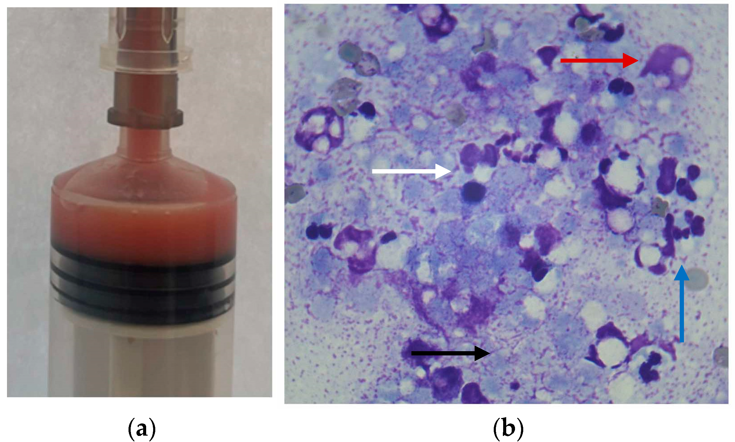
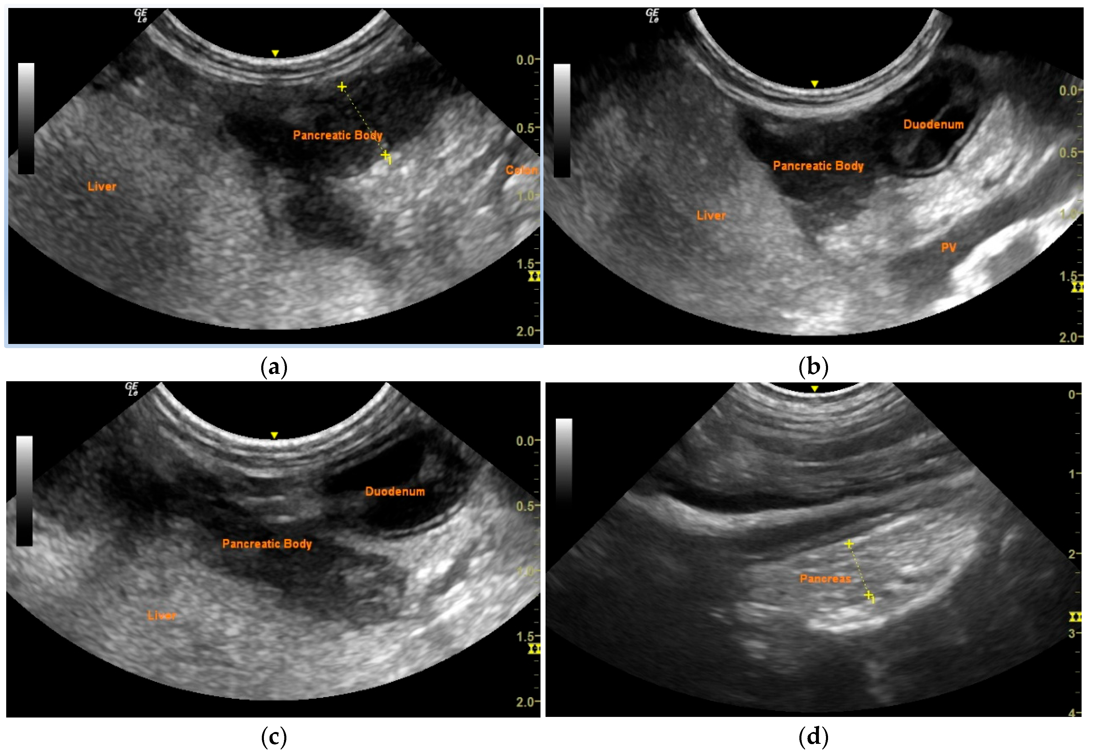
2. Case Description
3. Discussion
4. Conclusions
Author Contributions
Funding
Institutional Review Board Statement
Informed Consent Statement
Data Availability Statement
Conflicts of Interest
Abbreviations
| DM | Diabetes mellitus |
| CRT | Capillary refill time |
| Spec cPL | Specific pancreatic lipase |
| cTLI | Canine trypsin-like immunoreactivity |
| EPI | Exocrine pancreatic insufficiency |
References
- Warshaw, A.L. Inflammatory Masses Following Acute Pancreatitis: Phlegmon, Pseudocyst, and Abscess. Surg. Clin. N. Am. 1974, 54, 621–636. [Google Scholar] [CrossRef] [PubMed]
- Salisbury, S.K.; Lantz, G.C.; Nelson, R.W.; Kazacos, E.A. Pancreatic Abscess in Dogs: Six Cases (1978–1986). J. Am. Vet. Med. Assoc. 1988, 193, 1104–1108. [Google Scholar] [CrossRef] [PubMed]
- Edwards, D.; Bauer, M.; Walker, M.; Pardo, A.D.; McCracken, M.; Walker, T. Pancreatic Masses in Seven Dogs Following Acute Pancreatitis. J. Am. Anim. Hosp. Assoc. 1990, 26, 189–198. [Google Scholar]
- VanEnkevort, B.A.; O’Brien, R.T.; Young, K.M. Pancreatic Pseudocysts in 4 Dogs and 2 Cats: Ultrasonographic and Clinicopathologic Findings. J. Vet. Intern. Med. 1999, 13, 309–313. [Google Scholar] [CrossRef]
- Stimson, E.L.; Espada, Y.; Moon, M.; Troy, G.C. Pancreatic Abscess in Nine Dogs. J. Vet. Intern. Med. 1998, 9, 202. [Google Scholar]
- Coleman, M.; Robson, M. Pancreatic Masses Following Pancreatitis: Pancreatic Pseudocysts, Necrosis, and Abscesses. Contin. Educ. Vet. 2005, 27, 147–153. [Google Scholar]
- Johnson, M.D.; Mann, F.A. Treatment for Pancreatic Abscesses via Omentalization with Abdominal Closure versus Open Peritoneal Drainage in Dogs: 15 Cases (1994–2004). J. Am. Vet. Med. Assoc. 2006, 228, 397–402. [Google Scholar] [CrossRef]
- Anderson, J.R.; Cornell, K.K.; Parnell, N.K.; Salisbury, S.K. Pancreatic Abscess in 36 Dogs: A Retrospective Analysis of Prognostic Indicators. J. Am. Anim. Hosp. Assoc. 2008, 44, 171–179. [Google Scholar] [CrossRef]
- Akol, K.G.; Washabau, R.J.; Saunders, H.M.; Hendrick, M.J. Acute Pancreatitis in Cats with Hepatic Lipidosis. J. Vet. Intern. Med. 1993, 7, 205–209. [Google Scholar] [CrossRef]
- Saunders, H.M.; VanWinkle, T.J.; Drobatz, K.; Kimmel, S.E.; Washabau, R.J. Ultrasonographic Findings in Cats with Clinical, Gross Pathologic, and Histologic Evidence of Acute Pancreatic Necrosis: 20 Cases (1994–2001). J. Am. Vet. Med. Assoc. 2002, 221, 1724–1730. [Google Scholar] [CrossRef]
- Hecht, S.; Henry, G. Sonographic Evaluation of the Normal and Abnormal Pancreas. Clin. Tech. Small Anim. Pract. 2007, 22, 115–121. [Google Scholar] [CrossRef] [PubMed]
- Rademacher, N.; Ohlerth, S.; Scharf, G.; Laluhova, D.; Sieber-Ruckstuhl, N.; Alt, M.; Roos, M.; Grest, P.; Kaser-Hotz, B. Contrast-Enhanced Power and Color Doppler Ultrasonography of the Pancreas in Healthy and Diseased Cats. J. Vet. Intern. Med. 2008, 22, 1310–1316. [Google Scholar] [CrossRef] [PubMed]
- Bradley, E.L. A clinically based classification system for acute pancreatitis. Summary of the international symposium on acute pancreatitis, Atlanta, Ga, September 11 through 12, 1992. Arch. Surg. 1993, 128, 586–590. [Google Scholar] [CrossRef]
- van Santvoort, H.C.; Besselink, M.G.; Bakker, O.J.; Hofker, H.S.; Boermeester, M.A.; Dejong, C.H.; van Goor, H.; Schaapherder, A.F.; van Eijck, C.H.; Bollen, T.L.; et al. A Step-up Approach or Open Necrosectomy for Necrotizing Pancreatitis. N. Engl. J. Med. 2010, 362, 1491–1502. [Google Scholar] [CrossRef]
- Varadarajulu, S.; Bang, J.H.; Phadnis, M.A.; Christein, J.D.; Wilcox, C.M. Endoscopic Transmural Drainage of Peripancreatic Fluid Collections: Outcomes and Predictors of Treatment Success in 211 Consecutive Patients. J. Gastrointest. Surg. 2011, 15, 2080–2088. [Google Scholar] [CrossRef]
- Hollemans, R.A.; Bakker, O.J.; Boermeester, M.A.; Bollen, T.L.; Bosscha, K.; Bruno, M.J.; Buskens, E.; Dejong, C.H.; van Duijvendijk, P.; van Eijck, C.H.; et al. Superiority of Step-up Approach vs Open Necrosectomy in Long-Term Follow-up of Patients with Necrotizing Pancreatitis. Gastroenterology 2019, 156, 1016–1026. [Google Scholar] [CrossRef] [PubMed]
- Goyal, J.; Ramesh, J. Endoscopic Management of Peripancreatic Fluid Collections. Frontline Gastroenterol. 2014, 6, 199–207. [Google Scholar] [CrossRef]
- Talbot, C.T.; Cheung, R.; Holmes, E.J.; Cook, S.D. Medical and Surgical Management of Pancreatic Fluid Accumulations in Dogs: A Retrospective Study of 15 Cases. J. Vet. Intern. Med. 2022, 36, 919–926. [Google Scholar] [CrossRef]
- Nemoto, Y.; Haraguchi, T.; Shimokawa Miyama, T.; Kobayashi, K.; Hama, K.; Kurogouchi, Y.; Fujiki, N.; Baba, K.; Okuda, M.; Mizuno, T. Pancreatic Abscess in a Cat Due to Staphylococcus aureus Infection. J. Vet. Med. Sci. 2017, 79, 1146–1150. [Google Scholar] [CrossRef]
- Lee, M.; Kang, J.-H.; Chang, D.; Na, K.-J.; Yang, M.-P. Pancreatic Abscess in a Cat with Diabetes Mellitus. J. Am. Anim. Hosp. Assoc. 2015, 51, 180–184. [Google Scholar] [CrossRef]
- Smith, S.; Biller, D. Resolution of a Pancreatic Pseudocyst in a Dog Following Percutaneous Ultrasonographic-Guided Drainage. J. Am. Anim. Hosp. Assoc. 1998, 34, 515–522. [Google Scholar] [CrossRef] [PubMed]
- Greco, D.S.; Chastain, C.B. Endocrine and Metabolic System. In Veterinary Pediatrics: Dogs and Cats from Birth to Six Months, 3rd ed.; Hoskins, J.D., Ed.; Saunders: Philadelphia, PA, USA, 2001; pp. 344–370. [Google Scholar]
- Greco, D.S. Pediatric Endocrinology. Vet. Clin. Small Anim. Pract. 2006, 36, 549–556. [Google Scholar] [CrossRef]
- Gilor, C.; Niessen, S.J.M.; Furrow, E.; DiBartola, S.P. What’s in a Name? Classification of Diabetes Mellitus in Veterinary Medicine and Why It Matters. J. Vet. Intern. Med. 2016, 30, 927–940. [Google Scholar] [CrossRef] [PubMed]
- Warshaw, A.L.; Richter, J.M. A Practical Guide to Pancreatitis. Curr. Probl. Surg. 1984, 21, 6–79. [Google Scholar] [CrossRef]
- Warshaw, A.L.; Jin, G.L. Improved Survival in 45 Patients with Pancreatic Abscess. Ann. Surg. 1985, 202, 408–417. [Google Scholar] [CrossRef] [PubMed]
- Saxon, A.; Reynolds, J.T.; Doolas, A. Management of Pancreatic Abscesses. Ann. Surg. 1981, 194, 545–552. [Google Scholar] [CrossRef]
- Amano, H.; Takada, T.; Isaji, S.; Takeyama, Y.; Hirata, K.; Yoshida, M.; Mayumi, T.; Yamanouchi, E.; Gabata, T.; Kadoya, M.; et al. Therapeutic Intervention and Surgery of Acute Pancreatitis. J. Hepatobiliary Pancreat. Sci. 2009, 17, 53–59. [Google Scholar] [CrossRef]
- Schaer, M. Abscess, Necrosis, Pseudocyst, Phlegmon, and Infection. In Canine & Feline Gastroenterology; Washabau, R.J., Day, M.J., Eds.; Elsevier Saunders: St. Louis, MO, USA, 2013; pp. 829–834. [Google Scholar]
- Kang, J.-H.; Na, K.-J.; Mo, I.-P.; Chang, D.; Yang, M.-P. Juvenile Diabetes Mellitus Accompanied by Exocrine Pancreatic Insufficiency in a Dog. J. Vet. Med. Sci. 2008, 70, 1337–1340. [Google Scholar] [CrossRef]
- Mamom, T.; Rungpupradit, J. Diabetes Mellitus Concurrent with Exocrine Pancreatic Insufficiency in a Young Golden Retriever Dog: A Clinicopathological Report. J. Mahanakorn Vet. Med. 2010, 5, 51–60. [Google Scholar]
- Neiger, R.; Jaunin, V.B.; Boujon, C.E. Exocrine Pancreatic Insufficiency Combined with Insulin-Dependent Diabetes Mellitus in a Juvenile German Shepherd Dog. J. Small Anim. Pract. 1996, 37, 344–349. [Google Scholar] [CrossRef]
- Chen, J.; Fukami, N.; Li, Z. Endoscopic Approach to Pancreatic Pseudocyst, Abscess and Necrosis: Review on Recent Progress. Dig. Endosc. 2012, 24, 299–308. [Google Scholar] [CrossRef] [PubMed]
- Leppäniemi, A.; Tolonen, M.; Tarasconi, A.; Segovia-Lohse, H.; Gamberini, E.; Kirkpatrick, A.W.; Ball, C.G.; Parry, N.; Sartelli, M.; Wolbrink, D.; et al. 2019 WSES Guidelines for the Management of Severe Acute Pancreatitis. World J. Emerg. Surg. 2019, 14, 27. [Google Scholar] [CrossRef] [PubMed]
- Thompson, L.J.; Seshadri, R.; Raffe, M.R. Characteristics and Outcomes in Surgical Management of Severe Acute Pancreatitis: 37 Dogs (2001–2007). J. Vet. Emerg. Crit. Care 2009, 19, 165–173. [Google Scholar] [CrossRef] [PubMed]

Disclaimer/Publisher’s Note: The statements, opinions and data contained in all publications are solely those of the individual author(s) and contributor(s) and not of MDPI and/or the editor(s). MDPI and/or the editor(s) disclaim responsibility for any injury to people or property resulting from any ideas, methods, instructions or products referred to in the content. |
© 2025 by the authors. Licensee MDPI, Basel, Switzerland. This article is an open access article distributed under the terms and conditions of the Creative Commons Attribution (CC BY) license (https://creativecommons.org/licenses/by/4.0/).
Share and Cite
Daravigka, A.; Ninis, S.; Bourdekas, P.; Konstantinidis, A.O.; Ginoudis, A.; Adamama-Moraitou, K.K.; Lyraki, M.; Soubasis, N. Successful Management of a Pancreatic Abscess in a Dog with Juvenile Diabetes Mellitus Through Ultrasound-Guided Drainage and Medical Therapy. Vet. Sci. 2025, 12, 604. https://doi.org/10.3390/vetsci12070604
Daravigka A, Ninis S, Bourdekas P, Konstantinidis AO, Ginoudis A, Adamama-Moraitou KK, Lyraki M, Soubasis N. Successful Management of a Pancreatic Abscess in a Dog with Juvenile Diabetes Mellitus Through Ultrasound-Guided Drainage and Medical Therapy. Veterinary Sciences. 2025; 12(7):604. https://doi.org/10.3390/vetsci12070604
Chicago/Turabian StyleDaravigka, Alexandra, Stefanos Ninis, Panagiotis Bourdekas, Alexandros O. Konstantinidis, Argyrios Ginoudis, Katerina K. Adamama-Moraitou, Maria Lyraki, and Nektarios Soubasis. 2025. "Successful Management of a Pancreatic Abscess in a Dog with Juvenile Diabetes Mellitus Through Ultrasound-Guided Drainage and Medical Therapy" Veterinary Sciences 12, no. 7: 604. https://doi.org/10.3390/vetsci12070604
APA StyleDaravigka, A., Ninis, S., Bourdekas, P., Konstantinidis, A. O., Ginoudis, A., Adamama-Moraitou, K. K., Lyraki, M., & Soubasis, N. (2025). Successful Management of a Pancreatic Abscess in a Dog with Juvenile Diabetes Mellitus Through Ultrasound-Guided Drainage and Medical Therapy. Veterinary Sciences, 12(7), 604. https://doi.org/10.3390/vetsci12070604