Article

Probiotic Characterization of Lactiplantibacillus paraplantarum SDN1.2 and Its Anti-Inflammatory Effect on Klebsiella pneumoniae-Infected Mammary Glands

by
Jia Cheng
*,†,
Jingdi Tong
,
Can Li
,
Ziyan Wang
,
Hao Li
,
Meiyi Ren
,
Jinshang Song
,
Deyuan Song
,
Qinna Xie
and
Mingchao Liu
*
Department of Clinical Veterinary Medicine, College of Veterinary Medicine, Hebei Agricultural University, Baoding 071001, China
*
Authors to whom correspondence should be addressed.
These authors contributed equally to this work.
Vet. Sci. 2025, 12(4), 323; https://doi.org/10.3390/vetsci12040323
Submission received: 18 February 2025 / Revised: 23 March 2025 / Accepted: 28 March 2025 / Published: 1 April 2025
(This article belongs to the Special Issue Ruminant Mastitis: Therapies and Control)

Simple Summary

One of the most prevalent illnesses in dairy farms is mastitis. Although Lactiplantibacillus paraplantarum (L. paraplantarum), a significant probiotic with several uses, may have anti-inflammatory qualities, it remains unclear how it prevents mastitis. This study aimed to investigate the probiotic properties of L. paraplantarum SDN1.2 and its impact on mastitis caused by Klebsiella pneumoniae (K. pneumoniae) both in vitro and in vivo. Whole-genome sequencing analysis and in vitro tests confirmed that the application of L. paraplantarum SDN1.2 is safe and exhibits good probiotic properties. The results indicate that L. paraplantarum SDN1.2 mitigates K. pneumoniae-induced inflammation in mastitis, highlighting the therapeutic potential of L. paraplantarum in mastitis.

Abstract

K. pneumoniae is a major cause of bovine mastitis worldwide, making it difficult to control due to its resistance to multiple drugs. L. paraplantarum has been explored as a promising new approach to fighting bovine mastitis. In this study, the probiotic potential and safety of L. paraplantarum SDN1.2, as well as its ex vivo and in vivo anti-inflammatory effects against K. pneumoniae-induced mastitis, were comprehensively investigated using bioinformatics analyses and experimental validation methods. The results revealed that L. paraplantarum SDN1.2 exhibits non-hemolytic activity, is not cytotoxic, lacks virulence genes (e.g., adhesion factors, toxins, and invasion factors) and antibiotic resistance genes (e.g., beta-lactamases and tetracycline resistance genes), as supported by whole-genome sequencing, and significantly inhibits the growth of K. pneumoniae, as evaluated by antimicrobial tests. Following further validation in vitro, L. paraplantarum SDN1.2 demonstrated the capability to inhibit the adhesion and invasion of K. pneumoniae to bMECs. In a mouse model of K. pneumoniae-induced mastitis, L. paraplantarum SDN1.2 reduced the extent of neutrophil infiltration and inflammatory lesions. Furthermore, L. paraplantarum SDN1.2 pretreatment significantly reduced myeloperoxidase (MPO) activity and the expression of inflammatory cytokines (IL-6, IL-1β, and TNF-a) in mouse mammary gland tissue. In K. pneumoniae-infected bMECs, L. paraplantarum SDN1.2 significantly lowered lactate dehydrogenase (LDH) levels and expression of inflammatory cytokines such as IL-6, IL-1β, and TNF-α. The results demonstrated that the newly isolated L. paraplantarum SDN1.2 from bovine sources exhibits promising characteristics as a safe probiotic for the alleviation of bovine mastitis due to its safety profile and anti-inflammatory and antibacterial properties.

1. Introduction

Bovine mastitis, an infection of the mammary gland mostly caused by certain bacteria, poses a prevalent and challenging concern in dairy herds worldwide [1]. One important opportunistic bacterium that causes clinical mastitis is K. pneumoniae [2]. This bacterium, ubiquitously present in environmental reservoirs (e.g., soil and water), exhibits dual threats to both animal and human health through zoonotic potential and antimicrobial resistance dissemination [3]. Upon infection, an inflated immune response is frequently linked to K. pneumoniae-induced mastitis, which can result in severe clinical episodes, a marked decrease in milk output, and changes in inflammation of the udder [4]. In addition, K. pneumoniae rapidly adheres to and invades bovine mammary epithelial cells (bMECs) to evade antimicrobial therapy [5]. Furthermore, K. pneumoniae causes cell damage and apoptosis, including cell membrane rupture, nuclear membrane fragmentation, chromatin aggregation, mitochondrial swelling, and cell collapse, as well as an increase in concentrations of TNF-α, IL-1β, and IL-8 in the mammary gland [6].
Antibiotics are typically used as a routine treatment for bovine mastitis [7]. However, antibiotic resistance among the pathogenic bacteria that cause bovine mastitis has significantly increased due to the overuse and improper application of antibiotics in dairy herds [8]. This has resulted in treatment failures that pose a serious threat to the health of dairy cows. In addition, antibiotic residues have been detected in cow milk following treatment, which can result in economic burdens on dairy farms when contaminated milk is discarded [9]. The overuse of antibiotics not only contributes to environmental pollution but also leads an increased risk of premature slaughter (i.e., the early culling of cows due to chronic mastitis-related complications, such as irreversible udder damage or systemic infections) [10]. Moreover, K. pneumoniae control is impaired by multidrug-resistant bacteria, which pose a global threat to dairy production [11].
Probiotics are generally recognized as safe (GRAS) microorganisms [12]. Probiotics’ most important strain properties include antioxidant activity, reduced expression of inflammatory factors, antibacterial activity against potential pathogenic bacteria or fungi, and the ability to reduce pathogen adherence to surfaces [13]. Several studies have reported that probiotics, especially Lactobacillus spp. have anti-inflammatory effects in vivo and in vitro. For example, Lactobacillus plantarum regulates the NF-κB signaling activation pathway, reducing related inflammatory cytokines in mastitis [14]. Yue et al. [15] found that L. plantarum could reduce the expression of TLR4, IL6, and TNFα as well as jejunal injury and had a protective effect against diarrhea caused by enterotoxigenic Escherichia coli. Thus, we hypothesized that L. paraplantarum might have a protective effect against inflammatory injury in mastitis cows. However, our analysis of the present literature reveals that related basic research is still limited.
In this study, the probiotic L. paraplantarum SDN1.2 with potential anti-inflammatory activity was selected. The study aimed to investigate the potential protective role of this probiotic against K. pneumoniae-induced inflammation in bMECs and mammary gland inflammatory responses in mice, and to form a basis to develop effective microecological preparations.

2. Materials and Methods

2.1. Bacterial Strains

L. paraplantarum SDN1.2 (GenBank accession number: PRJNA1233565) was isolated by our laboratory and preserved at the China General Microbiological Culture Collection Center (CCTCC; No. 28881). L. paraplantarum SDN1.2 was cultured in de Man, Rogosa, and Sharpe (MRS) (Hopebio, Qingdao, China) broth and incubated at 37 °C under anaerobic conditions for 24 h. Subsequently, sequence similarity was assessed using the BLAST tool (version 2.13.0; NCBI), and a phylogenetic tree was constructed using the neighbor-joining method in MEGA 7.0 software based on 16S rRNA gene sequences from 15 Lactobacillus strains. K. pneumoniae was provided by the laboratory of Mr. Gao Jian, School of Animal Medicine, China Agricultural University, China. K. pneumoniae was cultured in Brain Heart Infusion (BHI) broth (Hopebio, Qingdao, China) and incubated at 37  °C for 12  h with aerobic shaking.

2.2. DNA Extraction and Whole-Genome Sequencing

The EasyPure® Bacteria Genomic DNA Kit (TransGen Biotech, Beijing, China) was used to extract the DNA from the host strain L. paraplantarum SDN1.2 according to the manufacturer’s instructions. The Nanodrop ND2000 spectrophotometer (ThermoFisher Scientific, Waltham, MA, USA) was used to evaluate the concentration and purity of the DNA, and electrophoresis on a 1% agarose gel was used to assess the quality of the DNA. The DNA library was constructed using the SQK-LSK109 ligation sequencing kit (Oxford Nanopore Technologies, Oxford, UK) to ensure that its concentration, purity, and integrity satisfied the requirements of the ensuing analysis. The library concentration was then measured using the Invitrogen Qubit 3.0 (Thermo Fisher Scientific, Waltham, MA, USA). The Illumina HiSeq™ 2000 sequencer (Illumina, San Diego, CA, USA) was used to sequence the final library. The predicted genes were aligned with multiple functional databases using BLAST (https://blast.ncbi.nlm.nih.gov/Blast.cgi accessed on 1 January 2020). Based on the highest BLAST score for each sequence, the alignment with the highest score (default identity ≥ 40%, coverage ≥ 40%) is annotated. The KEGG (Kyoto Encyclopedia of Genes and Genomes), eggNOG, CAZy (Carbohydrate-Active EnZymes Database), GO (Gene Ontology), and Pathogen–Host Interaction databases were annotated with the predicted coding genes.

2.3. Hemolytic Activity of L. paraplantarum SDN1.2

The hemolytic activity of L. paraplantarum SDN1.2 was assessed on blood agar supplemented with 5% (v/v) sheep blood (PB001, Land bridge, Beijing, China). Plates were streaked and incubated anaerobically at 37 °C for 24 h. Staphylococcus aureus served as the positive control. The hemolytic activity of the isolates was determined by the presence of β-hemolysis, as evidenced by a clear, colorless, or light-yellow zone surrounding the colonies.

2.4. Cytotoxicity Test

Cell viability was determined using Cell Counting Kit-8 (CCK-8; Solarbio Life Sciences, Beijing, China). The bMECs were seeded in 96-well plates (4 × 10⁴ cells per well) and cultured to 90% confluency at 5% CO2 and 37 °C under aerobic conditions. Cells were then treated with different concentrations of L. paraplantarum SDN1.2 (1 × 10⁵, 1 × 10⁶, 1 × 10⁷ CFU/mL) for 12 h. The untreated CONT group (BMECs without bacterial treatment) was included. After treatment, the medium was replaced with 100 μL of serum-free DMEM/F12, followed by the addition of 10 μL of CCK-8 solution. Thereafter, the cells were incubated at 37 °C for an additional 2 h. Absorbance was measured at 450 nm with a multimode microplate reader (Thermo Fisher Scientific, Waltham, MA, USA) to calculate cell viability.

2.5. Antibacterial Potential of L. paraplantarum SDN 1.2

The antibacterial activity of L. paraplantarum SDN1.2 was determined by the Oxford cup method [16] and the co-culture method [17]. For the Oxford cup method, L. paraplantarum SDN1.2 was anaerobically cultured in MRS broth at 37 °C for 24 h, centrifuged at 12,000 rpm for 20 min, and finally, the supernatant was filtered through a sterile filter with a pore size of 0.22 μm. A diluted K. pneumoniae culture (1:1000 dilution) was evenly spread on a Mueller–Hinton agar (MHA) (Hopebio, Qingdao, China) plate. Approximately 200 μL of L. paraplantarum SDN1.2 supernatant was added to each Oxford cup and incubated aerobically at 37 °C for 24 h using MRS medium (original pH 5.7) as a blank control, and the size of the inhibition zone was measured.
For the co-inoculated method, the concentrations of L. paraplantarum SDN1.2 and K. pneumoniae were adjusted to 1 × 10⁸ CFU/mL, and 100 μL of bacterial suspension of each of the two organisms was added together in 5 mL of MRS broth, while K. pneumoniae was used as a control, and incubated aerobically for 12 h and 24 h at 37 °C and 180 rpm, respectively. Viable counts of K. pneumoniae in the co-culture system were determined on the MacConkey inositol adonitol carbenicillin agar (MIAC) (Hopebio, Qingdao, China) plate.

2.6. Growth Curve of L. paraplantarum SDN1.2 in Different pH Levels

To evaluate the growth of L. paraplantarum SDN1.2 under different pH conditions, the pH of the MRS broth medium was adjusted from 4 to 7 (4.0, 4.5, 5.0, 5.5, 6.0, 6.5, and 7.0). The initial OD600 was measured immediately after inoculation of L. paraplantarum SDN1.2 into pH-adjusted MRS medium (approximately 0.2 at 0 h), and after 2, 4, 6, 8, 10, 12, 14, 16, 18, 20, 22, and 24 h of growth, the culture was vortexed to mix the cultures, and their absorbance at 600 nm was measured. MRS liquid medium not inoculated with L. paraplantarum SDN1.2 was selected as a blank control (OD600 value of approximately 0.17).

2.7. Antibacterial Activity Under Different pH Conditions

To investigate the antibacterial effects of L. paraplantarum SDN1.2 under different pH conditions, the MRS broth medium was adjusted to different pH values ranging from 4 to 7 (4, 4.5, 5.0, 5.5, 6.0, 6.5, and 7.0). L. paraplantarum SDN1.2 was inoculated in an MRS broth medium with different pH values. K. pneumoniae was adjusted to 0.5 McFarland (1 × 10⁸ CFU/mL), and 100 μL was plated on Mueller–Hinton agar (MHA). Next, 200 μL of cell-free supernatant was poured into the Oxford cups, while the untreated MRS broth medium (pH 5.7) served as a negative control. Following a 24-hour incubation period at 37 °C, the diameter of the inhibitory zones was measured.

2.8. Animal Experiments

The experimental study used SPF-grade Kunming mice from SPF (Beijing, China) Biotechnology Co., Ltd., that were around ten weeks old and weighed about fifty grams. Between three and seven days postpartum, a group of female mice were selected and divided into three groups: CONT, KP, and KP + L. paraplantarum SDN1.2 (n = 6 per group). After anesthesia using 2% Zoletil 50 (Sigma-Aldrich, St. Louis, MO, USA), the KP group as well as the KP + L. paraplantarum SDN1.2 group were injected with 100 μL of K. pneumoniae suspension (1 × 10⁴ CFU/mL), and the CONT group was injected with the same volume of PBS using a microsyringe through a nipple catheter; 100 μL of L. paraplantarum SDN1.2 bacterial suspension (1 × 10⁶ CFU/mL) was injected into the nipple of the KP + L. paraplantarum SDN1.2 group 24 h after the K. pneumoniae injection, and the same volume of PBS was injected into the KP group as well as the CONT group. Following a 24-h injection of L. paraplantarum SDN1.2, the mice were anesthetized and euthanized, and tissues from the mammary glands were gathered for additional examination. Hebei Agricultural University’s Experimental Animal Ethics Committee approved and allowed the procedures used on the animals in this study. The approval date was 15 June 2023, and the approval number is 2023089.

2.9. Histopathological Observations

Mammary glands of female mice were collected and fixed in a 4% formaldehyde solution. Mammary tissue was dehydrated in graded alcohol series, before being cleared in xylene and embedded in paraffin. Sections were stained with hematoxylin and eosin (H&E), and pathological and histological changes in the mammary glands were observed under a light microscope (100× and 400×). The extent of damage to mammary tissue was assessed using a scoring system based on a previous study [18]. Three independent analysts evaluating each section. The scoring system was based on the damage to the mammary tissue. There were five categories for each section: no impairment, mild impairment, moderate impairment, severe impairment, and very severe impairment, which are represented by scores 0–4.

2.10. Bacterial Load in the Mammary Glands

The number of K. pneumoniae colonies in mammary tissues was determined using a 10-fold serial dilution method to assess bacterial colonization. A tissue homogenizer was used to homogenize 0.1 g of the mammary gland in one milliliter of phosphate-buffered saline (PBS). The homogenate was then diluted using a 10-fold serial dilution procedure and incubated on MIAC plates at 37 °C for 12 h. The number of K. pneumoniae colonies was calculated.

2.11. Myeloperoxidase Evaluation

The mammary gland tissue was harvested and homogenized on ice with reaction buffer (weight/volume ratio 1:9). The detection method of MPO activity was carried out according to the manufacturer’s instructions (Nanjing Jiancheng Institute of Bioengineering, Nanjing, China). Enzyme activity was measured by measuring absorbance at 460 nm. (ODtest-ODcontrol)/[11.3 * weight (g)] is the MPO activity. The test used tissue homogenate, while the control used distilled water.

2.12. Cell Culture and Treatment

The cells were grown using the bovine mammary epithelial cell line MAC-T, which was acquired from the Shanghai Jingma Biological Technology (Shanghai, China). To maintain the cell culture, 89% DMEM/F12 medium (Procell, Wuhan, China) was mixed with 10% fetal bovine serum (FBS) (Meilun Bio, Dalian, China) and 1% penicillin–streptomycin solution (Solarbio, Beijing, China). bMECs were seeded in 6-well plates (8 × 10⁵ cells per well) or 96-well plates (4 × 10⁴ cells per well), grown to the logarithmic growth phase, and then incubated at 37 °C with 5% CO2. After achieving approximately 90% confluence by visual inspection, they were transferred to 6-well plates (approximately 2.4 × 10⁶ cells per well) or 96-well plates (approximately 2 × 10⁵ cells per well), and the cells were rinsed twice with phosphate-buffered saline (PBS) for subsequent experiments.
The cells were divided into four groups, each with three sample replicates: the control group (CONT), which did not receive any treatment; the K. pneumoniae group (KP), in which bMECs were infected with K. pneumoniae based on a 5:1 multiplicity of infection (MOI, or the ratio of K. pneumoniae to cells) for 6 h; the L. paraplantarum SDN1.2 group (L. paraplantarum SDN1.2), in which bMECs were infected with 1 × 10⁶ CFU/mL L. paraplantarum SDN1.2 for 9 h; and the K. pneumoniae + L. paraplantarum SDN1.2 group (KP + L. paraplantarum SDN1.2), in which bMECs were first pretreated with 1 × 10⁶ CFU/mL of L. paraplantarum SDN1.2 for 3 h, followed by infection with K. pneumoniae for 6 h.

2.13. Hematoxylin and Eosin Staining

The bMECs were seeded in 6-well plates with 8 × 10⁵ cells per well and grown to approximately 90% confluence. The bMECs were then pretreated with L. paraplantarum SDN1.2 for 3 h before adding K. pneumoniae and were then incubated at 37 °C with 5% CO2 for 3, 6, and 9 h. After being washed with PBS, the bMECs were fixed for 20 min in 4% paraformaldehyde, rinsed with PBS three times, and allowed to dry naturally for ten minutes. The manufacturer’s instructions were followed while performing hematoxylin–eosin (HE) (Beyotime Biotechnology, Shanghai, China) staining. Finally, cells in randomly selected fields were examined under an optical microscope (COIC, Chongqing, China) at 200× magnification.

2.14. Lactate Dehydrogenase (LDH) Release Assay LDH

The bMECs were seeded on 96-well plates and cultured until 90% confluency at 5% CO2 and 37 °C. The bMECs were pretreated with L. paraplantarum SDN1.2 for 3 h and/or infected with K. pneumoniae for 6 h. The supernatant from the bMECs was collected and measured using an LDH cytotoxicity assay kit (Beyotime Biotechnology Co., Ltd., Shanghai, China). A microplate reader was used to measure the absorbance value at 490 nm.

2.15. L. paraplantarum SDN1.2 Pretreatment on K. pneumoniae Adhesion and Invasion bMEC Assay Test

Adhesion and invasion of K. pneumoniae to bMECs were detected as previously described [5]. For the adhesion test, bMECs were inoculated into 6-well plates at 8 × 10⁵/well. The bMECs were cultured in 6-well plates until they reached about 90% confluence, washed three times with PBS, and the cells were pretreated for 3 h using DMEM/F12 basal medium with the concentration of L. paraplantarum SDN1.2 adjusted to 1 × 10⁶ CFU/mL, followed by the addition of K. pneumoniae according to the MOI = 5. K. pneumoniae co-interacted with bMECs for 3, 6, and 9 h. The K. pneumoniae alone treatment group was used as a positive control. The cells were then washed with PBS, and the cell suspension was doubly diluted and incubated on MIAC plates at 37 °C for 12 h. The number of K. pneumoniae colonies adhering to the surface of the bMECs was counted.
For the invasion test, bMECs were treated as described above. The bMECs were washed three times with PBS and treated with 50 µg/mL kanamycin per well for 2 h to kill extracellular K. pneumoniae. Then, 1 mL of 5% TritonX-100 (Solarbio) per well was added for 10 min to obtain the cell lysate, and the lysate was diluted in multiplicity and incubated on MIAC plates at 37 °C for 12 h. The number of K. pneumoniae bacteria invading the interior of the bMECs was counted by culture.

2.16. RNA Extraction and Real-Time PCR QPCR

Total RNA from bMECs and mouse mammary tissue was extracted using the TransZol Up Plus RNA kit (Quanshijin, Beijing, China). All RNA samples were subsequently reverse-transcribed into cDNA using a reverse transcription kit (Quanshijin, Beijing, China). Quantitative real-time RT-PCR was performed through using a CFX96 Real-Time PCR system (Bio-Rad, Hercules, CA, USA) with SYBR Green qPCR Master Mix (Quanshijin, Beijing, China). The mRNA expression of TNF-α, IL-6, and IL-1β was normalized to the mRNA expression of GAPDH. The primer sequences are given in Table 1. Using the instrument’s default melting curve acquisition software, the PCR amplification process involved predenaturing at 95 °C for 30 s, 95 °C for 10 s, 60 °C for 30 s, and 40 cycles. In this experiment, GAPDH was used as an internal reference gene, and the mRNA levels of the relevant target genes were calculated by the 2−ΔΔCt method.

2.17. Enzyme-Linked Immunosorbent Assay (ELISA)

Mammary tissue was homogenized with phosphate-buffered saline (PBS) and then centrifuged at 12,000 r/min for 20 min to collect the supernatants. After treating the bMECs, the cell supernatants were collected as instructed. The levels of IL-1β, TNF-α, and IL-6 in the mammary tissue and cells were quantified using a commercial ELISA kit (Shanghai Enzyme-linked Biotechnology Co., Ltd., Shanghai, China) following the manufacturer’s instructions. The absorbance was measured at 450 nm using a microplate reader.

2.18. Data Analysis

GraphPad Prism 8 was used for statistical analysis, which involved a one-way ANOVA with a significance level of p < 0.05. Results from all experiments are expressed as the mean ± SEM.

3. Results

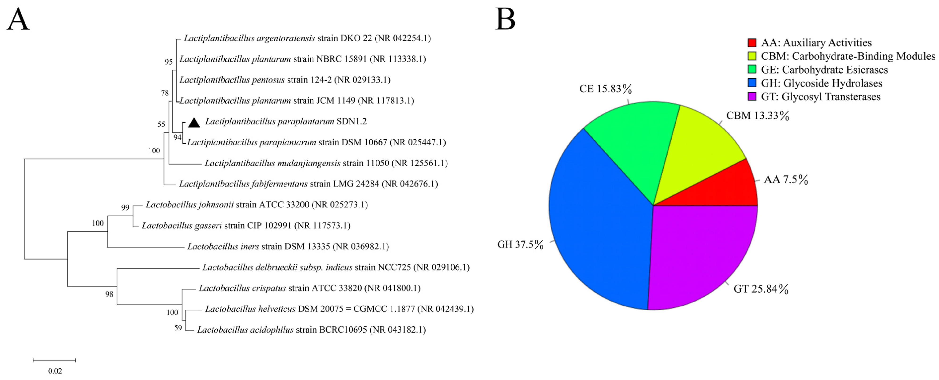
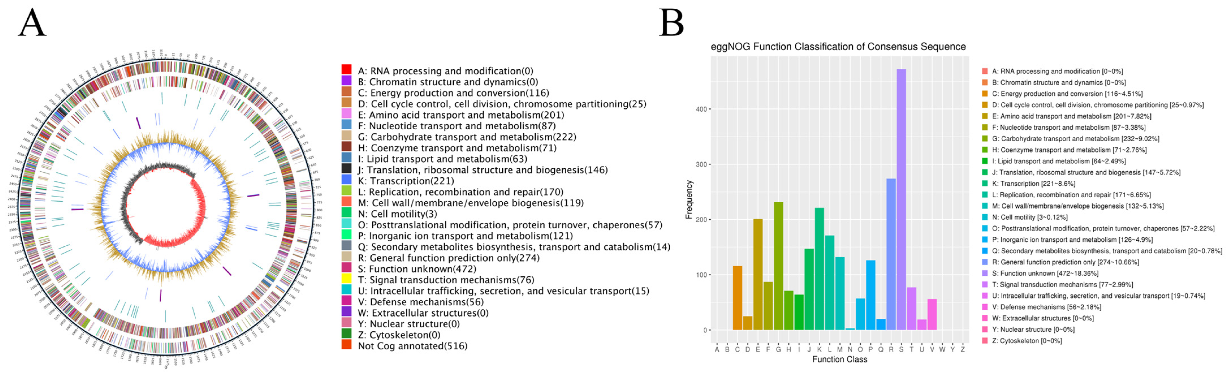
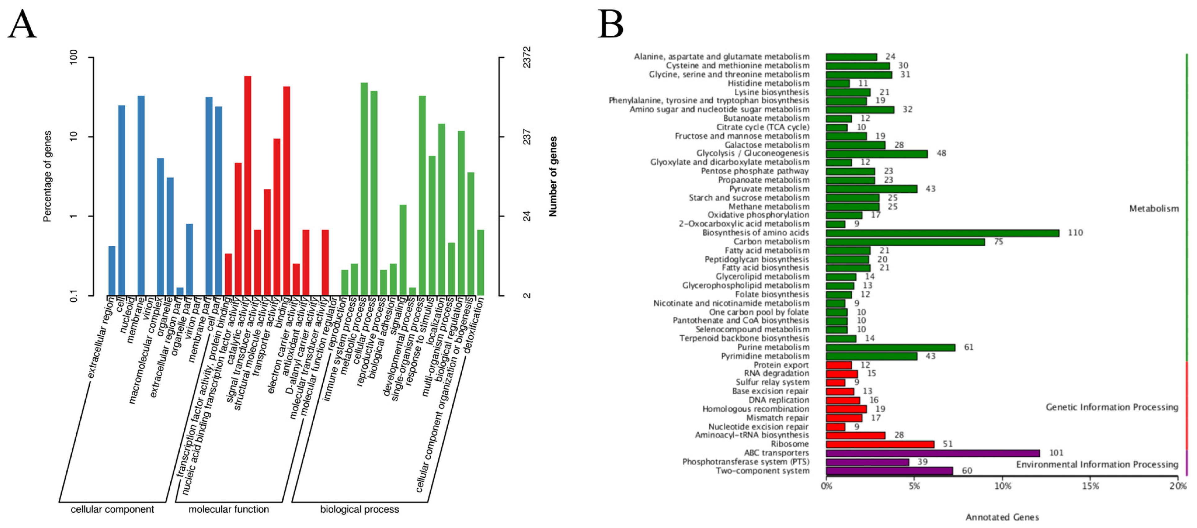
3.1. Whole-Genome Analysis of L. paraplantarum SDN1.2

The phylogenetic tree based on 16S rRNA sequences is shown in Figure 1A. The phylogenetic analysis indicated that SDN1.2 is closely related to the Lactiplantibacillus paraplantarum strain DSM 10667. Consequently, SDN1.2 was identified as L. paraplantarum and named Lactiplantibacillus paraplantarum SDN1.2.
Whole-genome sequencing was performed to explore the potential of L. paraplantarum SDN1.2. Genomic DNA was extracted using the EasyPure® Bacterial Genomic DNA Kit (TransGen Biotech, Beijing, China), with a concentration of 194.7 ng/μL and an A260/A280 ratio of 2.11, indicating high purity and minimal protein contamination (Supplementary Data S1). Figure 2A shows a complete circular genome map of L. paraplantarum SDN1.2, which comprises a single chromosome and a single plasmid. Table 2 summarizes the basic information on the genome of L. paraplantarum SDN1.2. The total length of the genome of L. paraplantarum SDN1.2 was 3,246,458 bp, with a GC content of 43.74%. The genome encodes 3045 genes, including 16 rRNAs (5S, 16S, 23S) and 69 tRNAs with an average length of 886 bp. These gene sequences span 2,699,214 bp, representing 83.14% of the total genome sequence. The KEGG, eggNOG, GO, and CAZy databases were used to analyze the gene functions of L. paraplantarum SDN1.2.
EggNOG annotation categorized the genes into 20 functional groups (Figure 2B). Carbohydrate transport and metabolism comprised 232 genes (9.02% of the annotated genes), which suggests that this host strain has a significant role in carbohydrate metabolism. A total of 132 genes (5.13% of all annotated genes) in the genome of L. paraplantarum SDN1.2 were annotated with cell wall/membrane/envelope biogenesis. These genes are likely involved in maintaining cell integrity and functionality, which may contribute to the strain’s ability to withstand environmental stresses. Fifty-six annotated genes were identified in the defense mechanism (2.18% of all annotated genes), indicating that the strain could resist the digestive tract environment and provide necessary conditions for stable colonization of bovine mammary epithelial cells.
Figure 1B displays the CAZy database annotation findings. The following five functions are annotated: carbohydrate esterases (CEs), glycoside hydrolases (GHs), glycosyl transferases (GTs), auxiliary activities (AAs), and carbohydrate-binding modules (CBMs). In that order, the percentage of functions with annotations is 37.5, 25.83, 15.83, 7.5, and 13.33%. The results indicate that the strain has a certain genetic basis and application potential in the degradation of carbohydrates such as cellulose and glycosides.
The GO database classification system covers three important activities: biological processes, molecular functions, and cellular components (Figure 3A). The top five categories include membrane (one of the 12 categories of cellular components), metabolic processes and cellular processes (two of the 15 categories of biological processes), catalytic activity and binding (two of the 12 categories of molecular activities), and more. The results of cell component annotation indicate that L. paraplantarum SDN12 has strong biofilm formation ability, which may enhance bacterial protection against the external environment.
In total, 1484 genes were annotated in the KEGG database (Figure 3B) and classified into three major categories. There were 200, 189, and 905 annotated genes associated with environmental information processing, genetic information processing, and metabolism, respectively. The biosynthesis of amino acid pathway contained the highest number of genes, with 110 genes. ABC transporters and carbon metabolism followed this, and 101 and 75 genes were annotated, respectively. The above results indicate that L. paraplantarum SDN1.2 has a strong capacity for carbohydrate metabolism, amino acid metabolism, and membrane transport.
Furthermore, a large number of antimicrobials, anti-inflammatory, and immunoregulatory-related genes and their pathway information in the KEGG database have been annotated for L. paraplantarum SDN1.2, which is shown in Table 3. The genome of L. paraplantarum SDN1.2 contains genes related to antibiotic biosynthesis: monobactam biosynthesis, streptomycin biosynthesis, and antipathogenic defense mechanisms: peptidoglycan biosynthesis, ubiquinone and other terpenoid-quinone biosynthesis, and other antimicrobial-related genes, as well as genes involved in the regulation of inflammatory mediators: arachidonic acid metabolism, secondary bile acid biosynthesis, and antioxidant and cytoprotective properties: glutathione metabolism, taurine and hypotaurine metabolism, and other anti-inflammatory related genes. It also contains genes related to immune cell metabolism and signaling: purine metabolism, arginine and proline metabolism, and symbiotic bacterial-dormitory interactions: aminoacyl-tRNA biosynthesis, vitamin B6 metabolism, and other immunomodulation-related genes.

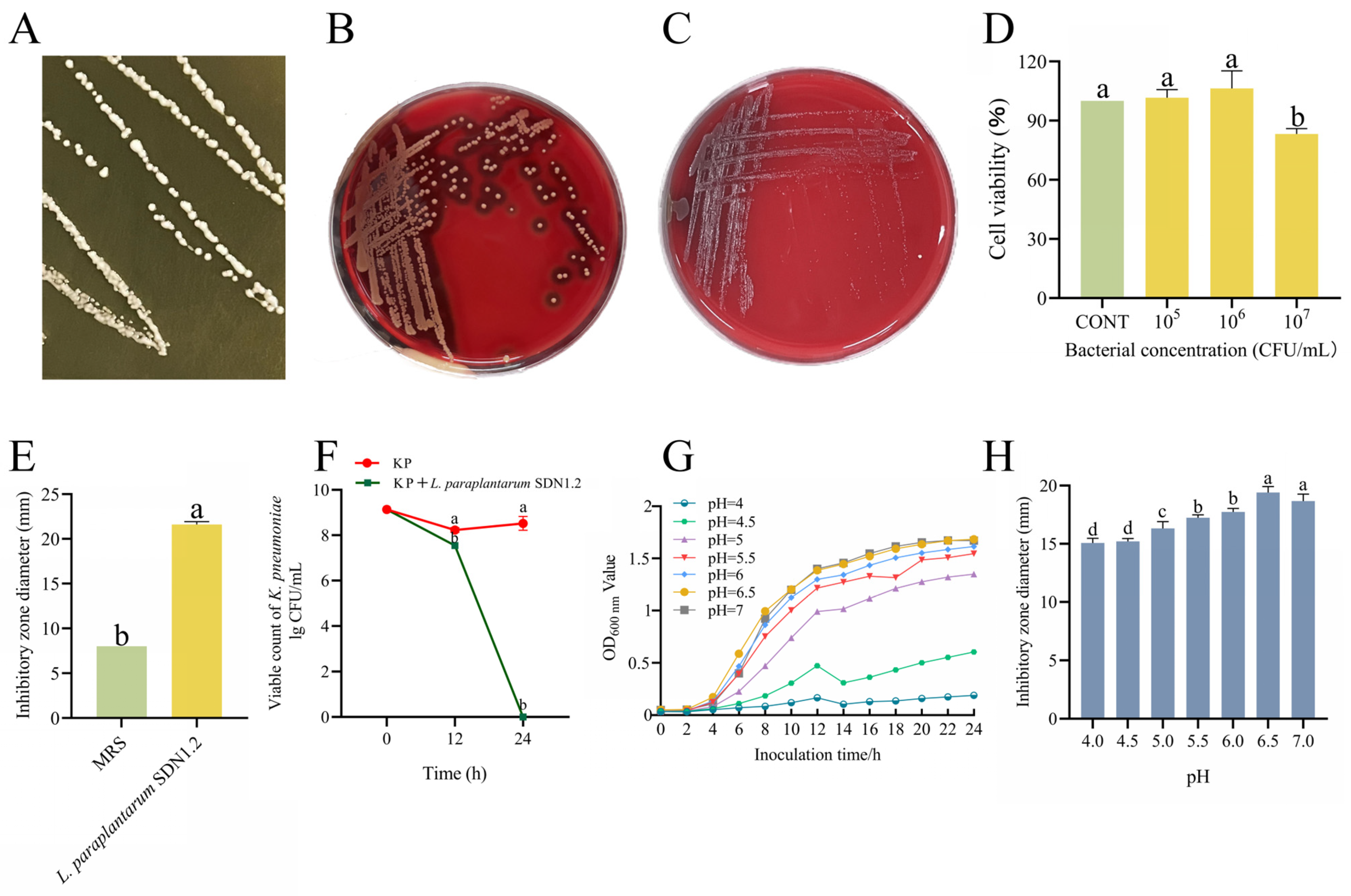
3.2. The Antibacterial Activity and Safety of L. paraplantarum SDN1.2

L. paraplantarum SDN1.2 forms small, smooth, milky round colonies in MRS agar medium (Figure 4A). In the hemolysis test, S. aureus showed a β-hemolytic zone (Figure 4B), while L. paraplantarum SDN1.2 had no hemolytic ring (Figure 4C), indicating L. paraplantarum SDN1.2 was non-hemolytic. The cytotoxicity assay results demonstrated that co-incubation of L. paraplantarum SDN1.2 at 1 × 10⁶ CFU/mL with bMECs for 12 h had no significant effect on cell viability (Figure 5D). The antibiotic resistance gene analysis of L. paraplantarum SDN1.2 was performed using CARD antibiotic resistance gene databases with identity >75%. Moreover, no resistance genes were annotated in L. paraplantarum SDN1.2.
Results of the Oxford cup experiment showed that L. paraplantarum SDN1.2 supernatant significantly inhibited the growth of K. pneumoniae (p < 0.05) (Figure 4E). K. pneumoniae counts in the co-culture group were significantly lower than the control group after both 12 and 24 h of co-cultivation (p < 0.05) (Figure 4F).
The growth curve of L. paraplantarum SDN1.2 at different pH levels is presented in Figure 4G. The growth performance of L. paraplantarum SDN1.2 is better at pH 6–7. With a continuous decrease in pH, the growth of L. paraplantarum SDN1.2 was inhibited to varying degrees. Figure 4H shows the antibacterial activity of L. paraplantarum SDN1.2 against K. pneumoniae under different pH conditions. Specifically, the maximum inhibitory diameter against K. pneumoniae is observed at pH 6.5.

3.3. L. paraplantarum SDN1.2 Ameliorates K. pneumoniae-Induced Injury to Mouse Mastitis

To confirm the effect of L. paraplantarum SDN1.2 on mastitis, we established a mouse mastitis model by injecting K. pneumoniae into the mammary ducts of mice (Figure 6A). Administration of L. paraplantarum SDN1.2 decreases the congestion and oedema of mammary tissue caused by K. pneumoniae (Figure 6B). The control group did not exhibit any bacterial colonization, as illustrated in Figure 6D. The L. paraplantarum SDN1.2 treatment groups had significantly lower bacterial loads in mammary tissue (p < 0.05) when compared to the KP group. H&E staining revealed that after the K. pneumoniae stimulation, infiltration of inflammatory cells and pathological damage were found in the mammary tissues. However, these changes were improved in the KP + L. paraplantarum SDN1.2 group, with a significant decrease in inflammation scores (p < 0.05) (Figure 6C,E). The MPO test also demonstrated that adding L. paraplantarum SDN1.2 may considerably lower the infiltration of inflammatory cells by K. pneumoniae (p < 0.05) (Figure 6F).

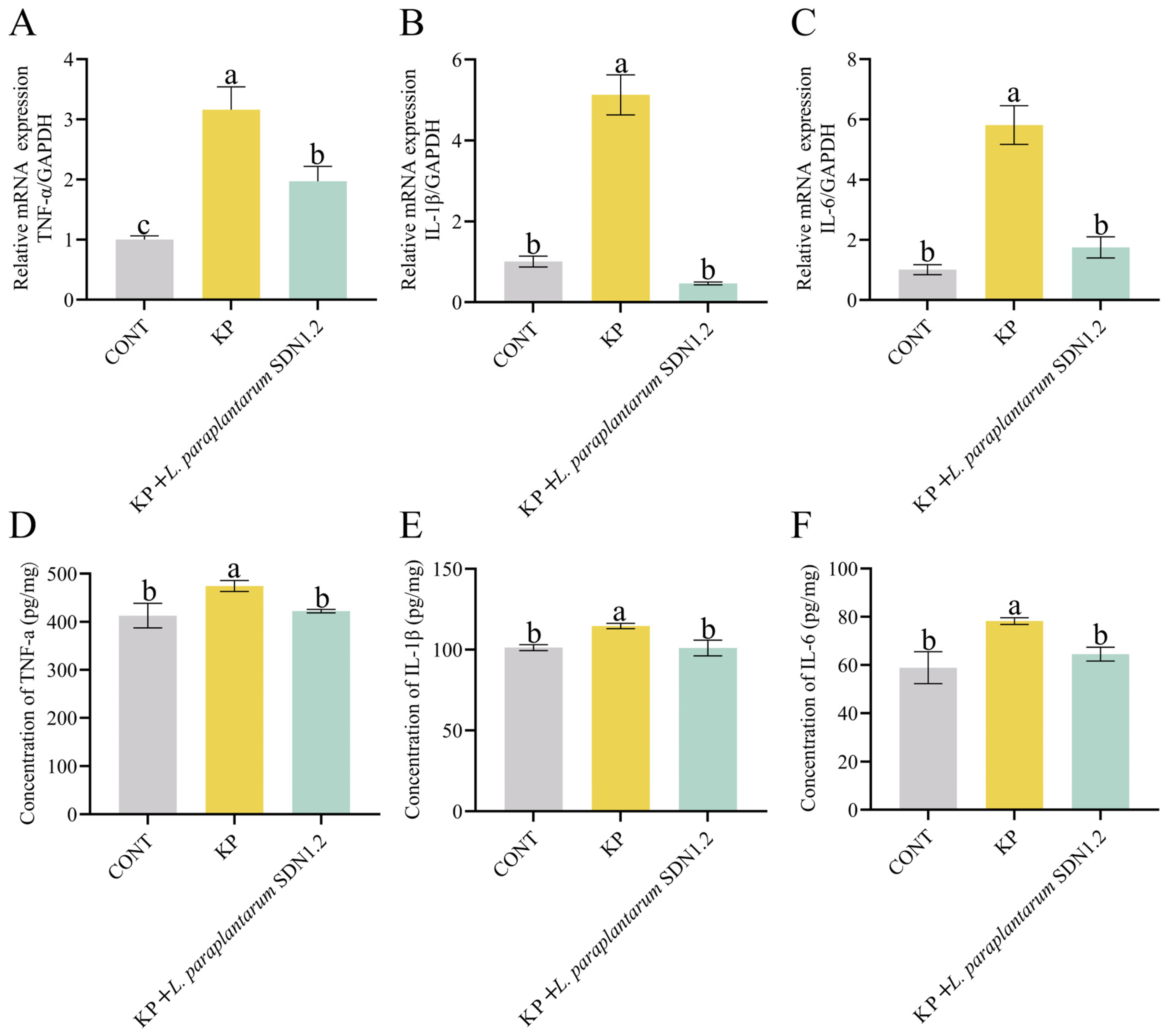
3.4. L. paraplantarum SDN1.2 Attenuates K. pneumoniae-Induced Inflammation in Mouse Mastitis

The mRNA expression levels of IL-1β, IL-6, and TNF-α in mammary tissues were measured using qRT–PCR. As shown in Figure 5A–C, the mRNA expression levels of IL-1β, IL-6, and TNF-α were higher in the KP group than in the CONT group (p < 0.05). The expression levels of IL-1β, IL-6, and TNF-α were lower in the KP + L. paraplantarum SDN1.2 group than in the KP group (p < 0.05).
IL-1β, IL-6, and TNF-α expression levels in mammary tissues were examined by ELISA. As shown in Figure 5D–F, the expression levels of IL-1β, IL-6, and TNF-α were higher in the KP group than in the CONT group (p < 0.05). The expression levels of IL-1β, IL-6, and TNF-α were lower in the KP + L. paraplantarum SDN1.2 group than in the KP group (p < 0.05). The ELISA and qRT–PCR results were consistent.

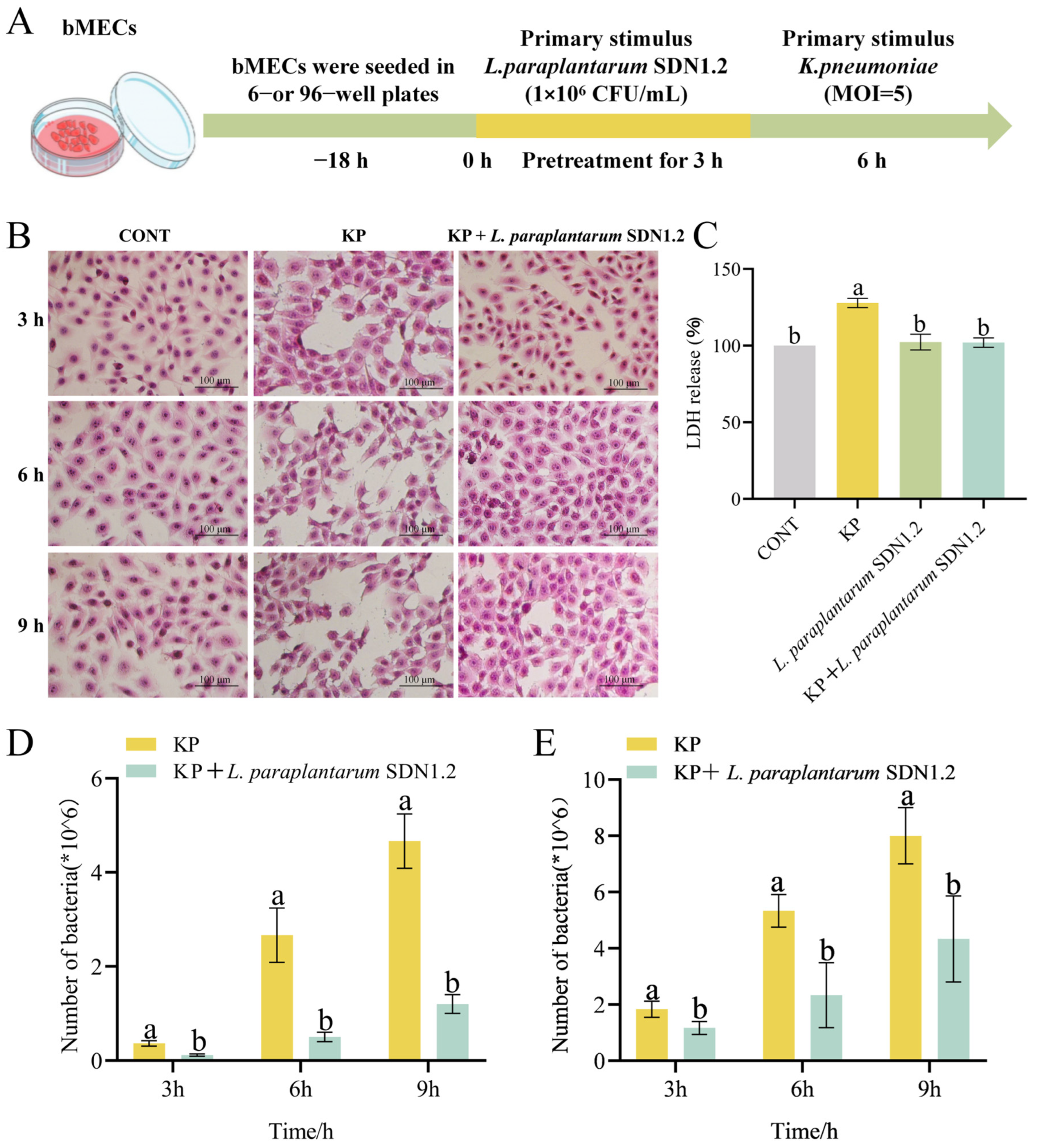
3.5. L. paraplantarum SDN1.2 Reduces the Cytotoxic Effects of bMECs in K. pneumoniae Infection

To evaluate the anti-inflammatory effects of L. paraplantarum SDN1.2 in vitro, we established a cellular mastitis model by challenging bMECs with K. pneumoniae (Figure 7A). Cell morphology and LDH assay were used to assess the cytotoxic effects of K. pneumoniae after incubation with L. paraplantarum SDN1.2 in bMECs. K. pneumoniae-infected bMECs exhibited morphological changes at 3 h post-infection (hpi), such as cellular enlargement (swelling), hyperchromatic nuclei (hyper-staining), and evidence of cell death (necrotic cell death). Furthermore, a reduction in bMEC number was observed. The extent of bMEC injury progressively increased with time. However, the group which had been pretreated with L. paraplantarum SDN1.2 showed significantly reduced swelling and necrosis in bMECs (Figure 7B). Regarding K. pneumoniae-infected bMECs, the KP group exhibited a significant increase in LDH release in comparison to the CONT group (p < 0.05), while the L. paraplantarum SDN1.2 group did not exhibit any significant change (p > 0.05). The LDH release of the SND1.2 + KP group was significantly lower in comparison to the KP group (p < 0.05) (Figure 7C).

3.6. L. paraplantarum SDN 1.2 Reduce Adhesion and Invasion of K. pneumoniae to bMECs

The finding of the current study showed that at three time points (3, 6, and 9 h), L. paraplantarum SDN1.2 significantly reduced the adhesion and invasion rate of K. pneumoniae in bMECs (p < 0.05) (Figure 7D,E).

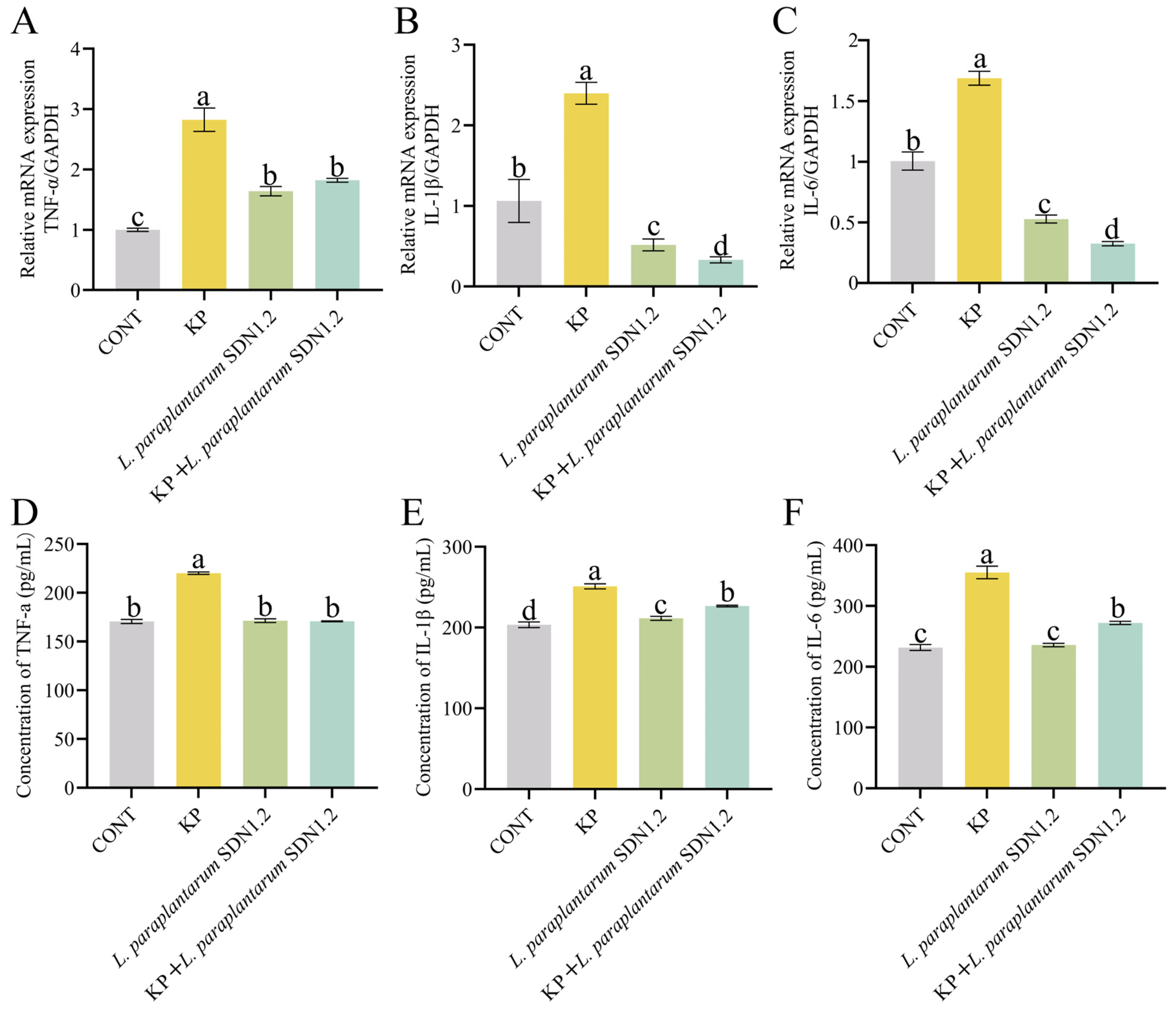
3.7. L. paraplantarum SDN1.2 Inhibits the Inflammatory Response of bMECs Infected with K. pneumoniae

K. pneumoniae substantially increased the mRNA expression levels of IL-6, IL-1β, and TNF-α (the KP group vs. the CONT group) in terms of anti-inflammatory potential (p < 0.05). On the other hand, the KP + L. paraplantarum SDN1.2 group, compared to the KP group, showed a significant decrease in the expression of these inflammatory markers (p < 0.05) (Figure 8A–C).
Following a K. pneumoniae infection, the supernatant’s levels of the inflammatory cytokines TNF-α, IL-1β, and IL-6 were significantly elevated compared to those in the CONT group (p < 0.05). Comparing the cells with L. paraplantarum SDN1.2 before and after the treatment group to those with K. pneumoniae-infected bMECs, the amounts of the cytokines TNF-α, IL-1β, and IL-6 were significantly lower (p < 0.05) (Figure 8D–F). According to the findings, L. paraplantarum SDN1.2 exerts anti-inflammatory effects in bMECs by reducing the expression of inflammatory factors produced by K. pneumoniae.

4. Discussion

K. pneumoniae is regarded as one of the principal causative agents of bovine mastitis, causing significant economic losses in the dairy sector and posing significant public health risks over the past decades [10]. The severe inflammation associated with K. pneumoniae mastitis is largely driven by the lipopolysaccharide (LPS) component of its cell wall. As a major constituent of the outer membrane of Gram-negative bacteria, LPS is known to trigger a robust immune response through recognition by Toll-like receptor 4 (TLR4) on host immune cells [19]. This interaction activates downstream signaling pathways, such as NF-κB and MAPK, leading to the production of pro-inflammatory cytokines (e.g., TNF-α, IL-1β, and IL-6) and chemokines [20]. In the context of mastitis, LPS from K. pneumoniae induces neutrophil infiltration, tissue damage, and systemic inflammatory responses, exacerbating the clinical manifestations of severe udder inflammation [21]. Furthermore, the structural variability of LPS, particularly the O-antigen, may influence the virulence and immune evasion strategies of K. pneumoniae strains, potentially affecting the severity of mastitis [22]. Currently, antibiotics are the primary therapies to treat K. pneumoniae mastitis. However, overuse of antibiotics and the slow development of novel antimicrobial agents are significant factors for the emergence of multidrug-resistant K. pneumoniae strains. Therefore, this critical situation demands urgent exploration of alternative treatment and prevention strategies. L. paraplantarum strains are gaining significant interest as potential antimicrobial alternatives due to their antimicrobial activity and immunomodulatory response [23]. To evaluate the potential of L. paraplantarum against K. pneumoniae infection, this study was designed. Our results demonstrate that L. paraplantarum SDN1.2 exhibited excellent biosafety, antibacterial activity, and anti-inflammatory properties against K. pneumoniae. However, the exact mechanism underlying the protective effects of L. paraplantarum SDN1.2 against K. pneumoniae mastitis requires further investigation.
Gene annotation enables the quick capture of relevant functional genes and safety data [24]. Thus, genome sequencing of L. paraplantarum SDN1.2 was carried out to assess its safety and anti-inflammatory potential using genes and genomes. Resistance assessment and hemolysis testing are significant strain screening parameters in the probiotic safety review procedure. The results of the testing revealed that L. paraplantarum SDN1.2 was not hemolytic and was not cytotoxic. Antibiotics are frequently used to kill invasive pathogens or inhibit their replication to control the spread of infection within the host. Antibiotic resistance ensures a probiotic’s survival within the host [25]. However, probiotics should be sensitive to at least two antibiotics or not carry intrinsic antimicrobial resistance genes, to reduce the risk of transmission. This investigation annotated target protein sequences using a BLAST-based CARD database based on whole-genome sequencing data from L. paraplantarum SDN1.2, and no resistance genes were identified. While drug susceptibility testing was not performed in the current study, our previous investigations demonstrated that L. paraplantarum SDN1.2 exhibited sensitivity to most antibiotics in standardized susceptibility assays [26]. Comprehensive hemolysis tests, drug sensitivity tests, and screening for resistance genes showed that the strain L. paraplantarum SDN1.2 was safe as a potential probiotic. In this work, the genome of L. paraplantarum SDN1.2 comprises genes encoding glycoside hydrolases (GHs) and glycosyl transferases (GTs). Glycoside hydrolases are catabolic enzymes of carbohydrate metabolic pathways, including glycosidases, which hydrolyze glycosidic bonds. The glycosidase glycosyl transferases are able to create glycosidic bonds from sugar donors having nucleoside phosphate or lipid phosphate leaving groups [27].
L. paraplantarum strains with antimicrobial properties generate significant interest in veterinary science and the livestock industry. Apart from its potential in veterinary medicine, L. paraplantarum has proven to be an important probiotic in human health. Previous studies have demonstrated that probiotics are effective in treating metabolic syndrome, diabetes, obesity, and gastrointestinal disorders [28]. It has been reported that L. paraplantarum 11 has broad-spectrum antimicrobial activity [29]. Our study shows that L. paraplantarum SDN1.2 exhibited excellent antibacterial activity against K. pneumoniae. Similarly, an already-reported investigation indicated that the cell-free supernatant of L. plantarum had a high antibacterial activity against numerous pathogenic enterobacteria (E. coli, Shigella flexneri, Salmonella typhimurium, Proteus mirabilis, and Campylobacter jejuni) [30]. Furthermore, Lactobacillus is known for its wide range of antimicrobial activities. It produces a variety of antimicrobial components, including organic acids, hydrogen peroxide, diacetyl, bacteriocins (plantaricins), and antimicrobial peptides, which are effective against a wide range of pathogenic microbes. Lactic acid bacteria are non-spore-forming, Gram-positive bacteria that can ferment sugar to produce a variety of organic acids [31]. A fall in pH can drastically reduce the development of other microorganisms. Furthermore, several investigations have indicated that H2O2, created during the metabolic process, can suppress microorganisms [32]. Therefore, we concluded that L. paraplantarum SDN1.2 may exert antimicrobial effects by producing some organic acids. Microorganisms produce specific proteins called secreted proteins during their life. These proteins can kill other organisms, enhancing environmental survival capabilities [33]. The host genomic sequence analysis showed that L. paraplantarum SDN1.2 84 potentially secreted proteins. These proteins may play a role in bacterium interactions with its environment, and some might have antimicrobial properties. Therefore, our future study objective is to determine the contribution of these secreted proteins to L. paraplantarum’s antimicrobial effects.
Controlling bacterial infections requires minimizing bacterial colonization. In this investigation, we discovered that udder tissues were highly colonized by bacteria, which is consistent with prior findings [34]. Supplementation with L. paraplantarum SDN1.2 effectively reduced bacterial colonization. Histopathological examination showed extensive infiltration of inflammatory cells and necrotic detached mammary epithelial cells in severely damaged mammary tissue. However, treatment with L. paraplantarum SDN1.2 decreased inflammatory cell infiltration and tissue damage. MPO is a marker for neutrophil invasion [35]. MPO levels in mammary tissue rose following K. pneumoniae infection, whereas supplementation with L. paraplantarum SDN1.2 significantly reduced MPO activity. As a result, supplementation with L. paraplantarum SDN1.2 can diminish inflammatory cell infiltration, hence alleviating the inflammatory response.
bMECs play a vital role in innate immunity, secreting cytokines like IL-1β, IL-6, and TNF-α, as well as enzymes like iNOS and COX-2 during mammary gland inflammation. This release supports the recruitment of immune cells, such as neutrophils and macrophages, for pathogenic microorganism elimination [36]. Maintaining proper amounts of inflammatory cytokines is critical for an efficient immune response against infections; however, excessive production exacerbates inflammation and worsens udder damage [37]. Consequently, limiting the excessive release of inflammatory mediators during mammary gland inflammation is critical. Our findings show that L. paraplantarum SDN1.2 effectively reduces the release of these mediators both in vivo and in vitro, thereby reducing K. pneumoniae-induced mastitis. This anti-inflammatory effect is consistent with previous studies on lactic acid bacteria strains. For instance, L. paraplantarum CRL 2051 reduces the production of pro-inflammatory cytokines such as TNF-α and IL-6 in a mouse model of metabolic disorders, highlighting its potential as an immunomodulator [38]. In the context of mastitis, L. plantarum KLDS 1.0344 has been reported to mitigate Escherichia coli-induced inflammation in bovine mammary epithelial cells by downregulating NF-κB-mediated signaling [14]. Additionally, the inhibition of L. paraplantarum SDN1.2-mediated inflammatory response was associated with the protection of bMECs from cytotoxicity. Lactate dehydrogenase (LDH) is a key indicator of bacterial cytotoxicity [39]. In this study, K. pneumoniae infection significantly increased LDH release from bMECs, indicating cellular damage and intracellular enzyme leakage. However, preincubation with L. paraplantarum SDN1.2 substantially reduced the elevated LDH release to normal levels, suggesting its protective effect against K. pneumoniae-induced cytotoxicity. This finding aligns with previous reports that L. plantarum strains can enhance epithelial barrier function and reduce cellular damage in inflammatory conditions [40].
Although probiotics have been proposed as alternatives in treating bovine mastitis, their limitations are well documented. Studies have shown that probiotics provide temporary protection for the breast by reducing the risk of infection and the severity of inflammation but do not achieve a cure for mastitis [41,42]. This transient efficacy highlights their role as a potential adjunct rather than a stand-alone solution for treating mastitis. However, probiotics might serve as an alternative to vaccination trials. Notably, probiotics may serve as a temporary alternative to vaccines in clinical trials but face challenges in inducing long-term adaptive immunity against multiple K. pneumoniae strains [43]. Despite these challenges, their capacity to transiently modulate immune responses and reduce acute infection highlights their potential as complementary tools in mastitis management.

5. Conclusions

In this study, we investigated the probiotic properties and anti-inflammatory capacity of L. paraplantarum SDN1.2. Through whole-genome sequencing analysis combined with in vitro experiments, we confirmed the biosafety of this strain and demonstrated an antimicrobial capacity. Furthermore, L. paraplantarum SDN1.2 reduced both the adhesion and invasion of K. pneumoniae in both in vitro and in vivo assays, and it attenuated K. pneumoniae-induced inflammatory responses by inhibiting the expression of inflammatory factors. This study provides evidence that L. paraplantarum ameliorates mastitis pathology and establishes a foundation for its potential application as a potential mastitis prevention strategy.

Supplementary Materials

The following supporting information can be downloaded at: https://www.mdpi.com/article/10.3390/vetsci12040323/s1, Supplementary Data S1: PRISMA 2020 Checklist; Supplementary Data S2: Nanodrop curve. Reference [44] is cited in Supplementary Materials.

Author Contributions

Conceptualization, M.L. and J.C.; methodology, J.C., J.T., C.L. and Z.W.; software, J.C. and J.T.; validation, J.C., J.T., C.L., Z.W. and H.L.; formal analysis, J.C., J.T., C.L., Z.W, H.L., M.R. and J.S.; investigation, J.C., J.T., C.L., Z.W., H.L., M.R. and J.S.; resources, M.L., D.S. and Q.X.; data curation, M.L., M.R., J.S. and D.S.; writing—original draft preparation, J.C. and J.T.; writing—review and editing, J.C., M.L., J.T., C.L., Z.W., H.L., M.R. and J.S.; visualization, J.C. and J.T.; supervision, M.L. and J.C; funding acquisition, M.L. All authors have read and agreed to the published version of the manuscript.

Funding

This research was funded by the biological agriculture joint fund of the Natural Science Foundation of Hebei Province (No. C2023204074), the Basic Scientific Research Foundation of Hebei Province (No. 1081002240), and the Natural Science Foundation of Hebei Agricultural University (No. 3118133).

Institutional Review Board Statement

The study protocol was approved by the Animal Care and Ethics Committee of Hebei Agricultural University (approval ID: 2023089).

Informed Consent Statement

Not applicable.

Data Availability Statement

The data presented in this study are contained within the article.

Acknowledgments

The authors greatly acknowledge the College of Animal Medicine, Hebei Agricultural University, for the use of experimental facilities.

Conflicts of Interest

The authors declare no conflicts of interest.

References

  1. Ashraf, A.; Imran, M. Causes, types, etiological agents, prevalence, diagnosis, treatment, prevention, effects on human health and future aspects of bovine mastitis. Anim. Health Res. Rev. 2020, 21, 36–49. [Google Scholar] [PubMed]
  2. Schukken, Y.; Chuff, M.; Moroni, P.; Gurjar, A.; Santisteban, C.; Welcome, F.; Zadoks, R. The “other” gram-negative bacteria in mastitis: Klebsiella, Serratia, and more. Vet. Clin. N. Am. Food Anim. Pract. 2020, 28, 239–256. [Google Scholar]
  3. Hu, Y.; Anes, J.; Devineau, S.; Fanning, S. Klebsiella pneumoniae: Prevalence, Reservoirs, Antimicrobial Resistance, Pathogenicity, and Infection: A Hitherto Unrecognized Zoonotic Bacterium. Foodborne Pathog. Dis. 2021, 18, 63–84. [Google Scholar]
  4. Fuenzalida, M.J.; Ruegg, P.L. Negatively-controlled, randomized clinical trial to evaluate intramammary treatment of nonsevere, gramnegative clinical mastitis. J. Dairy Sci. 2019, 102, 5438–5457. [Google Scholar]
  5. Yang, J.; Xiong, Y.; Barkema, H.W.; Tong, X.; Lin, Y.; Deng, Z.; Kastelic, J.P.; Nobrega, D.B.; Wang, Y.; Han, B.; et al. Comparative genomic analyses of Klebsiella pneumoniae K57 capsule serotypes isolated from bovine mastitis in China. J. Dairy Sci. 2024, 107, 3114–3126. [Google Scholar]
  6. Cheng, J.; Zhang, J.; Han, B.; Barkema, H.W.; Cobo, E.R.; Kastelic, J.P.; Zhou, M.; Shi, Y.; Wang, J.; Yang, R.; et al. Klebsiella pneumoniae isolated from bovine mastitis is cytopathogenic for bovine mammary epithelial cells. J. Dairy Sci. 2020, 103, 3493–3504. [Google Scholar] [CrossRef]
  7. Angelopoulou, A.; Warda, A.K.; Hill, C.; Ross, R.P. Non-antibiotic microbial solutions for bovine mastitis—Live biotherapeutics, bacteriophage, and phage lysins. Crit. Rev. Microbiol. 2019, 45, 564–580. [Google Scholar]
  8. Pal, C.; Bengtsson-Palme, J.; Kristiansson, E.; Larsson, D.G. The structure and diversity of human, animal and environmental resistomes. Microbiome 2016, 4, 54. [Google Scholar]
  9. Leite de Campos, J.; Gonçalves, J.L.; Kates, A.; Steinberger, A.; Sethi, A.; Suen, G.; Shutske, J.; Safdar, N.; Goldberg, T.; Ruegg, P.L. Variation in partial direct costs of treating clinical mastitis among 37 Wisconsin dairy farms. J. Dairy Sci. 2023, 106, 9276–9286. [Google Scholar]
  10. Ziesch, M.; Wente, N.; Zhang, Y.; Zaremba, W.; Engl, S.; Kromker, V. Noninferiority Trial Investigating the Efficacy of a Nonantibiotic Intramammary Therapy in the Treatment of Mild-to-Moderate Clinical Mastitis in Dairy Cows With Longer Lasting Udder Diseases. J. Vet. Pharmacol. Ther. 2018, 41, 11–21. [Google Scholar]
  11. Hoque, M.N.; Moyna, Z.; Faisal, G.M.; Das, Z.C.; Islam, T.; Newton, I.L.G. Whole-genome sequence of multidrug-resistant Klebsiella pneumoniae MNH_G2C5, isolated from bovine clinical mastitis milk. Microbiol. Resour. Announc. 2023, 12, e0007923. [Google Scholar] [CrossRef] [PubMed]
  12. Pineiro, M.; Stanton, C. Probiotic bacteria: Legislative framework—Requirements to evidence basis. J. Nutr. 2007, 137, 850S–853S. [Google Scholar] [CrossRef] [PubMed]
  13. Burkholder, K.M.; Fletcher, D.H.; Gileau, L.; Kandolo, A. Lactic acid bacteria decrease Salmonella enterica Javiana virulence and modulate host inflammation during infection of an intestinal epithelial cell line. Pathog. Dis. 2019, 77, ftz025. [Google Scholar] [CrossRef] [PubMed]
  14. Chen, Q.; Wang, S.; Guo, J.; Xie, Q.; Evivie, S.E.; Song, Y.; Li, B.; Huo, G. The Protective Effects of Lactobacillus plantarum KLDS 1.0344 on LPS-Induced Mastitis In Vitro and In Vivo. Front. Immunol. 2021, 12, 770822. [Google Scholar] [CrossRef] [PubMed]
  15. Yue, Y.; He, Z.; Zhou, Y.; Ross, R.P.; Stanton, C.; Zhao, J.; Zhang, H.; Yang, B.; Chen, W. Lactobacillus plantarum relieves diarrhea caused by enterotoxin-producing Escherichia coli through inflammation modulation and gut microbiota regulation. Food Funct. 2020, 11, 10362–10374. [Google Scholar] [CrossRef]
  16. Zhang, H.; HuangFu, H.; Wang, X.; Zhao, S.; Liu, Y.; Lv, H.; Qin, G.; Tan, Z. Activity of Lactic Acid Producing Leuconostoc mesenteroides QZ1178 Against Pathogenic Gallibacterium anatis. Front. Vet. Sci. 2021, 8, 630294. [Google Scholar] [CrossRef]
  17. Tsai, C.C.; Lai, T.M.; Hsieh, Y.M. Evaluation of Lactobacilli for Antagonistic Activity Against the Growth, Adhesion and Invasion of Klebsiella pneumoniae and Gardnerella vaginalis. Indian J. Microbiol. 2019, 59, 81–89. [Google Scholar] [CrossRef]
  18. Guo, W.; Liu, B.; Hu, G.; Kan, X.; Li, Y.; Gong, Q.; Xu, D.; Ma, H.; Cao, Y.; Huang, B.; et al. Vanillin protects the blood-milk barrier and inhibits the inflammatory response in LPS-induced mastitis in mice. Toxicol. Appl. Pharmacol. 2019, 365, 9–18. [Google Scholar] [CrossRef]
  19. Park, B.S.; Lee, J.O. Recognition of lipopolysaccharide pattern by TLR4 complexes. Immunol. Rev. 2013, 250, 143–153. [Google Scholar] [CrossRef]
  20. Liu, Y.; Chen, W.; Zheng, F.; Yu, H.; Wei, K. Xanthatin Alleviates LPS-Induced Inflammatory Response in RAW264.7 Macrophages by Inhibiting NF-κB, MAPK and STATs Activation. Molecules 2022, 27, 4603. [Google Scholar] [CrossRef]
  21. Burvenich, C.; Van Merris, V.; Mehrzad, J.; Diez-Fraile, A.; Duchateau, L. Severity of E. coli mastitis is mainly determined by cow factors. Vet. Res. 2003, 34, 521–564. [Google Scholar] [PubMed]
  22. Singh, S.; Wilksch, J.J.; Dunstan, R.A.; Mularski, A.; Wang, N.; Hocking, D.; Jebeli, L.; Cao, H.; Clements, A.; Jenney, A.W.J.; et al. LPS O Antigen Plays a Key Role in Klebsiella pneumoniae Capsule Retention. Microbiol. Spectr. 2022, 10, e0151721. [Google Scholar] [CrossRef] [PubMed]
  23. Li, K.; Yang, M.; Tian, M.; Jia, L.; Du, J.; Wu, Y.; Li, L.; Yuan, L.; Ma, Y. Lactobacillus plantarum 17-5 attenuates Escherichia coli-induced inflammatory responses via inhibiting the activation of the NF-κB and MAPK signalling pathways in bovine mammary epithelial cells. BMC Vet. Res. 2022, 18, 250. [Google Scholar] [CrossRef] [PubMed]
  24. Chen, Y.; Chen, M.; Jin, J.; Bai, Y.; Wang, Q.; Liao, Z.; Chen, Q. Isolation, identification, and whole genome sequence analysis of the alginate-degrading bacterium Cobetia sp. cqz5-12. Sci. Rep. 2020, 10, 10920. [Google Scholar]
  25. Holzapfel, W.; Arini, A.; Aeschbacher, M.; Coppolecchia, R.; Pot, B. Enterococcus faecium SF68 as a model for efficacy and safety evaluation of pharmaceutical probiotics. Benef. Microbes. 2018, 9, 375–388. [Google Scholar]
  26. Jin, T.; Feng, M.; Liang, H.; Liu, K.; Tong, J.; Ren, M.; Jiang, G.; Gao, J.; Liu, M.; Cheng, J. Isolation and Identification of 3 Lactic Acid Bacteria and Their Antibacterial Activity against Klebsiella pneumoniae Isolated from Bovine Mastitis. Chin. J. Vet. Med. 2025, 01, 013. [Google Scholar]
  27. Cuyvers, S.; Dornez, E.; Delcour, J.A.; Courtin, C.M. Occurrence and functional significance of secondary carbohydrate binding sites in glycoside hydrolases. Crit. Rev. Biotechnol. 2012, 2, 93–107. [Google Scholar]
  28. Liu, Y.W.; Liong, M.T.; Tsai, Y.C. New perspectives of Lactobacillus plantarum as a probiotic: The gut-heart-brain axis. J. Microbiol. 2018, 56, 601–613. [Google Scholar]
  29. Kalhoro, M.S.; Anal, A.K.; Kalhoro, D.H.; Hussain, T.; Murtaza, G.; Mangi, M.H. Antimicrobial Activities and Biopreservation Potential of Lactic Acid Bacteria (LAB) from Raw Buffalo (Bubalus bubalis) Milk. Oxidative Med. Cell. Longev. 2023, 2023, 8475995. [Google Scholar] [CrossRef]
  30. Danilova, T.A.; Adzhieva, A.A.; Mezentseva, M.V.; Suetina, I.A.; Danilina, G.A.; Minko, A.G.; Dmitrieva, M.L.; Zhukhovitsky, V.G. The inhibitory activity of Lactobacillus plantarum supernatant against Enterobacteria, Campylobacter, and tumor cells. Bull. Exp. Biol. Med. 2023, 176, 64–67. [Google Scholar] [CrossRef]
  31. Punia, B.S.; Suri, S.; Trif, M.; Ozogul, F. Organic acids production from lactic acid bacteria: A preservation approach. Food Biosci. 2022, 46, 101615. [Google Scholar]
  32. Pino, A.; Vaccalluzzo, A.; Caggia, C.; Balzaretti, S.; Vanella, L.; Sorrenti, V.; Ronkainen, A.; Satokari, R.; Randazzo, C.L. Lacticaseibacillus rhamnosus CA15 (DSM 33960) as a Candidate Probiotic Strain for Human Health. Nutrients 2022, 14, 4902. [Google Scholar] [CrossRef]
  33. Chernyatina, A.A.; Low, H.H. Core architecture of a bacterial type II secretion system. Nat. Commun. 2019, 10, 5437. [Google Scholar] [PubMed]
  34. Qiu, M.; Feng, L.; Yu, Z.; Zhao, C.; Gao, S.; Bao, L.; Zhang, N.; Fu, Y.; Hu, X. Probiotic Enterococcus mundtii H81 inhibits the NF-κB signaling pathway to ameliorate Staphylococcus aureus-induced mastitis in mice. Microb. Pathog. 2022, 164, 105414. [Google Scholar]
  35. Lin, W.; Chen, H.; Chen, X.; Guo, C. The Roles of Neutrophil-Derived Myeloperoxidase (MPO) in Diseases: The New Progress. Antioxidants 2024, 13, 132. [Google Scholar] [CrossRef]
  36. Sumaiya, K.; Langford, D.; Natarajaseenivasan, K.; Shanmughapriya, S. Macrophage migration inhibitory factor (MIF): A multifaceted cytokine regulated by genetic and physiological strategies. Pharmacol. Ther. 2022, 233, 108024. [Google Scholar]
  37. Bhol, N.K.; Bhanjadeo, M.M.; Singh, A.K.; Dash, U.C.; Ojha, R.R.; Majhi, S.; Duttaroy, A.K.; Jena, A.B. The interplay between cytokines, inflammation, and antioxidants: Mechanistic insights and therapeutic potentials of various antioxidants and anti-cytokine compounds. Biomed. Pharmacother. 2024, 178, 117177. [Google Scholar]
  38. Isas, A.S.; Balcells, M.F.; Maldonado Galdeano, C.; Palomo, I.; Rodriguez, L.; Fuentes, E.; Luna Pizarro, P.; Mateos Briz, R.; Mozzi, F.; Van Nieuwenhove, C. Fermented pomegranate juice enriched with pomegranate seed oil ameliorates metabolic disorders associated with a high-fat diet in C57BL/6 mice. Food Chem. 2025, 463, 141434. [Google Scholar]
  39. Wei, Z.; Wang, J.; Wang, Y.; Wang, C.; Liu, X.; Han, Z.; Fu, Y.; Zhang, N. Effects of neutrophil extracellular traps on bovine mammary epithelial cells in vitro. Front. Immunol. 2019, 10, 1003. [Google Scholar]
  40. Zhou, Y.; Wang, B.; Wang, Q.; Tang, L.; Zou, P.; Zeng, Z.; Zhang, H.; Gong, L.; Li, W. Protective Effects of Lactobacillus plantarum Lac16 on Clostridium perfringens Infection-Associated Injury in IPEC-J2 Cells. Int. J. Mol. Sci. 2021, 22, 12388. [Google Scholar] [CrossRef]
  41. Rainard, P.; Foucrasm, G. A Critical Appraisal of Probiotics for Mastitis Control. Front. Vet. Sci. 2018, 5, 251. [Google Scholar]
  42. Yu, Q.; Xu, C.; Wang, M.; Zhu, J.; Yu, L.; Yang, Z.; Liu, S.; Gao, X. The preventive and therapeutic effects of probiotics on mastitis: A systematic review and meta-analysis. PLoS ONE 2022, 17, e0274467. [Google Scholar]
  43. Petzl, W.; Zerbe, H.; Günther, J.; Seyfert, H.M.; Hussen, J.; Schuberth, H.J. Pathogen-specific responses in the bovine udder. Models and immunoprophylactic concepts. Res. Vet. Sci. 2018, 116, 55–61. [Google Scholar] [PubMed]
  44. Page, M.J.; McKenzie, J.E.; Bossuyt, P.M.; Boutron, I.; Hoffmann, T.C.; Mulrow, C.D.; Shamseer, L.; Tetzlaff, J.M..; Akl, E.A.; Brennan, S.E.; et al. The PRISMA 2020 statement: An updated guideline for reporting systematic reviews. BMJ 2021, 372, n71. [Google Scholar] [CrossRef]
Figure 1. (A) phylogenetic tree. (B) Carbohydrate-active enzyme (CAZy) analysis.
Vetsci 12 00323 g001
Figure 2. (A) A circular map of the chromosome. (B) Non-supervised orthologous groups (eggNOG) functional classification.
Vetsci 12 00323 g002
Figure 3. (A) Gene Ontology (GO) analysis. (B) Kyoto Encyclopedia of Genes and Genomes (KEGG) pathway enrichment.
Vetsci 12 00323 g003
Figure 4. The growth characterization and antibacterial activity of L. paraplantarum SDN1.2. (A) Morphology of L. paraplantarum SDN1.2 on MRS agar medium. (B,C) Hemolytic test of S. aureus and L. paraplantarum SDN1.2. (D) Cytotoxicity test. (E) Antibacterial results of L. paraplantarum SDN1.2 and K. pneumoniae Oxford cup. (F) The results of co-culture of L. paraplantarum SDN1.2 and K. pneumoniae (KP). (G) Antimicrobial effect of L. paraplantarum SDN1.2 on K. pneumoniae under different pH conditions. (H) L. paraplantarum SDN1.2 growth curves under different pH conditions. The mean ± SEM was used for all data presentations. Different lowercase letters mean significant differences (p < 0.05).
Vetsci 12 00323 g004
Figure 5. L. paraplantarum SDN1.2 attenuates K. pneumoniae-induced inflammation in mouse mastitis. (AC) Mammary tissue homogenate’s TNF-α, IL-1β, and IL-6 mRNA levels. (DF) TNF-α, IL-1β, and IL-6 protein levels in a homogenate of mammary tissue. The mean ± SEM was used for all data presentations. Different lowercase letters indicate significant differences (p < 0.05).
Vetsci 12 00323 g005
Figure 6. L. paraplantarum SDN1.2 ameliorates K. pneumoniae-induced injury to mouse mastitis. (A) Mouse therapeutic procedures. (B) Images of mouse mammary gland tissue from various treatment groups. (C) Mice in various treatment groups’ mammary gland tissue stained with HE. The arrows indicate the inflammatory cell infiltrates. (D) K. pneumoniae load in mammary tissue. (E) Mammary tissue histopathological score. (F) Mammary tissue MPO activity detection. The mean ± SEM was used for all data presentations. Different lowercase letters indicate significant differences (p < 0.05).
Vetsci 12 00323 g006
Figure 7. Pretreatment with L. paraplantarum SDN1.2 can improve bMEC damage in K. pneumoniae infection. (A) Cell culture and therapeutic procedures. (B) Morphological observations. (C) LDH release. (D,E) Adhesion (D) and invasion (E) of K. pneumoniae-infected bMECs in pretreatment of L. paraplantarum SDN1.2. The mean ± SEM was used for all data presentations. Different lowercase letters indicate significant differences (p < 0.05).
Vetsci 12 00323 g007
Figure 8. The bMECs infected with K. pneumoniae were prevented from releasing cytokines by L. paraplantarum SDN1.2. (AC) Using GAPDH as the internal reference gene, RT-qPCR was utilized to assess the mRNA of TNF-α, IL-1β, and IL-6. (DF) Levels of TNF-α, IL-1β, and IL-6 were measured through ELISA. The mean ± SEM was used for all data presentations. Different lowercase letters indicate significant differences (p < 0.05).
Vetsci 12 00323 g008
Table 1. List of primers for real-time PCR.
GenePrimer Sequence (5′-3′)Product Length (bp)Annealing Temperature (°C)Accession Number
Bos
IL-6ForwardGCTGAATCTTCCAAAAATGGAGG21560NM_173923.2
ReverseGCTTCAGGATCTGGATCAGTG
IL-1βForwardCCTCGGTTCCATGGGAGATG11960NM_174093.1
ReverseAGGCACTGTTCCTCAGCTTC
TNF-αForwardTCCAGAAGTTGCTTGTGCCT14460NM_173966.3
ReverseCAGAGGGCTGTTGATGGAGG
GAPDHForwardGTCTTCACTACCATGGAGAAGG20160NM_001034034.2
ReverseTCATGGATGACCTTGGCCAG
Mice
IL-1βForwardCCTGGGCTGTCCTGATGAGAG18860NM_008361.4
ReverseTCCACGGGAAAGACACAGGTA
IL-6ForwardTAGTCCTTCCTACCCCAATTTCC14260NM_001314054.1
ReverseTTGGTCCTTAGCCACTCCTTC
TNF-αForwardCAGGCGGTGCCTATGTCTC15560NM_001278601.1
ReverseCGATCACCCCGAAGTTCAGTAG
GAPDHForwardAGGTCGGTGTGAACGGATTTG13960NM_001289726.2
ReverseTGTAGACCATGTAGTTGAGGTCA
Table 2. General genome features of L. paraplantarum SDN1.2.
FeaturesResultsFeaturesResults
Genome size3,246,4585S rRNA6
GC content43.7416S rRNA5
Number of genes304523S rRNA5
Total gene length2,699,214tRNA69
Proportion of coding genes83.14eggNOG2529
Mean gene length886GO2372
Repeat sequence length2813KEGG1484
Repeat sequence content0.09VFDB0
Table 3. L. paraplantarum SDN1.2 genome’s antibacterial and anti-inflammatory pathway and related genes.
NoPathway IDDescriptionGene Number
1ko00261Monobactam biosynthesis7
2ko00521Streptomycin biosynthesis4
3ko00550Peptidoglycan biosynthesis20
4ko00130Ubiquinone and other terpenoid-quinone biosynthesis6
5ko00590Arachidonic acid metabolism1
6ko00121GOSecondary bile acid biosynthesis2
7ko00480Glutathione metabolism9
8ko00430Taurine and hypotaurine metabolism5
9ko00230Purine metabolism61
10ko00330Arginine and proline metabolism6
11ko00970Aminoacyl-tRNA biosynthesis28
12ko00750Vitamin B6 metabolism3
Disclaimer/Publisher’s Note: The statements, opinions and data contained in all publications are solely those of the individual author(s) and contributor(s) and not of MDPI and/or the editor(s). MDPI and/or the editor(s) disclaim responsibility for any injury to people or property resulting from any ideas, methods, instructions or products referred to in the content.

