Article

Characterization of the Population of Ovarian Preantral Follicles in Juvenile Six-Banded Armadillos Infected or Not by Mycobacterium leprae

by
Gabriela L. Lima
1,
Andreza V. Brasil
2,
Andreia M. Silva
2,
João Marcelo A. de P. Antunes
3,
Pierre Comizzoli
4 and
Alexandre R. Silva
2,*
1
Federal Institute of Education, Science and Technology of the Ceará State—IFCE, Crato 63100, Brazil
2
Laboratory on Animal Germplasm Conservation, Federal University of Semiarid Region—UFERSA, Mossoró 59600, Brazil
3
Veterinary Hospital, Federal University of Semiarid Region—UFERSA, Mossoró 59600, Brazil
4
Smithsonian’s National Zoo and Conservation Biology Institute, Washington, DC 20008, USA
*
Author to whom correspondence should be addressed.
Vet. Sci. 2025, 12(1), 37; https://doi.org/10.3390/vetsci12010037
Submission received: 17 November 2024 / Revised: 27 December 2024 / Accepted: 8 January 2025 / Published: 9 January 2025
(This article belongs to the Section Veterinary Reproduction and Obstetrics)

Simple Summary

Despite the ecological and public health importance of armadillos, the species is still poorly studied, especially regarding their reproductive aspects. In this work, we present the qualitative characteristics of preantral ovarian follicular population in juvenile six-banded armadillos infected or not by Mycobacterium leprae.

Abstract

The objective of this study was to characterize and estimate the population of ovarian preantral follicles in juvenile six-banded armadillos. Pairs of ovaries from five armadillos were collected during a routine epidemiological survey of leprosis (three healthy and two infected females). Ovaries weighed approximately 0.06 kg, representing a gonadosomatic index of 6.9 ± 0.8%. The mean ovarian follicular population was 15,567.2 preantral follicles per ovarian pair. For most females, follicular population was mainly composed of primordial follicles. No major histological ovarian parameter was affected by M. leprae infection. All females presented high rates of follicular degeneration, regardless of M. leprae infection. In conclusion, we present original particularities regarding the qualitative and quantitative characteristics of the ovarian population of juvenile six-banded armadillos.

1. Introduction

The six-banded armadillo (Euphractus sexcinctus Linnaeus, 1758) is a wild mammalian species belonging to the superorder Xenarthra that has considerable biological, socioeconomic and public health importance [1,2]. Armadillos play a role in public health since they function as reservoirs of Mycobacterium leprae, the infectious agent of leprosy. In Brazil, the country with the highest incidence of leprosis in the Americas [3], six-banded armadillos have been identified as natural carriers of M. leprae [4]. Despite this, the species plays a significant role in the balance of the ecosystems it inhabits, as it is responsible for controlling insects through its diet, as well as for soil aeration processes [1].
Although the species is classified as Least Concern [5], it is now facing threats and population decline in Brazil, due to intense hunting pressure, habitat loss and road kills [6,7]. However, conservation strategies are hampered by the lack of basic physiological data also for the E. sexcintus. Therefore, the development of conservation strategies has become a priority, including the study of its physiology [8] and conservation breeding strategies [9]. Importantly, approaches developed for the six-banded armadillo could be adapted to other related species, such as the three-banded armadillo (Tolypeutes tricinctus) [10] and the giant armadillo (Priodontes maximus) [11].
Many aspects of the reproductive physiology of the species still need to be elucidated, which has encouraged specific studies over the years [12]. Work aimed at collecting and conserving male gametes from six-banded armadillos has already been reported [13,14,15]. However, knowledge about female reproduction is limited to monitoring the estrous cycle, which has an average duration of 23.5 days [16]. The female reaches sexual maturity at 274 days of age and is capable of giving birth to two to three young after a gestation period of approximately 68 days [12]. Thus, knowing more about quantitative and qualitative aspects of ovarian anatomy and the follicular population is essential for future development of in vitro oocyte growth and maturation, gonadal tissue cryopreservation and transplantation protocols.
The aim of the study was to characterize and estimate the ovarian preantral follicle population in juvenile six-banded armadillos. In addition, we had the opportunity to describe the ovarian features in individuals infected or not infected with M. leprae.

2. Materials and Methods

2.1. Source of Animals

The animals used in the present study were part of an epidemiological investigation of the presence of M. leprae in six-banded armadillos in the state of Rio Grande do Norte, Brazil. The complete data from this investigation are published in the work of da Silva Ferreira et al. [3]. Five female six-banded armadillos (Euphractus sexcinctus) were obtained alive by wildlife veterinarians from separate locations near the city of Mossoró, Rio Grande do Norte state, Brazil, in May 2016. The animals were then characterized as young based on their body weight and based on the histological characteristics of their ovary relative to the emergence of secondary follicles, as previously described for another species of armadillo, Dasypus novemcinctus [17]. Here, we remember that, to date, there are no indications that the species presents any type of reproductive seasonality, in intertropical regions such as the one where the present work was conducted. At the UFERSA veterinary hospital, the animals were weighed and anesthetized with an intramuscular administration of tiletamine-zolazepam (Zoletil® 50, Virbac, Brazil), at a dose of 4 mg/kg to allow visual inspection of the entire body for the presence of lesions indicative of M. leprae infection as recommended by Sharma et al. [18]. The animals’ blood was collected by cardiac puncture, using a 10 mL syringe with a 22 G needle. Then, euthanasia was performed by the administration of potassium chloride via the femoral vein at a dose of 2.56 mEq/Kg (1 mL/kg of a 19.1% solution).

2.2. Detection of M. leprae Infection

Briefly, serum was obtained from the animals’ blood samples and aliquoted in volumes of 100 μL into Eppendorf centrifuge tubes (1.5 mL volume), which were frozen in dry ice for transport to the Laboratory of Cellular Microbiology (LCM), Instituto Oswaldo Cruz, FIOCRUZ, Rio de Janeiro. The tubes were stored in a freezer (−20 °C) until analysis. Serum was collected and examined using two “in-house” enzyme-linked immunosorbent assays (ELISAs) and via two commercially available (ML flow and NDO-LID®) immunochromatographic lateral flow (LF) tests, for detection of the PGL-I and/or LID-1 antigens of the bacterium [3]. Immunoenzymatic tests revealed that of the five animals, two were infected with M. leprae. Thus, the results of the study will be presented considering the uninfected animals, the infected animals, and the total values for the species.

2.3. Measurements of the Ovaries

After euthanasia, each individual’s pair of ovaries was collected and washed in 70% alcohol for 10 s, followed by two rinses in saline solution for 10 s each to eliminate blood and other contaminants. The ovaries were then measured for width, length, and thickness using a caliper and weighed on a digital scale. The gonadosomatic index (GSI) was determined based on the equation GSI = ovarian weight/animal weight × 100 [19].

2.4. Histological Processing

After processing, each pair of ovaries was fixed in Carnoy’s solution for 12 h, then dehydrated in a series of increasing ethanol concentrations, cleared in xylene, and embedded in histological paraffin. The resulting paraffin blocks were sectioned in a 5 μm series. Every 120th section was mounted on slides and stained with hematoxylin-eosin [20]. A mean of 180 and 188 histological sections were obtained from the right and left ovaries, respectively, and two sections per ovary were evaluated for the ovarian preantral follicle estimation.

2.5. Ovarian Preantral Follicles Morphometrics

For morphometry analysis, approximately 30 follicles were evaluated for each category (primordial, primary, and secondary) by light microscopy, under 400× or 1000× magnification (Zeiss, Muenchen, Germany), when microphotographs were taken. The mean of minimum and maximum (μm) diameters of the follicles, oocytes and nucleus from primordial, primary and secondary follicles were taken with the Image J free software (Version 1.54) [21].

2.6. Estimation of Ovarian Preantral Follicle Population

Follicles were identified and classified as primordial when they had an oocyte with a visible nucleus and were surrounded by a single layer of granulosa cells in a squamous shape; primary when they contained an oocyte surrounded by a layer of cubic-shaped cells; and secondary when the oocyte present was surrounded by two or more layers of cubic-shaped cells, without the presence of an antral cavity [22].
Preantral follicles (PFs) were counted according to their category and the numbers obtained were applied to the formula to estimate the ovarian preantral follicle (PF) population according to Gougeon and Chainy [20]:
PF Population = Nº of follicles × Nº of sections obtained × Thickness of sections
Nº of sections observed × Mean of oocyte nuclei diameter

2.7. Morphological Analysis of Ovarian Preantral Follicles

The preantral follicles (PFs) were identified and classified under light microscopy at 1000× magnification based on their structural integrity. Follicular morphology was assessed by examining the integrity of the oocyte, granulosa cells, and basement membrane. PFs were categorized as either morphologically normal or degenerated. Normal follicles contained an oocyte with a regular shape, uniform cytoplasm, and organized layers of granulosa cells. Degenerated follicles showed an oocyte with a pyknotic nucleus and/or shrunken ooplasm, in some cases, granulosa cell layers were disorganized, detached from the basement membrane, or included enlarged cells. To prevent duplicate evaluations, only sections with a visible oocyte nucleus were analyzed to count preantral follicles [22].

3. Results

3.1. Overall Anatomy of the Ovaries

The juvenile six-banded armadillo ovaries are round structures with smooth surfaces (Figure 1).
Values for body weight, ovary weight, ovary dimensions (width × length × thickness) and gonadosomatic index in six-banded armadillos infected (n = 2) or not (n = 3) by the Mycobacterium leprae are presented in Table 1.

3.2. Histological Features of the Ovarian Tissues and Preantral Follicles

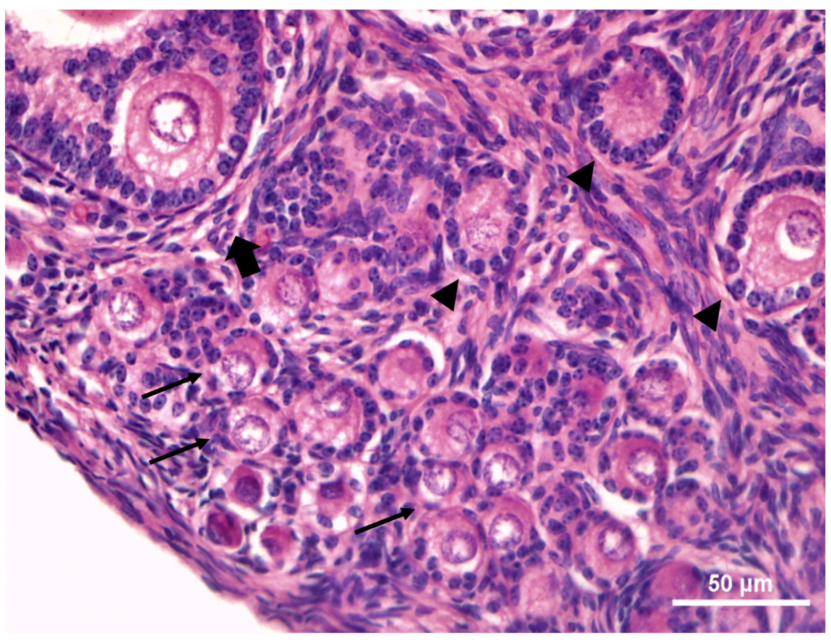
Microscopical analysis showed that ovaries contained a medulla and a cortical region with follicles at various stages of development (Figure 2). The primordial and primary follicles were located close to the germinative epithelium (superficial margins of the ovarian cortex), while the secondary ones were located in the inner part.
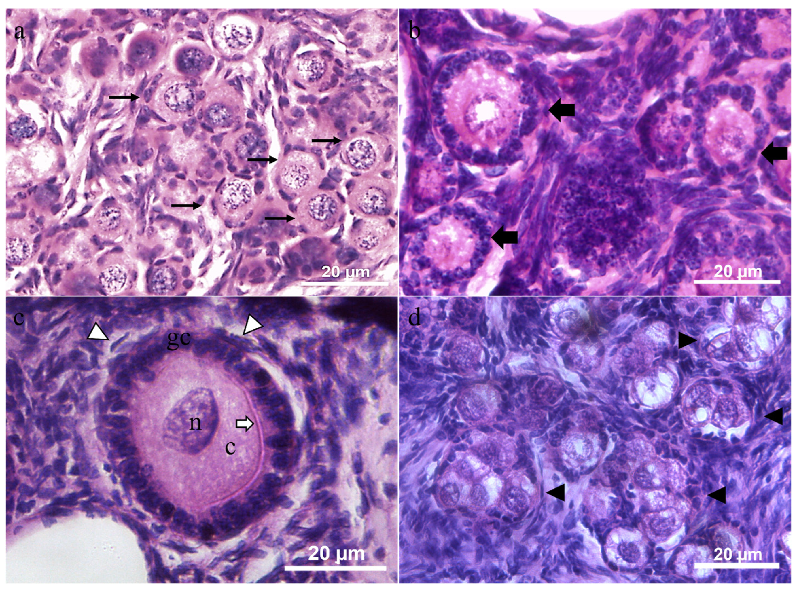
Primordial and primary follicles were characterized by an oocyte surrounded by a single layer of flattened or cuboidal granulosa cells, respectively (Figure 3a,b). Secondary follicles had two or more concentric layers of cuboidal granulosa cells around the oocyte. Externally of the granulosa cells, the internal theca was formed by elongated and fusiform cells, appearing as concentric layers. An evident zona pellucida was observed between granulosa cell layers and oocyte (Figure 3c). Oocytes presented heterogenic cytoplasm with some vacuoles. The nuclear membrane was visible. Scattered regions of chromatin and heterochromatin were located near the nucleus membrane. Multi-oocytes follicles (Figure 3d) were observed in all animals, near the germinative epithelium. Overall, a total of 4 to 150 multi-oocyte follicles per ovarian pair were observed (Table 2).

3.3. Ovarian Preantral Follicles Morphometrics and Estimation

Data related to preantral follicle morphometry are presented in Table 3.

3.4. Estimation of the Ovarian Preantral Follicle Population

Considering the five individuals, it could be inferred that the follicular population of the species would be 22,996.2 ± 7541.6 follicles per ovarian pair, as demonstrated in Table 4. When evaluating the data per animal in detail, however, we verified that individual A5 has a follicular population more than twice that of the other individuals. Therefore, when recalculating the follicular population based only on the other four individuals, we believe that the value of 15,567.2 preantral follicles per ovarian pair is more realistic to characterize the estimate of the follicular population of the species. Moreover, for four females, most of the follicular population was formed by primordial follicles, except in female A1, which presented a larger population of primary follicles.

3.5. Morphology of the Ovarian Preantral Follicles

Regardless of M. leprae infection, a high rate of ovarian follicle degeneration was observed in E. sexcinctus females, since only 41.4 ± 3.2% of follicles were classified as morphologically normal (Table 5).
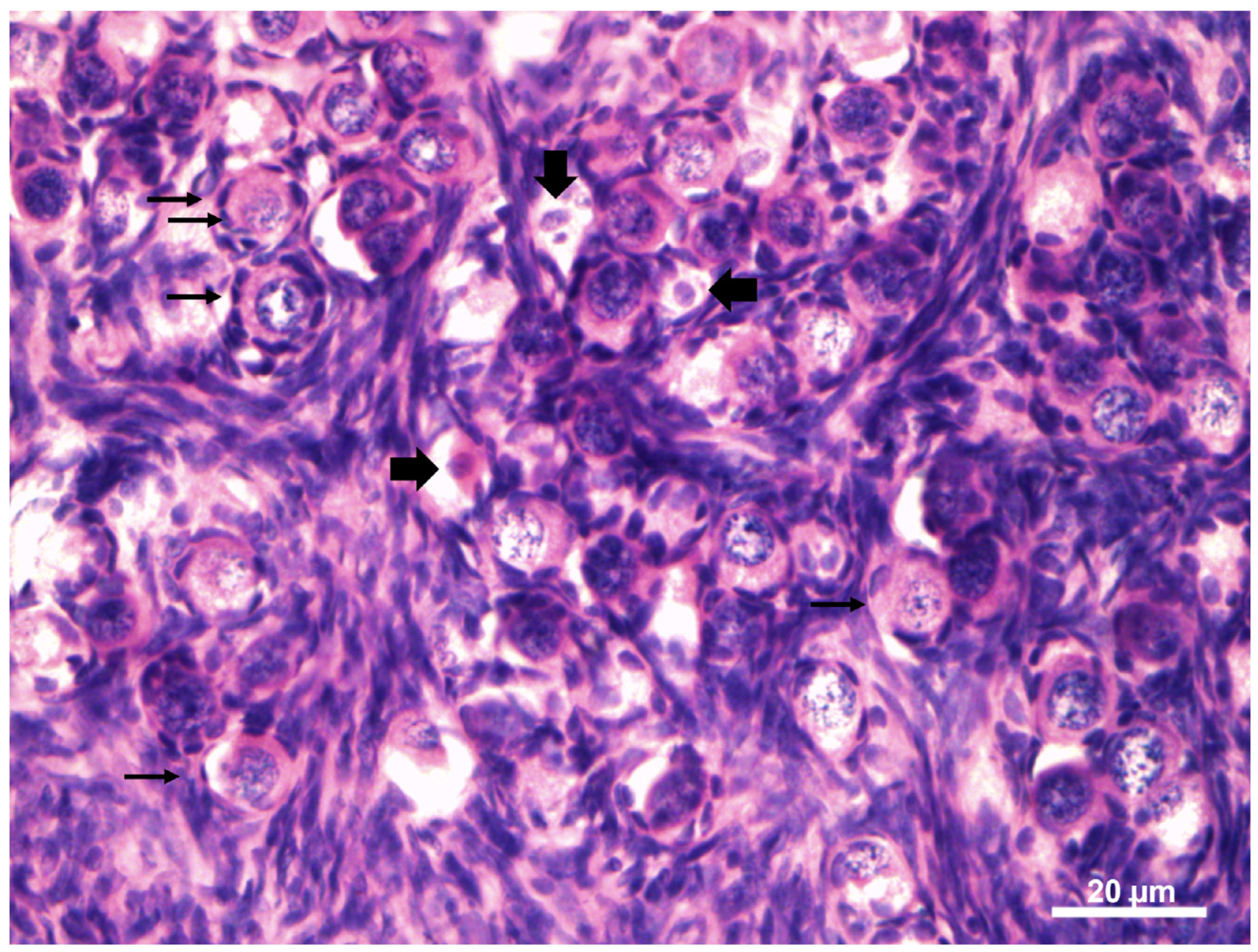
Morphologically normal PFs showed a spherical oocyte with an eosinophilic nucleus and heterogeneous cytoplasm. Granulosa cells without pycnotic nuclei were well-organized in layers surrounding the oocyte. Degenerated follicles showed a retraced oocyte with or without a pycnotic nucleus, accompanied or not by disorganized granulosa cells (Figure 4).

4. Discussion

The study provided important data on the characterization and estimation of the ovarian anatomy and preantral follicle populations in juvenile six-banded armadillos. Campos et al. [16] previously described the six-banded armadillo ovary appearance by ultrasound. The female gonads were well-defined structures, rounded and slightly hypoechoic in relation to the adjacent tissue. The ovary length measurement described by the authors was the same in the present study (0.90 cm); however, the width was larger (0.4 cm). It should be noted that during the process of forming the ultrasound image, there is the occurrence of the formation of image artifacts, which refer to the projection of images that do not match exactly with the true image of the location examined, mis-estimating the dimension of the ovary [16].
The ovarian measurements of the six-banded armadillo are similar to those of the lesser hairy (Chaetophractus vellerosus—0.5 cm) and the pichi (Zaedyus pichiy—0.4 cm) armadillos, larger than those of the pink fairy armadillo (Clamyphorus truncatus—0.2 to 0.3 cm), but smaller than those of the Azara’s domed armadillo (Tolypeutes matacus—0.8 to 1.0 cm), the large hairy armadillo (Chaetophractus villosus—0.6 to 1.0 cm) and the southern lesser long-nosed armadillo (Dasypus hybridus—0.6 cm to 0.9 cm) [23]. Studies have shown variations among the female reproductive organs of Dasypodidae species, mainly those related to the ovaries, uterus, and the lower portion of the reproductive tract [24,25,26]. These differences may be linked to the diversity of sperm types observed in Dasypodidae. In this family, the structure of the female genital tracts may create distinct functional and structural barriers for sperm, potentially leading to changes in sperm shape and size, suggesting a coevolutionary process between female reproductive traits and male gamete traits [23].
A large individual variation was verified for GSI (4.81–10.0) in juvenile E. sexcinctus. For other armadillos, data regarding GSI are not described, making it impossible to compare the results obtained with those of other species. In fact, GSI is not commonly described for mammalian females [19]. On the other hand, it is well described for aquatic species, such as fish and mollusks, being fundamental to understanding its reproductive status, oocyte size and ovarian maturity [27].
Microscopically, the analysis showed that the ovaries of six-banded armadillos were divided into a medulla and a cortical region, where ovarian follicles at various stages of development were found. Ovarian follicles are distributed in the ovarian cortical zone and are formed by an oocyte surrounded by a layer of flattened granulosa cells or one or more cuboidal cells, depending on the stage of follicle development. These characteristics are similar to those described for free-living six-banded armadillos from the central-western or southern regions of Brazil [8].
The follicular classification adopted in this study was in accordance with Silva et al. [22], classifying preantral follicles into three stages: primordial, primary, and secondary. This classification is different from that adopted by Rezende et al. [8] for the armadillo, in which preantral follicles were categorized as primordial, primary one-layer, primary two-layer, and secondary (with the initial formation of the antrum cavity). Despite the divergence in follicular classification, similarities were observed regarding the characteristics of the follicles described. In both studies, vacuoles were observed in the cytoplasm of oocytes, which presented evident nuclear membrane, with dispersed regions of chromatin and heterochromatin. Furthermore, the characterization of ovarian follicles has been previously performed in other armadillo species. The same ovarian follicle classification and similar characteristics were also described for the pichi, the lesser hairy and the southern lesser long-nosed armadillos [23]. In the hairy armadillo, for example, ovarian follicles were categorized as primordial, intermediate, early primary, late primary, secondary, tertiary, and Graafian or preovulatory [28] or into four stages (I–IV), based on follicle size and granulosa cell layers and shape [29].
As described here for the six-banded armadillo, the occurrence of multi-oocyte follicles was also observed for other species of the families Euphractinae (Ch. villosus, Ch. vellerosus, Z. pichiy and C. truncatus) and Tolypeutinae (T. matacus) [23,25,30]. In nine-banded armadillos, even when large follicles (>978 μm) were present, indicating proximity to ovulation, nests of primordial follicles were still found in the ovaries [17]. These multi-oocyte follicles were observed in all six-banded armadillos used in the present study, while in other species, such as Z. pichiy, it was not observed in all animals studied, inferring an interspecies variation [30]. The high variation of the multi-oocyte number observed among animals, both infected or not, may be attributed to the individual variation, as poly-ovular development seems not to be only a natural polymorphism [30], causing alterations of nest breakdown, but also is related to environmental endocrine disruptors and phytoestrogens [31]. All those factors may have contributed to the large individual difference observed among animals.
The reason for the occurrence of this phenomenon has not yet been completely elucidated. Theories have been suggested to explain its existence, such as mitotic division with a high rate of cell differentiation during follicle growth, from which several oocytes arise from primordial follicles [32], a failure of cell division in the initial stages of follicle development fusion of adjacent individual follicles [31].
In the large hairy armadillo, after light microscopy, the presence of multi-oocyte follicle-like structures was observed; however, ultrastructural and immunohistochemistry analysis showed the presence of ovarian germ cell (GC) cysts, interconnected by intercellular bridges and surrounded by a single layer of flat follicle cells [33]. In mammals, the GC cyst rupture process usually concludes around birth or shortly thereafter, with the development of primordial follicles containing single oocytes, which form the only gamete reservoir available throughout the female’s reproductive life [34]. The existence of GC cysts in adult animals is an evolutionary-conserved developmental event that enables a deeper investigation of the events surrounding folliculogenesis [33]. Further investigation is required to affirm if the structures classified as multi-oocyte follicles in six-banded armadillos are muti-oocytes or GC cysts involved in the formation of new primordial follicles after animal birth.
In the large hairy armadillo, using transmission electron microscopy, a distinct structure was described in the cytoplasm of oocytes in early growth, the multilamellar body—MLBs. The authors described this structure as an organelle with a transitory function, during the initial stages of follicular development, from follicle growth to the beginning of zona pellucida formation [29]. The MLBs are composed of lamellar units different from filaments, and these lamellae may coalesce and form large lamellar patches. A vital role of this organelle to the zona pellucida development is suggested. Some of the vacuoles observed in the six-banded armadillo’s oocyte cytoplasm may be related to the presence of the MLBs or to lipid droplets, as is observed in other mammals, such as peccaries [35]. However, further studies using transmission electron microscopy are required to confirm the hypothesis.
Regarding morphometry, the values obtained in the present work were closer to those described for a large hairy armadillo. Similarly, the oocytes increased their diameter according to the development of the follicle, while a smaller nuclear increase was observed [29]. Moreover, a positive and linear correlation between the diameter of the follicle and the oocyte was verified for large hairy armadillos [28]. Previous studies showed differences among the number and size of armadillos’ follicles throughout the year, indicating seasonal changes [17,33]. This can be related to the differences observed in this work and other studies. However, the existence of seasonal reproductive changes in the six-banded armadillo still needs to be investigated.
The follicular population observed here for six-banded armadillos was considerably larger than that observed in the greater hairy armadillo [33], in which the number of primordial follicles ranged from 1107.9 ± 362.4 (summer) to 2990.3 ± 306.3 (autumn), with the same number observed for primary follicles (257.5 ± 40.1 to 582.7 ± 33). It is worth highlighting that, in the present study, a formula was used to estimate the follicular population that has been previously validated and extensively reviewed for other domestic [36,37] and wild [35,38] species, as well as for humans [20]. As in the present study, in sloths (Bradypus variegatus), the majority of follicles found were characterized as primordial, forming the ovarian reserve pool [39].
In all-female six-banded armadillos, a high rate of ovarian follicular degeneration was observed, especially when compared with values found for other mammalian species [35,36,37]. Follicular development is known to depend on the rates of proliferation and atresia, which are regulated by many endocrine, paracrine, and autocrine factors [40]. Although atresia leads to the loss of numerous ovarian follicles, it plays a vital role in maintaining ovarian homeostasis in mammals, ensuring regular reproductive cyclicity [41]. In six-banded armadillos, however, further investigation is needed to elucidate whether such a high rate of degenerated follicles is physiological or related to external factors.
Regarding infection by M. leprae, no major changes were observed in the parameters evaluated in the ovary of six-banded armadillos. This information contradicts what was previously described for the nine-banded armadillo, in which the occurrence of considerable lepromatous infiltration in the ovaries of experimentally infected individuals was observed [42]. We, however, recognize the limitations of our findings, given the limited number of animals used. In any case, since this is a wild animal, the importance of the data should be considered, especially given the difficulties in obtaining access to such animals. This difficulty is also evident in the study that demonstrated for the first time the effect of M. leprae infection on the pregnancy of nine-banded armadillos, whose report was based on the description of three cases [43].

5. Conclusions

We demonstrated that the juvenile six-banded armadillo presents particularities regarding the characteristics and estimation of its ovarian preantral follicular population. Furthermore, we report that M. leprae infection apparently does not cause major changes in histological ovarian features in this species. With these data, we highlight that folliculogenesis in Xenarthra remains largely unexplored and studying it can provide valuable data on reproductive physiology, improving captive management, epidemiological studies and assisted reproductive technologies.

Author Contributions

G.L.L., A.V.B. and A.M.S.—methodology, validation, formal analysis, investigation and writing; J.M.A.d.P.A.—conceptualization, formal analysis, and writing; P.C.—formal analysis and writing; and A.R.S.—Conceptualization, methodology, valiation, formal analysis, investigation, writing, supervision and project administration. All authors have read and agreed to the published version of the manuscript.

Funding

Andreia M. Silva and Alexandre R. Silva are receipts of CNPq grants. Conselho Nacional de Desenvolvimento Científico—CNPq: 306409/2022-4.

Institutional Review Board Statement

The project was approved by the Ethics Committee of UFERSA (CEUA Proc. No. 23091.002959/2016-43) and authorized by the Chico Mendes Institute of Biodiversity—ICMBio (SISBIO 50564-1).

Informed Consent Statement

Informed consent was obtained from the animal owners for their animals to participate in the study.

Data Availability Statement

The data are available from the authors upon reasonable request.

Conflicts of Interest

The authors declare no conflicts of interest.

References

  1. Silva, A.R.; Sousa, P.C.; Freitas, C.I.A. Assisted reproduction in six-banded armadillos (Euphractus sexcinctus): Peculiarities and challenges. Rev. Bras. Reprod. Anim. 2015, 39, 61–65. [Google Scholar]
  2. Balamayooran, G.; Pena, M.; Sharma, R.; Truman, R.W. The armadillo as an animal model and reservoir host for Mycobacterium leprae. Clin. Dermatol. 2015, 33, 108–115. [Google Scholar] [CrossRef] [PubMed]
  3. da Silva Ferreira, J.; de Carvalho, F.M.; Vidal Pessolani, M.C.; de Paula Antunes, J.M.A.; de Medeiros Oliveira, I.V.P.; Ferreira Moura, G.H.; Truman, R.W.; Peña, M.T.; Sharma, R.; Duthie, M.S.; et al. Serological and molecular detection of infection with Mycobacterium leprae in Brazilian six banded armadillos (Euphractus sexcinctus). Comp. Immunol. Microbiol. Infect. Dis. 2020, 68, 101397. [Google Scholar] [CrossRef] [PubMed]
  4. Frota, C.C.; Lima, L.N.C.; Rocha, A.D.S.; Suffys, P.N.; Rolim, B.N.; Rodrigues, L.C.; Barreto, M.L.; Kendall, C.; Kerr, L.R.S. Mycobacterium leprae in six-banded (Euphractus sexcinctus) and nine-banded armadillos (Dasypus novemcinctus) in Northeast Brazil. Mem. Inst. Oswaldo Cruz 2012, 107 (Suppl. 1), 209–213. [Google Scholar] [CrossRef]
  5. Abba, A.M.; Lima, E.; Superina, M. Euphractus sexcinctus. In The IUCN Red List of Threatened Species 2014; IUCN Biodiversity Assessment & Knowledge Team: Red List Unit: Cambridge, UK, 2014; e.T8306A47441708. [Google Scholar] [CrossRef]
  6. Ribeiro, P.; Silveira Miranda, J.E.; Rodrigues de Araújo, D. The effect of roadkills on the persistence of xenarthran populations: The case of the Brazilian Cerrado. Edentata 2017, 18, 51–61. [Google Scholar] [CrossRef]
  7. Rodrigues, T.F.; Mantellatto, A.M.B.; Superina, M.; Chiarello, A.G. Ecosystem services provided by armadillos. Biol. Rev. 2019, 95, 1–21. [Google Scholar] [CrossRef] [PubMed]
  8. Rezende, L.; Kückelhaus, S.; Galdos-Riveros, A.; Ferreira, J.; Miglino, M. Vascularización, morfología e histología del ovario en el armadillo Euphractus sexcinctus (Linnaeus, 1758). Arch. Med. Vet. 2013, 45, 191–196. [Google Scholar] [CrossRef]
  9. Fernandes, D.P.; Praxedes, É.A.; Viana, J.V.D.S.; de Aquino, L.V.C.; Rodrigues, L.L.V.; Moura, Y.B.F.; de Oliveira, M.F.; Freitas, C.I.A.; Pereira, A.F. Influence of cryopreservation techniques and low concentrations of permeating cryoprotectants on the conservation of ear cartilage and skin derived from six-banded armadillos (Euphractus sexcinctus Linnaeus, 1758). Cryobiology 2023, 113, 104788. [Google Scholar] [CrossRef] [PubMed]
  10. Miranda, F.; Moraes-Barros, N.; Superina, M.; Abba, A.M. Tolypeutes tricinctus. In The IUCN Red List of Threatened Species 2014; IUCN Biodiversity Assessment & Knowledge Team: Red List Unit: Cambridge, UK, 2014; e.T21975A47443455. [Google Scholar] [CrossRef]
  11. Anacleto, T.C.S.; Miranda, F.; Medri, I.; Cuellar, E.; Abba, A.M.; Superina, M. Priodontes maximus. In The IUCN Red List of Threatened Species 2014; IUCN Biodiversity Assessment & Knowledge Team: Red List Unit: Cambridge, UK, 2014; e.T18144A47442343. [Google Scholar] [CrossRef]
  12. da Cunha Sousa, P.; Campos, L.B.; Silva, A.R.; Lima, G.L. The Armadillos. In Assisted Reproduction in Wild Mammals of South America; CRC Press: Boca Raton, FL, USA, 2023; pp. 291–305. [Google Scholar] [CrossRef]
  13. Serafim, M.K.B.; Lira, R.A.; Costa, L.L.M.; Gadelha, I.C.N.; Freitas, C.I.A.; Silva, A.R. Description of semen characteristics from six-banded armadillos (Euphractus sexcinctus) collected by electroejaculation. Anim. Reprod. Sci. 2010, 118, 362–365. [Google Scholar] [CrossRef]
  14. Santos, E.A.A.; Sousa, P.C.; Dias, C.E.V.; Castelo, T.S.; Peixoto, G.C.X.; Lima, G.L.; Ricarte, A.R.F.; Simão, B.R.; Freitas, C.I.A.; Silva, A.R. Assessment of sperm survival and functional membrane integrity of the six-banded armadillo (Euphractus sexcinctus). Theriogenology 2011, 76, 623–629. [Google Scholar] [CrossRef]
  15. Sousa, P.C.; Amorim, R.N.L.; Lima, G.L.; Paiva, A.L.C.; Paula, V.V.; Freitas, C.I.A.; Silva, A.R. Establishment of an anesthetic protocol for semen collection by electroejaculation in six-banded armadillos (Euphractus sexcinctus Linnaeus, 1758). Arq. Bras. Med. Vet. Zootec. 2016, 68, 1595–1601. [Google Scholar] [CrossRef]
  16. Campos, L.B.; Peixoto, G.C.X.; Lima, G.L.; Castelo, T.S.; Silva, A.R.; Freitas, C.I.A.; Silva, A.R. Monitoring the reproductive physiology of six-banded armadillos (Euphractus sexcinctus Linnaeus, 1758) through different techniques. Reprod. Domest. Anim. 2016, 51, 736–742. [Google Scholar] [CrossRef]
  17. Peppler, R.D.; Hossler, F.E.; Stone, S.C. Determination of reproductive maturity in the female nine-banded armadillo (Dasypus novemcinctus). Reproduction 1986, 76, 141–146. [Google Scholar] [CrossRef]
  18. Sharma, R.; Singh, P.; Loughry, W.J.; Lockhart, J.M.; Inman, W.B.; Duthie, M.S.; Pena, M.T.; Marcos, L.A.; Scollard, D.M.; Cole, S.T.; et al. Zoonotic Leprosy in the Southeastern United States. Emerg. Infect. Dis. 2015, 21, 2127–2134. [Google Scholar] [CrossRef] [PubMed]
  19. Lopes, K.R.F.; Lima, G.L.; Bezerra, L.G.P.; Barreto-Junior, R.A.; Oliveira, M.F.; Silva, A.R. Characterization of the ovarian preantral follicle populations and its correlation with age and nutritional status in Brazilian Northeastern donkeys (Equus assinus). Anim. Reprod. Sci. 2017, 187, 193–202. [Google Scholar] [CrossRef] [PubMed]
  20. Gougeon, A.; Chainy, G.B. Morphometric studies of small follicles in ovaries of women at different ages. J. Reprod. Fertil. 1987, 81, 433–442. [Google Scholar] [CrossRef] [PubMed]
  21. Lima, G.L.; Luz, V.B.; Lima, L.F.; Rocha, R.M.P.; Castro, S.V.; Castelo, T.S.; Rodrigues, A.P.R.; Figueiredo, J.R.; Silva, A.R. Interactions between different media and follicle-stimulating hormone supplementation on in vitro culture of preantral follicles enclosed in ovarian tissue derived from collared peccaries (Pecari tajacu Linneaus, 1758). Reprod. Domest. Anim. 2018, 53, 880–888. [Google Scholar] [CrossRef] [PubMed]
  22. Silva, J.R.V.; van den Hurk, R.; Costa, S.H.F.; Andrade, E.R.; Nunes, A.P.A.; Ferreira, F.V.A.; Lôbo, R.N.B.; Figueiredo, J.R. Survival and growth of goat primordial follicles after in vitro culture of ovarian cortical slices in media containing coconut water. Anim. Reprod. Sci. 2004, 81, 273–286. [Google Scholar] [CrossRef] [PubMed]
  23. Cetica, P.D.; Aldana Marcos, H.J.; Merani, M.S. Morphology of female genital tracts in Dasypodidae (Xenarthra, Mammalia): A comparative survey. Zoomorphology 2005, 124, 57–65. [Google Scholar] [CrossRef]
  24. Enders, A.C.; Buchanan, G.D. Some Effects of Ovariectomy and Injection of Ovarian Hormones in the Armadillo. J. Endocrinol. 1959, 19, 251–258. [Google Scholar] [CrossRef]
  25. Codón, S.M.; Casanave, E.B. Comparative morphology of the ovaries of three species of dasypodidae (Mammalia, Xenarthra). Rev. Chil. Anat. 2000, 18, 251–257. [Google Scholar] [CrossRef]
  26. Codón, S.M.; Casanave, E.B. Histology of the ovary of the armadillo Chaetophractus villosus (Mammalia, Dasypodidae). Rev. Brasil. Biol. 1996, 56, 599–604. [Google Scholar]
  27. Zeyl, J.; Love, O.; Higgs, D. Evaluating gonadosomatic index as an estimator of reproductive condition in the invasive round goby, Neogobius melanostomus. J. Great Lakes Res. 2014, 40, 164–171. [Google Scholar] [CrossRef]
  28. Codón, S.M.; Estecondo, S.G.; Galíndez, E.J.; Casanave, E.B. Ultrastructure and morphometry of ovarian follicles in the armadillo Chaetophractus villosus (Mammalia, Dasypodidae). Braz. J. Biol. 2001, 61, 485–496. [Google Scholar] [CrossRef]
  29. Rossi, L.F.; Solari, A.J. Large lamellar bodies and their role in the growing oocytes of the armadillo Chaetophractus villosus. J. Morphol. 2021, 282, 1330–1338. [Google Scholar] [CrossRef] [PubMed]
  30. Superina, M.; Carreño, N.; Jahn, G.A. Characterization of seasonal reproduction patterns in female pichis Zaedyus pichiy (Xenarthra: Dasypodidae) estimated by fecal sex steroid metabolites and ovarian histology. Anim. Reprod. Sci. 2009, 116, 358–369. [Google Scholar] [CrossRef]
  31. Gaytán, F.; Morales, C.; Manfredi-Lozano, M.; Tena-Sempere, M. Generation of multi-oocyte follicles in the peripubertal rat ovary: Link to the invasive capacity of granulosa cells? Fertil. Steril. 2014, 101, 1467–1476. [Google Scholar] [CrossRef] [PubMed]
  32. Kennedy, W. P, The occurrence of polyovular graafian follicles. J. Anat. 1924, 58, 328–334. [Google Scholar] [PubMed]
  33. Rossi, L.F.; Nottola, S.; Miglietta, S.; Macchiarelli, G.; Luaces, J.P.; Merico, V.; Merani, S.; Garagna, S.; Zuccotti, M. Germ cell cysts, a fetal feature in mammals, are constitutively present in the adult armadillo. Mol. Reprod. Dev. 2019, 87, 91–101. [Google Scholar] [CrossRef] [PubMed]
  34. Lei, L.; Spradling, A.C. Female mice lack adult germ-line stem cells but sustain oogenesis using stable primordial follicles. Proc. Natl. Acad. Sci. USA 2013, 110, 8585–8590. [Google Scholar] [CrossRef] [PubMed]
  35. Lima, G.L.; Santos, E.A.A.; Luz, V.B.; Rodrigues, A.P.R.; Silva, A.R. Morphological Characterization of the Ovarian Preantral Follicle Population of Collared Peccaries (Tayassu tajacu Linnaeus, 1758). Anat. Histol. Embryol. 2012, 42, 304–311. [Google Scholar] [CrossRef]
  36. Silva-Santos, K.C.; Santos, G.M.G.; Siloto, L.S.; Hertel, M.F.; Andrade, E.R.; Rubin, M.I.B.; Sturion, L.; Melo-Sterza, F.A.; Seneda, M.M. Estimate of the population of preantral follicles in the ovaries of Bos taurus indicus and Bos taurus taurus cattle. Theriogenology 2011, 76, 1051–1057. [Google Scholar] [CrossRef] [PubMed]
  37. Lucci, C.M.; Amorim, C.A.; Rodrigues, A.P.R.; Figueiredo, J.R.; Báo, S.N.; Silva, J.R.V.; Gonçalves, P.B.D. Study of preantral follicle population in situ and after mechanical isolation from caprine ovaries at different reproductive stages. Anim. Reprod. Sci. 1999, 56, 223–236. [Google Scholar] [CrossRef] [PubMed]
  38. Lopes, G.P.; Santos, R.R.; Almeida, D.V.; Brito, A.B.; Queiroz, H.L.; Domingues, S.F.S. Population estimate and morphometry of ovarian preantral follicles from three recently recognized squirrel monkey species: A comparative study. Zygote 2017, 25, 279–287. [Google Scholar] [CrossRef] [PubMed]
  39. Lima, A.K.F.; Felipe, C.B.S.; Silva, G.M.L. Biologia reprodutiva de Bradypus variegatus Schinz (1825): Desafios e perspectivas/Reproduction biology in Bradypus variegatus Schinz (1825): Challenges and perspectives. Rev. Bras. Reprod. Anim. 2018, 42, 109–113. [Google Scholar]
  40. Hsu, S.Y.; Hsueh, A.J. Tissue-specific Bcl-2 protein partners in apoptosis, p. An ovarian paradigm. Physiol. Rev. 2000, 80, 593–614. [Google Scholar] [CrossRef] [PubMed]
  41. McGee, E.A.; Hsueh, A.J.W. Initial and cyclic recruitment of ovarian follicles. Endocr. Rev. 2000, 21, 200–214. [Google Scholar] [PubMed]
  42. Binford, C.H.; Storrs, E.E.; Walsh, G.P. Disseminated infection in the nine-banded armadillo (Dasypus novemcinctus) resulting from inoculation with M. leprae. Observations made on 15 animals studied at autopsy. Int. J. Lepr. Other Mycobact. Dis. 1976, 44, 80–83. [Google Scholar] [PubMed]
  43. Job, C.K.; Sanchez, R.M.; Hastings, R.C. Lepromatous placentitis and intrauterine fetal infection in lepromatous nine-banded armadillos (Dasypus novemcinctus). Lab. Investig. 1987, 56, 44–48. [Google Scholar] [PubMed]
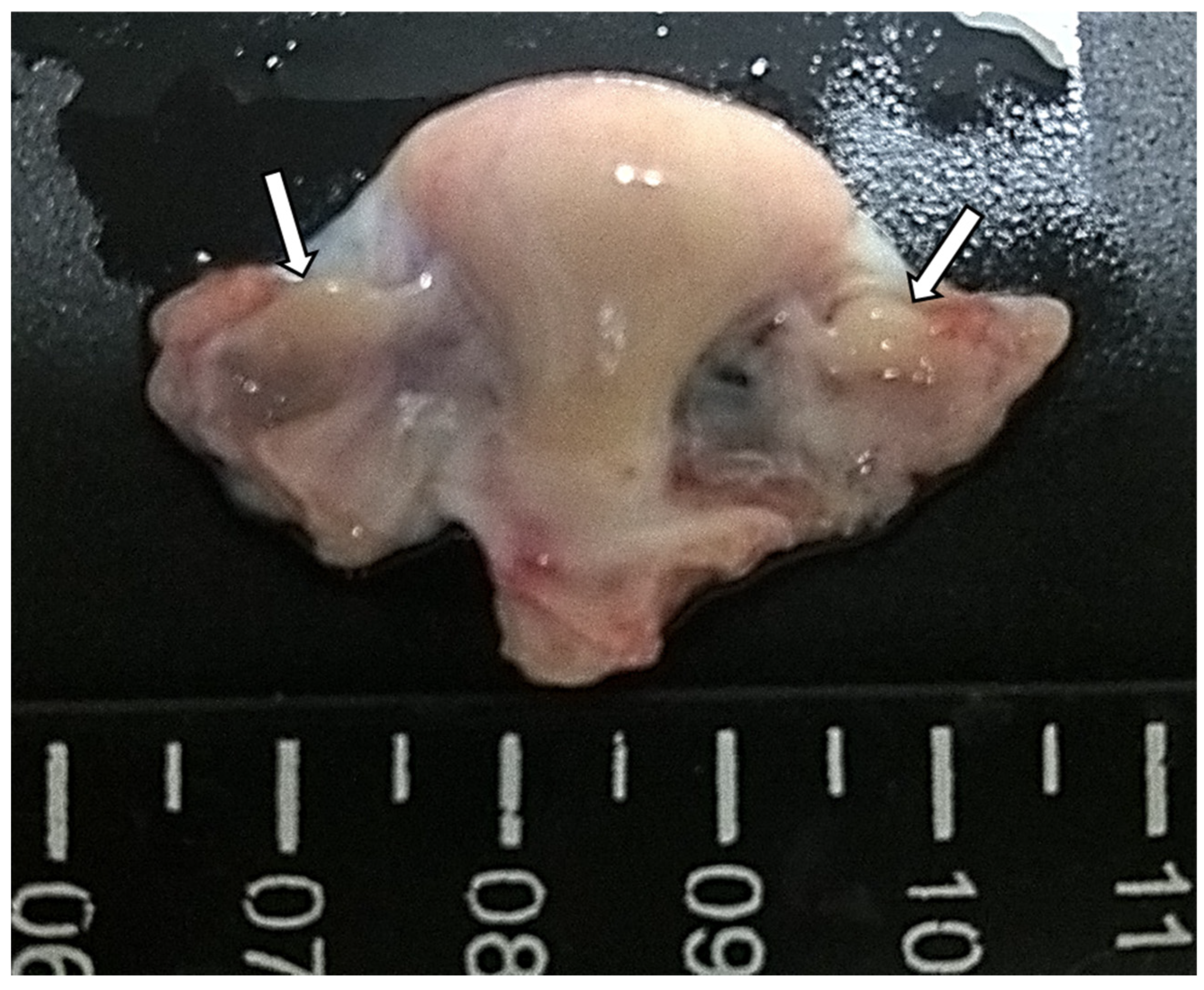
Figure 1. Macroscopic aspects of Euphractus sexcintus reproductive system (uterine body in the middle and ovaries on each side). Ovaries are round structures with smooth surfaces (white full arrows).
Vetsci 12 00037 g001
Figure 2. Histology of Euphractus sexcintus ovary with follicles at various stages of development. Primordial (arrows), primary (arrowhead) and secondary (full arrow) follicles. 400×.
Vetsci 12 00037 g002
Figure 3. Histological features in the juvenile six-banded armadillos (Euphractus sexcintus) preantral follicles. (a)—Primordial follicles (arrows) surrounded by a single layer of flattened granulosa cells. (b)—Primary follicles (full arrows) surrounded by a single layer of cuboidal granulosa cells, presenting some vacuoles at cytoplasm. (c)—Small secondary follicle showing two or more layers of cuboidal granulosa cells (n—nucleus, c—cytoplasm, gc—granulosa cells) and the presence of zona pellucida (white arrow). The theca cells were formed by elongated and fusiform cells (white arrowheads). (d)—multi-oocyte follicles (arrowheads). 400×.
Vetsci 12 00037 g003
Figure 4. Morphological normal follicles (arrows) exhibiting well-organized granulosa cells without pycnotic nuclei surrounding the oocyte. Degenerated follicles (full arrows) showing a retraced oocyte with a pycnotic nucleus, accompanied or not by disorganized granulosa cells. 400×.
Vetsci 12 00037 g004
Table 1. Body weight, ovary weight, ovary dimensions and gonadosomatic index in juvenile six-banded armadillos infected (n = 2) or not (n = 3) by the Mycobacterium leprae.
Animals Health ConditionBody Weight (Kg)Right OvaryLeft OvaryGonadosomatic Index (%)
Weight (Kg)Dimensions (mm)
(Width × Length × Thickness)
Weight (Kg)Dimensions (mm)
(Width × Length × Thickness)
A1Infected1.830.060.4 × 1 × 0.30.050.4 × 0.8 × 0.36.01
A2Infected1.930.070.4 × 1 × 0.20.060.4 × 1 × 0.26.74
A3Uninfected2.080.050.5 × 0.8 × 0.20.050.4 × 1 × 0.34.81
A4Uninfected1.600.060.4 × 0.8 × 0.30.100.5 × 1 × 0.310.0
A5Uninfected1.700.060.4 × 0.9 × 0.30.060.4 × 0.9 × 0.37.06
Means ± SEM1.8 ± 0.10.1 ± 0.00.4 × 0.9 × 0.30.1 ± 0.00.4 × 0.9 × 0.36.9 ± 0.8
Table 2. Number of multi-oocyte preantral follicles found in the ovarian pair of juvenile six-banded armadillos infected (n = 2) or not (n = 3) by the Mycobacterium leprae.
AnimalsHealth ConditionRight OvaryLeft OvaryTotal per Ovarian Pair
A1Infected7613
A2Infected9951150
A3Non-infected281240
A4Non-infected224
A5Non-infected702292
Means ± SEM 37.2 ± 18.818.6 ± 8.859.8 ± 27.3
Table 3. Morphometry (μm) means ± SEM of the oocyte nucleus, oocyte, and follicles (per category) in the ovary of juvenile six-banded armadillos infected (n = 2) or not (n = 3) by the Mycobacterium leprae.
AnimalsHealth ConditionFollicle CategoryNucleusOocyteFollicleNumber of Follicles Evaluated
A1InfectedPrimordial8.0 ± 0.313.4 ± 0.317.2 ± 0.330
Primary9.0 ± 0.316.3 ± 0.523.4 ± 0.930
Secondary12.9 ± 0.426.9 ± 1.342.9 ± 1.828
A2InfectedPrimordial7.2 ± 0.312.4 ± 0.216.1 ± 0.330
Primary8.9 ± 0.216.3 ± 0.423.5 ± 0.430
Secondary13.8 ± 1.232.5 ± 5.347.9 ± 7.310
A3Non-infectedPrimordial7.5 ± 0.110.5 ± 0.113.8 ± 0.230
Primary7.1 ± 0.210.2 ± 0.214.3 ± 0.230
Secondary8.1 ± 0.212.4 ± 0.818.7 ± 0.730
A4Non-infectedPrimordial6.9 ± 0.210.7 ± 0.214.4 ± 0.230
Primary8.7 ± 0.217.5 ± 0.524.2 ± 0.730
Secondary10.3 ± 1.324.0 ± 4.134.8 ± 7.910
A5Non-infectedPrimordial5.6 ± 0.18.5 ± 0.211.7 ± 0.230
Primary7.1 ± 0.314.1 ± 0.721.8 ± 1.129
Secondary9.3 ± 0.522.8 ± 1.438.0 ± 2.028
Means ± SEMPrimordial7.0 ± 0.211.1 ± 0.214.6 ± 0.2150
Primary8.2 ± 0.214.9 ± 0.521.4 ± 0.7149
Secondary10.9 ± 0.723.7 ± 2.636.5 ± 3.9106
Table 4. Estimation of preantral follicle (PF) population per ovary (right and left) and per ovarian pair (Total PF Population) of juvenile six-banded armadillos infected (n = 2) or not (n = 3) by the Mycobacterium leprae.
AnimalsHealth ConditionFollicle CategoryPF Population of Right OvaryPF Population of
Left Ovary
Total PF
Population
Proportions (%) of PF Category
A1InfectedPrimordial2929.22484.95414.135.4
Primary5371.93697.19068.959.4
Secondary315.3476.9792.185.2
Total8616.49143.717,760.1100.0
A2InfectedPrimordial7210.83708.710,919.464.4
Primary3222.62214.85437.432.1
Secondary287302.9589.83.5
Total10,720.36226.316,946.7100.0
A3UninfectedPrimordial4559.187168.611,727.869.0
Primary2517.82562.25082.029.9
Secondary120.561.8182.31.1
Total7197.49792.716,990.1100.0
A4UninfectedPrimordial4598.92426.67025.666.5
Primary1490.71908.03398.732.2
Secondary57.889.7147.41.3
Total6147.44424.310,571.8100.0
A5UninfectedPrimordial20,368.97628.727,997.553.1
Primary14,886.68812.723,699.344.9
Secondary756.0259.51015.62.0
Total36,011.516,700.952,712.4100.0
Means ± SEM 13,738.6 ± 5620.79257.6 ± 2100.622,996.2 ± 7541.6-
Table 5. Morphological integrity of preantral follicles in the ovaries of juvenile six-banded armadillos infected (n = 2) or not (n = 3) by the Mycobacterium leprae.
TotalProportions (%)
AnimalsHealth
Condition
Follicle CategoryNormalDegeneratedTotal Follicle/
Category
NormalDegenerated
A1InfectedPrimordial1334.64079.55414.124.775.4
Primary3120.85948.19069.034.465.6
Secondary265.5526.7792.233.566.49
A2InfectedPrimordial4162.76756.710,919.438.161.9
Primary2309.93127.55437.442.557.5
Secondary166.0423.8589.828.271.9
A3UninfectedPrimordial6190.85536.911,727.652.847.2
Primary2748.92333.15082.054.145.9
Secondary94.388182.351.748.3
A4UninfectedPrimordial3999.63026.07025.656.943.1
Primary1789.31609.53398.752.747.4
Secondary20.7126.7147.414.086.0
A5UninfectedPrimordial11,482.616,514.927,997.341.059.0
Primary11,293.912,405.423,699.347.752.3
Secondary490.9524.71015.648.351.7
Means ± SEM 3298.0 ± 965.74201.8 ± 1230.37499.9 ± 2169.641.4 ± 3.258.6 ± 3.2
Disclaimer/Publisher’s Note: The statements, opinions and data contained in all publications are solely those of the individual author(s) and contributor(s) and not of MDPI and/or the editor(s). MDPI and/or the editor(s) disclaim responsibility for any injury to people or property resulting from any ideas, methods, instructions or products referred to in the content.