
Advances in Detecting Cystic Echinococcosis in Intermediate Hosts and New Diagnostic Tools: A Literature Review

 , , ,
, , , 
Abstract
Simple Summary
Abstract
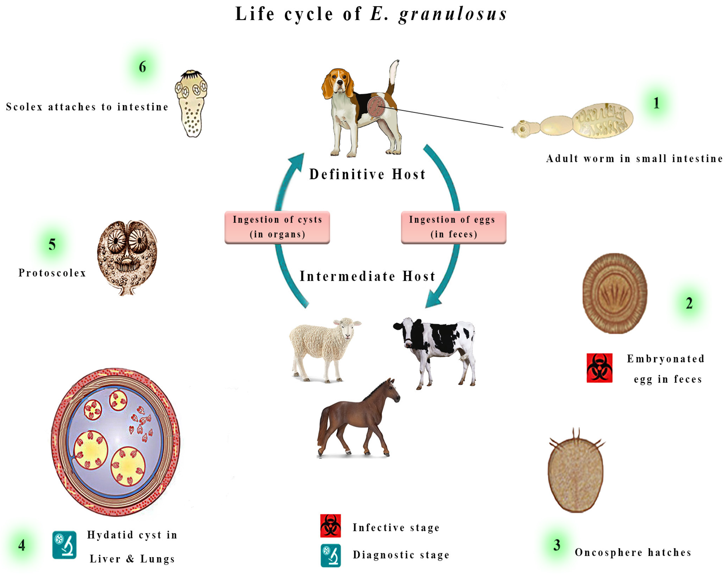
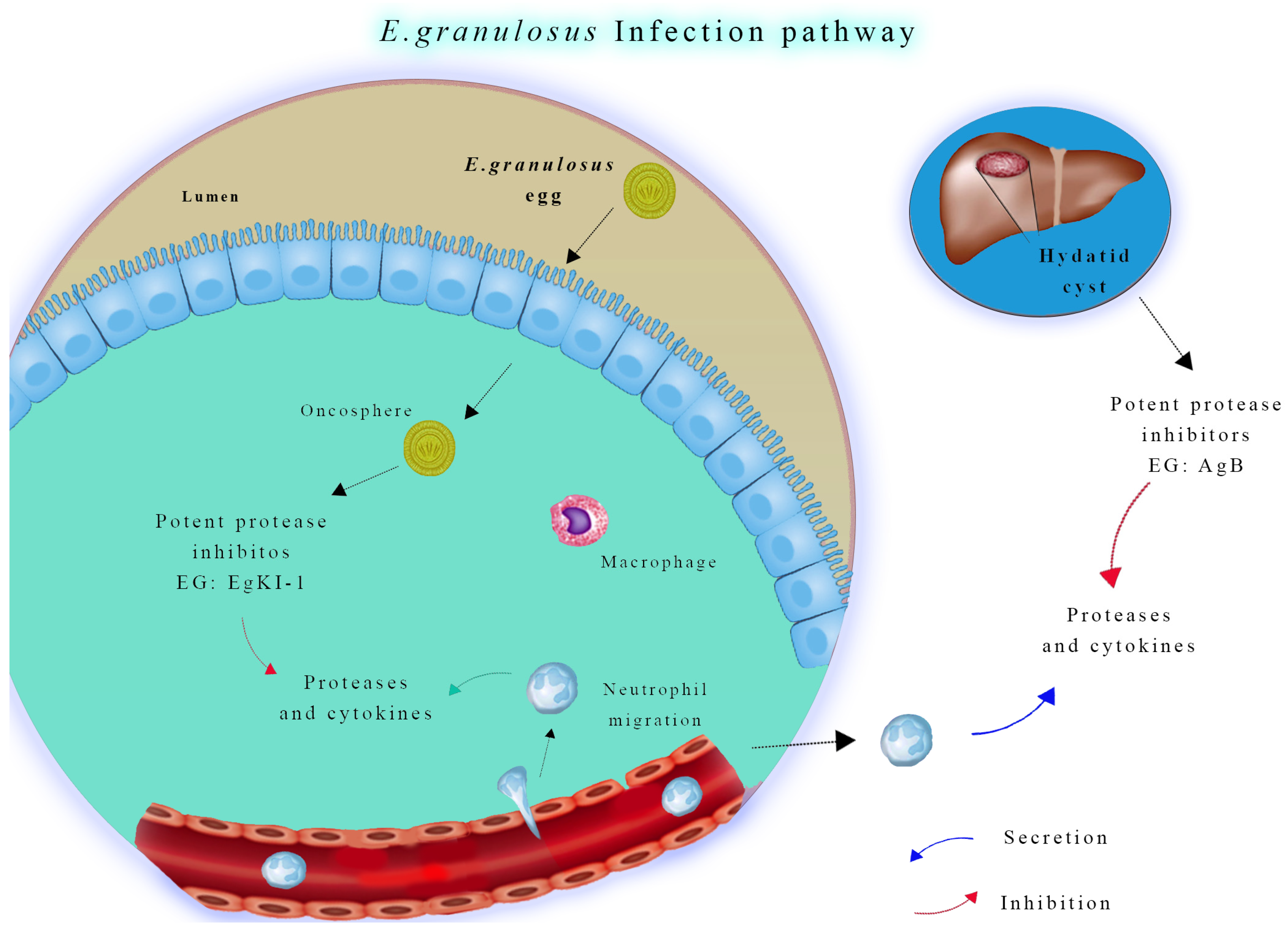
1. Introduction
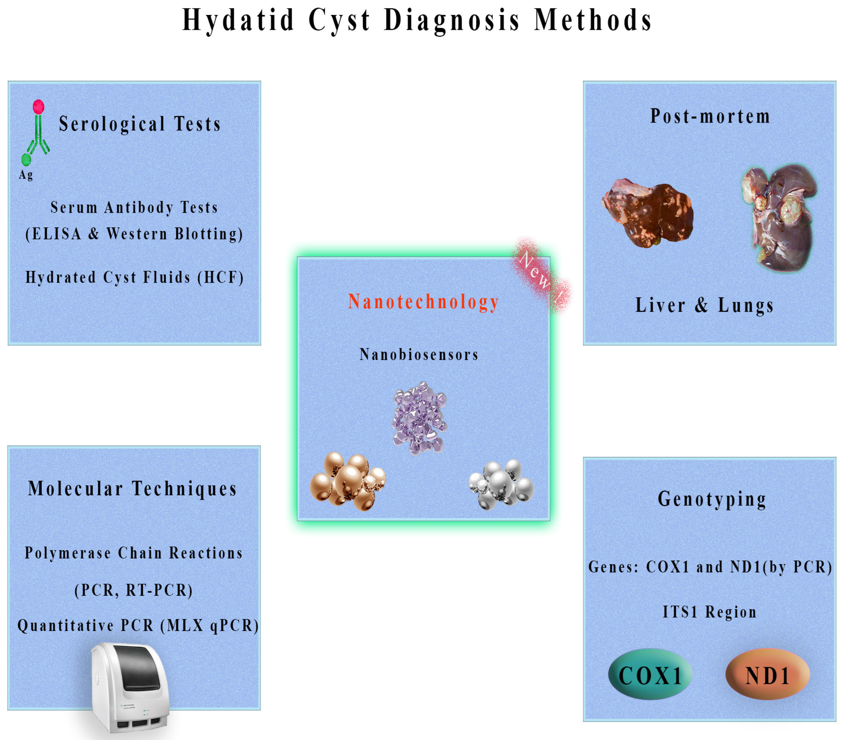
2. Methods for Diagnosis of Cystic Echinococcosis in Intermediate Hosts
2.1. Serological Tests
2.2. Post-Mortem Inspection
2.3. Molecular Techniques
2.3.1. Genotyping with Mitochondrial Genes
2.3.2. Genotyping with the ITS1 Region
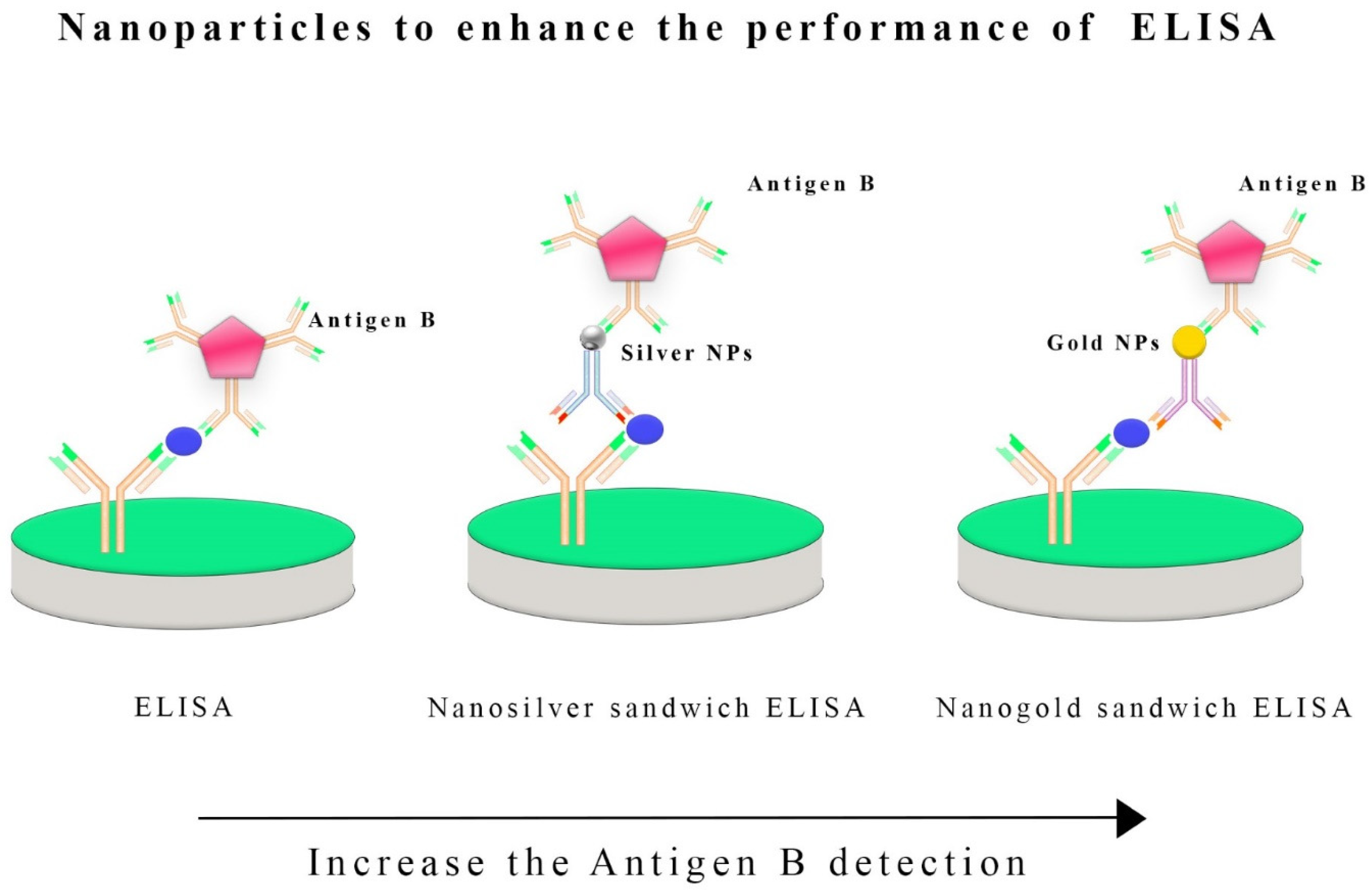
2.4. Nanobiosensors for Improved Hydatid Cyst Detection
3. Future and Prospective of Nanobiosensors
4. Discussion
5. Conclusions
Author Contributions
Funding
Institutional Review Board Statement
Informed Consent Statement
Data Availability Statement
Conflicts of Interest
References
- Thompson, R.C. Biology and systematics of echinococcus. Adv. Parasitol. 2017, 95, 65–109. [Google Scholar] [CrossRef] [PubMed]
- Deplazes, P.; Rinaldi, L.; Alvarez Rojas, C.A.; Torgerson, P.R.; Harandi, M.F.; Romig, T.; Antolova, D.; Schurer, J.M.; Lahmar, S.; Cringoli, G.; et al. Global distribution of alveolar and cystic echinococcosis. J. Adv. Parasitol. 2017, 95, 315–493. [Google Scholar] [CrossRef] [PubMed]
- Selcuk, M.A.; Celik, F.; Kesik, H.K.; Gunyakti Kilinc, S.; Ahmed, H.; Jiang, N.; Simsek, S.; Cao, J. In Silico evaluation of the haplotype diversity, phylogenetic variation and population structure of human E. granulosus sensu stricto (G1 Genotype) sequences. J. Pathog. 2022, 11, 1346. [Google Scholar] [CrossRef] [PubMed]
- Pavia, G.; De Gori, F.; Ciambrone, L.; De Gori, N.; Musarella, R.; Casalinuovo, F. Dispersal and molecular characterisation of the Echinococcus granulosus (Batsch, 1786) complex isolated from various intermediate hosts in the calabria region, southern Italy. Folia Parasitol. 2020, 67, 14. [Google Scholar] [CrossRef] [PubMed]
- Avcioglu, H.; Guven, E.; Balkaya, I.; Kirman, R.; Akyuz, M.; Mebarek Bia, M.; Gulbeyen, H.; Yaya, S. The situation of echinococcosis in stray dogs in Turkey: The first finding of echinococcus multilocularis and echinococcus ortleppi. Parasitololy 2021, 148, 1092–1098. [Google Scholar] [CrossRef] [PubMed]
- Laurimäe, T.; Kinkar, L.; Moks, E.; Bagrade, G.; Saarma, U. Exploring the genetic diversity of genotypes G8 and G10 of the Echinococcus canadensis cluster in Europe based on complete mitochondrial genomes (13,550–13,552 bp). Parasitololy 2023, 150, 631–637. [Google Scholar] [CrossRef] [PubMed]
- Casulli, A.; Massolo, A.; Saarma, U.; Umhang, G.; Santolamazza, F.; Santoro, A. Species and genotypes belonging to Echinococcus granulosus sensu lato complex causing human cystic echinococcosis in Europe (2000–2021): A systematic review. Parasites Vectors 2022, 15, 109. [Google Scholar] [CrossRef] [PubMed]
- Crotti, S.; Brustenga, L.; Cruciani, D.; Bonelli, P.; D’Avino, N.; Felici, A.; Morandi, B.; Sebastiani, C.; Spina, S.; Gobbi, M. Molecular Screening of Echinococcus spp. and Other Cestodes in Wild Carnivores from Central Italy. Vet. Sci. 2023, 10, 318. [Google Scholar] [CrossRef]
- Nakao, M.; Lavikainen, A.; Hoberg, E. Is Echinococcus intermedius a valid species? Trends Parasitol. 2015, 31, 342–343. [Google Scholar] [CrossRef]
- Nakao, M.; McManus, D.P.; Schantz, P.M.; Craig, P.S.; Ito, A. A molecular phylogeny of the genus echinococcus inferred from complete mitochondrial genomes. Parasitololy 2007, 134 Pt 5, 713–722. [Google Scholar] [CrossRef]
- Nakao, M.; Yanagida, T.; Konyaev, S.; Lavikainen, A.; Odnokurtsev, V.A.; Zaikov, V.A.; Ito, A. Mitochondrial phylogeny of the genus echinococcus (Cestoda: Taeniidae) with emphasis on relationships among Echinococcus canadensis genotypes. Parasitololy 2013, 140, 36–1625. [Google Scholar] [CrossRef]
- Thompson, R.C. The molecular epidemiology of echinococcus infections. Pathogens 2020, 9, 453. [Google Scholar] [CrossRef] [PubMed]
- Alvi, M.A.; Alshammari, A.; Ali, R.M.; Ul Haq, S.; Bashir, R.; Li, L.; Saqib, M.; Sajid, M.S.; Ghafoor, M.; Imran, M.; et al. Revealing novel cytb and nad5 genes-based population diversity and benzimidazole resistance in Echinococcus granulosus of bovine origin. Front. Vet. Sci. 2023, 10, 1191271. [Google Scholar] [CrossRef] [PubMed]
- Massolo, A.; Simoncini, A.; Romig, T. The ‘bridge effect’ by intermediate hosts may explain differential distributions of Echinococcus species. Trends Parasitol. 2022, 38, 501–512. [Google Scholar] [CrossRef] [PubMed]
- Zanini, F.; Di Salvo, V.; Pierangeli, N.; Lazzarini, L.; Curto, E. Presence of Echinococcus granulosus sensu lato in the endoparasitic fauna of feral dogs in Tierra del Fuego, Argentina. Vet. Parasitol. Reg. Stud. 2023, 44, 100916. [Google Scholar] [CrossRef] [PubMed]
- Serra, E.; Masu, G.; Chisu, V.; Cappai, S.; Masala, G.; Loi, F.; Piseddu, T. Environmental contamination by Echinococcus spp. Eggs as a risk for human health in educational farms of Sardinia, Italy. Vet. Sci. 2022, 9, 143. [Google Scholar] [CrossRef] [PubMed]
- Chacha, C.S.; Stephano, M.A.; Irunde, J.I.; Mwasunda, J.A. Cystic echinococcosis dynamics in dogs, humans and cattle: Deterministic and stochastic modeling. Results Phys. 2023, 51, 106697. [Google Scholar] [CrossRef]
- Parija, S.C.; Pramodhini, S. Echinococcosis; Springer: Berlin/Heidelberg, Germany, 2022; pp. 353–368. [Google Scholar] [CrossRef]
- Kolapo, T.U.; Oksanen, A.; Davidson, R.; Jenkins, E.J. Cystic and Alveolar Echinococcosis Caused by Echinococcus Canadensis and E. Multilocularis in the Arctic; Springer: Berlin/Heidelberg, Germany, 2022; pp. 279–295. [Google Scholar] [CrossRef]
- Alho, A.M.; Dias, M.C.; Cardo, M.; Aguiar, P.; de Carvalho, L.M. The evolution of cystic echinococcosis in Humans and Ruminants in Portugal—A One Health Approach. Vet. Sci. 2023, 10, 584. [Google Scholar] [CrossRef] [PubMed]
- Widdicombe, J.; Basáñez, M.G.; Entezami, M.; Jacksonmm, D.; Larrieu, E.; Prada, J.M. The economic evaluation of cystic echinococcosis control strategies focused on zoonotic hosts: A scoping review. PLoS Negl. Trop. Dis. 2022, 16, e0010568. [Google Scholar] [CrossRef]
- Alvi, M.A.; Alsayeqh, A.F. Food-borne zoonotic echinococcosis: A review with special focus on epidemiology. Front. Vet. Sci. 2022, 9, 1072730. [Google Scholar] [CrossRef]
- Casulli, A.; Abela-Ridder, B.; Petrone, D.; Fabiani, M.; Bobić, B.; Carmena, D.; Šoba, B.; Zerem, E.; Gargaté, M.J.; Kuzmanovska, G.; et al. Unveiling the incidences and trends of the neglected zoonosis cystic echinococcosis in Europe: A systematic review from the MEmE project. Lancet Inf. Dis. 2023, 23, E95–E107. [Google Scholar] [CrossRef] [PubMed]
- Khalili, N.; Iranpour, P.; Khalili, N.; Haseli, S. Hydatid disease: A pictorial review of uncommon locations. Iran. J. Med. Sci. 2023, 48, 118–129. [Google Scholar] [CrossRef] [PubMed]
- Sadr, S.; Lotfalizadeh, N.; Abbasi, A.M.; Soleymani, N.; Hajjafari, A.; Roohbaksh Amooli Moghadam, E.; Borji, H. Challenges and prospective of enhancing hydatid cyst chemotherapy by nanotechnology and the future of nanobiosensors for diagnosis. Trop. Med. Infect. Dis. 2023, 8, 494. [Google Scholar] [CrossRef]
- Dietrich, C.F.; Douira-Khomsi, W.; Gharbi, H.; Sharma, M.; Cui, X.W.; Sparchez, Z.; Richter, J.; Kabaalioğlu, A.; Atkinson, N.S.; Schreiber-Dietrich, D.; et al. Cystic echinococcosis, review and illustration of non-hepatic manifestations. Med. Ultrason. 2020, 22, 319–324. [Google Scholar] [CrossRef]
- Calame, P.; Weck, M.; Busse-Cote, A.; Brumpt, E.; Richou, C.; Turco, C.; Doussot, A.; Bresson-Hadni, S.; Delabrousse, E. Role of the radiologist in the diagnosis and management of the two forms of hepatic echinococcosis. Insights Imaging 2022, 13, 68. [Google Scholar] [CrossRef] [PubMed]
- Brunetti, E.; Kern, P.; Vuitton, D.A. Expert consensus for the diagnosis and treatment of cystic and alveolar echinococcosis in humans. Acta Trop. 2010, 114, 1–16. [Google Scholar] [CrossRef]
- Brunetti, E.; Tamarozzi, F.; Macpherson, C.; Filice, C.; Piontek, M.S.; Kabaalioglu, A.; Dong, Y.; Atkinson, N.; Richter, J.; Schreiber-Dietrich, D.; et al. Ultrasound and cystic echinococcosis. Ultrasound Int. Open (UIO) 2018, 4, E70–E78. [Google Scholar] [CrossRef] [PubMed]
- Peruzzu, A.; Mastrandrea, S.; Fancellu, A.; Bonelli, P.; Muehlethaler, K.; Masala, G.; Santucciu, C. Comparison and evaluation of analytic and diagnostic performances of four commercial kits for the detection of antibodies against Echinococcus granulosus and multilocularis in human sera. Comp. Immunol. Microbiol. Infect. Dis. 2022, 86, 101816. [Google Scholar] [CrossRef]
- Moghadam, Z.K.; Ghaffarifar, F.; Khalilpour, A.; Aziz, F.A.; Saadatnia, G.; Noordin, R. IgG4 Detection of Echinococcus granulosus Paramyosin Is a Useful Diagnostic Test for Human Hydatidosis. Clin. Vaccine Immunol. 2013, 20, 501–505. [Google Scholar] [CrossRef]
- Tokarska-Rodak, M.; Plewik, D.; Michalski, A.J.; Kołodziej, M.; Mełgieś, A.; Pańczuk, A.; Konon, H.; Niemcewicz, M. Serological surveillance of vector-borne and zoonotic diseases among hunters in eastern Poland. J. Vector Borne Dis. 2016, 53, 355–361. [Google Scholar]
- Tamarozzi, F.; Covini, I.; Mariconti, M.; Narra, R.; Tinelli, C.; De Silvestri, A.; Manzoni, F.; Casulli, A.; Ito, A.; Neumayr, A.; et al. Comparison of the diagnostic accuracy of three rapid tests for the serodiagnosis of hepatic cystic echinococcosis in humans. PLoS Negl. Trop. Dis. 2016, 10, e0004444. [Google Scholar] [CrossRef]
- Tamarozzi, F.; Longoni, S.S.; Vola, A.; Degani, M.; Tais, S.; Rizzi, E.; Prato, M.; Scarso, S.; Silva, R.; Brunetti, E.; et al. Evaluation of nine commercial serological tests for the diagnosis of human hepatic cyst echinococcosis and the differential diagnosis with other focal liver lesions: A diagnostic accuracy study. Diagnostics 2021, 11, 167. [Google Scholar] [CrossRef]
- Tamarozzi, F.; Silva, R.; Fittipaldo, V.A.; Buonfrate, D.; Gottstein, B.; Siles-Lucas, M. Serology for the diagnosis of human hepatic cystic echinococcosis and its relation with cyst staging: A systematic review of the literature with meta-analysis. PLoS Negl. Trop. Dis. 2021, 15, e0009370. [Google Scholar] [CrossRef]
- Pagnozzi, D.; Addis, M.F.; Biosa, G.; Roggio, A.M.; Tedde, V.; Mariconti, M.; Tamarozzi, F.; Meroni, V.; Masu, G.; Masala, G.; et al. Diagnostic accuracy of antigen 5-based ELISAs for human cystic echinococcosis. PLoS Negl. Trop. Dis. 2016, 10, e0004585. [Google Scholar] [CrossRef] [PubMed]
- Lima, R.R.; Lima, J.V.; Ribeiro, J.F.; Nascimento, J.B.; Oliveira, W.F.; Cabral Filho, P.E.; Fontes, A. Emerging biomedical tools for biomarkers detection and diagnostics in schistosomiasis. Talanta 2023, 265, 124900. [Google Scholar] [CrossRef]
- Achi, F.; Attar, A.M.; Lahcen, A.A. Electrochemical nanobiosensors for the detection of cancer biomarkers in real samples: Trends and challenges. TrAC Trends Anal. Chem. 2023, 170, 117423. [Google Scholar] [CrossRef]
- Ahmad, A.; Imran, M.; Ahsan, H. Biomarkers as biomedical bioindicators: Approaches and techniques for the detection, analysis, and validation of novel biomarkers of diseases. Pharmaceutics 2023, 15, 1630. [Google Scholar] [CrossRef] [PubMed]
- Kulkarni, M.B.; Ayachit, N.H.; Aminabhavi, T.M. Recent advancements in nanobiosensors: Current trends, challenges, applications, and future scope. Biosensors 2022, 12, 892. [Google Scholar] [CrossRef]
- Hassan, A.A.; Sayed-ElAhl, R.M.; El Hamaky, A.M.; Mansour, M.K.; Oraby, N.H.; Barakat, M.H. Nanodiagnostics: New Tools for Detection of Animal Pathogens; Springer: Berlin/Heidelberg, Germany, 2022; pp. 299–325. [Google Scholar] [CrossRef]
- Cerbu, C.; White, J.C.; Sabliov, C.M. Nanotechnology in Livestock: Improving Animal Production and Health; Elsevier: Amsterdam, The Netherlands, 2023; pp. 181–213. [Google Scholar] [CrossRef]
- Gil Rosa, B.; Akingbade, O.E.; Guo, X.; Gonzalez-Macia, L.; Crone, M.A.; Cameron, L.P.; Freemont, P.; Choy, K.L.; Güder, F.; Yeatman, E.; et al. Multiplexed immunosensors for point-of-care diagnostic applications. Biosens. Bioelectron. 2022, 203, 114050. [Google Scholar] [CrossRef]
- Abdelbaset, A.E.; Yagi, K.; Nonaka, N.; Nakao, R. Cystic echinococcosis in humans and animals in Egypt: An epidemiological overview. Curr. Res. Parasitol. Vector Borne Dis. 2021, 1, 100061. [Google Scholar] [CrossRef]
- Vaisi-Raygani, A.; Mohammadi, M.; Jalali, R.; Salari, N.; Hosseinian-Far, M. Prevalence of cystic echinococcosis in slaughtered livestock in Iran: A systematic review and meta-analysis. BMC Infect. Dis. 2021, 21, 429. [Google Scholar] [CrossRef]
- Toaleb, N.I.; Aboelsoued, D.; Abdel Megeed, K.N.; Hekal, S.H. A novel designed sandwich ELISA for the detection of Echinococcus granulosus antigen in camels for diagnosis of cystic echinococcosis. Trop. Med. Infect. Dis. 2023, 8, 400. [Google Scholar] [CrossRef]
- Siddartha, P.V.; Babu, A.J.; Rao, T.M.; Rayulu, V.C.; Swetha, C.S. A comparative evaluation of four different immunoassays in the diagnosis of cystic echinococcosis using a crude and purified hydatid cyst fluid antigen. Acta Parasitol. 2022, 67, 1667–1679. [Google Scholar] [CrossRef]
- Barnes, T.S.; Deplazes, P.; Gottstein, B.; Jenkins, D.J.; Mathis, A.; Siles-Lucas, M.; Torgerson, P.R.; Ziadinov, I.; Heath, D.D. Challenges for diagnosis and control of cystic hydatid disease. Acta Trop. 2012, 123, 1–7. [Google Scholar] [CrossRef]
- Darabi, E.; Motevaseli, E.; Mohebali, M.; Rokni, M.B.; Khorramizadeh, M.R.; Zahabiun, F.; Heidari, S.; Kia, E.B. Evaluation of a novel Echinococcus granulosus recombinant fusion B-EpC1 antigen for the diagnosis of human cystic echinococcosis using indirect ELISA in comparison with a commercial diagnostic ELISA kit. Exp. Parasitol. 2022, 240, 108339. [Google Scholar] [CrossRef]
- Jahani, Z.; Meshgi, B.; Rajabi-Bzl, M.; Jalousian, F.; Hasheminasab, S. Improved serodiagnosis of hydatid cyst disease using gold nanoparticle labeled antigen B in naturally infected sheep. Iran. J. Parasitol. 2014, 9, 218. [Google Scholar] [PubMed]
- Shirazi, S.; Hoghooghi-Rad, N.; Madani, R. The comparison between AgB-ELISA and a new method of Nano-ELISA for diagnosis of hydatidosis in sheep. J. Hell. Vet. Med. Soc. 2022, 73, 3629–3634. [Google Scholar] [CrossRef]
- Hassanain, M.A.; Toaleb, N.I.; Shaapan, R.M.; Hassanain, N.A.; Maher, A.; Yousif, A.B. Immunological detection of human and camel cystic echinococcosis using different antigens of hydatid cyst fluid, protoscoleces, and germinal layers. Vet. World. 2021, 14, 270. [Google Scholar] [CrossRef]
- Sykes, A.L.; Larrieu, E.; Poggio, T.V.; Céspedes, M.G.; Mujica, G.B.; Basáñez, M.G.; Prada, J.M. Modelling diagnostics for Echinococcus granulosus surveillance in sheep using latent class analysis: Argentina as a case study. One Health 2021, 14, 100359. [Google Scholar] [CrossRef] [PubMed]
- Entezami, M.; Nocerino, M.; Widdicombe, J.; Bosco, A.; Cringoli, G.; Casulli, A.; Lo Iacono, G.; Rinaldi, L.; Prada, J.M. The spatial distribution of cystic echinococcosis in Italian ruminant farms from routine surveillance data. Front. Trop. Dis. 2022, 3, 1034572. [Google Scholar] [CrossRef]
- Issa, A.R.; Arif, S.H.; Mohammed, A.A.; Santolamazza, F.; Santoro, A.; Mero, W.M.; Casulli, A. Insights into Human Cystic Echinococcosis in the Kurdistan Region, Iraq: Characteristics and Molecular Identification of Cysts. Pathogens 2022, 11, 408. [Google Scholar] [CrossRef]
- Rodarte, K.A.; Fair, J.M.; Bett, B.K.; Kerfua, S.D.; Fasina, F.O.; Bartlow, A.W. A scoping review of zoonotic parasites and pathogens associated with abattoirs in Eastern Africa and recommendations for abattoirs as disease surveillance sites. Public Health Front. 2023, 11, 1194964. [Google Scholar] [CrossRef]
- Eaton, S.L.; Murdoch, F.; Rzechorzek, N.M.; Thompson, G.; Hartley, C.; Blacklock, B.T.; Proudfoot, C.; Lillico, S.G.; Tennant, P.; Ritchie, A.; et al. Modelling neurological diseases in large animals: Criteria for model selection and clinical assessment. Cells 2022, 11, 2641. [Google Scholar] [CrossRef] [PubMed]
- Muniruzzaman, M.D.; Hossain, M.D.; Labony, S.; Alim, M.; Anisuzzaman. Clinical presentation and diagnosis of pulmonary hydatidosis in a cow mimicking to aspiration pneumonia. Vet. Rec. Case Rep. 2023, 11, e553. [Google Scholar] [CrossRef]
- Shoulah, S.A.; Gaballa, M.M.; Marawan, M.A.; Saqr, S.A.; Abdelhady, A.; Alzahrani, H.A.; Wakid, M.H.; Al-Jabr, O.A.; Selim, A. Pathological findings and oxidative stress status associated with hydatidosis in dromedary camels. Vet. Sci. 2023, 10, 74. [Google Scholar] [CrossRef] [PubMed]
- Khan, J.; Basharat, N.; Khan, S.; Jamal, S.M.; Rahman, S.U.; Shah, A.A.; Khan, S.; Ali, R.; Khan, S.N.; Ali, I. Prevalence and molecular characterization of cystic echinococcosis in livestock population of the malakand division, khyber pakhtunkhwa, Pakistan. Front. Vet. Sci. 2021, 8, 757800. [Google Scholar] [CrossRef]
- Díaz, Á.; Barrios, A.A.; Grezzi, L.; Mouhape, C.; Jenkins, S.J.; Allen, J.E.; Casaravilla, C. Immunology of a unique biological structure: The Echinococcus laminated layer. Protein Cell 2023, 14, 87–104. [Google Scholar] [CrossRef]
- Shabani, M.; Solhjoo, K.; Taghipour, A.; Jahromi, A.S.; Karami, S.; Armand, B. The occurrence of cystic echinococcosis in slaughtered livestock in Jahrom, south of Iran. Parasite Epidemiol. Control. 2022, 19, e00274. [Google Scholar] [CrossRef]
- Dey, P. Polymerase Chain Reaction: Principle, Technique and Applications in Pathology; Springer: Berlin/Heidelberg, Germany, 2022; pp. 215–228. [Google Scholar] [CrossRef]
- Król, G.; Fortunka, K.; Majchrzak, M.; Piktel, E.; Paprocka, P.; Mańkowska, A.; Lesiak, A.; Karasiński, M.; Strzelecka, A.; Durnaś, B.; et al. Metallic Nanoparticles and Core-Shell Nanosystems in the Treatment, Diagnosis, and Prevention of Parasitic Diseases. Pathogens 2023, 12, 838. [Google Scholar] [CrossRef] [PubMed]
- Bradbury, R.S.; Sapp, S.G.; Potters, I.; Mathison, B.A.; Frean, J.; Mewara, A.; Sheorey, H.; Tamarozzi, F.; Couturier, M.R.; Chiodini, P.; et al. Where have all the diagnostic morphological parasitologists gone? J. Clin. Microbiol. 2022, 60, e00986-22. [Google Scholar] [CrossRef] [PubMed]
- Knapp, J.; Lallemand, S.; Monnien, F.; Felix, S.; Courquet, S.; Umhang, G.; Millon, L. Real-time multiplex PCR for human echinococcosis and differential diagnosis. Parasite 2023, 30, 3. [Google Scholar] [CrossRef] [PubMed]
- Santoro, A.; Santolamazza, F.; Cacciò, S.M.; La Rosa, G.; Antolová, D.; Auer, H.; Bagrade, G.; Bandelj, P.; Basso, W.; Beck, R.; et al. Mitochondrial genetic diversity and phylogenetic relationships of Echinococcus multilocularis in Europe. Int. J. Parasitol. 2024, 54, 233–245. [Google Scholar] [CrossRef] [PubMed]
- Zhang, X.; Jian, Y.; Li, Z.; Duo, H.; Guo, Z.; Fu, Y. Optimization of single-tube nested PCR for the detection of Echinococcus spp. Exp. Parasitol. 2023, 247, 108494. [Google Scholar] [CrossRef] [PubMed]
- Hadipour, M.; Darani, H.Y.; Talebzadeh, H.; Eslamian, M.; Aboutalebian, S.; Harandi, M.F.; Mirhendi, H. Sensitive detection of specific cell-free DNA in serum samples from sheep with cystic echinococcosis. PLoS Negl. Trop. Dis. 2023, 17, e0011715. [Google Scholar] [CrossRef] [PubMed]
- Zhang, X.; Jian, Y.; Guo, Z.; Duo, H.; Wei, Y. development of a triplex real-time pcr assay to detect Echinococcus species in canid fecal samples. J. Parasitol. 2022, 108, 79–87. [Google Scholar] [CrossRef] [PubMed]
- Ohiolei, J.A.; Yan, H.B.; Odeniran, P.O.; Li, L.; Shumuye, N.A.; Qurishi, S.A.; Isaac, C.; Fu, B.Q.; Jia, W.Z. Echinococcus granulosus sensu lato in animal intermediate hosts: What is with the organ location? Vet. Parasitol. 2022, 304, 109695. [Google Scholar] [CrossRef] [PubMed]
- Zhao, Y.; Gesang, D.; Wan, L.; Li, J.; Qiangba, G.; Danzeng, W.; Basang, Z.; Renzhen, N.; Yin, J.; Gongsang, Q.; et al. Echinococcus spp. and genotypes infecting humans in Tibet Autonomous Region of China: A molecular investigation with near-complete/complete mitochondrial sequences. Parasit. Vectors 2022, 15, 75. [Google Scholar] [CrossRef] [PubMed]
- Mori, K.; Imamura, A.; Hirayama, I.; Minamoto, T. Detection of Echinococcus multilocularis in repurposed environmental DNA samples from river water. PeerJ 2023, 11, e15431. [Google Scholar] [CrossRef] [PubMed]
- Jiménez-Avalos, G.; Soto-Obando, A.; Solis, M.; Gilman, R.H.; Cama, V.; Gonzalez, A.E.; García, H.H.; Sheen, P.; Requena, D.; Zimic, M. Assembly and phylogeographical analysis of novel Taenia solium mitochondrial genomes suggest stratification within the African-American genotype. Parasit. Vectors 2023, 16, 349. [Google Scholar] [CrossRef]
- Mahmud, S.; Biswas, S.; Afrose, S.; Mita, M.A.; Hasan, M.R.; Shimu, M.S.; Paul, G.K.; Chung, S.; Saleh, M.A.; Alshehri, S.; et al. Use of next-generation sequencing for Identifying mitochondrial disorders. Curr. Issues Mol. Biol. 2022, 44, 1127–1148. [Google Scholar] [CrossRef]
- Karamon, J.; Samorek-Pieróg, M.; Bilska-Zając, E.; Korpysa-Dzirba, W.; Sroka, J.; Bełcik, J.; Zdybel, J.; Cencek, T. Echinococcus multilocularis genetic diversity based on isolates from pigs confirmed the characteristic haplotype distribution and the presence of the Asian-like haplotype in Central Europe. J. Vet. Res. 2023, 67, 567–574. [Google Scholar] [CrossRef]
- Shumuye, N.A.; Li, L.; Li, W.H.; Zhang, N.Z.; Wu, Y.T.; Wu, Y.D.; Tian, W.J.; Zhang, L.S.; Nian, X.F.; Dai, G.D.; et al. Infection of sheep by Echinococcus multilocularis in Gansu, China: Evidence from mitochondrial and nuclear DNA analysis. Infect. Dis. Poverty 2023, 12, 72. [Google Scholar] [CrossRef] [PubMed]
- Sanchez, L.; Mayta, H.; Jara, L.M.; Verástegui, M.; Gilman, R.H.; Gómez-Puerta, L.A.; Gavidia, C.M. Echinococcus granulosus sensu stricto and E. canadensis are distributed in livestock of highly endemic area in the Peruvian highlands. Acta Trop. 2022, 225, 106178. [Google Scholar] [CrossRef]
- Shahbazi, A.E.; Saidijam, M.; Maghsood, A.H.; Matini, M.; Haghi, M.M.; Fallah, M. Genotyping of fresh and parafinized human Hydatid cysts using nad1 and cox1 genes in Hamadan Province, West of Iran. Iran. J. Parasito. 2020, 15, 259–265. [Google Scholar] [CrossRef]
- Satam, H.; Joshi, K.; Mangrolia, U.; Waghoo, S.; Zaidi, G.; Rawool, S.; Thakare, R.P.; Banday, S.; Mishra, A.K.; Das, G.; et al. Next-Generation sequencing technology: Current trends and advancements. Biology 2023, 12, 997. [Google Scholar] [CrossRef]
- Hua, R.Q.; Du, X.D.; He, X.; Gu, X.B.; Xie, Y.; He, R.; Xu, J.; Peng, X.R.; Yang, G.Y. Genetic diversity of Echinococcus granulosus sensu lato in China: Epidemiological studies and systematic review. Transbound. Emerg. Dis. 2022, 69, e1382–e1392. [Google Scholar] [CrossRef]
- Rahman, M.T.; Sobur, M.A.; Islam, M.S.; Ievy, S.; Hossain, M.J.; El Zowalaty, M.E.; Rahman, A.T.; Ashour, H.M. Zoonotic diseases: Etiology, impact, and control. Microorganisms 2020, 8, 1405. [Google Scholar] [CrossRef] [PubMed]
- Moghaddam, S.M.; Picot, S.; Ahmadpour, E. Interactions between hydatid cyst and regulated cell death may provide new therapeutic opportunities. Parasite 2019, 26, 70. [Google Scholar] [CrossRef] [PubMed]
- Arya, S.S.; Dias, S.B.; Jelinek, H.F.; Hadjileontiadis, L.J.; Pappa, A.M. The convergence of traditional and digital biomarkers through AI-assisted biosensing: A new era in translational diagnostics? Biosens. Bioelectron. 2023, 235, 115387. [Google Scholar] [CrossRef]
- Bird, B.H.; Mazet, J.A. Detection of emerging zoonotic pathogens: An integrated one health approach. Annu. Rev. Anim. Biosci. 2018, 15, 121–139. [Google Scholar] [CrossRef]
- Sharifi, Y.; Sadjjadi, S.M.; Jafari, S.H.; Nikoupour Deilami, H.; Mardani, P.; Solgi, R. Application and evaluation of native antigen B from Echinococcus granulosus sensu stricto and E. canadensis alone or mixture for serodiagnosis of human G1-G3 and G6/G7 genotypes cystic echinococcosis sera, using ELISA and Western blotting. Parasitol. Res. 2023, 122, 2227–2236. [Google Scholar] [CrossRef]
- Mahmoudvand, H.; Al-Abodi, H.R.; Zolfagharkhani, P.; Ghasemian Yadegari, J. Anti-helminthic effects and cellular mechanisms of Astragalus ecbatanus extract against Echinococcus granulosus protoscoleces. J. Parasit. Dis. 2022, 46, 1047–1054. [Google Scholar] [CrossRef]
- Jafari, F.; Maghsood, A.H.; Fallah, M.; Jalilvand, A.; Matini, M.; Amini, B. Design of highly sensitive nano-biosensor for diagnosis of hydatid cyst based on gold nanoparticles. Photodiagnosis Photodyn. Ther. 2022, 38, 102786. [Google Scholar] [CrossRef]
- El-Sherbini, M.; Abdelaal, A.A.; Ramdan, A.; Rifaie, S.A.; Ahmed, R.; Abdelraouf, A.; Abdelaal, A.A.; El Hamshary, M.; Badr, M.S. Molecular characterization of Echinococcus granulosus isolates from human cases using gold nanoparticles-based DNA microarray with silver enhancement simple colorimetric technology. Egypt. Acad. J. Biol. Sci. C Physiol. Mol. Biol. 2022, 14, 245–256. [Google Scholar] [CrossRef]
- Safarpour, H.; Majdi, H.; Masjedi, A.; Pagheh, A.S.; Pereira, M.L.; Rodrigues Oliveira, S.M.; Ahmadpour, E. Development of optical biosensor using protein A-Conjugated chitosan–gold nanoparticles for diagnosis of cystic echinococcosis. Biosensors 2021, 11, 134. [Google Scholar] [CrossRef]
- Shakra, M.Y.; Abou-Sheishaa, G.A.; Hafez, A.O.; Shalash, I.R. Conjugated silver nanoparticles as a diagnostic tool for circulating hydatid antigens. Egypt. J. Immunol. 2022, 29, 84–93. [Google Scholar] [CrossRef] [PubMed]
- Khayyal, A.E.; Mahmoud, M.S.; Ibrahim, R.B.; Bayoumi, I.R.; Badawy, A.F. Diagnosis of human cystic echinococcosis by detecting antigen B in serum and urine using nanomagnetic beads-enzyme linked immunosorbent assay. J. Egypt Soc. Parasitol. 2022, 52, 491–500. [Google Scholar] [CrossRef]
- Ramesh, M.; Janani, R.; Deepa, C.; Rajeshkumar, L. Nanotechnology-Enabled biosensors: A review of fundamentals, design principles, materials, and applications. Biosensors 2023, 13, 40. [Google Scholar] [CrossRef]
- Santucciu, C.; Bonelli, P.; Peruzzu, A.; Fancellu, A.; Marras, V.; Carta, A.; Mastrandrea, S.; Bagella, G.; Piseddu, T.; Profili, S.; et al. Cystic Echinococcosis: Clinical, immunological, and biomolecular evaluation of patients from Sardinia (Italy). Pathogens 2020, 9, 907. [Google Scholar] [CrossRef]
- Alvi, M.A.; Ali, R.M.; Khan, S.; Saqib, M.; Qamar, W.; Li, L.; Fu, B.Q.; Yan, H.B.; Jia, W.Z. Past and present of diagnosis of echinococcosis: A review (1999–2021). Acta Trop. 2023, 243, 106925. [Google Scholar] [CrossRef]
- Alba-Patiño, A.; Vaquer, A.; Barón, E.; Russell, S.M.; Borges, M.; de la Rica, R. Micro- and nanosensors for detecting blood pathogens and biomarkers at different points of sepsis care. Mikrochim. Acta 2022, 189, 74. [Google Scholar] [CrossRef] [PubMed]
- Khazaei, M.; Sadat Hosseini, M.; Moshfegh Haghighi, A.; Misaghi, M. Nanosensors and their applications in early diagnosis of cancer. Sens. Bio Sens. Res. 2023, 41, 100569. [Google Scholar] [CrossRef]
- Jin, C.; Wu, Z.; Molinski, J.H.; Zhou, J.; Ren, Y.; Zhang, J.X. Plasmonic nanosensors for point-of-care biomarker detection. Mater. Today. Bio. 2022, 14, 100263. [Google Scholar] [CrossRef] [PubMed]
- Ngashangva, L.; Hemdan, B.A.; El-Liethy, M.A.; Bachu, V.; Minteer, S.D.; Goswami, P. Emerging bioanalytical devices and platforms for rapid detection of pathogens in environmental samples. Micromachines 2022, 13, 1083. [Google Scholar] [CrossRef] [PubMed]
- Mishra, A.R.; Rai, A.; Ansari, M.D.; Singh, R.P. Technical trends in public healthcare and medical engineering. Vet. Med. 2023, 3, 45–71. [Google Scholar] [CrossRef]
- Kamel, H.H.; Elleboudy, N.A.; Hasan, A.N.; Ali, I.R.; Mohammad, O.S. Nano magnetic-based ELISA and nano magnetic-based latex agglutination test for diagnosis of experimental trichinellosis. J. Parasit. Dis. 2023, 47, 400–409. [Google Scholar] [CrossRef] [PubMed]
- Saelens, G.; Robertson, L.; Gabriël, S. Diagnostic tools for the detection of taeniid eggs in different environmental matrices: A systematic review. Food Waterborne Parasitol. 2022, 26, e00145. [Google Scholar] [CrossRef] [PubMed]
- Davenport, E.S.; Dziuba, M.K.; Jacobson, L.E.; Calhoun, S.K.; Monell, K.J.; Duffy, M.A. How does parasite environmental transmission stage concentration change before, during, and after disease outbreaks? Ecololy 2024, 105, e4235. [Google Scholar] [CrossRef] [PubMed]
- Smith, A.A.; Li, R.; Tse, Z.T. Reshaping healthcare with wearable biosensors. Sci. Rep. 2023, 13, 4998. [Google Scholar] [CrossRef] [PubMed]
- Markandan, K.; Tiong, Y.W.; Sankaran, R.; Subramanian, S.; Markandan, U.D.; Chaudhary, V.; Numan, A.; Khalid, M.; Walvekar, R. Emergence of infectious diseases and role of advanced nanomaterials in point-of-care diagnostics: A review. Biotechnol. Genet. Eng. Rev. 2022; ahead of print. [Google Scholar] [CrossRef]
- Xiao, J.; Zhu, Y.; Wu, J.; Bai, M.; Xin, Y.; Wang, Q.; Zhao, J. Expression profiling of exosomal miRNAs derived from different stages of infection in mice infected with Echinococcus granulosus protoscoleces using high-throughput sequencing. Parasitol. Res. 2022, 121, 1993–2008. [Google Scholar] [CrossRef]
- Paul, P.K. Aspects of Biosensors with Refers to Emerging Implications of Artificial Intelligence, Big Data and Analytics: The Changing Healthcare–A General Review; Springer: Berlin/Heidelberg, Germany, 2023; pp. 1–18. [Google Scholar] [CrossRef]
- Kumar, P.P.; Kaushal, S.; Lim, D.K. Recent advances in nano/microfabricated substrate platforms and artificial intelligence for practical surface-enhanced Raman scattering-based bioanalysis. TrAC Trends Anal. Chem. 2023, 168, 117341. [Google Scholar] [CrossRef]
- Broza, Y.Y.; Haick, H. Biodiagnostics in an era of global pandemics—From biosensing materials to data management. VIEW 2022, 3, 20200164. [Google Scholar] [CrossRef]
- Wang, J.; Chen, L.; Wang, X. Health Crowd Sensing and Computing: From Crowdsourced Digital Health Footprints to Population Health Intelligence; Springer: Berlin/Heidelberg, Germany, 2023; pp. 387–408. [Google Scholar] [CrossRef]
- Jain, U.; Shakya, S.; Saxena, K. Nano-Biosensing Devices Detecting Biomarkers of Communicable and Non-Communicable Diseases of Animals; Springer: Berlin/Heidelberg, Germany, 2021; pp. 415–434. [Google Scholar] [CrossRef]
- Khizar, S.; Elaissari, A.; Al-Dossary, A.A.; Zine, L.; Jaffrezic-Renault, N.; Errachid, A. Advancement in Nanoparticle-based biosensors for point-of-care In Vitro Diagnostics. Curr. Top. Med. Chem. 2022, 22, 807–833. [Google Scholar] [CrossRef]
- Bhattacharya, M.; Dey Paul, I.; Jayachandran, L.; Halder, S.; Banerjee, S. Nanobiosensors: Principles, techniques, and innovation in nanobiosensors. In Biosensors in Food Safety and Quality; CRC Press: Boca Raton, FL, USA, 2022; pp. 85–110. [Google Scholar] [CrossRef]
- Arshad, R.; Sargazi, S.; Fatima, I.; Mobashar, A.; Rahdar, A.; Ajalli, N.; Kyzas, G.Z. Nanotechnology for therapy of zoonotic diseases: A comprehensive overview. ChemistrySelect 2022, 7, e202201271. [Google Scholar] [CrossRef]
- Hajjafari, A.; Sadr, S.; Rahdar, A.; Bayat, M.; Lotfalizadeh, N.; Dianaty, S.; Rezaei, A.; Moghaddam, S.P.; Hajjafari, K.; Simab, P.A.; et al. Exploring the integration of nanotechnology in the development and application of biosensors for enhanced detection and monitoring of colorectal cancer. Inorg. Chem. Commun. 2024, 164, 112409. [Google Scholar] [CrossRef]
- Dhahi, T.S.; Dafhalla, A.K.; Saad, S.A.; Zayan, D.M.; Ahmed, A.E.; Elobaid, M.E.; Adam, T.; Gopinath, S.C. The importance, benefits, and future of nanobiosensors for infectious diseases. Biotechnol. Appl. Biochem. 2024, 71, 429–445. [Google Scholar] [CrossRef]
- Deng, J.; Zhao, S.; Liu, Y.; Liu, C.; Sun, J. Nanosensors for diagnosis of infectious diseases. ACS Appl. Bio Mater. 2021, 4, 3863–3879. [Google Scholar] [CrossRef]
- Sadr, S.; Charbgoo, A.; Borji, H.; Hajjafari, A. Interactions between innate immunity system and Echinococcus granulosus: Permission for vaccine development. Series. Med. Sci. 2022, 3, 1–18. [Google Scholar]
- Noori, R.; Ahmad, R.; Sardar, M. Nanobiosensor in health sector: The milestones achieved and future prospects. In Nanobiosensors for Agricultural, Medical and Environmental Applications; Springer: Berlin/Heidelberg, Germany, 2020; pp. 63–90. [Google Scholar] [CrossRef]
- Atkinson, J.A.; Gray, D.J.; Clements, A.C.; Barnes, T.S.; McManus, D.P.; Yang, Y.R. Environmental changes impacting Echinococcus transmission: Research to support predictive surveillance and control. Glob. Chang. Biol. 2013, 19, 677–688. [Google Scholar] [CrossRef]
- Ghatee, M.A.; Nikaein, K.; Taylor, W.R.; Karamian, M.; Alidadi, H.; Kanannejad, Z.; Sehatpour, F.; Zarei, F.; Pouladfar, G. Environmental, climatic and host population risk factors of human cystic echinococcosis in southwest of Iran. BMC Public Health 2020, 20, 1–13. [Google Scholar] [CrossRef]
- Salouti, M.; Khadivi Derakhshan, F. Biosensors and nanobiosensors in environmental applications. In Biogenic Nano-Particles and Their Use in Agro-Ecosystems; Ghorbanpour, M., Bhargava, P., Varma, A., Choudhary, D., Eds.; Springer: Singapore, 2020; pp. 515–591. [Google Scholar] [CrossRef]
- Banda, G.T.; Deribe, K.; Davey, G. How can we better integrate the prevention, treatment, control and elimination of neglected tropical diseases with other health interventions? A systematic review. BMJ. Glob. Health 2021, 6, e006968. [Google Scholar] [CrossRef]
- Sadr, S.; Lotfalizadeh, N.; Ghafouri, S.A.; Delrobaei, M.; Komeili, N.; Hajjafari, A. Nanotechnology innovations for increasing the productivity of poultry and the prospective of nanobiosensors. Vet. Med. Sci. 2023, 9, 2118–2131. [Google Scholar] [CrossRef]

Disclaimer/Publisher’s Note: The statements, opinions and data contained in all publications are solely those of the individual author(s) and contributor(s) and not of MDPI and/or the editor(s). MDPI and/or the editor(s) disclaim responsibility for any injury to people or property resulting from any ideas, methods, instructions or products referred to in the content. |
© 2024 by the authors. Licensee MDPI, Basel, Switzerland. This article is an open access article distributed under the terms and conditions of the Creative Commons Attribution (CC BY) license (https://creativecommons.org/licenses/by/4.0/).
Share and Cite
Hajjafari, A.; Sadr, S.; Santucciu, C.; Masala, G.; Bayat, M.; Lotfalizadeh, N.; Borji, H.; Partovi Moghaddam, S.; Hajjafari, K. Advances in Detecting Cystic Echinococcosis in Intermediate Hosts and New Diagnostic Tools: A Literature Review. Vet. Sci. 2024, 11, 227. https://doi.org/10.3390/vetsci11060227
Hajjafari A, Sadr S, Santucciu C, Masala G, Bayat M, Lotfalizadeh N, Borji H, Partovi Moghaddam S, Hajjafari K. Advances in Detecting Cystic Echinococcosis in Intermediate Hosts and New Diagnostic Tools: A Literature Review. Veterinary Sciences. 2024; 11(6):227. https://doi.org/10.3390/vetsci11060227
Chicago/Turabian StyleHajjafari, Ashkan, Soheil Sadr, Cinzia Santucciu, Giovanna Masala, Mansour Bayat, Narges Lotfalizadeh, Hassan Borji, Soroush Partovi Moghaddam, and Khashayar Hajjafari. 2024. "Advances in Detecting Cystic Echinococcosis in Intermediate Hosts and New Diagnostic Tools: A Literature Review" Veterinary Sciences 11, no. 6: 227. https://doi.org/10.3390/vetsci11060227
APA StyleHajjafari, A., Sadr, S., Santucciu, C., Masala, G., Bayat, M., Lotfalizadeh, N., Borji, H., Partovi Moghaddam, S., & Hajjafari, K. (2024). Advances in Detecting Cystic Echinococcosis in Intermediate Hosts and New Diagnostic Tools: A Literature Review. Veterinary Sciences, 11(6), 227. https://doi.org/10.3390/vetsci11060227