
Characterizations of Elephant Endotheliotropic Herpesvirus Type 1A and 4 Co-Infections in Asian Elephant (Elephas maximus) Calves

 ,
,  and
and
Abstract
Simple Summary
Abstract
1. Introduction
2. Case Information
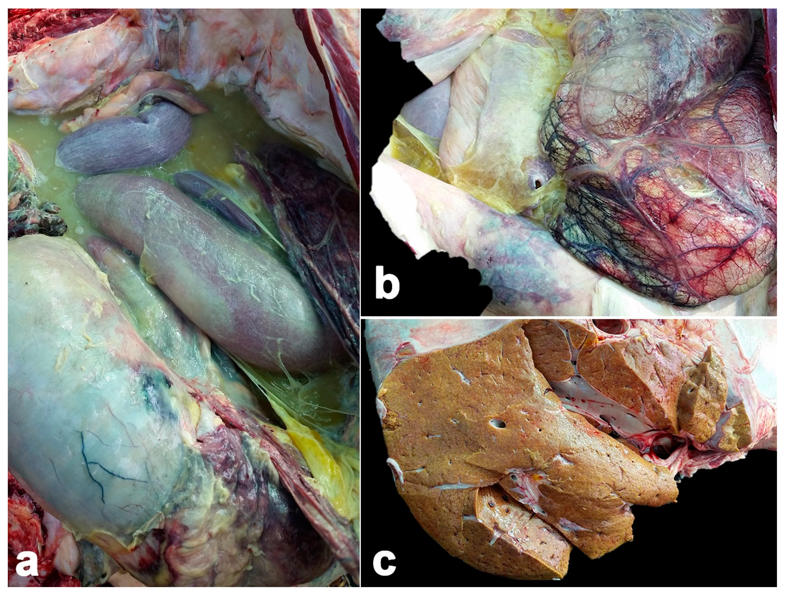
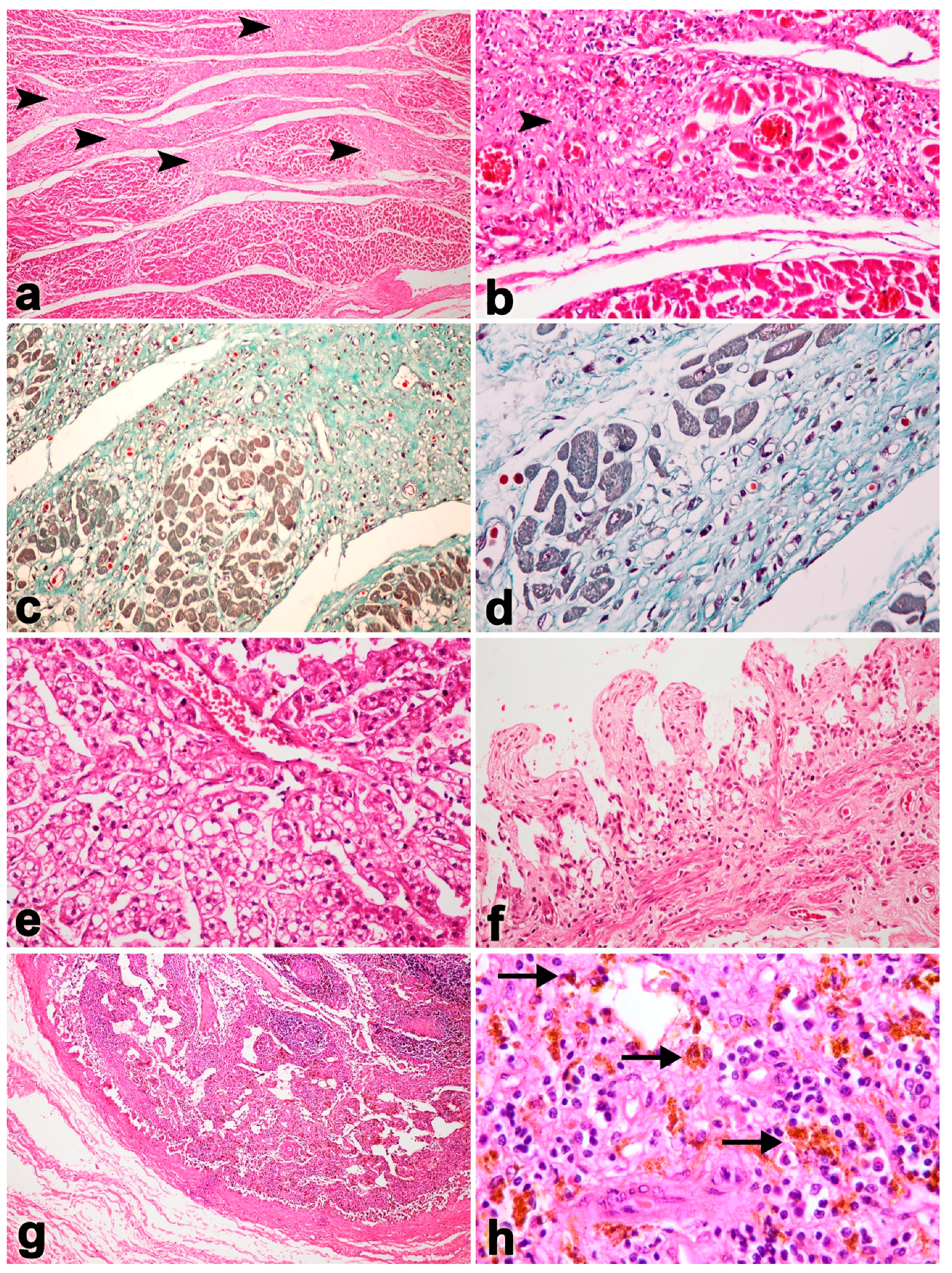
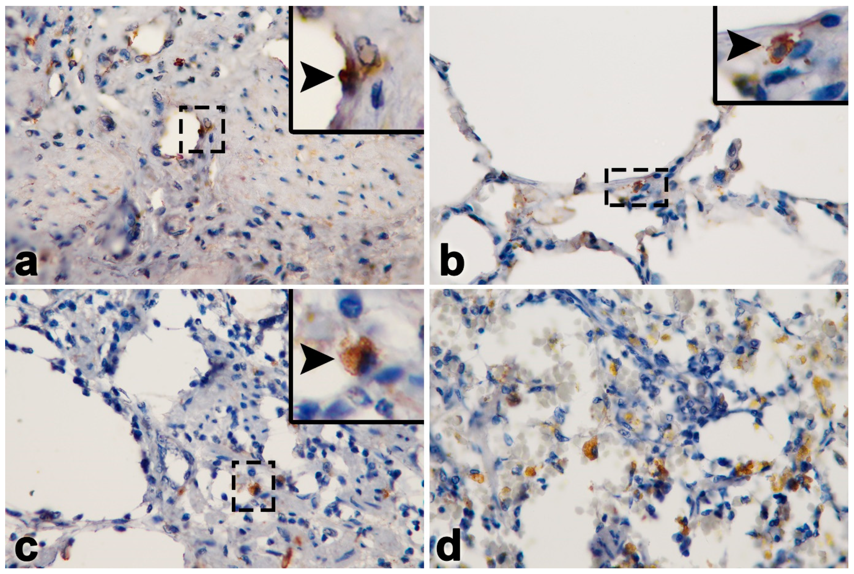
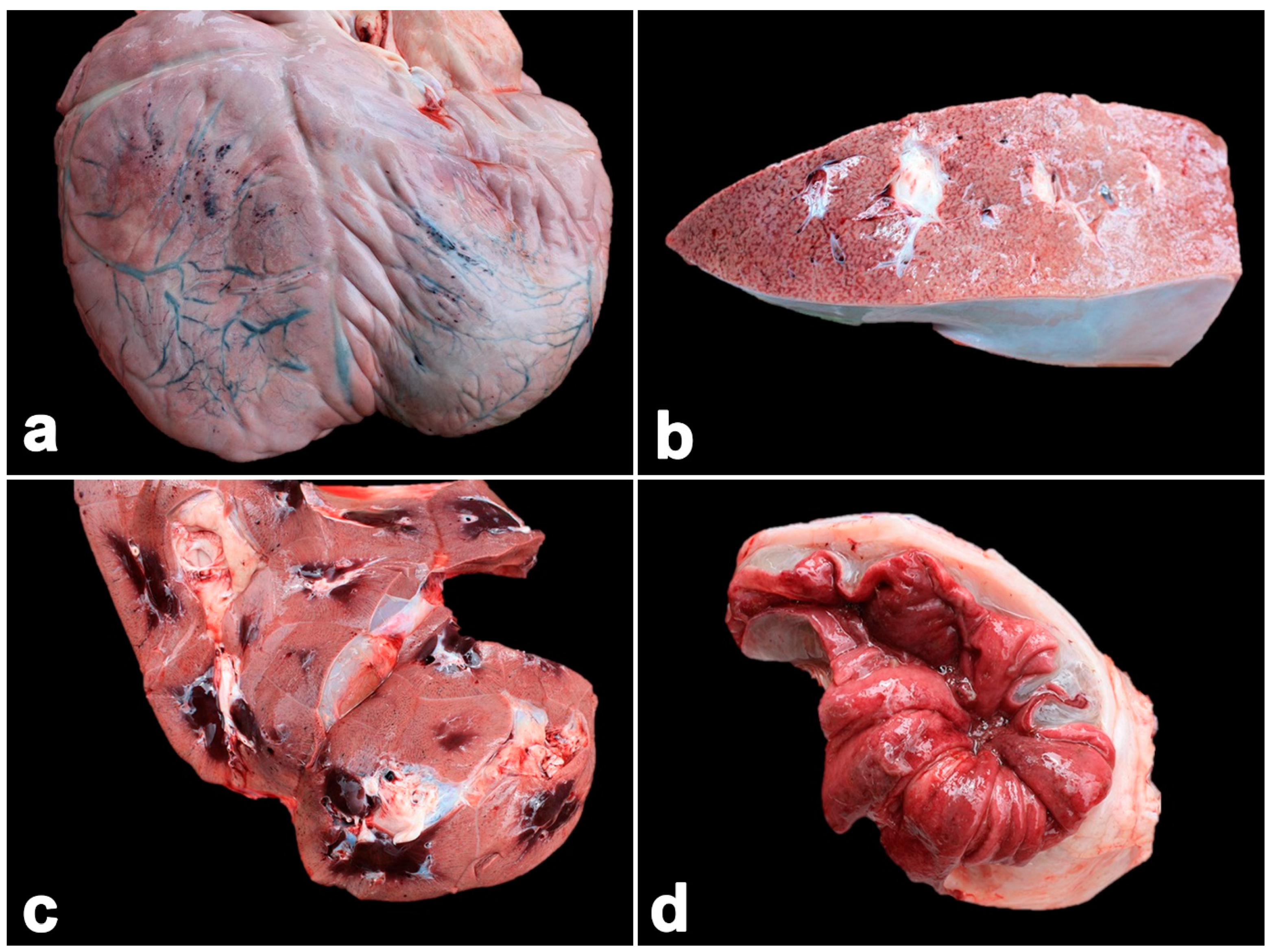
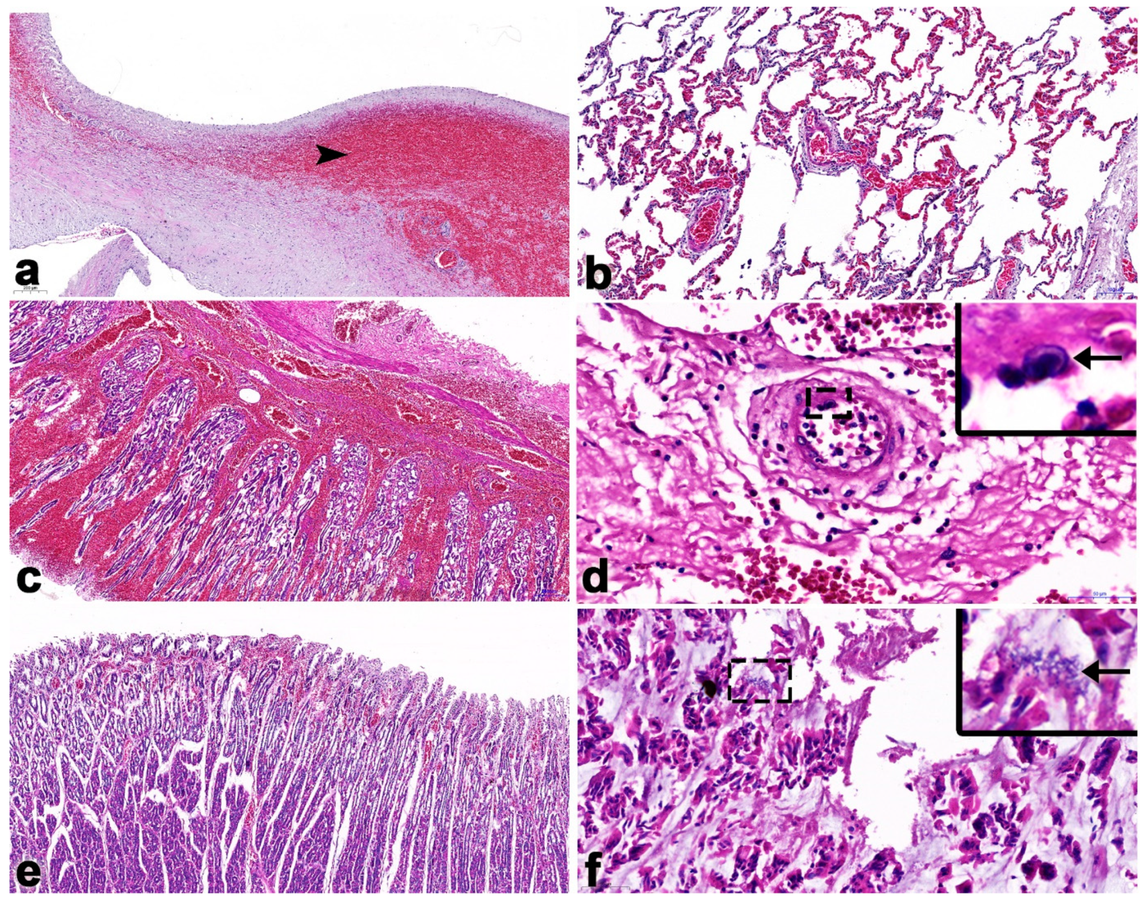
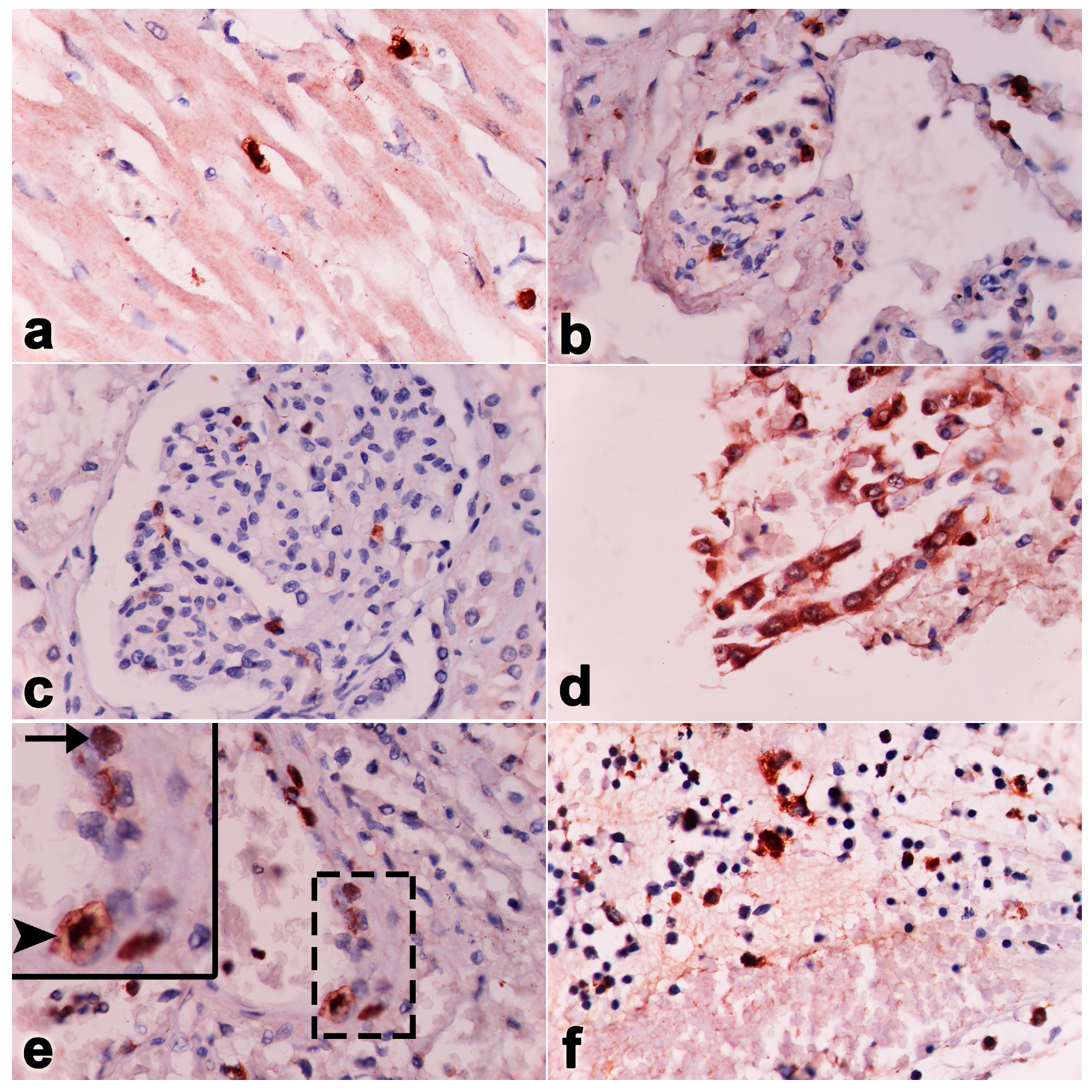
2.1. Case No. 1
2.2. Case No. 2
2.3. Case No. 3
3. Discussion
4. Conclusions
Author Contributions
Funding
Institutional Review Board Statement
Informed Consent Statement
Data Availability Statement
Acknowledgments
Conflicts of Interest
References
- McCully, R.M.; Basson, P.A.; Pienaar, J.G.; Erasmus, B.J.; Young, E. Herpes nodules in the lung of the African elephant (Loxodonta africana (Blumebach, 1792)). Onderstepoort J. Vet. Res. 1971, 38, 225–235. [Google Scholar]
- Garner, M.M.; Helmick, K.; Ochsenreiter, J.; Richman, L.K.; Latimer, E.; Wise, A.G.; Maes, R.K.; Kiupel, M.; Nordhausen, R.W.; Zong, J.C.; et al. Clinico-pathologic features of fatal disease attributed to new variants of endotheliotropic herpesviruses in two Asian elephants (Elephas maximus). Vet. Pathol. 2009, 46, 97–104. [Google Scholar] [CrossRef]
- Richman, L.K.; Montali, R.J.; Cambre, R.C.; Schmitt, D.; Hardy, D.; Hildbrandt, T.; Bengis, R.G.; Hamzeh, F.M.; Shahkolahi, A.; Hayward, G.S. Clinical and pathological findings of a newly recognized disease of elephants caused by endotheliotropic herpesviruses. J. Wildl. Dis. 2000, 36, 1–12. [Google Scholar] [CrossRef]
- Schmitt, D.L.; Hardy, D.A.; Montali, R.J.; Richman, L.K.; Lindsay, W.A.; Isaza, R.; West, G. Use of famciclovir for the treatment of endotheliotrophic herpesvirus infections in Asian elephants (Elephas maximus). J. Zoo Wildl. Med. 2000, 31, 518–522. [Google Scholar]
- Sripiboon, S.; Jackson, B.; Ditcham, W.; Holyoake, C.; Robertson, I.; Thitaram, C.; Tankaew, P.; Letwatcharasarakul, P.; Warren, K. Molecular characterisation and genetic variation of Elephant Endotheliotropic Herpesvirus infection in captive young Asian elephants in Thailand. Infect. Genet. Evol. 2016, 44, 487–494. [Google Scholar] [CrossRef]
- Richman, L.K.; Montali, R.J.; Garber, R.L.; Kennedy, M.A.; Lehnhardt, J.; Hildebrandt, T.; Schmitt, D.; Hardy, D.; Alcendor, D.J.; Hayward, G.S. Novel endotheliotropic herpesviruses fatal for Asian and African elephants. Science 1999, 283, 1171–1176. [Google Scholar] [CrossRef]
- Fickel, J.; Richman, L.K.; Montali, R.; Schaftenaar, W.; Goritz, F.; Hildebrandt, T.; Pitra, C. A variant of the endotheliotropic herpesvirus in Asian elephants (Elephas maximus) in European zoos. Vet. Microbiol. 2001, 82, 103–109. [Google Scholar] [CrossRef]
- Latimer, E.; Zong, J.C.; Heaggans, S.Y.; Richman, L.K.; Hayward, G.S. Detection and evaluation of novel herpesviruses in routine and pathological samples from Asian and African elephants: Identification of two new probosciviruses (EEHV5 and EEHV6) and two new gammaherpesviruses (EGHV3B and EGHV5). Vet. Microbiol. 2011, 147, 28–41. [Google Scholar] [CrossRef] [PubMed]
- Wilkie, G.S.; Davison, A.J.; Watson, M.; Kerr, K.; Sanderson, S.; Bouts, T.; Steinbach, F.; Dastjerdi, A. Complete genome sequences of elephant endotheliotropic herpesviruses 1A and 1B determined directly from fatal cases. J. Virol. 2013, 87, 6700–6712. [Google Scholar] [CrossRef] [PubMed]
- Long, S.Y.; Latimer, E.M.; Hayward, G.S. Review of Elephant Endotheliotropic Herpesviruses and Acute Hemorrhagic Disease. ILAR J. 2016, 56, 283–296. [Google Scholar] [CrossRef] [PubMed]
- Dastjerdi, A.; Seilern-Moy, K.; Darpel, K.; Steinbach, F.; Molenaar, F. Surviving and fatal Elephant Endotheliotropic Herpesvirus-1A infections in juvenile Asian elephants-lessons learned and recommendations on anti-herpesviral therapy. BMC Vet. Res. 2016, 12, 178. [Google Scholar] [CrossRef] [PubMed]
- Sripiboon, S.; Tankaew, P.; Lungka, G.; Thitaram, C. The occurrence of elephant endotheliotropic herpesvirus in captive Asian elephants (Elephas maximus): First case of EEHV4 in Asia. J. Zoo. Wildl. Med. 2013, 44, 100–104. [Google Scholar] [CrossRef] [PubMed]
- Boonprasert, K.; Punyapornwithaya, V.; Tankaew, P.; Angkawanish, T.; Sriphiboon, S.; Titharam, C.; Brown, J.L.; Somgird, C. Survival analysis of confirmed elephant endotheliotropic herpes virus cases in Thailand from 2006. PLoS ONE 2019, 14, e0219288. [Google Scholar] [CrossRef]
- Yun, Y.; Sripiboon, S.; Pringproa, K.; Chuammitri, P.; Punyapornwithaya, V.; Boonprasert, K.; Tankaew, P.; Angkawanish, T.; Namwongprom, K.; Arjkumpa, O.; et al. Clinical characteristics of elephant endotheliotropic herpesvirus (EEHV) cases in Asian elephants (Elephas maximus) in Thailand during 2006–2019. Vet Q. 2021, 41, 268–279. [Google Scholar] [CrossRef]
- Jesus, S.A.; Doherr, M.G.; Hildebrandt, T.B. Elephant Endotheliotropic Herpesvirus Impact in the European Asian Elephant (Elephas maximus) Population: Are Hereditability and Zoo-Associated Factors Linked with Mortality? Animals 2021, 11, 2816. [Google Scholar] [CrossRef]
- Seilern-Moy, K.; Haycock, J.; Dastjerdi, A.; Leifsson, P.S.; Bertelsen, M.F.; Perrin, K.L. Fatal elephant endotheliotropic herpesvirus-1 and -4 co-infection in a juvenile Asian elephant in Europe. JMM Case Rep. 2016, 3, 2. [Google Scholar] [CrossRef]
- Guntawang, T.; Sittisak, T.; Srivorakul, S.; Kochagul, V.; Photichai, K.; Thitaram, C.; Sthitmatee, N.; Hsu, W.-L.; Pringproa, K. In vivo characterization of target cells for acute elephant endotheliotropic herpesvirus (EEHV) infection in Asian elephants (Elephas maximus). Sci. Rep. 2020, 10, 11402. [Google Scholar] [CrossRef]
- Srivorakul, S.; Guntawang, T.; Kochagul, V.; Photichai, K.; Sittisak, T.; Janyamethakul, T.; Boonprasert, K.; Khammesri, S.; Langkaphin, W.; Punyapornwithaya, V.; et al. Possible roles of monocytes/macrophages in response to elephant endotheliotropic herpesvirus (EEHV) infections in Asian elephants (Elephas maximus). PLoS ONE 2019, 14, e0222158. [Google Scholar] [CrossRef]
- Stanton, J.J.; Zong, J.C.; Latimer, E.; Tan, J.; Herron, A.; Hayward, G.S.; Ling, P.D. Detection of pathogenic elephant endotheliotropic herpesvirus in routine trunk washes from healthy adult Asian elephants (Elephas maximus) by use of a real-time quantitative polymerase chain reaction assay. Am. J. Vet. Res. 2010, 71, 925–933. [Google Scholar] [CrossRef]
- Flower, M.E.; Mikota, S.K.; Hedges, S. Biology, Medicine, and Surgery of Elephants; Blackwell Publishing Ltd.: Ames, IA, USA, 2006; pp. 325–345. [Google Scholar]
- Boonprasert, K.; Yun, Y.; Kosaruk, W.; Towiboon, P.; Tankaew, P.; Punyapornwithaya, V.; Janyamathakul, T.; Muanghong, P.; Brown, J.L.; Thitaram, C. A Longitudinal Study of Hematology and Stress Biomarker Profiles in Young Asian Elephants (Elephas maximus) in Relation to Elephant Endotheliotropic Herpesvirus (EEHV) in Thailand. Animals 2021, 11, 2530. [Google Scholar] [CrossRef]
- Kochagul, V.; Srivorakul, S.; Boonsri, K.; Somgird, C.; Sthitmatee, N.; Thitaram, C.; Pringproa, K. Production of antibody against elephant endotheliotropic herpesvirus (EEHV) unveils tissue tropisms and routes of viral transmission in EEHV-infected Asian elephants. Sci. Rep. 2018, 8, 4675. [Google Scholar] [CrossRef]
- Boonsri, K.; Somgird, C.; Noinafai, P.; Pringproa, K.; Janyamethakul, T.; Angkawanish, T.; Brown, J.L.; Tankaew, P.; Srivorakul, S.; Thitaram, C. Elephant Endotheliotropic Herpesvirus Associated with Clostridium Perfringens Infection in Two Asian Elephant (Elephas maximus) Calves. J. Zoo. Wildl. Med. 2018, 49, 178–182. [Google Scholar] [CrossRef]
- van Asten, A.J.; van der Wiel, C.W.; Nikolaou, G.; Houwers, D.J.; Grone, A. A multiplex PCR for toxin typing of Clostridium perfringens isolates. Vet. Microbiol. 2009, 136, 411–412. [Google Scholar] [CrossRef]
- Kendall, R.; Howard, L.; Masters, N.; Grant, R. The Impact of Elephant Endotheliotropic Herpesvirus on the Captive Asian Elephant (Elephas maximus) Population of the United Kingdom and Ireland (1995–2013). J. Zoo Wildl. Med. 2016, 47, 405–418. [Google Scholar] [CrossRef]
- Fuery, A.; Tan, J.; Peng, R.; Flanagan, J.P.; Tocidlowski, M.E.; Howard, L.L.; Ling, P.D. Clinical Infection of Two Captive Asian Elephants (Elephas maximus) with Elephant Endotheliotropic Herpesvirus 1b. J. Zoo. Wildl. Med. 2016, 47, 319–324. [Google Scholar] [CrossRef] [PubMed]
- Fuery, A.; Browning, G.R.; Tan, J.; Long, S.; Hayward, G.S.; Cox, S.K.; Flanagan, J.P.; Tocidlowski, M.E.; Howard, L.L.; Ling, P.D. Clinical Infection of Captive Asian Elephants (Elephas maximus) with Elephant Endotheliotropic Herpesvirus. J. Zoo Wildl. Med. 2016, 47, 311–318. [Google Scholar] [CrossRef]
- Wissink-Argilaga, N.; Dastjerdi, A.; Molenaar, F.M. Using in-House Hematology to Direct Decision-Making in the Successful Treatment and Monitoring of a Clinical and Subsequently Subclinical Case of Elephant Endotheliotropic Herpesvirus 1b. J. Zoo Wildl. Med. 2019, 50, 498–502. [Google Scholar] [PubMed]
- Stacy, N.I.; Isaza, R.; Wiedner, E. First report of changes in leukocyte morphology in response to inflammatory conditions in Asian and African elephants (Elephas maximus and Loxodonta africana). PLoS ONE 2017, 12, e0185277. [Google Scholar] [CrossRef]
- Guntawang, T.; Sittisak, T.; Kochagul, V.; Srivorakul, S.; Photichai, K.; Boonsri, K.; Janyamethakul, T.; Boonprasert, K.; Langkaphin, W.; Thitaram, C.; et al. Pathogenesis of hemorrhagic disease caused by elephant endotheliotropic herpesvirus (EEHV) in Asian elephants (Elephas maximus). Sci. Rep. 2021, 11, 12998. [Google Scholar] [CrossRef] [PubMed]
- Ossent, P.; Guscetti, F.; Metzler, A.E.; Lang, E.M.; Rubel, A.; Hauser, B. Acute and fatal herpesvirus infection in a young Asian elephant (Elephas maximus). Vet. Pathol. 1990, 27, 131–133. [Google Scholar] [CrossRef] [PubMed]
- Wilkie, G.S.; Davison, A.J.; Kerr, K.; Stidworthy, M.F.; Redrobe, S.; Steinbach, F.; Dastjerdi, A.; Denk, D. First fatality associated with elephant endotheliotropic herpesvirus 5 in an Asian elephant: Pathological findings and complete viral genome sequence. Sci. Rep. 2014, 4, 6299. [Google Scholar] [CrossRef] [PubMed]
- Luz, S.; Howard, L.L. Guidline for management. In Elephant Endotheliotropic Herpesvirus (EEHV) in Asia, Recommendations from the 1st Asian EEHV strategy Meeting, 2nd ed.; Wildlife Reserves Singapore Group: Singapore, 2016. [Google Scholar]
- Brock, A.P.; Isaza, R.; Hunter, R.P.; Richman, L.K.; Montali, R.J.; Schmitt, D.L.; Koch, D.E.; Lindsay, W.A. Estimates of the pharmacokinetics of famciclovir and its active metabolite penciclovir in young Asian elephants (Elephas maximus). Am. J. Vet. Res. 2012, 73, 1996–2000. [Google Scholar] [CrossRef] [PubMed]
- Boonprasert, K.; Mahasawangkul, S.; Angkawanish, T.; Jansittiwej, S.; Langkaphin, W.; Sombutputorn, P.; Bampenpol, P.; Mahanil, W. Endotheliotropic Herpesvirus Treatments in a Wild Orphan Baby Asian Elephant (Elephas maximus); Crown Plaza River Oaks: Houston, TX, USA, 2015. [Google Scholar]
- Khammesri, S.; Mathura, Y.; Boonprasert, K.; Ampasavate, C.; Hongwiset, D.; Brown, J.L.; Thitaram, C. Successful treatment of elephant endotheliotropic herpesvirus infection in an Asian elephant (Elephas maximus) calf by oral acyclovir medication: Case report. J. Vet. Med. Sci. 2021, 83, 125–129. [Google Scholar] [CrossRef] [PubMed]
- Khammesri, S.; Ampasavate, C.; Hongwiset, D.; Mektrirat, R.; Sangsrijan, S.; Brown, J.L.; Thitaram, C. Pharmacokinetics and analytical determination of acyclovir in Asian elephant calves (Elephas maximus). Vet. Anim. Sci. 2022, 15, 100227. [Google Scholar] [CrossRef]

| Parameter | Normal Range a | Normal Range b | Day 3 | Day 5 | Day 8 | Day 11 | Day 19 |
|---|---|---|---|---|---|---|---|
| PCV (%) | 30–40 | 32.25–43.80 | 24 | 16 | 17.5 | 14 | 15.3 |
| HGB (g/dL) | 11–15 | - | 9.5 | 8.9 | 6.8 | 5.8 | 6 |
| RBC (×106 cells/µL) | 2.5–5.0 | 2.17–3.47 | 2.20 | 2.06 | 1.60 | 1.35 | 1.4 |
| WBC (cells/µL) | 10,000–18,000 | 2000–4000 | 23,360 | 19,550 | 22,900 | 15,540 | 13,650 |
| Heterophil (cells/µL) | 1.18–3.57 | 0.41–1.17 | 0.30 | 0.24 | 0.31 | 0.28 | 0.28 |
| Lymphocyte (cells/µL) | 5000–8000 | 6918–13,980 | 13,548 | 9970 | 6412 | 4972 | 4360 |
| Monocyte (cells/µL) | 2000–4000 | 2316–5578 | 2102 | 1564 | 2290 | 2175 | 1898 |
| Eosinophils (cells/µL) | 100–1000 | 0–535 | - | 1368 | 6897 | 621 | 546 |
| M:H ratio | 1.18–3.57 | 0.41–1.17 | 0.30 | 0.24 | 0.31 | 0.28 | 0.28 |
| Platelet (×103 cells/µL) | 200–600 | 374–554 | 97 | 205 | 377 | 466 | 369 |
| Creatinine (mg/dL) | 1.0–2.0 | - | 3.8 | 6.6 | 13.6 | 1.3 | 3 |
| BUN (mg/dL) | 5–20 | - | 30 | 35 | 39 | 12 | 63 |
| AST (U/L) | 15–35 | - | 31 | 35 | 39 | 12 | 63 |
| ALP (U/L) | 60–450 | - | 98 | 146 | 176 | 15 | - |
| GLU (mg/dL) | 60–116 | - | 52 | 21 | 64 | 10 | - |
| TP (g/dL) | 6–12 | - | 6.6 | 8 | 8.2 | 8.1 | 6.9 |
| CK (U/L) | 5–250 | - | - | - | - | - | 395 |
| Day Test | Ct (EEHV1A) | Ct (EEHV4) |
|---|---|---|
| Day 1 | 28.06 | 37.14 |
| Day 3 | 28.97 | 35.17 |
| Day 4 | 29.11 | 35.87 |
| Day 5 | 29.00 | Undetected |
| Day 6 | 31.05 | Undetected |
| Day 7 | 30.37 | 37.20 |
| Day 8 | 30.69 | Undetected |
| Day 9 | 32.63 | 38.15 |
| Day 10 | 32.18 | 37.27 |
| Day 14 | 33.05 | Undetected |
| Day 17 | 33.19 | 37.08 |
| Day 28 | 31.34 | Undetected |
| Day 34 | 31.05 | 36.83 |
| Tissue | Ct (EEHV1A) | Ct (EEHV4) |
|---|---|---|
| Heart | 28.67 | Undetected |
| Liver | 29.80 | 33.45 |
| Intestine | 32.85 | 38.08 |
| Lymph node | 32.31 | Undetected |
| Feces | 25.97 | 31.15 |
| Parameter | Normal Range a | Normal Range b | Day 2 |
|---|---|---|---|
| PCV (%) | 30–40 | 32.25–43.80 | 55 |
| HGB (g/dL) | 11–15 | - | 20.4 |
| RBC (×106 cells/µL) | 2.5–5.0 | 2.17–3.47 | 4.73 |
| WBC (cells/µL) | 10,000–18,000 | 2000–4000 | 22,060 |
| Heterophil (cells/µL) | 1.18–3.57 | 0.41–1.17 | 12,795 |
| Lymphocyte (cells/µL) | 5000–8000 | 6918–13,980 | 5736 |
| Monocyte (cells/µL) | 2000–4000 | 2316–5578 | 3530 |
| Eosinophils (cells/µL) | 100–1000 | 0–535 | - |
| M:H ratio | 1.18–3.57 | 0.41–1.17 | 0.28 |
| Platelets (×103 cells/µL) | 200–600 | 374–554 | 173 |
| Creatinine (mg/dL) | 1.0–2.0 | - | 1.93 |
| BUN (mg/dL) | 5–20 | - | 20.1 |
| AST (U/L) | 15–35 | - | 54 |
| ALP (U/L) | 60–450 | - | 129 |
| GLU (mg/dL) | 60–116 | - | - |
| TP (g/dL) | 6–12 | - | 7.1 |
| CK (U/L) | 5–250 | - | - |
Disclaimer/Publisher’s Note: The statements, opinions and data contained in all publications are solely those of the individual author(s) and contributor(s) and not of MDPI and/or the editor(s). MDPI and/or the editor(s) disclaim responsibility for any injury to people or property resulting from any ideas, methods, instructions or products referred to in the content. |
© 2024 by the authors. Licensee MDPI, Basel, Switzerland. This article is an open access article distributed under the terms and conditions of the Creative Commons Attribution (CC BY) license (https://creativecommons.org/licenses/by/4.0/).
Share and Cite
Boonprasert, K.; Srivorakul, S.; Monchaivanakit, N.; Langkaphin, W.; Sripiboon, S.; Janyamethakul, T.; Srisa-ad, C.; Guntawang, T.; Brown, J.L.; Thitaram, C.; et al. Characterizations of Elephant Endotheliotropic Herpesvirus Type 1A and 4 Co-Infections in Asian Elephant (Elephas maximus) Calves. Vet. Sci. 2024, 11, 147. https://doi.org/10.3390/vetsci11040147
Boonprasert K, Srivorakul S, Monchaivanakit N, Langkaphin W, Sripiboon S, Janyamethakul T, Srisa-ad C, Guntawang T, Brown JL, Thitaram C, et al. Characterizations of Elephant Endotheliotropic Herpesvirus Type 1A and 4 Co-Infections in Asian Elephant (Elephas maximus) Calves. Veterinary Sciences. 2024; 11(4):147. https://doi.org/10.3390/vetsci11040147
Chicago/Turabian StyleBoonprasert, Khajohnpat, Saralee Srivorakul, Natcha Monchaivanakit, Warangkhana Langkaphin, Supaphen Sripiboon, Thittaya Janyamethakul, Channarong Srisa-ad, Thunyamas Guntawang, Janine L. Brown, Chatchote Thitaram, and et al. 2024. "Characterizations of Elephant Endotheliotropic Herpesvirus Type 1A and 4 Co-Infections in Asian Elephant (Elephas maximus) Calves" Veterinary Sciences 11, no. 4: 147. https://doi.org/10.3390/vetsci11040147
APA StyleBoonprasert, K., Srivorakul, S., Monchaivanakit, N., Langkaphin, W., Sripiboon, S., Janyamethakul, T., Srisa-ad, C., Guntawang, T., Brown, J. L., Thitaram, C., & Pringproa, K. (2024). Characterizations of Elephant Endotheliotropic Herpesvirus Type 1A and 4 Co-Infections in Asian Elephant (Elephas maximus) Calves. Veterinary Sciences, 11(4), 147. https://doi.org/10.3390/vetsci11040147