
Analysis of Trypanosoma equiperdum Recombinant Proteins for the Serological Diagnosis of Dourine

 , , , , , , , , , , , , and
Simple Summary
Abstract
1. Introduction
2. Materials and Methods
2.1. Expression and Production of T. equiperdum Recombinant Proteins
2.2. Serum Panel
2.3. Indirect ELISA
2.4. Immunoblotting
3. Results
3.1. Expression and Production of T. equiperdum Recombinant Proteins
3.2. Indirect ELISA
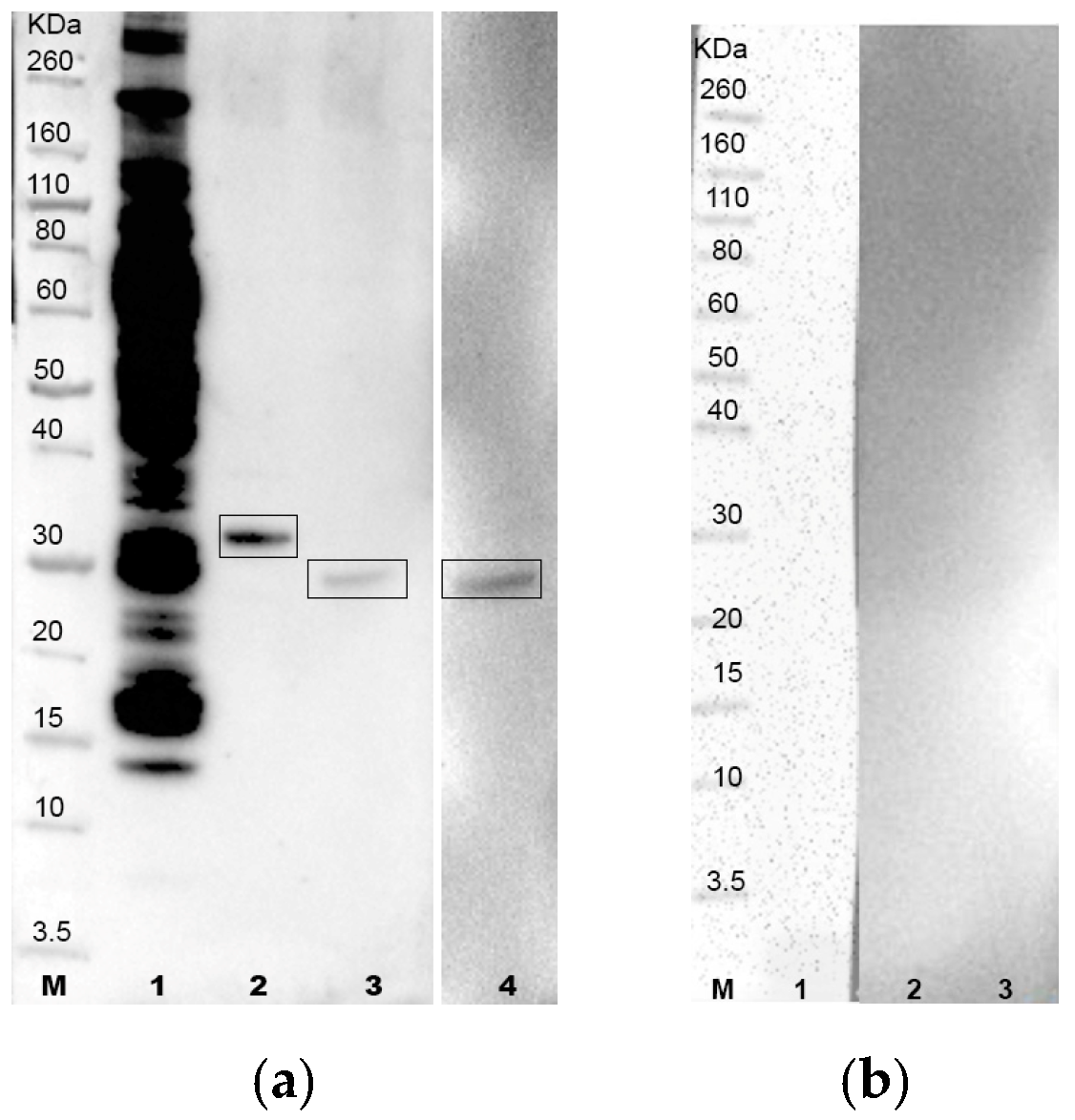
3.3. Immunoblotting
4. Discussion
5. Conclusions
Supplementary Materials
Author Contributions
Funding
Institutional Review Board Statement
Informed Consent Statement
Data Availability Statement
Conflicts of Interest
References
- World Organisation for Animal Health (Ed.) Chapter 3.6.3. Dourine in horses (Trypanosoma equiperdum infection). In Manual of Diagnostic Tests and Vaccines for Terrestrial Animals; World Organisation for Animal Health: Paris, France, 2021; pp. 1–13. [Google Scholar]
- Sidney, R.; Andrew, M.G.; James, C.; Richard, N. Dourine–an emerging venereal threat to European horses. AHT/BEVA/DEFrA Equine Q. Dis. Surveill. Rep. 2013, 6, 7. [Google Scholar]
- Gizaw, Y.; Megersa, M.; Fayera, T. Dourine: A neglected disease of equids. Trop. Anim. Health Prod. 2017, 49, 887–897. [Google Scholar] [CrossRef]
- Brun, R.; Hecker, H.; Lun, Z.R. Trypanosoma evansi and T-equiperdum: Distribution, biology, treatment and phylogenetic relationship (a review). Vet. Parasitol. 1998, 79, 95–107. [Google Scholar] [CrossRef]
- Desquesnes, M.; Holzmuller, P.; Lai, D.; Dargantes, A.; Lun, Z.; Jittaplapong, S. Trypanosoma evansi and Surra: A Review and Perspectives on Origin, History, Distribution, Taxonomy, Morphology, Hosts, and Pathogenic Effects. Biomed Res. Int. 2013, 2013, 194176. [Google Scholar] [CrossRef]
- Carnes, J.; Anupama, A.; Balmer, O.; Jackson, A.; Lewis, M.; Brown, R.; Cestari, I.; Desquesnes, M.; Gendrin, C.; Hertz-Fowler, C.; et al. Genome and Phylogenetic Analyses of Trypanosoma evansi Reveal Extensive Similarity to T. brucei and Multiple Independent Origins for Dyskinetoplasty. PLoS Neglect. Trop. Dis. 2015, 9, e3404. [Google Scholar] [CrossRef]
- Calistri, P.; Narcisi, V.; Atzeni, M.; De Massis, F.; Tittarelli, M.; Mercante, M.T.; Ruggieri, E.; Scacchia, M. Dourine Reemergence in Italy. J. Equine Vet. Sci. 2013, 33, 83–89. [Google Scholar] [CrossRef]
- Hébert, L.; Moumen, B.; Madeline, A.; Steinbiss, S.; Lakhdar, L.; Van Reet, N.; Büscher, P.; Laugier, C.; Cauchard, J.; Petry, S. First Draft Genome Sequence of the Dourine Causative Agent: Trypanosoma Equiperdum Strain OVI. J. Genom. 2017, 5, 1–3. [Google Scholar] [CrossRef] [PubMed]
- Mendoza, E.; Bubis, J.; Pérez-Rojas, Y.; Montilla, A.J.; Spencer, L.M.; Bustamante, F.; Martínez, J.C. High immunological response against a Trypanosoma equiperdum protein that exhibits homology with the regulatory subunits of mammalian cAMP-dependent protein kinases. J. Immunoass. Immunochem. 2018, 39, 451–469. [Google Scholar] [CrossRef]
- Luciani, M.; Di Pancrazio, C.; Di Febo, T.; Tittarelli, M.; Vulpiani, M.P.; Puglielli, M.O.; Naessens, J.; Sacchini, F. IgG antibodies from dourine infected horses identify a distinctive Trypanosoma equiperdum antigenic pattern of low molecular weight molecules. Vet. Immunol. Immunopathol. 2013, 151, 140–146. [Google Scholar] [CrossRef] [PubMed]
- Luciani, M.; Di Febo, T.; Orsini, M.; Krasteva, I.; Cattaneo, A.; Vulpiani, M.P.; Di Pancrazio, C.; Bachi, A.; Tittarelli, M. Trypanosoma equiperdum Low Molecular Weight Proteins As Candidates for Specific Serological Diagnosis of Dourine. Front. Vet. Sci. 2018, 5, 40. [Google Scholar] [CrossRef] [PubMed]
- Hopkins, R.; Esposito, D. A rapid method for titrating baculovirus stocks using the Sf-9 Easy Titer cell line. BioTechniques 2009, 47, 785–788. [Google Scholar] [CrossRef]
- Di Febo, T.; Luciani, M.; Ciarelli, A.; Bortone, G.; Di Pancrazio, C.; Rodomonti, D.; Teodori, L.; Tittarelli, M. Production and characterization of monoclonal antibodies against horse immunoglobulins useful for the diagnosis of equine diseases. J. Immunoass. Immunochem. 2015, 36, 253–264. [Google Scholar] [CrossRef] [PubMed]
- Zou, K.H.; O’Malley, A.J.; Mauri, L. Receiver-operating characteristic analysis for evaluating diagnostic tests and predictive models. Circulation 2007, 115, 654–657. [Google Scholar] [CrossRef]
- World Organisation for Animal Health. Trypanosoma evansi infections (including Surra). In Manual of Diagnostic Tests and Vaccines for Terrestrial Animals, OIE Technical Disease Cards; World Organisation for Animal Health: Paris, France, 2013; pp. 1–4. [Google Scholar]
- Vulpiani, M.P.; Carvelli, A.; Giansante, D.; Iannino, F.; Paganico, D.; Ferri, N. Reemergence of Dourine in Italy: Clinical Cases in Some Positive Horses. J. Equine Vet. Sci. 2012, 33, 468–474. [Google Scholar] [CrossRef]
- Desquesnes, M.; Bossard, G.; Patrel, D.; Herder, S.; Patout, O.; Lepetitcolin, E.; Thevenon, S.; Berthier, D.; Pavlovic, D.; Brugidou, R.; et al. First outbreak of Trypanosoma evansi in camels in metropolitan France. Vet. Rec. 2008, 162, 750–752. [Google Scholar] [CrossRef] [PubMed]
- Ferreira, A.W.; Belem, Z.R.; Lemos, A.A.; Reed, S.G.; Campos-Neto, A. Enzyme-linked immunosorbent assay for serological diagnosis of chagas’ disease employing a Trypanosoma cruzi recombinant antigen that consists of four different peptides. J. Clin. Microbiol. 2001, 39, 4390–4395. [Google Scholar] [CrossRef]
- Hernandez, P.; Heimann, M.; Riera, C.; Solano, M.; Santalla, J.; Luquetti, A.O.; Beck, E. Highly Effective Serodiagnosis for Chagas’ Disease. Clin. Vaccine Immunol. 2010, 17, 1598–1604. [Google Scholar] [CrossRef] [PubMed]
- Camussone, C.; Gonzalez, V.; Belluzo, M.S.; Pujato, N.; Ribone, M.E.; Lagier, C.M.; Marcipar, I.S. Comparison of Recombinant Trypanosoma cruzi Peptide Mixtures versus Multiepitope Chimeric Proteins as Sensitizing Antigens for Immunodiagnosis. Clin. Vaccine Immunol. 2009, 16, 899–905. [Google Scholar] [CrossRef]
- Maria Peverengo, L.; Garcia, V.; Maria Rodeles, L.; Mendicino, D.; Vicco, M.; Lagier, C.; Gonzalez, V.; Gugliotta, L.; Marcipar, I. Development and assessment of an improved recombinant multiepitope antigen-based immunoassay to diagnose chronic Chagas disease. Parasitology 2018, 145, 1594–1599. [Google Scholar] [CrossRef]
- Caicedo Diaz, R.A.; Forsyth, C.; Bernal, O.A.; Marchiol, A.; Beltran Duran, M.; Batista, C.; Herazo, R.; Javier Vera, M.; Pachon Abril, E.; Andres Valencia-Hernandez, C.; et al. Comparative evaluation of immunoassays to improve access to diagnosis for Chagas disease in Colombia. Int. J. Infect. Dis. 2019, 87, 100–108. [Google Scholar] [CrossRef]
- Kruecken, J.; Greif, G.; von Samson-Himmelstjerna, G. In silico analysis of the cyclophilin repertoire of apicomplexan parasites. Parasites Vectors 2009, 2, 27. [Google Scholar] [CrossRef]
- Ünal, C.M.; Steinert, M. Microbial peptidyl-prolyl cis/trans isomerases (PPIases): Virulence factors and potential alternative drug targets. Microbiol. Mol. Biol. Rev. 2014, 78, 544–571. [Google Scholar] [CrossRef]
- UniProt Consortium UniProt: A worldwide hub of protein knowledge. Nucleic Acids Res. 2019, 47, D506–D515. [CrossRef]
- Lipatova, Z.; Hain, A.U.; Nazarko, V.Y.; Segev, N. Ypt/Rab GTPases: Principles learned from yeast. Crit. Rev. Biochem. Mol. Biol. 2015, 50, 203–211. [Google Scholar] [CrossRef] [PubMed]
- Kim, J.J.; Lipatova, Z.; Segev, N. TRAPP Complexes in Secretion and Autophagy. Front. Cell Dev. Biol. 2016, 4, 20. [Google Scholar] [CrossRef] [PubMed]
- Leibly, D.J.; Nguyen, T.N.; Kao, L.T.; Hewitt, S.N.; Barrett, L.K.; Van Voorhis, W.C. Stabilizing additives added during cell lysis aid in the solubilization of recombinant proteins. PLoS ONE 2012, 7, e52482. [Google Scholar] [CrossRef] [PubMed]
- Verney, M.; Gautron, M.; Lemans, C.; Rince, A.; Hans, A.; Hebert, L. Development of a microsphere-based immunoassay for the serological diagnosis of equine trypanosomosis. Sci. Rep. 2022, 12, 1308. [Google Scholar] [CrossRef]
- Russell, W.M.S.; Burch, R.L. The Principles of Humane Experimental Technique; Methuen: London, UK, 1959. [Google Scholar]


| Protein | Sequence (FASTA) | Protein Length (Aminoacids) |
|---|---|---|
| A0A1G4I8N3 | MPKVSVPPKTERHGKIEAPETNNPKVFFDVSINEKAAGRIVMELYADTVPRTA ENFRALCTGEKGRGKSGKPLHYKGCIFHRVIPGFMLQGGDITRGNGTGGESIYGTTFR DESFAGKAGKHTGLGCLSMANAGPNTNGSQFFICTANTPWLNGKHVVFGRVTE GIDVVKSIERLGSDSGKTRGRIIIANCGELQTQKEGAAKKEKVKAKEGNNAGKLSTI VEGTGGAKRPREDVEDLEERKKRIREKRERIAKLRAQAEEKHHHHHH | 268 |
| A0A1G4I464 | MRALTFRSSLCAGRTAVGAMCFGRLWASSTSPTEGSEKQNVTED SETVSVAPVSPEAYAKLEKELSDAKERIAELKKEVLYRAADAENARRIGSEDVTKAKAY GITSFGKDMLDVVDTLERGLEAITKLPQAEVEGHKTLSSIHTGIKLSLKLLLNNLA KHGIEKLDVAVGAKFDPNFHDALLKVPPTAEAPPGHISTVLKTGYKIQDRVLRA SQVGVASDDHHHHHH | 228 |
| A0A1G4I740 | MSQHGLVSESVVSELSLELVSYALRGNSQKKEINFCRNKEVDTEFG SKGIERLGLLVGLRSAERLLYREATFGGSTPNDVARFVGQHLWKTVFGKKVDRMKH MDKIYFCLIDNNFRWLQGFSDAKSDQTVSAVDGYPYDSSEKYCGGDGPTDQGKESG SVLPPDSDVLRYAVSILRGFVQVMYPSGPIKIQASRNEKGETQFVLDFRSVA THHHHHH | 217 |
| Protein | Cloning Strategy |
|---|---|
| A0A1G4I8N3 | EcoRI-Kozak sequence-A0A1G4I8N3-His—stop codon—HindIII |
| A0A1G4I464 | EcoRI-Kozak sequence-A0A1G4I464-His—stop codon—HindIII |
| A0A1G4I740 | NdeI-ATG-A0A1G4I740-His tag—Stop codon—HindIII |
| Complement Fixation | ||||
|---|---|---|---|---|
| Positive | Negative | Total | ||
| Indirect ELISA A0A1G4I8N3 | Positive | 13 | 37 | 50 |
| Negative | 2 | 43 | 45 | |
| Total | 15 | 80 | 95 | |
| Diagnostic sensitivity | 86.7% | C.L. (95%) 53.7–96.0 | ||
| Diagnostic specificity | 53.8% | C.L. (95%) 42.9–64.3 | ||
| Diagnostic accuracy | 59.0% | C.L. (95%) 48.9–68.3 | ||
| Complement Fixation | ||||
|---|---|---|---|---|
| Positive | Negative | Total | ||
| Indirect ELISA A0A1G4I464 | Positive | 8 | 33 | 41 |
| Negative | 7 | 47 | 54 | |
| Total | 15 | 80 | 95 | |
| Diagnostic sensitivity | 53.3% | C.L. (95%) 23.6–75.3 | ||
| Diagnostic specificity | 58.7% | C.L. (95%) 47.8–68.9 | ||
| Diagnostic accuracy | 57.9% | C.L. (95%) 47.8–67.3 | ||
| Complement Fixation | ||||
|---|---|---|---|---|
| Positive | Negative | Total | ||
| Indirect ELISA A0A1G4I740 | Positive | 11 | 28 | 39 |
| Negative | 4 | 52 | 56 | |
| Total | 15 | 80 | 95 | |
| Diagnostic sensitivity | 73.3% | C.L. (95%) 47.6–89.0 | ||
| Diagnostic specificity | 65.0% | C.L. (95%) 54.0–74.6 | ||
| Diagnostic accuracy | 66.3% | C.L. (95%) 56.3–75.0 | ||
| Complement Fixation | ||||
|---|---|---|---|---|
| Positive | Negative | Total | ||
| Immunoblotting A0A1G4I8N3 | Positive | 13 | 6 | 19 |
| Negative | 2 | 74 | 76 | |
| Total | 15 | 80 | 95 | |
| Diagnostic sensitivity | 86.7% | C.L. (95%) 61.7–96.0 | ||
| Diagnostic specificity | 92.5% | C.L. (95%) 84.6–96.5 | ||
| Diagnostic accuracy | 91.6% | C.L. (95%) 84.2–95.6 | ||
| Complement Fixation | ||||
|---|---|---|---|---|
| Positive | Negative | Total | ||
| Immunoblotting A0A1G4I464 | Positive | 7 | 15 | 22 |
| Negative | 8 | 65 | 73 | |
| Total | 15 | 80 | 95 | |
| Diagnostic sensitivity | 46.7% | C.L. (95%) 24.7–70.1 | ||
| Diagnostic specificity | 81.3% | C.L. (95%) 71.3–88.3 | ||
| Diagnostic accuracy | 75.8% | C.L. (95%) 66.3–83.3 | ||
| Complement Fixation | ||||
|---|---|---|---|---|
| Positive | Negative | Total | ||
| Immunoblotting A0A1G4I740 | Positive | 12 | 29 | 41 |
| Negative | 3 | 51 | 54 | |
| Total | 15 | 80 | 95 | |
| Diagnostic sensitivity | 80.0% | C.L. (95%) 54.4–92.7 | ||
| Diagnostic specificity | 63.8% | C.L. (95%) 52.8–73.4 | ||
| Diagnostic accuracy | 66.3% | C.L. (95%) 56.3–75.0 | ||
Disclaimer/Publisher’s Note: The statements, opinions and data contained in all publications are solely those of the individual author(s) and contributor(s) and not of MDPI and/or the editor(s). MDPI and/or the editor(s) disclaim responsibility for any injury to people or property resulting from any ideas, methods, instructions or products referred to in the content. |
© 2024 by the authors. Licensee MDPI, Basel, Switzerland. This article is an open access article distributed under the terms and conditions of the Creative Commons Attribution (CC BY) license (https://creativecommons.org/licenses/by/4.0/).
Share and Cite
Luciani, M.; Armillotta, G.; Di Febo, T.; Krasteva, I.; Ulisse, S.; Di Pancrazio, C.; Laguardia, C.; Perletta, F.; Serroni, A.; Maggetti, M.; et al. Analysis of Trypanosoma equiperdum Recombinant Proteins for the Serological Diagnosis of Dourine. Vet. Sci. 2024, 11, 127. https://doi.org/10.3390/vetsci11030127
Luciani M, Armillotta G, Di Febo T, Krasteva I, Ulisse S, Di Pancrazio C, Laguardia C, Perletta F, Serroni A, Maggetti M, et al. Analysis of Trypanosoma equiperdum Recombinant Proteins for the Serological Diagnosis of Dourine. Veterinary Sciences. 2024; 11(3):127. https://doi.org/10.3390/vetsci11030127
Chicago/Turabian StyleLuciani, Mirella, Gisella Armillotta, Tiziana Di Febo, Ivanka Krasteva, Simonetta Ulisse, Chiara Di Pancrazio, Caterina Laguardia, Fabrizia Perletta, Anna Serroni, Marta Maggetti, and et al. 2024. "Analysis of Trypanosoma equiperdum Recombinant Proteins for the Serological Diagnosis of Dourine" Veterinary Sciences 11, no. 3: 127. https://doi.org/10.3390/vetsci11030127
APA StyleLuciani, M., Armillotta, G., Di Febo, T., Krasteva, I., Ulisse, S., Di Pancrazio, C., Laguardia, C., Perletta, F., Serroni, A., Maggetti, M., Testa, L., Sacchini, F., Iorio, M., Rodomonti, D., Tittarelli, M., & Mercante, M. T. (2024). Analysis of Trypanosoma equiperdum Recombinant Proteins for the Serological Diagnosis of Dourine. Veterinary Sciences, 11(3), 127. https://doi.org/10.3390/vetsci11030127