Article

Epidemiology and Molecular Characterisation of Multidrug-Resistant Escherichia coli Isolated from Cow Milk

1
Faculty of Veterinary Medicine, Chattogram Veterinary and Animal Sciences University, Chattogram 4225, Bangladesh
2
Faculty of Life Sciences: Food, Nutrition and Health, University of Bayreuth, 95447 Bayreuth, Germany
3
School of Psychology, University of New England, Armidale, NSW 2350, Australia
4
Nutritional Sciences Graduate Program, Margaret Ritchie School of Family and Consumer Sciences, College of Agricultural & Life Sciences, University of Idaho, Moscow, ID 83844, USA
5
Global Health and Development Program, Laney Graduate School, Emory University, Atlanta, GA 30322, USA
6
Faculty of Science and Engineering, Southern Cross University, East Lismore, NSW 2480, Australia
7
Plant Biosecurity and Product Integrity, Biosecurity Queensland, Department of Agriculture and Fisheries, Brisbane, QLD 4000, Australia
8
Queensland Alliance for One Health Sciences, School of Veterinary Science, The University of Queensland, Gatton, QLD 4343, Australia
*
Authors to whom correspondence should be addressed.
These authors contributed equally to this work.
Vet. Sci. 2024, 11(12), 609; https://doi.org/10.3390/vetsci11120609
Submission received: 27 October 2024 / Revised: 26 November 2024 / Accepted: 27 November 2024 / Published: 29 November 2024

Simple Summary

Antimicrobial resistance (AMR) is a growing global issue that poses serious public health risks. This study investigated the prevalence and pattern of antimicrobial resistance of Escherichia coli in raw cow milk from 18 farms in Chattogram, Bangladesh. Out of 450 samples, 134 (29.77%) tested positive for E. coli. Antimicrobial susceptibility testing revealed high resistance rates (69.40%) to ampicillin, amoxicillin–clavulanic acid, cephalothin, and cephalexin, while resistance to norfloxacin was lowest (21.64%). All isolates were multidrug-resistant (MDR), showing resistance to three or more antimicrobial classes, with a multiple resistance index >0.2. PCR testing detected the blaTEM gene in 74.19% of isolates, the highest among extended-spectrum beta-lactamase (ESBL) genes. The blaCMY-1 gene was less prevalent (6.45%), and the tetD gene was rare (2.9%). Positive correlations were noted between antimicrobial resistance and resistance gene presence, with a strong link (r = 1) between ciprofloxacin and ceftazidime resistance. This study highlights the significant presence of MDR E. coli in raw milk, posing a potential public health threat through the food chain. It calls for urgent measures to manage AMR, including prudent antimicrobial use, enhanced surveillance, and targeted interventions in Bangladesh’s dairy sector.

Abstract

Antimicrobial resistance (AMR) is a growing global concern and poses a significant threat to public health. The emergence of multidrug-resistant organisms, including Escherichia coli, also presents a risk of transmission to humans through the food chain, including milk. This study aimed to investigate the prevalence of E. coli in raw milk in the Chattogram metropolitan area (CMA) of Bangladesh and their phenotypic and genotypic antimicrobial resistance patterns. A total of 450 raw cow milk samples were collected from 18 farms within the CMA. The isolation and identification of E. coli were performed following standard bacteriological methods. Antimicrobial susceptibility testing (AST) was conducted using the Kirby–Bauer disc diffusion method. Molecular detection of E. coli and antimicrobial resistance genes was performed using the Polymerase Chain Reaction (PCR). This study found 134 (29.77%) milk samples that tested positive for E. coli. Antimicrobial susceptibility testing (AST) revealed the highest resistance rates (69.40%) to be for ampicillin, amoxicillin–clavulanic acid, cephalothin, and cephalexin, with the lowest resistance (21.64%) being for norfloxacin. A significant correlation (r = 1) was observed between ciprofloxacin and ceftazidime resistance among the antimicrobials tested. All E. coli isolates were classified as multidrug-resistant (MDR), being resistant to three or more antimicrobial classes, with a multiple resistance index >0.2. PCR amplification showed that the blaTEM gene had the highest prevalence (74.19%) among the ESBL and antimicrobial resistance genes tested. In contrast, the blaCMY-1 gene had a lower prevalence (6.45%) among the ESBL genes, while the tetD gene had the lowest prevalence (2.9%) among the resistance genes tested. Positive correlations were observed between antimicrobial resistance and the presence of these resistance genes. This study emphasises the high prevalence of MDR E. coli in raw cow milk and its significant potential impact on public health. It underscores the urgent need for strategic interventions to effectively manage and mitigate AMR in the Bangladeshi dairy sector, focusing on the prudent use of antimicrobials and implementing enhanced AMR surveillance.

1. Introduction

Antimicrobial resistance (AMR) is a major global health threat that diminishes the effectiveness of bacterial disease treatments, leading to more complex, prolonged, costly, and difficult healthcare interventions. It is estimated that AMR may result in an extra 10 million deaths per year, 100 trillion USD in economic cost, and an 11% decline in livestock productivity by 2050 [1]. AMR is defined as the resistance of microorganisms to clinically relevant antimicrobial medications at standard doses [2]. Furthermore, bacteria are called multidrug-resistant (MDR) when they are resistant to at least three classes of antimicrobials [3]. One of the most significant consequences of the indiscriminate use of antimicrobials, which is known as the “Silent Pandemic”, may be the global spread of MDR strains [4]. Since the discovery of the first antimicrobial therapy, resistance to antimicrobials has been considered a natural process in which microbes evolve to resist the effects of drugs [5]. The combination of the overuse of antimicrobials leading to reduced treatment efficacy and the lack of new antimicrobial development to combat these new superbugs has progressively increased the risks of AMR in recent years [6].
AMR is a significant and prevalent issue in food-producing animals but receives insufficient attention. In general, antimicrobials are used in the dairy industry to treat diseases like mastitis and for both therapeutic and preventive purposes [7]. The use of sub-therapeutic doses of antimicrobials for prophylactic and growth promotion is a risk for AMR development in animal production environments, including the dairy industry. By 2030, the use of antimicrobials (AMU) in food-producing animals will increase by more than 67% to meet this demand [8]. The primary concern about AMR in animals is that resistant strains of bacteria could spread zoonotically from animals to humans [9]. Humans may be exposed to resistant strains and genes if they consume contaminated food, such as contaminated meat, unpasteurised milk, and milk products, or if resistant strains and genes spread through the environment, such as animal waste and runoff water from agricultural sites, or via direct animal contact [10,11]. Milk and milk products can harbour diverse microorganisms and serve as significant sources of pathogens that propagate through food. In Bangladesh, widespread consumption of raw milk, often produced without strict sanitary controls, raises concerns as antibiotic use in livestock can promote resistant strains like E. coli, posing significant health risks to consumers. Milk can become contaminated with foodborne pathogens like E. coli through direct contact with infected sources on a dairy farm or the introduction of udder debris (bovine faeces, environmental contaminants) from an infected animal, posing a risk of infection to humans [12].
The emergence of antibiotic-resistant E. coli presents a major global health threat, posing significant challenges to veterinary care, public health, and dairy cattle producers by complicating treatment efforts [13]. Various AMR genes are responsible for antimicrobial resistance, which bacteria can readily acquire through horizontal gene transfer mechanisms [14,15,16]. Over time, a key risk is the accumulation of resistance genes that will confer a broad range of AMR phenotypes, including MDR [17]. Beyond the foodborne risk, the spread of MDR E. coli is a public health concern because it poses a risk to farm workers and other people who encounter animals [18]. In E. coli, resistance to a broad spectrum of β-lactam antibiotics is often spread via horizontal gene transfer of extended-spectrum beta-lactamase (ESBL) genes, with ESBL-producing strains more likely to exhibit multidrug resistance, making infections more difficult to treat [19]. Many ESBL genes, such as blaCTX-M, blaTEM, PampC, blaOXA, blaCMY, and blaACC1, have also been found in faecal samples from pigs, cattle, chickens, and sheep [20,21]. This is because a lack of knowledge and uncontrolled access to medicines can lead to increased use and more inappropriate use of antimicrobials [22]. The utilisation of antimicrobials in Bangladesh’s livestock industry is entirely irrational, which makes the spread of AMR more likely [23]. AMR problems can also emerge in developing countries like Bangladesh due to the lack of adequate healthcare infrastructure [24]. The current investigation aims to ascertain the pattern and prevalence of antimicrobial resistance in E. coli strains isolated from raw milk sources in Bangladesh. The key research questions are to determine the prevalence of E. coli in raw milk, analyse their resistance profiles against key antimicrobials of public health relevance, and identify associated resistance genes using genetic analysis. This study aims to provide valuable insights into the public health risks of antimicrobial resistance transmission from dairy milk to humans, particularly within the context of a developing country.

2. Materials and Methods

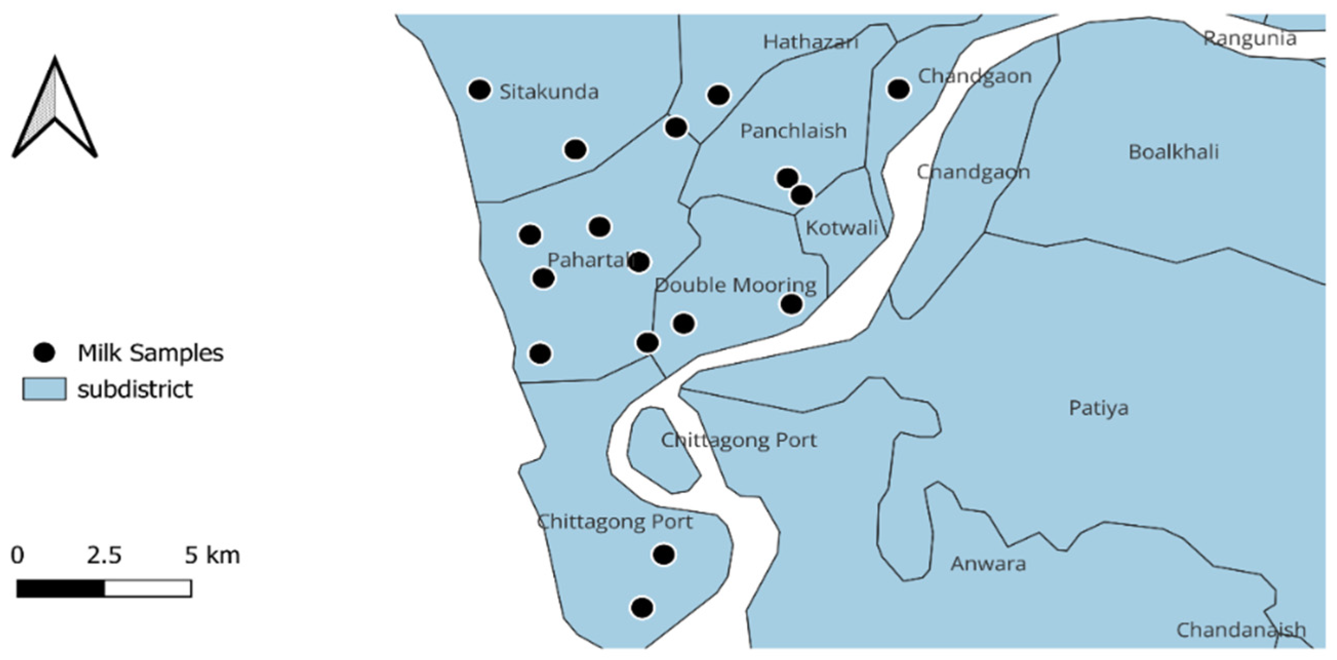
2.1. Study Design and Sample Collection

This study was conducted between September 2021 and August 2022 in the Chattogram metropolitan area (CMA) within the Chattogram district of Bangladesh. A total of 18 large-scale dairy farms hosting ≥50 dairy cows from seven locations, including Patenga, Akbershah, Dewanhut, Foys Lake, Sadarghat, Pahartali, and Wireless Area, were selected randomly for sample collection. These farm locations are illustrated in Figure 1. From each farm, 25 raw milk samples were collected from randomly selected cows after cleansing the udders and teat-ends with cotton soaked in 70% isopropanol to ensure aseptic condition. This study did not include cows with a recent treatment history and any active diseases, including mastitis. A total of 450 raw milk samples were collected in separate Falcon tubes using an aseptic technique.

2.2. Sample Preparation

After collection, samples were transferred to the Department of Physiology, Biochemistry and Pharmacology (DPBP), CVASU, for further investigation whilst maintaining a cold chain. For primary enrichment, samples were diluted with buffered peptone water (BPW) (HIMEDA, Mumbai, India), maintaining a ratio of 9:1 (BPW: cow milk sample), and incubated at 37 °C overnight.

2.3. Phenotypic Isolation and Identification of E. coli

To isolate E. coli, a loopful of enriched broth (BPW) was inoculated onto MacConkey agar (HIMEDIA, Mumbai, India) and incubated at 37 °C for 24 h. Suspected colonies were inoculated onto Eosin Methylene Blue (EMB) agar (HIMEDIA, Mumbai, India) and incubated at 37 °C for 24 h for biochemical confirmation. Confirmed colonies were then inoculated onto blood agar (HIMEDIA, Mumbai, India) and incubated at 37 °C for 24 h. All phenotypically confirmed E. coli isolates were incubated overnight at 37 °C in brain heart infusion (BHI) broth (HIMEDIA, Mumbai, India). After incubation, 700 µL of BHI broth was added to 300 µL of 15% glycerol in an Eppendorf tube for each isolate and stored at −80 °C for further investigations.

2.4. Molecular Confirmation of E. coli

All phenotypically positive E. coli isolates were subjected to molecular identification by multiplex PCR using species-specific primers targeting uidA and uspA genes. The genomic DNA was extracted following the crude boiling method [25]. The multiplex PCR assay was conducted using the following primes: uspA (F) CCGATACGCTGCCAATCAGT; uspA (R) ACGCAGACCGTAGGCCAGAT; uidA (F)TATGGAATTTCGCCGATTTT; and uidA (R) TGTTTGCCTCCCTGCTGCGG, maintaining the initial denaturation at 94 °C for 5 min and final extension at 72 °C for 10 min with the 35 cycles of denaturation at 94 °C for 10 s, annealing at 52.2 °C for 10 s, and extension at 72 °C for 1 min [26]. All PCR reactions were performed on a thermal cycler (DLAB Scientific Inc., Alhambra, CA, USA) with a final volume of 25 µL containing 12.5 µL DreamTaq 2X master mix (Thermofisher Scientific, Waltham, MA, USA), 1 µL forward and reverse primer (10 pmol/µL), 2 µL template DNA, and ~8.5 µL Nuclease-free water, in the research lab under the DPBP, CVASU. All the amplified PCR products were screened by electrophoresis with a 1.5% agarose gel (MP Biomedicals, Santa Ana, CA, USA) for 25 min at 120 V in 1x TAE buffer and visualised using ethidium bromide (Sigma Aldrich, Burlington, MA, USA) on a gel documentation system (UVP UVsolo touch-Analytik Jena AG, Thermo Fisher Scientific, Waltham, MA, USA). ATCC 25922 E. coli strains were used as a positive control, and nuclease-free water (NFW) as a negative control.

2.5. Phenotypic Antimicrobial Resistance Profiles

All E. coli isolates were screened for antimicrobial susceptibility against a panel of antimicrobials using the Kirby–Bauer disc diffusion method [27]. A total of 17 antimicrobials representing seven different antimicrobial groups (penicillins, cephalosporins, phenicols, tetracyclines, aminoglycosides, fluoroquinolones, and sulfonamides) with public health significance were selected. The following antimicrobial agents were used: AMP: ampicillin (10 µg); AUG: amoxicillin–clavulanic acid (30 µg); KF: cephalothin (30 µg); CL: cephalexin (30 µg); FOX: cefoxitin (30 µg); CTX: cefotaxime (30 µg); CAZ: ceftazidime (30 µg); FFC: Florfenicol (30 µg); TE: tetracycline (30 µg); DO: doxycycline (30 µg); CN: gentamicin (10 µg); N: neomycin (30 µg); CIP: ciprofloxacin (5 µg); LEV: levofloxacin (5 µg); ENR: enrofloxacin (5 µg); NOR: norfloxacin (10 µg); and SXT: sulfamethoxazole–trimethoprim (23.75 + 1.25 µg) (Oxoid Ltd., Hampshire, UK). The bacterial suspension was adjusted to the turbidity of 0.5 McFarland standard (equivalent to growth of 1–2 × 10⁸ CFU/mL) and streaked over the entire dry surface of Mueller Hinton agar (Oxoid Ltd.®, pH 7.3 ± 0.1) three times, rotating the plate approximately at 60 degrees by a sterile swab stick. Following incubation, the diameter of the disc and the extent of the inhibition zone (measured in millimetres) were recorded, and the results were interpreted according to the Clinical Laboratory Standards Institute’s guidelines [28]. Isolates that were resistant to more than 3 antimicrobial classes were termed multidrug-resistant (MDR) [29]. The multiple antibiotic resistance (MAR) index was estimated using the formula described previously by Algammal et al. [30].

2.6. Detection of AMR Genes

A total of 15 AMR genes were screened in this study, including those conferring ESBL-resistance (blaTEM, blaSHV, blaCTX-M, blaOXA-1, blaOXA-2, blaCMY-1, blaCMY-2, blaACC-1, PampC), sulfonamides resistance (sul-1, sul-2), and tetracycline resistance (tetA, tetB, tetC, tetD), by PCR. We selected those genes based on the WHO classification of higher public health risk and the commonly used antimicrobials in dairy practice in Bangladesh. The oligonucleotide primer sequences, annealing temperature, and amplicon size are shown in Table 1. The pan drug-susceptible E. coli ATCC 25922 strain was used as a negative control in each PCR to detect AMR genes.

2.7. Statistical Analysis

All data were recorded and organised in Microsoft Excel 2019 for statistical analysis. The data were then analysed in STATA/IC-15 (Stata Corp, 4905 Lakeway Drive, College Station, TX, USA) to estimate the prevalence and 95% confidence intervals (CI). The correlation coefficients among antimicrobials, phenotypic AMR, and resistance genes were calculated and illustrated using R software (version 4.4.1; https://www.r-project.org/; accessed on 20 October 2024) with the ggplot2 (ggcorrplot version 0.1.4.1) package. The geographical map was constructed using ArcGIS version 10.8.

3. Results

3.1. Prevalence of E. coli

A total of 450 milk samples from 18 CMA farms were investigated in this study. The demographic information for this study is summarised in Supplementary Table S1, which lists the variables, sample categories, and sample sizes for each category. The detection rate of E. coli was highest on Farm 18 (64%, 95% CI: 42.52–82.03) and lowest at 12% (95% CI: 2.55–31.22) on both Farms 7 and 10 (Supplementary Table S1). A total of 134 isolates (29.77%; 95% CI: 25.59–34.24) were confirmed as E. coli from raw milk samples.

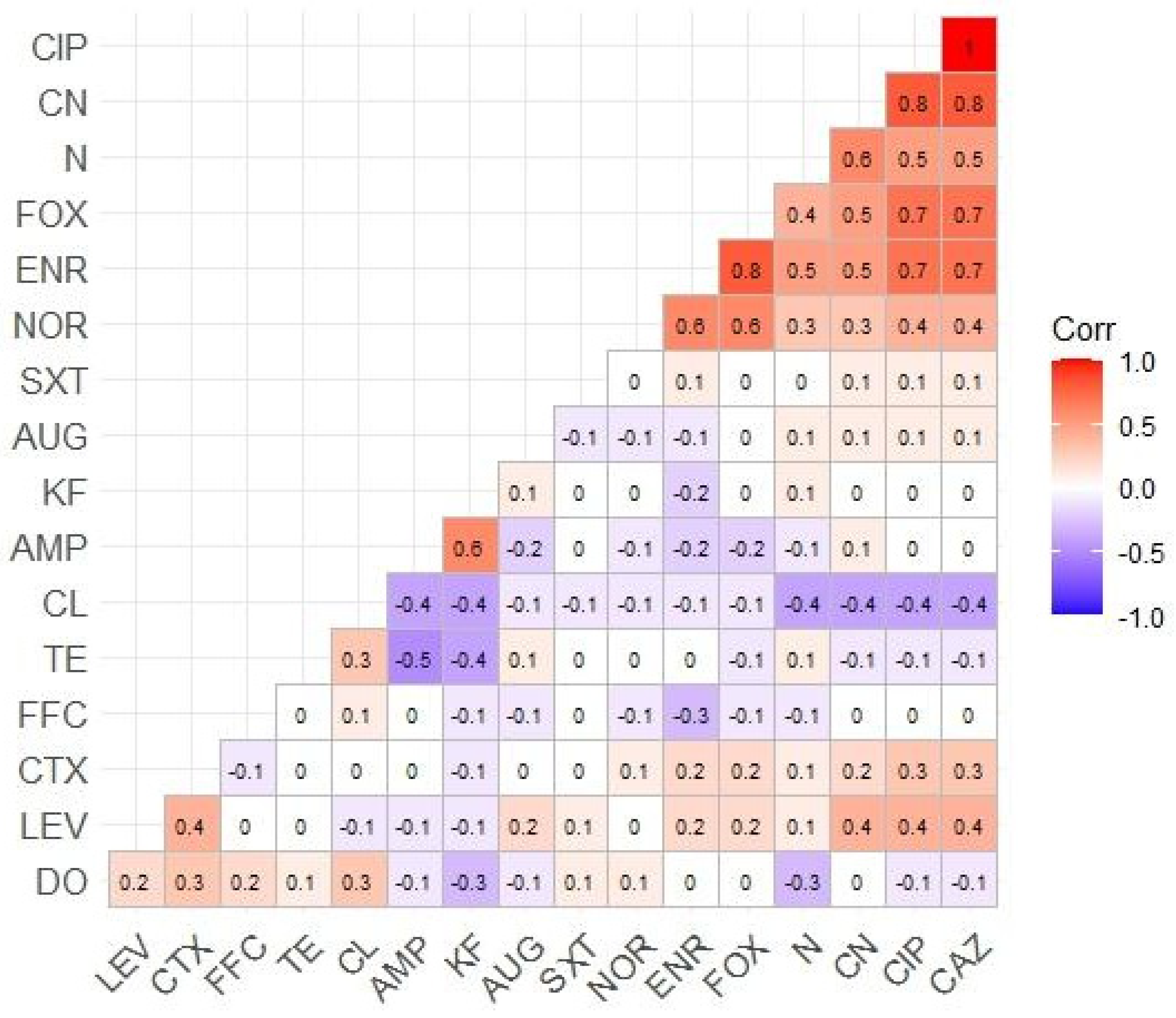
3.2. AMR Profiles of Isolated E. coli

The AST revealed that four beta-lactam antimicrobials, including ampicillin, amoxicillin–clavulanic acid, cephalothin, and cephalexin, showed the highest rates of resistance (69.40%), followed by sulfamethoxazole–trimethoprim (68.65%) and florfenicol (55.97%). In contrast, the lowest resistance rate was observed in the fluoroquinolones, ciprofloxacin (23.88%), levofloxacin (23.88%), and norfloxacin (21.64%), but not enrofloxacin (41.79%). The tetracyclines exhibited a similarly high resistance rate (51.49%). ESBL antibiotics, such as cefotaxime and ceftazidime, had similar resistance rates to each other (23.88%), except for cefoxitin (51.49%). Resistance rates for aminoglycosides, such as gentamicin and neomycin, were 51.49% and 32.84%, respectively. The AMR profiles are shown in Table 2. Correlation coefficients among antimicrobials tested in this study displayed a significant level of correlation between resistance rates for many antimicrobials, e.g., CIP and CAZ (r = 1); CIP, CAZ, and CN (r = 0.8); ENR and FOX (r = 0.8); CIP, CAZ, FOX, and ENR (r = 0.7); N and CN (r = 0.6), as illustrated in Figure 2.

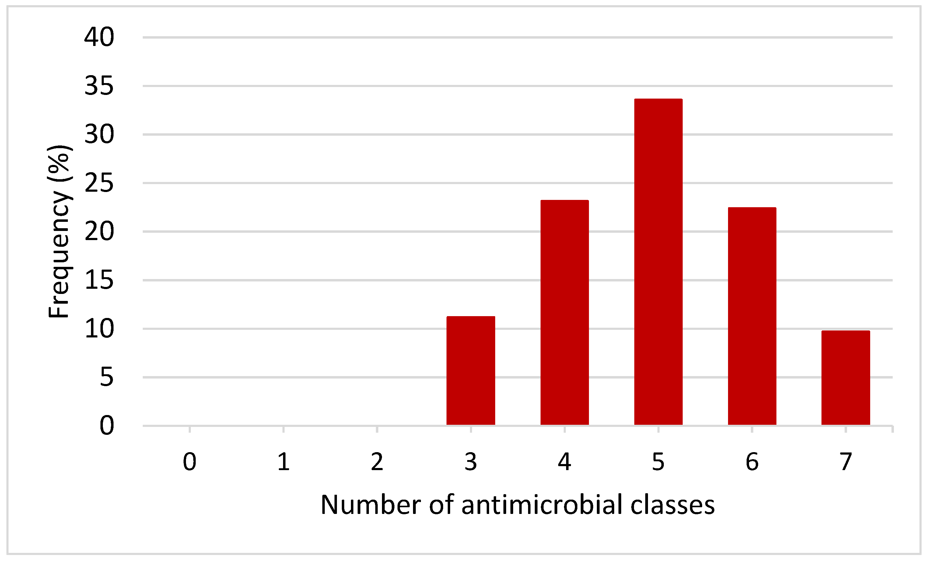
3.3. Phenotypic MDR Patterns of E. coli Isolates

All the E. coli isolates from raw cow milk were classified as MDR (resistant to at least three or more antimicrobial classes) (Figure 3). The MDR patterns varied between isolates, with most showing unique resistance profiles. Only 2.24% (3/134) of the isolates exhibited the same resistance pattern (LEV, SXT, DO, AMP, AUG, KF, CL, FFC). The MDR patterns are illustrated in Table 3. The MAR index in this study ranged from 0.24 to 0.82 (Table 3).

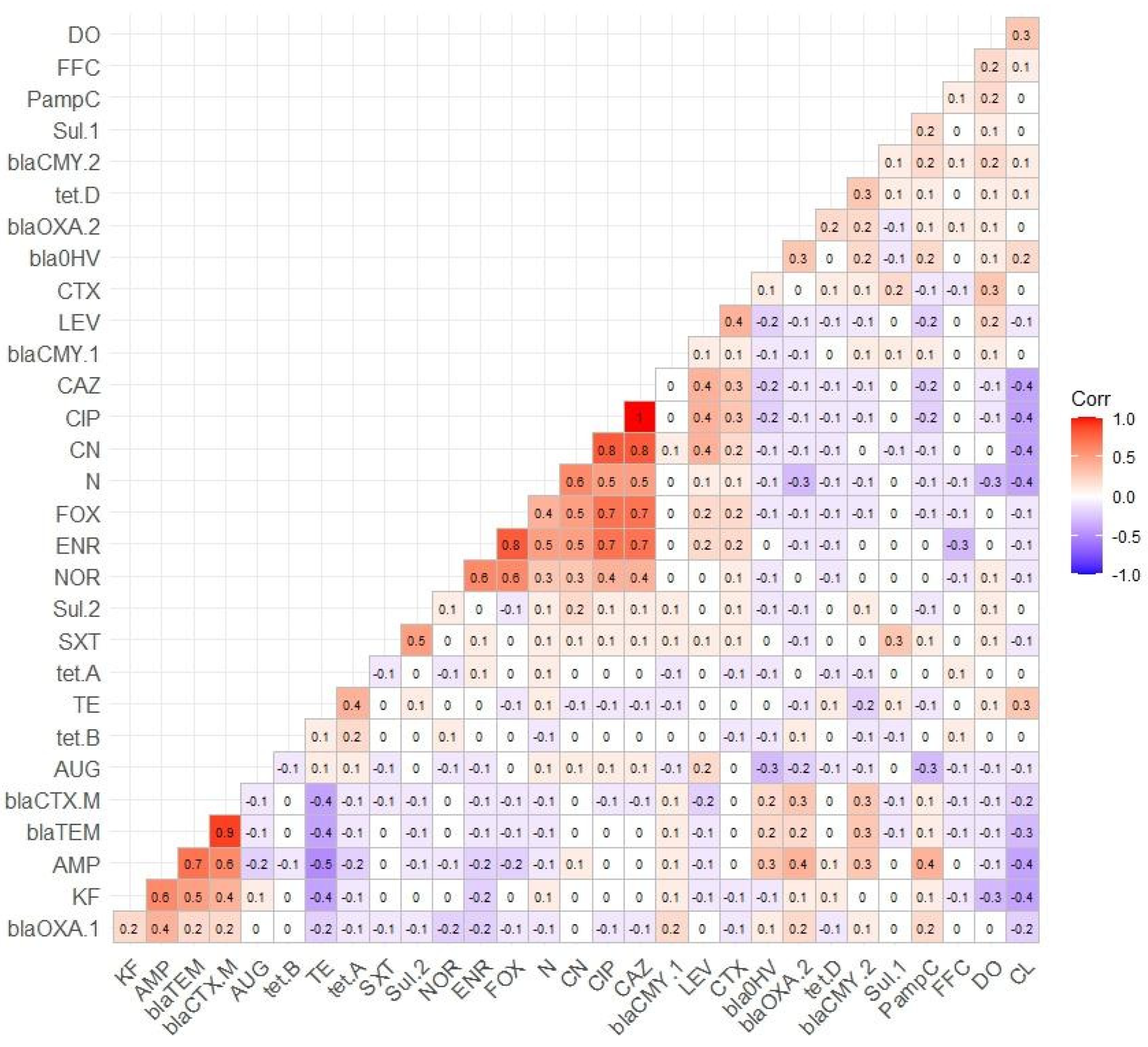
3.4. Distribution of AMR Genes

A total of nine ESBL genes were tested. Among them, the prevalence of the blaTEM gene was the highest (74.19%), followed by blaCTX-M (69.89%), blaOXA-2 (40.86%), PampC (37.63%), and blaOXA-1 (33.33%). In contrast, the prevalence of the blaCMY-1 gene was lower (6.45%), while blaACC-1 was absent. Among the tetracycline resistance genes tested, the prevalence of the tetA gene was highest (30.43%), followed by tetB (5.8%) and tetD (2.9%), and the tetC gene was absent. Similarly, the prevalence of sulfonamide resistance genes, sul-1 and sul-2, was 25% and 55.43%, respectively. The prevalence of antimicrobial resistance genes is shown in Table 4. A positive correlation was observed between phenotypic AMR and resistance genes, including AMP and blaTEM (r = 0.7), AMP and blaCTX-a (r = 0.6), KF and blaTEM (r = 0.5), KF and blaCTX-M (r = 0.4), SXT and sul-2 (r = 0.5), TE and tetA (r = 0.4), AMP and PampC (r = 0.4), as illustrated in Figure 4.

4. Discussion

The current study revealed a high prevalence of E. coli in the milk of Bangladeshi farms collected from various dairy cattle, with the isolates frequently displaying resistance to multiple antimicrobial classes. At the farm level, the prevalence of E. coli varied between 12 to 64%. In this study, the overall prevalence of E. coli in farm milk is approximately 30%, less than the 42% prevalence reported in Iran by Vahedi et al. [34]. The variation in the prevalence of E. coli in the present study might be due to variations in hygiene and managemental practices in different farms. Another study reported the same 42% prevalence of E. coli in milk in Ethiopia [35]. Studies indicated that the prevalence of E. coli contamination in raw milk in Bangladesh is notably high, ranging from 50% to 92%, often linked to poor hygiene practices during milking and handling [36]. Global contamination rates vary significantly, with lower incidences in developed countries, such as the US, where E. coli contamination in milk is generally below 10% due to stricter regulations and better sanitation [37]. This disparity suggests that faecal contamination and hygiene standards are poorer in Bangladesh than in many other regions. In this study, 17 antimicrobials were tested using the AST, and 15 resistance genes were tested against those antimicrobials. The AST of the isolates revealed that E. coli resistant to ampicillin, amoxicillin–clavulanic acid, cephalothin, and cephalexin was the most prevalent, followed by sulfamethoxazole–trimethoprim and florfenicol. A recent study showed 15% resistance to sulfonamide and 3% resistance to trimethoprim [38]. Another one reported the prevalence of E. coli in calves from a dairy farm was 37.5% [39]. In contrast, this study revealed that out of 92 sulfamethoxazole–trimethoprim isolates, 23 were resistant to the sul-1 gene, and 51 were resistant to the sul-2 gene.
E. coli antimicrobial resistance for the isolates was found to be conferred by a wide range of resistance genes. These genes include the tet genes (tetA, tetB, tetC, tetD), gene for Tetracycline resistance, the blaTEM, blaSHV, PampC, blaOXA, blaACC, blaCMY, blaCTX-M genes for Ampicillin, and Sul-1, Sul-2 genes for Trimethoprim-sulfamethoxazole [14,15,16]. Evidence shows that AMR patterns in dairy farms in Bangladesh reflect the high usage of specific antimicrobials, particularly β-lactams, commonly used in treating mastitis and other infections [40]. High rates of extended-spectrum β-lactamase (ESBL)-producing E. coli have been reported in dairy environments, likely due to the extensive use of β-lactam antibiotics [16]. This supports the hypothesis that increased antimicrobial use correlates with higher resistance rates. This pattern aligns with global observations that frequent use of antimicrobials drives the development of resistance in microbial populations [41]. In Bangladesh, certain antimicrobials, including fluoroquinolones and aminoglycosides, are not strictly regulated for use in dairy cattle, despite their potential to contribute to AMR, unlike in countries like Australia [42], where their use in food-producing animals is banned. The relevance to the current study is significant, as the continued use of these drugs in Bangladesh likely contributes to the high resistance rates observed, particularly for classes like fluoroquinolones.
A previous study reported that 13.4% of E. coli isolated from milk were resistant to tetracycline [43], but in this study, the resistance was higher (51%). Among 69 tetracycline-resistant isolates, tetA was the most frequently detected gene, accounting for 21 (86.5%), while tetB was detected in only 4 (8.1%) isolates. Only two resistant genes for tetC and tetD were detected. According to [14], only 0.4% of faecal samples were positive for ESBL-producing E. coli isolated from a lactating bovine, while 6.5% of the farm environment samples were positive. Hassan [16] reported that 62.50% of milk samples contained ESBL E. coli with the gene combination blaTEM + blaCTX-M. Correlations between resistance rates for different antimicrobials suggest co-selection, where resistance to one antimicrobial may confer resistance to others due to shared resistance mechanisms, such as plasmids carrying multiple resistance genes. This phenomenon is exacerbated by polypharma practices, where the overuse of multiple antimicrobials in dairy farming can lead to the selection of multidrug-resistant bacteria. Studies have shown that the widespread use of various antimicrobials in regions like Bangladesh promotes co-selection, driving the persistence of multidrug-resistant organisms in agricultural environments [44,45].
The World Health Organization (WHO) categorised antimicrobials into three groups to mitigate the situation: access; monitor; and reserve groups [46]. Access group antimicrobials are available to be prescribed to patients by physicians. If this group fails due to resistant genes in organisms, it is recommended that the patient be monitored in a separate group [47]. Reserve categories of antimicrobials are for future use if others become resistant. Scientific knowledge and evidence are required to mitigate AMR issues before they become widespread crises. AMR is among the deadliest threats to human and animal health. To alleviate the AMR health threat before it manifests in large-scale medical emergencies, it is necessary to identify risks and appropriate mitigation strategies based on scientific evidence and knowledge. Our finding showed that most isolates in this study were MDR, suggesting a much higher prevalence of MDR in livestock-associated E. coli in dairy farms. This poses serious concerns for both animal and public health, as MDR bacteria reduce treatment options for infections in dairy cattle, leading to potential economic losses and increased animal suffering. Additionally, the spread of MDR pathogens from animals to humans through direct contact or consuming contaminated dairy products represents a significant public health risk, particularly in regions like Bangladesh.

5. Limitations

This study’s scope is confined to farms within the Chattogram metropolitan area of Bangladesh, limiting the generalisability of the findings to be applied to other regions or dairy farming practices nationally. The focus on E. coli alone may overlook the presence of other critical antimicrobial-resistant bacteria in raw milk that could pose a public health risk. However, E. coli is a widely used indicator organism in food microbiology, such that levels of resistance in E. coli are reflective of general antimicrobial selection pressures and levels of resistance amongst other enteric microbes, including foodborne pathogens.

6. Conclusions

This study reveals a high prevalence of AMR and MDR E. coli in raw cow milk from dairy farms in the Chattogram metropolitan area, with significant resistance to widely used antimicrobials such as ampicillin and amoxicillin–clavulanic acid. The findings show a strong link between resistance patterns and the presence of resistance genes, particularly blaTEM, highlighting the risk of resistant bacteria transmission to humans via the dairy food chain, a serious public health concern. Urgent strategic interventions, including prudent antimicrobial use in dairy farming and enhanced AMR surveillance, are essential to control AMR spread in the dairy sector and protect public health.

Supplementary Materials

The following supporting information can be downloaded at: https://www.mdpi.com/article/10.3390/vetsci11120609/s1, Supplementary Table S1: Descriptive demography of the present study.

Author Contributions

Conceptualization, M.M.H. and Z.T.M.; methodology, M.M.H., Z.T.M. and C.N.; formal analysis, M.M.H. and C.N.; writing—original draft preparation, Z.T.M., M.M.H. and A.A.S.; writing—review and editing, M.H.A., R.R., S.A.K., M.A.K., S.S., R.C., J.I.A. and M.M.H. All authors have read and agreed to the published version of the manuscript.

Funding

Bangladesh Bureau of Education Information and Statistics (BANBEIS), Ministry of Education, People’s Republic of Bangladesh, project number #SD-2019967.

Institutional Review Board Statement

This study was conducted by following the Declaration of Helsinki, and the protocol was approved by the Ethics Committee of the Chattogram Veterinary and Animal Sciences University, Bangladesh (permit reference number: CVASU/Dir (R and E) EC/2019/126 (02), Date: 29 December 2019).

Informed Consent Statement

Informed consent was obtained from all subjects involved in the study.

Data Availability Statement

The data presented in this study are available in this article and Supplementary Material.

Acknowledgments

The authors sincerely appreciate all the office staff and lab assistants of the Department of Physiology, Biochemistry, and Pharmacology, Chattogram Veterinary and Animal Sciences University, Chattogram, Bangladesh. The authors also acknowledge the dairy farmers’ support in providing the necessary information and milk samples.

Conflicts of Interest

The authors declare no conflicts of interest.

References

  1. O’Neill, J. Tackling Drug-Resistant Infections Globally: Final Report and Recommendations; Government of the United Kingdom: London, UK, 2016.
  2. Sakalauskienė, G.V.; Radzevičienė, A. Antimicrobial Resistance: What Lies Beneath This Complex Phenomenon? Diagnostics 2024, 14, 2319. [Google Scholar] [CrossRef] [PubMed]
  3. de Brito, F.A.; de Freitas, A.P.; Nascimento, M.S. Multidrug-resistant biofilms (MDR): Main mechanisms of tolerance and resistance in the food supply chain. Pathogens 2022, 11, 1416. [Google Scholar] [CrossRef] [PubMed]
  4. Sharma, C.; Rokana, N.; Chandra, M.; Singh, B.P.; Gulhane, R.D.; Gill, J.P.S.; Ray, P.; Puniya, A.K.; Panwar, H. Antimicrobial resistance: Its surveillance, impact, and alternative management strategies in dairy animals. Front. Vet. Sci. 2018, 4, 237. [Google Scholar] [CrossRef] [PubMed]
  5. Annunziato, G. Strategies to overcome antimicrobial resistance (AMR) making use of non-essential target inhibitors: A review. Int. J. Mol. Sci. 2019, 20, 5844. [Google Scholar] [CrossRef]
  6. Murugaiyan, J.; Kumar, P.A.; Rao, G.S.; Iskandar, K.; Hawser, S.; Hays, J.P.; Mohsen, Y.; Adukkadukkam, S.; Awuah, W.A.; Jose, R.A.M. Progress in alternative strategies to combat antimicrobial resistance: Focus on antibiotics. Antibiotics 2022, 11, 200. [Google Scholar] [CrossRef]
  7. Abebe, R.; Hatiya, H.; Abera, M.; Megersa, B.; Asmare, K. Bovine mastitis: Prevalence, risk factors and isolation of Staphylococcus aureus in dairy herds at Hawassa milk shed, South Ethiopia. BMC Vet. Res. 2016, 12, 270. [Google Scholar] [CrossRef]
  8. Mulchandani, R.; Wang, Y.; Gilbert, M.; Van Boeckel, T.P. Global trends in antimicrobial use in food-producing animals: 2020 to 2030. PLoS Glob. Public Health 2023, 3, e0001305. [Google Scholar] [CrossRef]
  9. Loo, E.; Lai, K.S.; Mansor, R. Antimicrobial usage and resistance in dairy cattle production. Vet. Med. Pharm. 2019, 7, 7–82. [Google Scholar]
  10. Lhermie, G.; Gröhn, Y.T.; Raboisson, D. Addressing antimicrobial resistance: An overview of priority actions to prevent suboptimal antimicrobial use in food-animal production. Front. Microbiol. 2017, 7, 2114. [Google Scholar] [CrossRef]
  11. Ayukekbong, J.A.; Ntemgwa, M.; Atabe, A.N. The threat of antimicrobial resistance in developing countries: Causes and control strategies. Antimicrob. Resist. Infect. Control 2017, 6, 47. [Google Scholar] [CrossRef]
  12. Batabyal, K.; Banerjee, A.; Pal, S.; Dey, S.; Joardar, S.N.; Samanta, I.; Isore, D.P.; Singh, A.D. Detection, characterization, and antibiogram of extended-spectrum beta-lactamase Escherichia coli isolated from bovine milk samples in West Bengal, India. Vet. World 2018, 11, 1423. [Google Scholar] [CrossRef] [PubMed]
  13. Almansour, A.M.; Alhadlaq, M.A.; Alzahrani, K.O.; Mukhtar, L.E.; Alharbi, A.L.; Alajel, S.M. The Silent Threat: Antimicrobial-Resistant Pathogens in Food-Producing Animals and Their Impact on Public Health. Microorganisms 2023, 11, 2127. [Google Scholar] [CrossRef]
  14. Skocková, A.; Cupáková, S.; Karpísková, R.; Janstová, B. Detection of tetracycline resistance genes in Escherichia coli from raw cow’s milk. J. Microbiol. Biotechnol. Food Sci. 2012, 1, 777. [Google Scholar]
  15. Metzger, S.; Hogan, J. Antimicrobial susceptibility and frequency of resistance genes in Escherichia coli isolated from bovine mastitis. J. Dairy Sci. 2013, 96, 3044–3049. [Google Scholar] [CrossRef] [PubMed]
  16. Kamaruzzaman, E.A.; Abdul Aziz, S.; Bitrus, A.A.; Zakaria, Z.; Hassan, L. Occurrence and characteristics of extended-spectrum β-lactamase-producing Escherichia coli from dairy cattle, milk, and farm environments in Peninsular Malaysia. Pathogens 2020, 9, 1007. [Google Scholar] [CrossRef]
  17. Bajaj, P.; Singh, N.S.; Virdi, J.S. Escherichia coli β-lactamases: What really matters. Front. Microbiol. 2016, 7, 417. [Google Scholar] [CrossRef]
  18. Walther, B.; Tedin, K.; Lübke-Becker, A. Multidrug-resistant opportunistic pathogens challenging veterinary infection control. Vet. Microbiol. 2017, 200, 71–78. [Google Scholar] [CrossRef]
  19. Karkaba, A.; Grinberg, A.; Benschop, J.; Pleydell, E. Characterisation of extended-spectrum β-lactamase and AmpC β-lactamase-producing Enterobacteriaceae isolated from companion animals in New Zealand. N. Z. Vet. J. 2017, 65, 105–112. [Google Scholar] [CrossRef]
  20. Mandujano, A.; Cortés-Espinosa, D.V.; Vásquez-Villanueva, J.; Guel, P.; Rivera, G.; Juárez-Rendón, K.; Cruz-Pulido, W.L.; Aguilera-Arreola, G.; Guerrero, A.; Bocanegra-García, V.; et al. Extended-Spectrum β-Lactamase-Producing Escherichia coli Isolated from Food-Producing Animals in Tamaulipas, Mexico. Antibiotics 2023, 12, 1010. [Google Scholar] [CrossRef]
  21. Ali, T.; ur Rahman, S.; Zhang, L.; Shahid, M.; Zhang, S.; Liu, G.; Gao, J.; Han, B. ESBL-producing Escherichia coli from cows suffering mastitis in China contain clinical class 1 integrons with CTX-M linked to IS CR1. Front. Microbiol. 2016, 7, 1931. [Google Scholar] [CrossRef]
  22. Estany-Gestal, A.; Salgado-Barreira, A.; Vazquez-Lago, J.M. Antibiotic Use and Antimicrobial Resistance: A Global Public Health Crisis. Antibiotics 2024, 13, 900. [Google Scholar] [CrossRef] [PubMed]
  23. Sobur, M.A.; Sabuj, A.A.M.; Sarker, R.; Rahman, A.T.; Kabir, S.L.; Rahman, M.T. Antibiotic-resistant Escherichia coli and Salmonella spp. associated with dairy cattle and farm environment having public health significance. Vet. World 2019, 12, 984. [Google Scholar] [CrossRef]
  24. Khan, S.A.; Imtiaz, M.A.; Sayeed, M.A.; Shaikat, A.H.; Hassan, M.M. Antimicrobial resistance pattern in domestic animal-wildlife-environmental niche via the food chain to humans with a Bangladesh perspective; a systematic review. BMC Vet. Res. 2020, 16, 302. [Google Scholar] [CrossRef] [PubMed]
  25. Sarker, M.S.; Mannan, M.S.; Ali, M.Y.; Bayzid, M.; Ahad, A.; Bupasha, Z.B. Antibiotic resistance of Escherichia coli isolated from broilers sold at live bird markets in Chattogram, Bangladesh. J. Adv. Vet. Anim. Res. 2019, 6, 272–277. [Google Scholar] [CrossRef] [PubMed]
  26. Godambe, L.P.; Bandekar, J.; Shashidhar, R. Species specific PCR based detection of Escherichia coli from Indian foods. 3 Biotech 2017, 7, 130. [Google Scholar] [CrossRef] [PubMed]
  27. Bauer, A.; Kirby, W.; Sherris, J.C.; Turck, M. Antibiotic susceptibility testing by a standardized single disk method. Am. J. Clin. Pathol. 1966, 45, 493–496. [Google Scholar] [CrossRef]
  28. CLSI. Performance Standards for Antimicrobial Susceptivility Testing, 33rd ed.; CLSI: Pittsburgh, PA, USA, 2023; Volume 43, CLSI Supplement M100; Available online: https://clsi.org/ (accessed on 5 March 2024).
  29. Magiorakos, A.-P.; Srinivasan, A.; Carey, R.B.; Carmeli, Y.; Falagas, M.; Giske, C.; Harbarth, S.; Hindler, J.; Kahlmeter, G.; Olsson-Liljequist, B. Multidrug-resistant, extensively drug-resistant and pandrug-resistant bacteria: An international expert proposal for interim standard definitions for acquired resistance. Clin. Microbiol. Infect. 2012, 18, 268–281. [Google Scholar] [CrossRef]
  30. Algammal, A.M.; El-Tarabili, R.M.; Alfifi, K.J.; Al-Otaibi, A.S.; Hashem, M.E.A.; El-Maghraby, M.M.; Mahmoud, A.E. Virulence determinant and antimicrobial resistance traits of Emerging MDR Shiga toxigenic E. coli in diarrheic dogs. AMB Express 2022, 12, 34. [Google Scholar] [CrossRef]
  31. Koo, H.-J.; Woo, G.-J. Distribution and transferability of tetracycline resistance determinants in Escherichia coli isolated from meat and meat products. Int. J. Food Microbiol. 2011, 145, 407–413. [Google Scholar] [CrossRef]
  32. Lanz, R.; Kuhnert, P.; Boerlin, P. Antimicrobial resistance and resistance gene determinants in clinical Escherichia coli from different animal species in Switzerland. Vet. Microbiol. 2003, 91, 73–84. [Google Scholar] [CrossRef]
  33. Hasman, H.; Mevius, D.; Veldman, K.; Olesen, I.; Aarestrup, F.M. β-Lactamases among extended-spectrum β-lactamase (ESBL)-resistant Salmonella from poultry, poultry products and human patients in The Netherlands. J. Antimicrob. Chemother. 2005, 56, 115–121. [Google Scholar] [CrossRef] [PubMed]
  34. Vahedi, M.; Nasrolahei, M.; Sharif, M.; Mirabi, A. Bacteriological study of raw and unexpired pasteurized cow’s milk collected at the dairy farms and super markets in Sari city in 2011. J. Prev. Med. Hyg. 2013, 54, 120. [Google Scholar] [PubMed]
  35. Megersa, R.; Mathewos, M.; Fesseha, H. Isolation and identification of Escherichia coli from dairy cow raw milk in Bishoftu town, Central Ethiopia. Arch. Vet. Anim. Sci. 2019, 1, 1–7. [Google Scholar]
  36. Mahmud, S.; Ali, M.F.; Faruque, M.O.; Wasim, M.; Evamoni, F.Z.; Chowdhury, K.; Napis, S.; Mohiuddin, A. Prevalence and Molecular Characterization of Microbial Contaminants in Raw Cow Milk of Tangail District in Bangladesh. Curr. Nutr. Food Sci. 2022, 18, 220–230. [Google Scholar]
  37. Williams, E.N.; Van Doren, J.M.; Leonard, C.L.; Datta, A.R. Prevalence of Listeria monocytogenes, Salmonella spp., Shiga toxin-producing Escherichia coli, and Campylobacter spp. in raw milk in the United States between 2000 and 2019: A systematic review and meta-analysis. J. Food Prot. 2023, 86, 100014. [Google Scholar] [CrossRef]
  38. Agatha, T.M.; Wibawati, P.A.; Izulhaq, R.I.; Agustono, B.; Prastiya, R.A.; Wardhana, D.K.; Abdramanov, A.; Lokapirnasari, W.P.; Lamid, M. Antibiotic resistance of Escherichia coli from the milk of Ettawa crossbred dairy goats in Blitar Regency, East Java, Indonesia. Vet. World 2023, 16, 168. [Google Scholar] [CrossRef]
  39. Astorga, F.; Navarrete-Talloni, M.J.; Miró, M.P.; Bravo, V.; Toro, M.; Blondel, C.J.; Hervé-Claude, L.P. Antimicrobial resistance in E. coli isolated from dairy calves and bedding material. Heliyon 2019, 5, e02773. [Google Scholar] [CrossRef]
  40. Islam, S. Coliform and Staphylococcal Mastitis in Cows: Risk Factors and Trends in Antimicrobial Resistance. 2020. Available online: http://archive.saulibrary.edu.bd:8080/xmlui/handle/123456789/4787 (accessed on 12 January 2024).
  41. Collis, R.M.; Burgess, S.A.; Biggs, P.J.; Midwinter, A.C.; French, N.P.; Toombs-Ruane, L.; Cookson, A.L. Extended-spectrum beta-lactamase-producing Enterobacteriaceae in dairy farm environments: A New Zealand perspective. Foodborne Pathog. Dis. 2019, 16, 5–22. [Google Scholar] [CrossRef]
  42. Cheng, A.C.; Turnidge, J.; Collignon, P.; Looke, D.; Barton, M.; Gottlieb, T. Control of fluoroquinolone resistance through successful regulation, Australia. Emerg. Infect. Dis. 2012, 18, 1453. [Google Scholar] [CrossRef]
  43. Liu, H.; Meng, L.; Dong, L.; Zhang, Y.; Wang, J.; Zheng, N. Prevalence, antimicrobial susceptibility, and molecular characterization of Escherichia coli isolated from raw milk in dairy herds in Northern China. Front. Microbiol. 2021, 12, 730656. [Google Scholar] [CrossRef]
  44. Miller, S.A.; Ferreira, J.P.; LeJeune, J.T. Antimicrobial use and resistance in plant agriculture: A one health perspective. Agriculture 2022, 12, 289. [Google Scholar] [CrossRef]
  45. Ahmad, I.; Malak, H.A.; Abulreesh, H.H. Environmental antimicrobial resistance and its drivers: A potential threat to public health. J. Glob. Antimicrob. Resist. 2021, 27, 101–111. [Google Scholar]
  46. Gandra, S.; Kotwani, A. Need to improve availability of “access” group antibiotics and reduce the use of “watch” group antibiotics in India for optimum use of antibiotics to contain antimicrobial resistance. J. Pharm. Policy Pract. 2019, 12, 20. [Google Scholar] [CrossRef] [PubMed]
  47. Hsia, Y.; Lee, B.R.; Versporten, A.; Yang, Y.; Bielicki, J.; Jackson, C.; Newland, J.; Goossens, H.; Magrini, N.; Sharland, M. Use of the WHO Access, Watch, and Reserve classification to define patterns of hospital antibiotic use (AWaRe): An analysis of paediatric survey data from 56 countries. Lancet Glob. Health 2019, 7, e861–e871. [Google Scholar] [CrossRef]
Figure 1. Geographical locations of the farms randomly selected for sampling in this study.
Vetsci 11 00609 g001
Figure 2. Heatmap showing the correlation coefficient among antimicrobials tested in this study.
Vetsci 11 00609 g002
Figure 3. MDR profiles of E. coli isolates from raw cow milk.
Vetsci 11 00609 g003
Figure 4. The correlation coefficient between phenotypic AMR and resistance genes.
Vetsci 11 00609 g004
Table 1. The oligonucleotide primer sequences for the detection of AMR genes in E. coli isolated from raw dairy milk.
Antimicrobial AgentsTarget GenePrimer NAMEPrimer Sequence
(5′-3′)
Annealing Temp.Amplicon Size (bp)References
TetracyclinestetAtetA-FCGCCTTTCCTTTGGGTTCTCTATATC55 °C182[31]
tetA-RCAGCCCACCGAGCACAGG
tetBtetB-FGCCAGTCTTGCCAACGTTAT975
tetB-RATAACACCGG TTGCATTGGT
tetCtetC-FTTCAACCCAGTCAGCTCCTT560
tetC-RGGGAGGCAGACAAGGTATAGG
tetDtetD-FGAGCGTACCGCCTGGTTC780
tetD-RTCTGATCAGCAGACAGATTGC
Sulphonamidessul-1sul-1-FCGGCGTGGGCTACCTGAACG68 °C779[32]
sul-1-RGCCGATCGCGTGAAGTTCCG
sul-2sul-2-FCCTGTTTCGTCCGACACAGA66 °C721
sul-2-RGAAGCGCAGCCGCAATTCAT
ESBLsblaTEMblaTEM-FATAAAATTCTTGAAGACGAAA54 °C964[33]
blaTEM-RGACAGTTACCAATGCTTAATC
blaSHVblaSHV-FGCTTTCCCATGATGAGCACC50 °C854
blaSHV-RAGGCGGGTGACGTTGTCGC
PampCPampC-FGTGAATACAGAGCCAGACGC50 °C343
PampC-RGTTGTTTCCGGGTGATGC
blaOXA-1blaOXA-1-RGTGTGTTTAGAATGGTGATCGCATT62 °C820
blaOXA-1-RGTGTGTTTAGAATGGTGATCGCATT
blaOXA-2blaOXA-2-FACGATAGTTGTGGCAGACGAAC62 °C602
blaOXA-2-RATYCTGTTTGGCGTATCRATATTC
blaCTX-MblaCTX-M-FATGTGCAGYACCAGTAARGTKATGGC60 °C593
blaCTX-M-FTGGGTRAARTARGTSACCAGAAYCAGCGG
blaCMY-1blaCMY-1-FGTGGTGGATGCCAGCATCC58 °C915
blaCMY-1-RGGTCGAGCCGGTCTTGTTGAA
blaCMY-2blaCMY-2-FGCACTTAGCCACCTATACGGCAG58 °C758
blaCMY-2-RGCTTTTCAAGAATGCGCCAGG
blaACC-1blaACC-1-FATYCTGTTTGGCGTATCRATATTC53 °C818
blaACC-1-RAGCCTCAGCAGCCGGTTAC
Table 2. Phenotypic antimicrobial susceptibility test profiles of E. coli isolates.
Antimicrobial GroupsAntimicrobial AgentsSusceptible (S)
N (%)
Intermediate (I)
N (%)
Resistant (R)
N (%)
PenicillinsAMP (10 µg)36 (26.86)5 (3.73)93 (69.40)
AUG (30 µg)36 (26.86)5 (3.73)93 (69.40)
CephalosporinsKF (30 µg)40 (29.85)1 (0.75)93 (69.40)
CL (30 µg)41 (30.59)0 (0)93 (69.40)
FOX (30 µg)34 (25.37)44 (32.84)56 (41.79)
CTX (30 µg)53 (39.55)49 (36.57)32 (23.88)
CAZ (30 µg)53 (39.55)49 (36.57)32 (23.88)
PhenicolsFFC (30 µg)49 (36.56)10 (7.46)75 (55.97)
TetracyclinesTE (30 µg)40 (29.85)25 (18.66)69 (51.49)
DO (30 µg)52 (38.81)13 (9.70)69 (51.49)
AminoglycosidesCN (30 µg)45 (33.58)45 (33.58)44 (32.84)
N (30 µg)25 (18.65)40 (29.85)69 (51.49)
FluoroquinolonesCIP (5 µg)53 (39.55)49 (36.57)32 (23.88)
LEV (5 µg)80 (59.70)22 (16.41)32 (23.88)
ENR (5 µg)34 (25.37)44 (32.83)56 (41.79)
NOR (10 µg)85 (63.43)20 (14.92)29 (21.64)
SulfonamidesSXT (23.75 + 1.25 µg)40 (29.85)2 (1.5)92 (68.65)
Table 3. The phenotypic MDR patterns of E. coli isolates in this study.
Phenotypic Multidrug Resistance PatternsNo. of Isolates (%)MAR Index
SXT, DO, AUG, KF, CL, FFC1 (0.75)0.35
DO, AUG, CL, FFC1 (0.75)0.24
ENR, NOR, SXT, TE, DO, AMP, AUG, KF, CL, FOX1 (0.75)0.59
N, ENR, SXT, AMP, CL1 (0.75)0.29
AUG, KF, CL, FOX, FFC2 (1.49)0.29
SXT, KF, CL, FFC1 (0.75)0.24
SXT, DO, AMP, KF, FFC1 (0.75)0.29
ENR, NOR, SXT, AMP, CL, FOX1 (0.75)0.35
TE, DO, AMP, AUG, KF, CL, FFC1 (0.75)0.41
DO, AMP, CL, FOX, FFC1 (0.75)0.29
ENR, NOR, DO, CL, FOX1 (0.75)0.29
TE, DO, AMP, KF, CL, FFC1 (0.75)0.35
TE, DO, AMP, CL, FFC1 (0.75)0.29
ENR, NOR, TE, DO, AMP, CL, FOX1 (0.75)0.41
N, ENR, TE, DO, AMP, CL, FFC1 (0.75)0.41
CN, N, ENR, NOR, DO, AMP, KF, CL, FOX, FFC1 (0.75)0.59
N, AMP, AUG, KF, CL, FOX, CTX, FFC1 (0.75)0.47
SXT, DO, AMP, KF, CL, CTX, FFC1 (0.75)0.41
ENR, NOR, SXT, DO, AMP, KF, CL, FOX, CTX1 (0.75)0.53
N, ENR, SXT, TE, AMP, KF, CL, CTX1 (0.75)0.47
AMP, AUG, KF, CL, FFC1 (0.75)0.29
SXT, AMP, KF, CL, FFC1 (0.75)0.29
LEV, SXT, TE, DO, AMP, KF, CL, CTX, FFC1 (0.75)0.53
ENR, NOR, DO, AMP, KF, CL, FOX, FFC1 (0.75)0.47
ENR, SXT, DO, AMP, KF, CL, FOX, FFC1 (0.75)0.47
SXT, TE, AMP, AUG, KF, CL, FFC1 (0.75)0.41
TE, AMP, KF, CL, FFC1 (0.75)0.29
N, ENR, NOR, SXT, TE, DO, KF, CL, FOX1 (0.75)0.53
CN, N, SXT, TE, AMP, KF, CL2 (1.49)0.41
AMP, AUG, KF, CL, FOX, FFC1 (0.75)0.35
SXT, AMP, AUG, KF, CL, FOX, FFC1 (0.75)0.41
SXT, DO, AMP, KF, CL, FFC2 (1.49)0.35
ENR, NOR, SXT, DO, AMP, CL, FOX, CTX, FFC1 (0.75)0.53
ENR, SXT, DO, AMP, CL, CTX1 (0.75)0.35
SXT, TE, DO, AMP, CL, CTX1 (0.75)0.35
CN, N, LEV, SXT, DO, AMP, AUG, KF, CL, CTX, FFC1 (0.75)0.65
CN, N, CIP, LEV, ENR, SXT, DO, AMP, AUG, KF, FOX, CTX, CAZ, FFC1 (0.75)0.82
SXT, DO, AMP, AUG, KF, CL2 (1.49)0.35
CN, N, CIP, LEV, ENR, NOR, SXT, DO, AMP, AUG, KF, FOX, CTX, CAZ1 (0.75)0.82
SXT, TE, DO, AMP, AUG, KF, CL, CTX1 (0.75)0.47
CN, N, SXT, TE, DO, AMP, AUG, CL, FFC1 (0.75)0.53
CN, N, CIP, LEV, ENR, NOR, TE, DO, AUG, CL, FOX, CTX, CAZ, FFC1 (0.75)0.82
CN, N, CIP, ENR, NOR, SXT, TE, DO, AUG, KF, FOX, CTX, CAZ, FFC1 (0.75)0.82
CN, N, DO, AMP, AUG, KF, CL, CTX1 (0.75)0.47
CN, N, CIP, ENR, SXT, DO, AMP, AUG, KF, FOX, CTX, CAZ, FFC1 (0.75)0.76
LEV, SXT, DO, AMP, AUG, KF, CL, CTX1 (0.75)0.47
CN, N, CIP, ENR, NOR, SXT, DO, AMP, AUG, KF, FOX, CTX, CAZ1 (0.75)0.76
LEV, ENR, SXT, TE, DO, AUG, CL, FOX, CTX1 (0.75)0.53
ENR, TE, DO, AUG, CL, CTX1 (0.75)0.35
CN, N, CIP, SXT, LEV, ENR, NOR, TE, DO, AUG, CL, FOX, CTX, CAZ, FFC1 (0.75)0.88
LEV, SXT, TE, DO, AUG, CL, CTX1 (0.75)0.41
CN, N, TE, AMP, AUG, KF, FFC1 (0.75)0.41
N, ENR, SXT, TE, DO, AUG, KF, CL, FOX2 (1.49)0.53
N, SXT, TE, AMP, AUG, KF1 (0.75)0.35
LEV, SXT, TE, DO, AMP, AUG, KF, CL, CTX1 (0.75)0.53
TE, AMP, AUG, KF2 (1.49)0.24
SXT, AMP, AUG, KF2 (1.49)0.24
LEV, SXT, AMP, AUG, KF1 (0.75)0.29
N, ENR, SXT, TE, AUG, KF, CL, FOX1 (0.75)0.47
CN, N, SXT, AMP, AUG, KF1 (0.75)0.35
CN, N, CIP, LEV, ENR, NOR, TE, DO, AUG, CL, FOX, CTX, CAZ1 (0.75)0.76
LEV, TE, DO, AUF, CL, CTX, FFC1 (0.75)0.41
N, TE, DO, AUG, CL, FFC1 (0.75)0.35
CN, N, CIP, LEV, ENR, AMP, AUG, KF, FOX, CAZ, FFC1 (0.75)0.65
LEV, SXT, DO, AMP, AUG, KF, CL, FFC3 (2.24)0.47
CN, N, SXT, DO, AMP, AUG, KF, FFC1 (0.75)0.47
CN, N, CIP, LEV, ENR, NOR, TE, DO, AUG, CL, FOX, CAZ, FFC1 (0.75)0.76
CN, N, CIP, ENR, SXT, TE, DO, AUG, CL, FOX, CAZ1 (0.75)0.65
N, SXT, AMP, KF, FFC1 (0.75)0.29
CN, LEV, SXT, TE, DO, AUG, CL, FFC1 (0.75)0.47
SXT, TE, DO, AMP, AUG, KF, CL, FFC2 (1.49)0.47
CN, N, CIP, LEV, ENR, NOR, SXT, AMP, AUG, KF, FOX, CAZ1 (0.75)0.71
CN, N, CIP, ENR, SXT, TE, AUG, CL, FOX, CAZ1 (0.75)0.59
CN, N, LEV, DO, AMP, AUG, KF, CL1 (0.75)0.47
N, SXT, TE, AUG, CL, FFC1 (0.75)0.35
CN, N, CIP, ENR, SXT, TE, DO, AUG, CL, FOX, CAZ, FFC1 (0.75)0.71
SXT, DO, AMP, AUG, KF, CL, FFC2 (1.49)0.41
N, SXT, AMP, AUG, KF, FFC2 (1.49)0.35
LEV, SXT, TE, DO, CL, FFC1 (0.75)0.35
N, SXT, TE, CL, FFC1 (0.75)0.29
SXT, TE, DO, CL, FFC2 (1.49)0.29
CN, N, CIP, LEV, ENR, NOR, SXT, AMP, KF, FOX, CAZ, FFC1 (0.75)0.71
CN, N, CIP, ENR, SXT, AMP, KF, FOX, CAZ1 (0.75)0.53
CN, N, CIP, ENR, NOR, SXT, AMP, KF, FOX, CAZ1 (0.75)0.59
SXT, TE, CL, FFC1 (0.75)0.24
SXT, TE, DO, AUG, CL, FFC1 (0.75)0.29
CN, N, CIP, LEV, ENR, SXT, AMP, AUG, KF, FOX, CTX, CAZ, FFC1 (0.75)0.76
N, SXT, TE, DO, AMP, AUG, KF, FFC1 (0.75)0.47
CN, N, CIP, ENR, NOR, SXT, TE, AMP, AUG, KF, FOX, CAZ, FFC1 (0.75)0.76
CN, N, CIP, LEV, ENR, NOR, SXT, TE, DO, AUG, CL, FOX, CTX, CAZ1 (0.75)0.82
N, AMP, AUG, KF2 (1.49)0.24
CN, N, CIP, ENR, NOR, SXT, AMP, AUG, KF, FOX, CAZ, FFC1 (0.75)0.71
N, SXT, TE, AMP, AUG, KF, FFC1 (0.75)0.41
CN, N, CIP, ENR, SXT, TE, AMP, AUG, FOX, CTX, CAZ, FFC1 (0.75)0.71
CN, N, CIP, ENR, NOR, SXT, AMP, AUG, KF, FOX, CAZ1 (0.75)0.65
CN, N, CIP, LEV, ENR, SXT, TE, AUG, CL, FOX, CAZ1 (0.75)0.65
TE, DO, AUG, CL, FFC1 (0.75)0.29
N, TE, AUG, CL, FFC1 (0.75)0.29
CN, N, CIP, ENR, NOR, AMP, AUG, KF, FOX, CAZ, FFC1 (0.75)0.65
CN, N, CIP, LEV, ENR, AMP, AUG, KF, FOX, CAZ1 (0.75)0.59
SXT, AMP, AUG, KF, CL2 (1.49)0.29
SXT, TE, AMP, AUG, KF, CL1 (0.75)0.35
N, ENR, NOR, SXT, TE, AUG, KF, CL, FOX1 (0.75)0.53
ENR, SXT, TE, AUG, CL, FOX1 (0.75)0.35
N, LEV, ENR, SXT, TE, AUG, CL1 (0.75)0.41
SXT, TE, AUG, CL1 (0.75)0.41
N, ENR, NOR, TE, AUG, KF, CL, FOX1 (0.75)0.47
N, SXT, TE, AMP, KF1 (0.75)0.29
TE, AMP, AUG, KF, CL, FFC2 (1.49)0.35
CN, N, CIP, LEV, ENR, SXT, AMP, KF, FOX, CTX, CAZ1 (0.75)0.65
CN, N, CIP, ENR, NOR, SXT, DO, AMP, KF, FOX, CAZ, FFC1 (0.75)0.71
CN, N, AMP, KF, CL, CTX1 (0.75)0.35
CN, N, CIP, LEV, ENR, AMP, KF, FOX, CTX, CAZ1 (0.75)0.59
CN, N, CIP, LEV, ENR, TE, DO, AMP, AUG, KF, FOX, CTX, CAZ, FFC1 (0.75)0.82
N, ENR, TE, AUG, KF, CL, FOX1 (0.75)0.41
N, TE, AMP, AUG, KF1 (0.75)0.29
Note: AMP = ampicillin; AUG = amoxicillin–clavulanic acid; KF = cephalothin; CL: cephalexin; FOX = cefoxitin; CTX = cefotaxime; CAZ = ceftazidime; FFC = Florfenicol; TE = tetracycline; DO = doxycycline; CN = gentamicin; N = neomycin; CIP = ciprofloxacin; LEV = levofloxacin; ENR = enrofloxacin; NOR= norfloxacin; SXT: sulfamethoxazole–trimethoprim; and MAR= multiple antibiotic resistance.
Table 4. Prevalence of antimicrobial resistance genes detected in E. coli isolates.
Resistance GenesNo. of Resistance Genes PresentNumber of Phenotypic-Resistant Isolates (n = 134)Resistance Gene,
%, 95%CI
tetA216930.43 (20.80–42.13)
tetB4695.8 (1.85–14.40)
tetD2692.9 (0.2–10.57)
sul-1239225 (17.22–34.78)
sul-2519255.43 (45.26–65.17)
blaTEM699374.19 (64.42–82.05)
blaSHV179318.28 (11.64–27.43)
PampC359337.63 (28.45–47.80)
blaOXA-1319333.33 (24.56–43.43)
blaOXA-2389340.86 (31.42–51.03)
blaCTX-M659369.89 (59.90–78.31)
blaCMY-16936.45 (2.72–13.64)
blaCMY-2169317.20 (10.77–26.24)
Disclaimer/Publisher’s Note: The statements, opinions and data contained in all publications are solely those of the individual author(s) and contributor(s) and not of MDPI and/or the editor(s). MDPI and/or the editor(s) disclaim responsibility for any injury to people or property resulting from any ideas, methods, instructions or products referred to in the content.

