Article

Comparative Genomics of Staphylococcus rostri, an Undescribed Bacterium Isolated from Dairy Mastitis

by
Desiree Corvera Kløve
1,
Michael Farre
2,
Mikael Lenz Strube
3 and
Lærke Boye Astrup
2,*
1
Department of Health Technology, Technical University of Denmark, Kemitorvet, 2800 Kongens Lyngby, Denmark
2
SEGES Innovation P/S, Agro Food Park, 8200 Aarhus, Denmark
3
Department of Biotechnology and Biomedicine, Technical University of Denmark, Søltofts Plads, 2800 Kongens Lyngby, Denmark
*
Author to whom correspondence should be addressed.
Vet. Sci. 2023, 10(9), 530; https://doi.org/10.3390/vetsci10090530
Submission received: 1 July 2023 / Revised: 12 August 2023 / Accepted: 19 August 2023 / Published: 22 August 2023
(This article belongs to the Special Issue Spotlight on Mastitis of Dairy Cows)

Abstract

:

Simple Summary

Mastitis is one of the most common diseases in dairy cows. To control mastitis in dairy cows, it is vital to understand the causative agents including their respective mode of action. Several bacterial species may cause bovine mastitis although non-aureus Staphylococci (NAS) are often reported as the most frequently observed cause of subclinical mastitis in dairy cows. As species of NAS may differ in their epidemiology and pathogenicity, performing diagnostics at the species level is crucial. This study is about Staphylococcus rostri, which is a newly identified NAS. Staphylococcus rostri is rarely reported or characterized in domestic animals, and, to our knowledge, never as a causative agent of dairy mastitis. With the present study, we report the finding of 81 Staphylococcus rostri isolates from nine dairy herds and mainly from subclinical mastitis. We characterized these S. rostri isolates with whole genome sequencing analysis. The results showed a limited distribution of known genes associated with virulence (clpP and clpC, n = 81 isolates) and antibiotic resistance (str, n = 1 isolate and lnuA n = 2 isolates). However, we found that the S. rostri isolates consisted of herd-specific clones, indicating that each herd had separate introduction source(s). Overall, this study suggests that S. rostri acts as a mastitis pathogen, despite the exact pathogenetic mechanisms of S. rostri still requiring full characterization and remaining unknown.

Abstract

This study characterizes 81 S. rostri isolates from bovine mastitis (of which 80 were subclinical). The isolates were first identified as S. microti by MALDI-TOF MS, but later whole genome sequencing analysis allowed reclassification as S. rostri. The isolates were derived from 52 cows and nine dairy herds in Denmark. To describe the pathogenicity of S. rostri, we used whole genome sequencing to infer the distribution of genes associated with virulence, antibiotic resistance, and mobile genetic elements. Also, we performed a core-genome phylogeny analysis to study the genetic relatedness among the isolates. All 81 isolates expressed the same virulence profile comprising two putative virulence genes, clpP and clpC. Three isolates carried a resistance gene encoding streptomycin (str) or lincomycin (lnuA) resistance. The distribution of plasmids suggested the detected antibiotic resistance genes to be plasmid-mediated. Phages were abundant among the isolates, and the single isolate from clinical mastitis acquired a phage disparate from the rest, which potentially could be involved with virulence in S. rostri. The core genome phylogeny revealed a strong genetic intra-herd conservation, which indicates the source of introduction being herd-specific and might further imply the ability of S. rostri to adapt to the bovine niche and spread from cow-to-cow in a contagious manner. With this study, we aim to acquaint clinicians and professionals with the existence of S. rostri which might have been overlooked so far.

1. Introduction

Mastitis is one of the most common diseases in dairy cows, and Staphylococci are key agents in this regard [1]. In relation to bovine mastitis, Staphylococcus aureus is considered one of the major pathogens owing to its frequency and severity as a mastitis agent. All the non-aureus Staphylococci (NAS) are oppositely referred to as minor pathogens since they are primarily associated with subclinical (SCM) or mild clinical mastitis (CM). In terms of numbers, however, NAS are reported as the most prevalent mastitis-associated bacteria causing increased somatic cell counts (SCCs) despite the absence of severe infection. Thereby, NAS can negatively affect the milk quality and subsequently comprise a burden to milk production [2,3].
Traditionally, NAS has been considered a homogeneous group; hence, species differentiation has not been a routine procedure at diagnostic laboratories or in research. More recent research, however, demonstrates that species-specific differences among NAS exist, e.g., regarding their epidemiology, virulence factors, and antibiotic susceptibility. Therefore, it is now recommended to consider NAS as a heterogeneous group, which starts with studying each member individually and accordingly conducting diagnostics on species level [4,5]. Conventional mastitis diagnostics rely on culture and phenotypic characterization often based on commercial biochemical kits. Usage of such diagnostics is known to be laborious, time-consuming, and (most importantly) often fails to distinguish among many mastitis bacteria, including the NAS species, subsequently failing to provide correct diagnoses [6]. Increased application of molecular methods and matrix-assisted laser desorption ionization–time of flight mass spectrometry (MALDI-TOF MS) reduces such challenges, and when implemented in the laboratory, these techniques are generally considered faster, more cost-effective, and more reliable for the identification of mastitis bacteria on species level [6,7,8,9].
To date, more than 20 different NAS species have been isolated from bovine mastitis, including S. chromogenes, S. simulans, S. haemolyticus, S. xylosus, and S. epidermidis as the most frequently identified species [4]. On the contrary, Staphylococcus microti and Staphylococcus rostri are two examples of novel NAS members. They were first discovered in 2010 from common voles (Microtus arvalis) with Brucella infection and noses of healthy pigs, respectively [10,11,12]. While S. microti up till now has been reported in milk from bovine mastitis three times (Poland 2016, United States 2019, and Germany 2020) [5,13,14], S. rostri is only once encountered in the literature in association with bovine mastitis (United States 2019) [14], although S. rostri has been isolated related to dairy cows previously. First, in a single bulk-tank milk sample (Belgium 2017) [15], and second in fecal samples from healthy dairy cows (Belgium 2019) [16]. As such, findings of S. microti and S. rostri are rarely reported in the literature and characterization studies on these NAS species in relation to bovine mastitis remain few. This might suggest that these species are seldom found in the milk from bovine mastitis or alternatively that they have simply been overlooked hitherto (perhaps due to limited species differentiation by conventional diagnostic methods).
The present study provides a genotypic description of 81 S. rostri isolates from bovine mastitis (mainly SCM) in Denmark. The isolates were initially identified as S. microti by MALDI-TOF MS followed by whole genome sequencing (WGS), which revealed that the isolates were misclassified as S. microti at first. The present characterization of S. rostri focuses on genetic relatedness among the isolates, and the presence of virulence genes, antibiotic resistance genes, and mobile genetic elements (MGEs). To our knowledge, this study comprises the first genotypic characterization of S. rostri in bovine mastitis.

2. Materials and Methods

2.1. Isolate Collection and Identification

Most of the isolates in this study (n = 80/81) originated from another research study focusing on bacteria associated with bovine SCM in Denmark, which was conducted at Centre for Diagnostics, Technical University of Denmark (CfD, DTU) (unpublished). In that study, quarter milk samples were collected during 2019 to 2020 from dairy cows with SCM, i.e., cows with a SCC of ≥200.00 cells/mL and no visible signs of infection. The samples were collected just after the monthly milk recording, performed by the national Registration and Milk Recording organization (RYK) which monitors all DHI herds in Denmark. The milk samples were classified as either new or persistent infections. We defined persistent infection as cows having had an increased SCC of ≥200.000 cells/mL measured at least two times during the last three consecutive and monthly RYK recordings. New infection was defined as cows having had one SCC ≥200.00 cells/mL measured for the last three RYK recordings. The last isolate (n = 1/81) in the present study, came from a CM milk sample, submitted to CfD, DTU in 2019 by a veterinary clinic for diagnostic analysis.
For all milk samples, 10 µL was cultured on blood agar with 5% calf blood (SSI Diagnostica A/S, Hillerød, Denmark) and incubated overnight at 37 °C. Pure subcultures were identified by MALDI-TOF MS (Bruker Daltonics, Bremen, Germany) as described by Astrup and associates, 2022 [6]. A MALDI-TOF MS score ≥2.00 was considered an accurate identification on the species level, a score between 1.70 and 1.99 as an accurate identification on the genus level, and a score ≤1.69 as a non-reliable identification [6,17], and in this study, such cases were classified as ‘No ID’. The identified isolates were stored in LB bouillon with 15% glycerol at −80 °C until further analysis. Isolates found in mixed cultures with >2 different bacterial species were classified as contaminated and omitted from the study [18].

2.2. Whole-Genome Sequencing Analysis

All isolates identified as S. microti by MALDI-TOF MS at CfD, DTU from 2019 to June 2020 were whole genome sequenced to perform the genotypic characterization. DNA extraction and sequencing were outsourced to Novogene (Novogene (UK) Co., Ltd., Cambridge, UK). In brief, DNA extraction was performed using an AllPrep DNA/RNA kit (Qiagen, Hilden, Germany) according to the manufacturer’s instructions. Extracted DNA was sequenced using the Illumina NovaSeq6000 platform in a 2 × 150 bp paired-end configuration.
The quality of the raw reads was examined using FastQC (v.0.11.9). SPAdes (v.3.13.1) was used for genome assembly, applying the settings “-k 21,33,55,77 --careful” [19]. The assembled genomes were inspected using QUAST (v.5.0.2) [20]. Taxonomic identification was determined by average nucleotide identity (ANI) analysis of the assembled genomes and the three closest reference genomes: S. microti DSM 22147 (GenBank Accession no. GCA_002902635), S. rostri DSM 21968 (GenBank Accession no. GCA_002902145), and S. muscae ATCC 49910 (GenBank Accession no. GCA_003019275) [10,11] using Pyani with ANIb [21]. Data handling and visualization was conducted in R (v. 4.2.3) using the pheatmap package library [22].
Prokka (v. 1.14.6) was used for gene annotation using the S. rostri DSM 21968 as reference [23]. The GFF3 files generated by prokka were used by roary (v. 3.7.0) to define the core and accessory genomes (95% blastp cutoff) [24]. Here, the core genome consists of genes shared between all isolates, while the accessory genome is non-core genes carried by at least one isolate. Based on the core genome alignment, a phylogenetic tree was generated using RAxML-NG and the GTR + G model [25], which was then visualized in iTol [26]. The core genome alignment was further converted to a single-nucleotide polymorphism (SNP) matrix using snp-dist (v. 0.8.2) (https://github.com/tseemann/snp-dists (accessed on 2 June 2023)). The GFF3 files were additionally used with the query_pan_genome function with roary to study potential gene differences between sets of isolates [24]. Chi-square test was applied for the descriptive statistics in R (p < 0.05) [24].
PhiSpy (v. 4.2.21) was used to predict prophage sequences [27] and vContact2 (v. 0.11.3) to obtain clustering information of the identified prophages [28], which was finally mapped as a clustering network in Cytoscape (https://cytoscape.org/ (accessed on 8 May 2023)). The Abricate pipeline (https://github.com/tseemann/abricate (accessed on 21 April 2023)) was applied combined with VFDB (70% identity and coverage) [29], ResFinder (90% identity and 60% coverage) [30,31,32], and PlasmidFinder (95% identity and 60% coverage) [32,33] to investigate the presence of virulence, antibiotic resistance, and plasmid-associated genes.

3. Results

3.1. Bacterial Species Delineation

In the period 2019 to June 2020, a total number of 81 isolates from bovine mastitis were identified as S. microti at the diagnostic laboratory of CfD, DTU. As S. microti is a NAS rarely encountered nor described in the literature, we performed WGS on the isolates to characterize this novel species. The mean assembly length and GC-content were 2.30 Mb and 38.47%, respectively (Table S1).
To confirm the taxonomy of the isolates, an ANI analysis was performed including all 81 isolates and genomes of reference strains of S. microti, S. rostri, and S. muscae (Figure S1). These reference species were chosen due to previous studies demonstrating them to be the nearest relatives to each other [10,11,13]. The similarity of the 81 test isolates was found closest to S. rostri DSM 21968 with ANI values >99.0%, whereas the ANI values for S. microti DSM 22147 and S. muscae ATCC 49910 were ~83% and ~80%, respectively. Following the species boundary cut-off of >95% [21,34], the 81 isolates were taxonomy classified as S. rostri accordingly.
To enable future comparative studies on S. rostri, we deposited all 81 genomes from this study to the National Center for Biotechnology Information (NCBI) under BioProject no. PRJNA988321.

3.2. S. rostri Isolate Distribution

Of the 80 S. rostri SCM isolates, 35 isolates were classified as new SCM and 45 as persistent SCM. The last isolate (n = 1/81) came from CM (Table S2).
Overall, the isolates originated from 52 cows distributed among nine Danish dairy herds. Besides a single herd (herd 5) being located in Zealand, Denmark, all the other herds are distributed across Jutland, Denmark which represents the main geographical area of dairy herds in Denmark. The number of isolates among each herd varied from one isolate up to twenty-five isolates (Table 1). For 30 cows, S. rostri was detected in a single quarter, while 21 cows had S. rostri detected in two or three quarters. Finally, a single cow had S. rostri in all four quarters (Table 1).
Twenty-four isolates (29.6%) were found in pure cultures and 57 isolates (70.3%) in mixed cultures, i.e., cultures containing two different bacterial species (Table S2). The CM isolate was found in pure culture, and no significant association was found between type of culture (pure/mixed) and type of mastitis (new SCM/persistent SCM) (p = 0.4741).
The mixed cultures showed that S. rostri was identified in combination with 16 different bacterial species in total (Table 2). The most frequently observed species in the mixed cultures were other NAS (n = 34/57), including S. simulans, S. epidermidis, and S. haemolyticus (Table 2). These three NAS species are generally considered among the most prevalent ones causing bovine SCM and furthermore, they are often recognized as being involved with persistent SCM [4]. For the latter reason, we investigated if the occurrence of S. rostri in mixed culture with another NAS was associated with the type of mastitis (new SCM/persistent SCM), but no statistical significance was found (p = 0.2025). For all herds, a mixed culture containing S. rostri with another NAS, was found in a minimum of one case, and none of the NAS species were exclusively related to specific herds, i.e., S. simulans, S. epidermidis, and S. haemolyticus, etc., were identified from several herds. At cow-level, 22 cows had S. rostri detected in >2 quarters. Out of these twenty-two cows, only four cows showed the same pair of S. rostri + another NAS in >2 quarters; one cow had S. rostri + S. chromogenes in two quarters, one cow had S. rostri + S. simulans in two quarters, while two other cows had S. rostri in three quarters where either S. epidermidis or S. simulans, respectively, were detected in two of the same quarters (Table S2). For the remaining 18 cases of S. rostri in ≥2 quarters pr. cow and in mixed culture with another NAS, different NAS species were detected among each quarter.

3.3. Pan-Genome Analysis

A pan-genome analysis was completed to deduce the genetic content and relatedness among the 81 isolates. For this, the porcine S. rostri DSM 21968 strain was included as a reference. This strain was selected due to it being the only reference genome of S. rostri available in the NCBI genome database at the time we conducted this study (https://www.ncbi.nlm.nih.gov (accessed on 3 August 2022)).
Overall, a pan-genome of 2761 genes was identified in the isolates, of which 1509 genes were assigned a name and function whilst 1252 genes were annotated as hypothetical proteins (with unknown function). The core genome comprised 1941 genes and thereby formed 70% of the pan-genome, which is within the range of core genome sizes observed for other NAS from bovine mastitis [35]. The core genome decreased to 650 genes when performing a similar pan-genome analysis replacing the S. rostri DSM21968 strain with S. microti DSM 22147 as a reference, and it additionally dropped to 370 genes when using S. muscae ATCC 49910 as the reference. These observations suggest a somewhat high level of interspecies diversity among these three NAS species, although they are each other’s nearest relatives [10,11,13], and despite a reasonable level of gene conservation within S. rostri.

3.4. Virulence Factors, Antibiotic Resistance and Mobile Genetic Elements

To explore the pathogenicity of S. rostri, we evaluated the distribution of genes associated with virulence and antibiotic resistance. In this regard, we further looked for MGEs and discussed their potential association to virulence and antibiotic resistance.
All 81 isolates and the S. rostri DSM 21968 strain carried two putative virulence genes: the ATP-dependent protease subunits clpP and clpC (Table S4). Overall, the Clp family of ATPases plays a crucial role in the folding, assembly, and degradation of proteins, and thereby maintenance of homeostasis [36]. While the clpP gene is well-conserved in most bacterial species, the clpC gene is usually found in Gram-positive bacteria specifically. The two genes can associate to form a proteolytic complex (i.e., ClpCP), which can regulate virulence in Gram-positive bacteria regarding, e.g., stress tolerance, biofilm formation, and motility [36,37]. However, in the present study, the clpP and clpC genes were not detected in the same contig for any of the isolates (nor in S. rostri DSM 21968 strain), and thereby they were presumably not forming such a complex; thus, their exact virulence properties in S. rostri remain uncertain.
Three isolates were found to carry a resistance gene (Figure 1, Table S4). These genes were, respectively, str (encoding streptomycin resistance) carried by SR51_herd3.cow13, and lnuA (encoding lincomycin resistance) carried by SR54_herd5.cow2 and SR77_herd8.cow6. The resistant isolates were further found to carry at least one plasmid replicon in the same contig as the resistance gene (Figure 1, Table S4), which might indicate mediation of plasmid-borne resistance through horizontal gene transfer. The streptomycin resistant isolate, SR51_herd3.Cow13, yielded a rep7a_4_repD(pK214) replicon, which has previously been associated with streptomycin resistance [38]. The lincomycin resistant isolates, SR54_herd5_cow2 and SR77_herd8.cow6, carried replicon genes of rep13_7_rep(pLNU9) and rep21_27_rep(pLNU4), respectively. The lincosamide resistance plasmids were first recovered in bovine S. chromogenes SCM isolates in 2007 [39], but both SR54_herd5.cow2 and SR77_herd8.cow6 were found in pure cultures (Table S2). The SR54_herd5.cow2 isolate further carried a rep10_3_pNE131p1(pNE131) replicon, and finally the S. rostri DSM 21968 reference strain also yielded the rep13_7_rep(pLNU9) replicon although no resistance genes were observed here.
To summarize, a limited extent of known virulence and antibiotic resistance genes were found in the S. rostri isolates. We therefore speculated if other genes would be present with pathogenetic influence in S. rostri. To explore this, we evaluated the gene differences among the bovine isolates according to infection type, i.e., new SCM vs. persistent SCM and CM vs. SCM; additionally, differences in gene content between the bovine isolates and the porcine S. rostri DSM 21968 reference strain were evaluated. The latter, to explore the presence of genes potentially involved with host specificity. In this context, we did not predict any genes exclusively associated with SCM overall, or new SCM or persistent SCM (Table S3).
Interestingly, 61 genes were found to be unique for the CM isolate (SR81_herd9.cow1) (Table S3). While 21/61 genes had an annotated function, mostly related to phage elements, the remaining 40/61 genes were annotated as hypothetical proteins which may be candidates for further examination on virulence properties. Furthermore, 26 genes were shared among all the 81 bovine isolates but were absent in the porcine S. rostri DSM 21968 strain (Table S3). Of these 26 genes, only 10 were loosely annotated as being involved in gene transfer. Taking this together, we speculated whether the S. rostri isolates were carrying phages with influence on (a) the pathogenicity of S. rostri and (b) the host-specificity of S. rostri. To explore this, we investigated the overall distribution of phages among the S. rostri population and evaluated their relation to each other via a viral clustering network (Figure 2).
One to five phages were predicted for each of all the S. rostri isolates, with the exception of a single isolate from herd 3 (SR_36_herd3.cow6) where no phages were found. Overall, the phage prediction suggested nine different phage-species each distributed as a viral cluster (VC) (Figure 2). No phage was found in all the bovine study isolates as speculated, since the largest VC (VC0.0) covered 56 phages identified in isolates representing only 6/9 herds (herds 1, 2, 3, 4, 7, and 8) and furthermore in the S. rostri DSM 21968 strain. However, three VCs were found covering phages solely from bovine isolates (VC1.0, VC7.0, and VC8.0), thereby suggesting some bovine-unique phages for S. rostri. Within these three VCs, more herds (herd 1, 3, 8, and 9) were represented, although the VCs were all small (VC1.0 = 11 isolates, VC7.0 = 2 isolates, and VC8.0 = 4 isolates). Five phages were observed in the CM isolate (SR81_herd9.cow1), where four out of five clustered together with other phages from SCM isolates and/or the reference strain. The remaining 1/5 predicted phage was a singleton, i.e., was not part of a VC as it did not share gene similarity with any other of the predicted phages. This could demonstrate a phage with impact on the pathogenicity of S. rostri, but this needs further investigation.

3.5. Core genome Phylogeny

To further investigate the genetic relatedness among the S. rostri isolates, a phylogenetic tree was constructed based on the core genome alignment of the entire S. rostri population, including the porcine S. rostri DSM 21968 strain (Figure 1). Two overall lineages were found: one, consisting only of the S. rostri DSM 21968 strain and a second comprising all the 81 bovine isolates. The bovine isolates were distributed in two clades (C1 and C2) (with a clade referring to a grouping of isolates composed of a common ancestor and all the descendants from that common ancestor). The C1 clade consisted solely of the two isolates from herd 7 and the C2 clade of the remaining seventy-nine bovine isolates and eight herds. The C2 clade further displayed two subclades (C2a and C2b), with C2a covering all isolates from herd 1 (n = 25 isolates) and C2b covering the isolates from the remaining herds (herds 2, 3, 4, 5, 6, 8, 9) (n = 54 isolates). Of note, the isolates in C2 clustered together into smaller groups (subclades) according to their herd of origin, which indicates the presence of herd-specific S. rostri strains.
Clade C2a, covering all isolates from herd 1 (n = 25), included a large subclade containing 22/25 isolates, which could demonstrate more S. rostri genotypes within herd 1. For this large subclade, the intra SNP value was 33 while it increased to 77 when including all isolates from herd 1 (the entire clade C2a). Clade C2b consisted of two subclades named clade C2b1 (red branches, n = 10 isolates) and C2b2 (blue branches, n = 44 isolates) (Figure 1). C2b1 included the CM isolate (SR81_herd9.cow1), the single isolate from herd 4 (SR52_herd4.cow1), and 8/10 isolates from herd 8, while C2b2 covered the single isolate from herd 2 (SR26_herd2.cow1) and all isolates from herds 3, 5, 6, and 2/8 isolates from herd 8 (Figure 1). The intra SNP for the entire C2b1 was 162, and it was 10 for the subclade consisting of 8/10 isolates from herd 8. The intra SNP for the entire C2b2 was 299, and it was 55 for the subclade containing all isolates from herd 3 (n = 25), 11 for the subclade including the two isolates from herd 5, and 6 for the subclade covering all isolates from herd 6 (n = 14). The clearly observed clustering pattern of the isolates according to herd of origin, combined with the low intra-herd SNPs, revealed that the S. rostri strains consisted of herd-specific clones.

4. Discussion

This study presents the first characterization of S. rostri isolated from bovine mastitis. The isolates were initially identified as S. microti by MALDI-TOF MS, but reclassified as S. rostri following WGS and ANI analysis including the S. microti DSM 22147, S. rostri DSM 21968, and S. muscae ATCC 49910 as controls. These reference species were chosen due to previous studies demonstrating them to be the nearest relatives to each other [10,11,13]. Of note, we later found that S. rostri was not present in the applied MALDI-TOF MS spectrum library, which probably led to the misidentification of the isolates at first. We therefore accentuate the importance of integrating spectra of novel bovine strains to MALDI-TOF MS libraries to enhance species identification and future diagnostic performance based on MALDI-TOF MS.
To explore the pathogenesis of S. rostri as a possible mastitis agent, we investigated the distribution of virulence and antibiotic resistance genes. Only two putative virulence genes were detected among the S. rostri isolates (clpP and clpC). Despite the fact that several virulence genes have been observed among NAS overall, some studies report virulence genes being less frequent among NAS compared to the major pathogen S. aureus [4,35], and that the presence, or absence, of virulence genes among NAS does not directly correlate with the severity of mastitis they might cause [35]. We performed similar observations as the latter suggestion, since the identified virulence genes were part of the core genome, hence no differences in the presence, or absence, of virulence genes were found according to the type of mastitis (CM vs. SCM or new SCM vs. persistent SCM) in our study.
In Denmark, bovine mastitis is the main indication for antibiotic usage in adult dairy cows (cows > 1 year old), and beta-lactamase sensitive penicillins is the class counting for most of the antibiotic usage in this regard [40]. However, no resistance genes encoding penicillin resistance were found in any of the isolates in this study. Overall, the occurrence of genetic antibiotic resistance among the isolates was low as only three isolates were carrying a resistance gene encoding streptomycin or lincomycin resistance, respectively. Antibiotic resistance trends among S. rostri have been studied previously, but for isolates with origins other than bovine mastitis and the observed resistance levels seem to vary. Stegmann and associates found S. rostri isolates from pigs resistant towards tetracycline, penicillin, streptomycin, clindamycin, erythromycin, and trimethoprim [41]. Vanderhaeghen and associates reported two methicillin-resistant S. rostri from pigs. These isolates were positive for the mecA gene, but they were beta-lactam-susceptible phenotypically [42]. Locatelli and associates found S. rostri from dairy water buffaloes susceptible to all antibiotics tested [43]. Finally, Wuytack and associates studied the resistance trends among bovine fecal S. rostri isolates and found all isolates expressing both phenotypic and genotypic resistance towards beta-lactams [16]. We did not perform phenotypic resistance determination in the present study due to funding limitations and therefore we cannot know if the genotypic resistance profiles detected are expressed phenotypically. But since antibiotic resistance has indeed been reported among S. rostri previously [16,41], surveilling phenotypic resistance trends among S. rostri from bovine mastitis may become a relevant topic in future research.
To summarize the virulence and resistance gene distribution, a limited distribution of virulence and antibiotic resistance genes were found among the S. rostri isolates. However, it is worth mentioning that a possible explanation for these observations could be due to the sequencing strategy applied, i.e., 2. generation (short-read) sequencing which, unlike 3. generation (long-read) sequencing, provides contigs instead of a closed genome; hence, some genes might not be detected due to fragment overlap or sequence gaps.
Regarding the classification of mastitis, NAS are often involved in persistent SCM, but the genetic factors behind this capability are still not clear. Importantly, in this study, persistent SCM isolates were classified as such, when isolates derived from cows with an increased SCC measured for at least 2/3 of the last RYK recordings (corresponding to approximately the last 2–3 months). By this, the historic SCC data served as an indicator of SCM persistency, but whether the previously measured increased SCCs were due to one continuous, possibly intermittent infection, or to several new infections remains unclear. Hence, it is unknown if the persistent SCM was due to S. rostri alone, another pathogen than S. rostri, or by the combined presence of S. rostri and other pathogen(s) over time. Therefore, a possible reason for not finding any genetic differences explicitly among the group of new versus persistent SCM isolates in this study might be that the persistent SCM cases were not related to S. rostri alone. Likewise, our study cannot exclude that cow-factors are more predictive for the outcome of acute versus persistent SCM, nor for the outcome of SCM versus CM when S. rostri is present in the udder, as cow-factors have not been included in our study. Also, the low number of clinical isolates hampers the comparison between CM versus SCM.
Considering the possible transmission routes, the core genome phylogeny combined with the low intra SNPs measured suggested the presence of herd-specific clones. Such findings are of epidemiological importance and imply that each herd probably had its own introductory source(s). In addition, such findings may suggest two possible scenarios: (a) that S. rostri was infecting the cows separately from a single unknown source specific for each herd, or (b) that S. rostri can adapt to the bovine mammary gland following cow-to-cow transmission, and thereby that S. rostri can act as a contagious mastitis pathogen.
Reported findings of S. rostri from domestic animals and in general are very sparse, although S. rostri has been described as a natural colonizer of the nasal cavities in healthy pigs [41]. Hence, one could speculate if the bovine S. rostri were acquired from a porcine-related source. However, none of the herds in the present study were performing pig husbandry at the time this study was conducted, even though herds 1, 3, 5, and 6 were previously farming pigs according to the national Central Husbandry Register (CHR) database [44]. But for these four herds, the pig husbandry ended at least a decade ago. Also, the phylogenetic analysis showed that the two isolates from herd 7 (SR69_herd7.cow1 and SR70_herd7.cow2) were closest related to the porcine S. rostri DSM 21968 reference (Figure 1), but according to the CHR database, herd 7 was never associated with pig farming. Taking this together, pigs cannot be pointed out as the direct source of introduction due to spill-over to the herd environment from previous pig husbandry. However, it cannot be precluded that the introduction sources were related to some spill-over related to pigs in another manner (e.g., transmission from other hosts associated with pigs such as farm staff.). In addition, the isolates from herd 8 clustered separately in two different clades (C2b1 and C2b2) suggesting more genotypes within herd 8 as similar for herd 1 (Figure 1). This could reflect the introduction and spread of S. rostri more than once, e.g., due to animal trading, new farm personnel, etc. According to the CHR database, animal movement did take place during the last decade for all herds included in this study, but not between any of the test herds. Nor did any of the test herds move animals to or from the same herds as the other test herds.
Discussing the pathogenetic potential of S. rostri, Wuytack and associates previously found S. rostri as the most prevalent NAS among fecal samples from healthy dairy cows, hypothesizing S. rostri to act as an indicator of fecal contamination to bulk tank milk like E. coli [16]. We cannot preclude S. rostri as an environmental contaminator in our milk samples. However, we found 24/80 (30%) of the S. rostri isolates from SCM as pure cultures, and of note, the single S. rostri isolate from CM as a pure culture as well, which indicate the ability of S. rostri to act as a mastitis pathogen itself. Furthermore, we identified 12 additional S. rostri isolates, which were excluded from the overall study analysis, as they were found in samples with ≥2 other bacterial species, hence classified as contaminated. These isolates were from some of the present study herds (herds 1, 3, 4, and 8) and a single isolate was from a herd not represented in the present study (herd 10). When performing a pan-genome analysis followed by a phylogenetic tree of the core genome alignment after adding these 12 additional S. rostri isolates, we did not observe any distinctive changes in the clustering pattern (i.e., the contamination-related isolates clustered together with the non-contamination-related isolates according to herd of origin). Furthermore, no genes were found to be unique for the group of contamination-related S. rostri isolates (Table S3). Accordingly, the contaminated samples support the conclusion that S. rostri is farm-specific. Therefore, it is not possible to discern whether S. rostri in mixed culture samples are present due to contamination of the milk sample or not. Yet, it is expected that if a milk sample is contaminated, there would be >2 bacterial species present [18]. Hence, the high number of mixed culture samples (i.e., 2 bacterial species) (n = 57) versus the lower number of contaminated samples (n = 12), that contain S. rostri indicates that S. rostri is more likely to be related to the milk rather than to contamination of it. If S. rostri should be considered a contaminant rather than a pathogen, we would expect more contaminated samples and fever mixed and pure cultures.
Last but not least, when S. rostri was found with another pathogen (mixed samples) there was no pattern of what pathogens were found together with S. rostri, except that the other pathogen was most often another NAS. Hence, there was no indication that S. rostri was merely accompanying flora of more important pathogens.
Summing up on the pathogenetic potential, the relatively high proportions of pure- and mixed culture samples, the relatively low proportion of contaminated samples, and the lack of pattern in the mixed samples indicates that S. rostri should be considered at least an opportunistic mastitis pathogen.

5. Conclusions

The present study reports the finding and characterization of 81 S. rostri isolates from bovine mastitis. The isolates were identified as S. microti by MALDI-TOF MS followed by WGS and later classified as S. rostri by ANI analysis. The isolates came from 52 cows from nine Danish dairy herds. Overall, the results showed limited presence of virulence and resistance genes. All isolates carried two putative genes involved with virulence, clpP and clpC, and three isolates carried a single resistance gene encoding streptomycin or lincomycin resistance, respectively. A pan-genome analysis revealed an overall high gene conservation in S. rostri, although a core genome phylogeny analysis showed that the S. rostri isolates consisted of herd-specific clones. Also, we observed that S. rostri was found only rarely in contaminated samples. Altogether, our study raises the hypothesis that S. rostri might act as a mastitis pathogen and that it persists on farms either via cow-to-cow transmission or via unknown environmental niche(s).

Supplementary Materials

The following supporting information can be downloaded at: https://www.mdpi.com/article/10.3390/vetsci10090530/s1, Table S2. Overview of S. rostri isolates from dairy cows with mastitis; Table S3. Query_pan_genome results; Table S4. Presence of virulence, resistance and plasmid genes; Figure S1. Average Nucleotide Identity of S. rostri; Table S1. Assembly metrics of S. rostri isolates; Figure S2. Core genome phylogeny of 93 S. rostri isolates.

Author Contributions

Conceptualization, M.F. and L.B.A.; methodology, D.C.K. and M.L.S.; data curation, D.C.K., M.F. and L.B.A.; writing—original draft preparation, D.C.K.; writing—review and editing, D.C.K., M.F., M.L.S. and L.B.A.; project administration, M.F. and L.B.A. All authors have read and agreed to the published version of the manuscript.

Funding

This project was funded by SEGES Innovation P/S.

Institutional Review Board Statement

Ethical review and approval were waived for this study as sampling of material from diseased animals for laboratory examination and diagnosis is not considered an animal experiment, but a part of ordinary veterinary treatment practices. These samples do not require ethical approvement, according to Danish or EU legislations (2010/63/EU, article 1:5: “practices not likely to cause pain, suffering, distress or lasting harm equivalent to, or higher than, that caused by the introduction of a needle in accordance with good veterinary practice”).

Informed Consent Statement

Informed Consent Statement has obtained from all subjects involved in the study.

Data Availability Statement

Genomes from this study have been deposited with NCBI under BioProject no. PRJNA988321. Datasets generated and analyzed during the study, which are not provided as Supplementary Materials, are available from the corresponding author upon reasonable request.

Acknowledgments

The authors would like to thank Bettina Nonnemann, Karina Kristensen, and Frederik Damsgaard for the excellent laboratory work and technical support regarding MALDI-TOF MS analysis and Novogene (Novogene (UK) Co., Ltd.) for the sequencing collaboration.

Conflicts of Interest

The authors declare no conflict of interest.

References

  1. De Buck, J.; Ha, V.; Naushad, S.; Nobrega, B.D.; Luby, C.; Middleton, R.J.; De Vliegher, S.; Barkema, H.W. Non-aureus Staphylococci and Bovine Udder Health: Current Understanding and Knowledge Gaps. Front. Vet. Sci. 2021, 8, 658031. [Google Scholar] [CrossRef] [PubMed]
  2. Tenhagen, B.A.; Köster, G.; Wallmann, J.; Heuwieser, W. Prevalence of mastitis pathogens and their resistance against antimicrobial agents in dairy cows in Brandenburg, Germany. J. Dairy Sci. 2006, 89, 2542–2551. [Google Scholar] [CrossRef]
  3. Pitkälä, A.; Haveri, M.; Pyörälä, S.; Myllys, V.; Honkanen-Buzalski, T. Bovine mastitis in Finland 2001—Prevalence, distribution of bacteria, and antimicrobial resistance. J. Dairy Sci. 2004, 87, 2433–2441. [Google Scholar] [CrossRef] [PubMed]
  4. Vanderhaeghen, W.; Piepers, S.; Leroy, F.; Van Coillie, E.; Haesebrouck, F.; De Vliegher, S. Invited review: Effect, persistence, and virulence of coagulase-negative Staphylococcus species associated with ruminant udder health. J. Dairy Sci. 2014, 97, 5275–5293. [Google Scholar] [CrossRef] [PubMed]
  5. Hamel, J.; Zhang, Y.; Wente, N.; Krömker, V. Non-S. aureus staphylococci (NAS) in milk samples: Infection or contamination? Vet. Microbiol. 2020, 242, 108594. [Google Scholar] [CrossRef] [PubMed]
  6. Astrup, L.B.; Pedersen, K.; Farre, M. Microbiological Diagnoses on Clinical Mastitis-Comparison between Diagnoses Made in Veterinary Clinics versus in Laboratory Applying MALDI-TOF MS. Antibiotics. 2022, 11, 271. [Google Scholar] [CrossRef]
  7. Cremonesi, P.; Monistero, V.; Moroni, P.; Barberio, A.; Almeida, R.; Latorre, A.A.; Castiglioni, B. Detection methods. In Encyclopedia of Dairy Sciences, 3rd ed.; Bansal, N., Lance, H.B., Everett, L.D.D., Harte, F., Lean, I.J., McNamara, J.P., Smithers, G.W., Tsakalidou, E., Eds.; Elsevier Ltd.: Amsterdam, The Netherlands, 2022; Volume 4, pp. 457–468. [Google Scholar]
  8. Nonnemann, B.; Lyhs, U.; Svennesen, L.; Kristensen, K.A.; Klaas, I.C.; Pedersen, K. Bovine mastitis bacteria resolved by MALDI-TOF mass spectrometry. J. Dairy Sci. 2019, 102, 2515–2524. [Google Scholar] [CrossRef]
  9. Mahmmod, Y.S.; Nonnemann, B.; Svennesen, L.; Pedersen, K.; Klaas, I.C. Typeability of MALDI-TOF assay for identification of non-aureus staphylococci associated with bovine intramammary infections and teat apex colonization. J. Dairy Sci. 2018, 101, 9430–9438. [Google Scholar] [CrossRef]
  10. Nováková, D.; Pantůček, R.; Hubálek, Z.; Falsen, E.; Busse, H.J.; Schumann, P.; Sedláček, I. Staphylococcus microti sp. nov., isolated from the common vole (Microtus arvalis). Int. J. Syst. Evol. Microbiol. 2010, 60, 566–573. [Google Scholar] [CrossRef]
  11. Riesen, A.; Perreten, V. Staphylococcus rostri sp. nov., a haemolytic bacterium isolated from the noses of healthy pigs. Int. J. Syst. Evol. Microbiol. 2010, 60, 2042–2047. [Google Scholar] [CrossRef]
  12. Strube, M.L.; Hansen, J.E.; Rasmussen, S.; Pedersen, K. A detailed investigation of the porcine skin and nose microbiome using universal and Staphylococcus specific primers. Sci. Rep. 2018, 8, 12751. [Google Scholar] [CrossRef] [PubMed]
  13. Król, J.; Wanecka, A.; Twardoń, J.; Mrowiec, J.; Dropińska, A.; Bania, J.; Podkowik, M.; Korzeniowska-Kowal, A.; Paściak, M. Isolation of Staphylococcus microti from milk of dairy cows with mastitis. Vet. Microbiol. 2016, 182, 163–169. [Google Scholar] [CrossRef] [PubMed]
  14. Jenkins, S.N.; Okello, E.; Rossitto, P.V.; Lehenbauer, T.W.; Champagne, J.; Penedo, M.C.T.; Arruda, A.G.; Godden, S.; Rapnicki, P.; Gorden, P.J.; et al. Molecular epidemiology of coagulase-negative Staphylococcus species isolated at different lactation stages from dairy cattle in the United States. PeerJ 2019, 7, e6749. [Google Scholar] [CrossRef] [PubMed]
  15. De Visscher, A.; Piepers, S.; Haesebrouck, F.; Supré, K.; De Vliegher, S. Coagulase-negative Staphylococcus species in bulk milk: Prevalence, distribution, and associated subgroup- and species-specific risk factors. J. Dairy Sci. 2017, 100, 629–642. [Google Scholar] [CrossRef] [PubMed]
  16. Wuytack, A.; De Visscher, A.; Piepers, S.; Boyen, F.; Haesebrouck, F.; De Vliegher, S. Non-aureus staphylococci in fecal samples of dairy cows: First report and phenotypic and genotypic characterization. J. Dairy Sci. 2019, 102, 9345–9359. [Google Scholar] [CrossRef] [PubMed]
  17. Bizzini, A.; Durussel, C.; Bille, J.; Greub, G.; Prod’hom, G. Performance of matrix-assisted laser desorption ionization-time of flight mass spectrometry for identification of bacterial strains routinely isolated in a Clinical Microbiology Laboratory. J. Clin. Microbiol. 2010, 48, 1549–1554. [Google Scholar] [CrossRef]
  18. Bulletin of the International Dairy Federation 448/2011. Suggested Interpretation of Mastitis Terminology (Revision of Bulletin of IDF N◦ 338/1999). List of Terms and Interpretations. Available online: https://shop.fil-idf.org/products/suggestedinterpretation-of-mastitis-terminology-revision-of-bulletin-of-idf-n-3381999 (accessed on 12 September 2022).
  19. Nurk, S.; Bankevich, A.; Antipov, D.; Gurevich, A.; Korobeynikov, A.; Lapidus, A.; Prjibelsky, A.; Pyshkin, A.; Sirotkin, A.; Sirotkin, Y.; et al. Assembling Single-Cell Genomes and Mini-Metagenomes from Chimeric MDA Products. J. Comput. Biol. 2013, 20, 714–737. [Google Scholar] [CrossRef]
  20. Gurevich, A.; Saveliev, V.; Vyahhi, N.; Tesler, G. QUAST: Quality assessment tool for genome assemblies. Bioinformatics 2013, 29, 1072–1075. [Google Scholar] [CrossRef]
  21. Pritchard, L.; Glover, R.H.; Humphris, S.; Elphinstone, J.G.; Toth, I.K. Genomics and taxonomy in diagnostics for food security: Soft-rotting enterobacterial plant pathogens. Anal. Methods 2016, 8, 12–24. [Google Scholar] [CrossRef]
  22. R Core Team. R: A Language and Environment for Statistical Computing. Available online: https://www.r-project.org/ (accessed on 1 May 2023).
  23. Seemann, T. Prokka: Rapid prokaryotic genome annotation. Bioinformatics 2014, 30, 2068–2069. [Google Scholar] [CrossRef]
  24. Page, A.J.; Cummins, C.A.; Hunt, M.; Wong, V.K.; Reuter, S.; Holden, M.T.G.; Fookes, M.; Falush, D.; Keane, J.A.; Parkhill, J. Roary: Rapid large-scale prokaryote pan genome analysis. Bioinformatics 2015, 31, 3691–3693. [Google Scholar] [CrossRef] [PubMed]
  25. Kozlov, A.M.; Darriba, D.; Flouri, T.; Morel, B.; Stamatakis, A. RAxML-NG: A fast, scalable, and user-friendly tool for maximum likelihood phylogenetic inference. Bioinformatics 2019, 35, 4453–4455. [Google Scholar] [CrossRef] [PubMed]
  26. Letunic, I.; Bork, P. Interactive Tree Of Life (iTOL) v5: An online tool for phylogenetic tree display and annotation. Nucleic Acids Res. 2021, 49, W293–W296. [Google Scholar] [CrossRef] [PubMed]
  27. Akhter, S.; Aziz, R.K.; Edwards, R.A. PhiSpy: A novel algorithm for finding prophages in bacterial genomes that combines similarity- and composition-based strategies. Nucleic Acids Res. 2012, 40, e126. [Google Scholar] [CrossRef]
  28. Bin, H.J.; Bolduc, B.; Zablocki, O.; Kuhn, J.H.; Roux, S.; Adriaenssens, E.M.; Brister, J.R.; Kropinski, A.M.; Krupovic, M.; Lavigne, R.; et al. Taxonomic assignment of uncultivated prokaryotic virus genomes is enabled by gene-sharing networks. Nat. Biotechnol. 2019, 37, 632–639. [Google Scholar] [CrossRef]
  29. Liu, B.; Zheng, D.D.; Zhou, S.Y.; Chen, L.H.; Yang, J. VFDB 2022: A general classification scheme for bacterial virulence factors. Nucleic Acids Res. 2022, 50, D912–D917. [Google Scholar] [CrossRef]
  30. Bortolaia, V.; Kaas, R.F.; Ruppe, E.; Roberts, M.C.; Schwarz, S.; Cattoir, V.; Philippon, A.; Allesoe, R.L.; Rebelo, A.R.; Florensa, A.R.; et al. ResFinder 4.0 for predictions of phenotypes from genotypes. J. Antimicrob. Chemother. 2020, 75, 3491–3500. [Google Scholar] [CrossRef]
  31. Zankari, E.; Allesøe, R.; Joensen, K.G.; Cavaco, L.M.; Lund, O.; Aarestrup, F.M. PointFinder: A novel web tool for WGS-based detection of antimicrobial resistance associated with chromosomal point mutations in bacterial pathogens. J. Antimicrob. Chemother. 2020, 72, 2764–2768. [Google Scholar] [CrossRef]
  32. Camacho, C.; Coulouris, G.; Avagyan, V.; Ma, N.; Papadopoulos, J.; Bealer, K.; Madden, T.L. BLAST+: Architecture and applications. BMC Bioinform. 2009, 10, 421. [Google Scholar] [CrossRef]
  33. Carattoli, A.; Zankari, E.; Garcia-Fernandez, A.; Voldby, L.M.; Lund, O.; Villa, L.; Aarestrup, F.M.; Hasman, H. PlasmidFinder and pMLST: In silico detection and typing of plasmids. Antimicrob. Agents Chemother. 2014, 58, 3895–3903. [Google Scholar] [CrossRef]
  34. Yoon, S.H.; Ha, S.-m.; Lim, J.; Kwon, S.; Chun, J. A large-scale evaluation of algorithms to calculate average nucleotide identity. Antonie Van Leeuwenhoek Vol. 2017, 110, 1281–1286. [Google Scholar] [CrossRef] [PubMed]
  35. Åvall-Jääskeläinen, S.; Taponen, S.; Kant, R.; Paulin, L.; Blom, J.; Palva, A.; Koort, J. Comparative genome analysis of 24 bovine-associated Staphylococcus isolates with special focus on the putative virulence genes. PeerJ 2018, 2018, e4560. [Google Scholar] [CrossRef] [PubMed]
  36. Queraltó, C.; Álvarez, R.; Ortega, C.; Díaz-Yáñez, F.; Paredes-Sabja, D.; Gil, F. Role and Regulation of Clp Proteases: A Target against Gram-Positive Bacteria. Bacteria 2023, 2, 21–36. [Google Scholar] [CrossRef]
  37. Aljghami, M.E.; Barghash, M.M.; Majaesic, E.; Bhandari, V.; Houry, W.A. Cellular functions of the ClpP protease impacting bacterial virulence. Front. Mol. Biosci. 2022, 9, 1054408. [Google Scholar] [CrossRef]
  38. Viñes, J.; Fàbregas, N.; Pérez, D.; Cuscó, A.; Fonticoba, R.; Francino, O.; Ferrer, L.; Migura-Garcia, L. Concordance between Antimicrobial Resistance Phenotype and Genotype of Staphylococcus pseudintermedius from Healthy Dogs. Antibiotics 2022, 11, 1625. [Google Scholar] [CrossRef]
  39. Lüthje, P.; von Köckritz-Blickwede, M.; Schwarz, S. Identification and characterization of nine novel types of small staphylococcal plasmids carrying the lincosamide nucleotidyltransferase gene lnu(A). J. Antimicrob. Chemother. 2007, 59, 600–606. [Google Scholar] [CrossRef]
  40. DANMAP 2021. Use of Antimicrobial Agents and Occurrence of Antimicrobial Resistance in Bacteria from Food Animals, Food and Humans in Denmark. 4.3.2 Antimicrobial Consumption in Cattle. Available online: https://www.danmap.org/reports/2021 (accessed on 26 June 2023).
  41. Stegmann, R.; Perreten, V. Antibiotic resistance profile of Staphylococcus rostri, a new species isolated from healthy pigs. Vet. Microbiol. 2010, 145, 165–171. [Google Scholar] [CrossRef]
  42. Vanderhaeghen, W.; Vandendriessche, S.; Crombé, F.; Dispas, M.; Denis, O.; Hermans, K.; Haesebrouck, F.; Butaye, P. Species and staphylococcal cassette chromosome mec (SCCmec) diversity among methicillin-resistant non-Staphylococcus aureus staphylococci isolated from pigs. Vet. Microbiol. 2012, 158, 123–128. [Google Scholar] [CrossRef]
  43. Locatelli, C.; Piepers, S.; De Vliegher, S.; Barberio, A.; Supré, K.; Scaccabarozzi, L.; Pisoni, G.; Bronzo, V.; Haesebrouck, F.; Moroni, P. Effect on quarter milk somatic cell count and antimicrobial susceptibility of Staphylococcus rostri causing intramammary infection in dairy water buffaloes. J. Dairy Sci. 2013, 96, 3799–3805. [Google Scholar] [CrossRef]
  44. Centrale Husdyrbrugsregister—CHR. Ministeriet for Fødevarer, Landbrug og Fiskeri. Fødevarestyrelsen. Available online: https://chr.fvst.dk/chri/faces/frontpage (accessed on 21 March 2023).
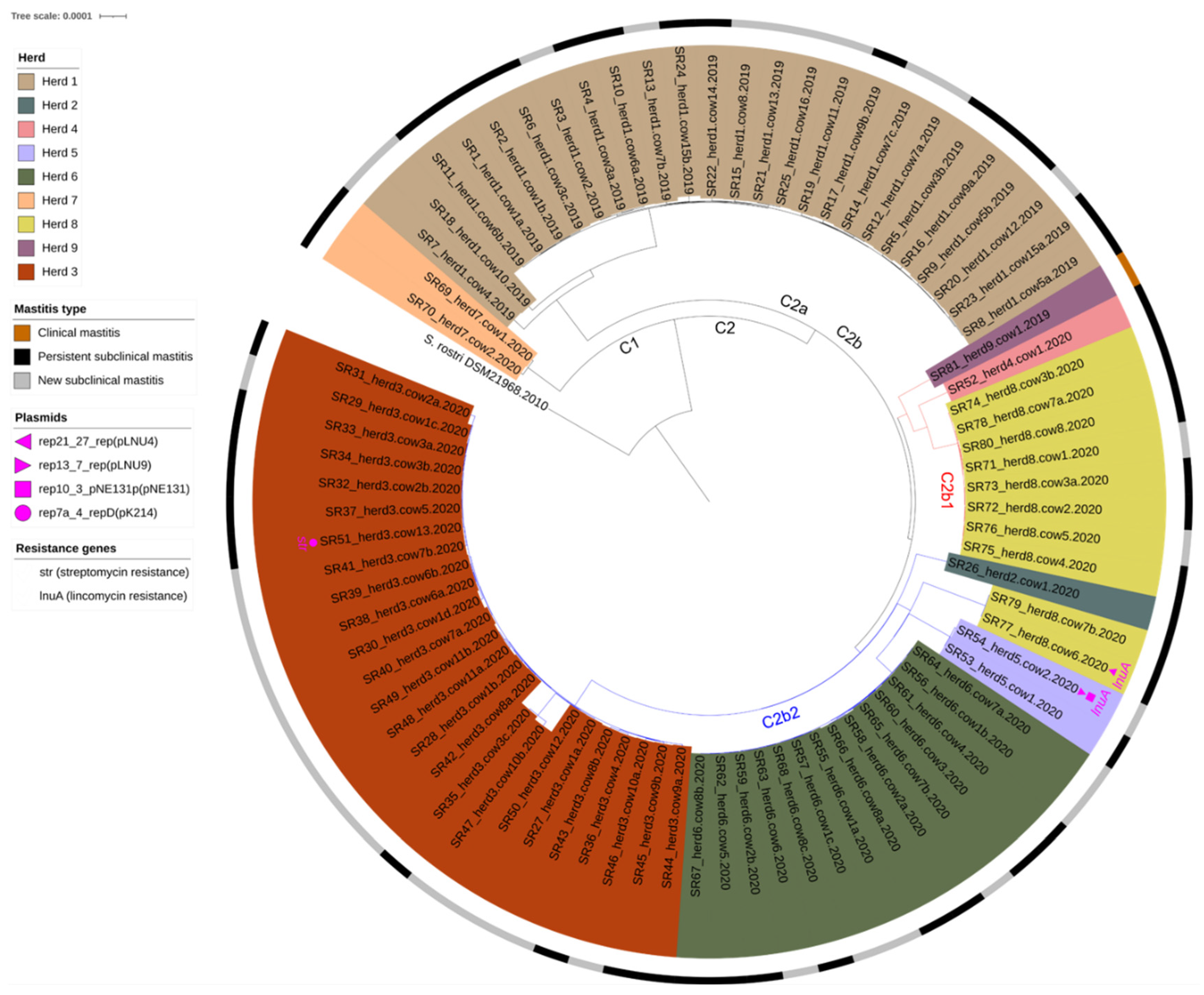
Figure 1. Maximum-likelihood phylogenetic tree based on the core genome of 81 S. rostri isolates from bovine mastitis and S. rostri DSM 21968 (GenBank Accession no. GCF002902145) as a reference (tree re-rooted from the reference). The test isolates were labeled from SR1-SR81 (S for “Staphylococcus”, R for “rostri”) and assigned a herd and cow number. In cows with S. rostri in >1 quarters, the isolates were additionally named a–d depending on the number of S. rostri-positive quarters as follows: “a” for the isolate from the S. rostri-positive quarter number 1; “b” for the positive quarter number 2; and so forth.
Vetsci 10 00530 g001
Figure 2. Network of viral clusters (VCs) representing the predicted phages among the S. rostri isolates. “Overlap”: phages sharing overlap with other phages from multiple VCs; “Other”: phages classified as singletons or outliers; “singletons”: phages with only a few or no gene similarity against other phages; “outliers”: phages with some genes shared with other phages, but not enough to be placed within a VC [28]. Reference strain*: the S. rostri DSM 21968.
Vetsci 10 00530 g002
Table 1. Overview of S. rostri isolates from bovine mastitis and the number of cows infected by it from 2019 to 2020.
Herdn
Isolates
n
Animals
n Animals
One Quarter
n Animals
Two Quarters
n Animals
Three Quarters
n Animals
All Quarters
herd 12516952-
herd 2111---
herd 325134711
herd 4111---
herd 5222---
herd 6148422-
herd 7222---
herd 810862--
herd 9111---
Table 2. Bacterial species found in mixed cultures with S. rostri isolates. From a total of 57 milk samples, 16 different species were identified in combination with S. rostri.
Bacterial Speciesn
Staphylococcus simulans14
Staphylococcus epidermidis9
Staphylococcus haemolyticus7
Corynebacterium amycolatum6
Lactococcus garvieae4
Aerococcus viridans3
Staphylococcus chromogenes3
Streptococcus gallolyticus2
Streptococcus uberis1
Streptococcus canis1
Staphylococcus muscae1
Enterococcus faecalis1
Escherichia coli1
Lactococcus lactis1
Citobacter koseri1
Kocuria rhizophila1
“No ID”1
Disclaimer/Publisher’s Note: The statements, opinions and data contained in all publications are solely those of the individual author(s) and contributor(s) and not of MDPI and/or the editor(s). MDPI and/or the editor(s) disclaim responsibility for any injury to people or property resulting from any ideas, methods, instructions or products referred to in the content.