Article

Isolation, Identification and Drug Resistance Rates of Bacteria from Pigs in Zhejiang and Surrounding Areas during 2019–2021

1
Institute of Animal Husbandry and Veterinary Medicine, Zhejiang Academy of Agricultural Sciences, Hangzhou 310021, China
2
College of Veterinary Medicine, Huazhong Agricultural University, Wuhan 430070, China
*
Authors to whom correspondence should be addressed.
These authors contributed equally to this work.
Vet. Sci. 2023, 10(8), 502; https://doi.org/10.3390/vetsci10080502
Submission received: 25 June 2023 / Revised: 18 July 2023 / Accepted: 21 July 2023 / Published: 3 August 2023
(This article belongs to the Special Issue Advances in Veterinary Clinical Microbiology)

Abstract

:

Simple Summary

This study aimed to determine the prevalence of bacterial diseases in pig farms in various regions of Zhejiang Province and surrounding areas. A total of 526 samples were collected from 85 pig farms in Zhejiang Province and surrounding areas. In this study, samples were analyzed using bacterial isolation and purification, Gram staining, PCR amplification, and antimicrobial susceptibility testing. The isolated bacteria were mainly as follows: Pasteurella multocida (Pm), Bordetella bronchiseptica (Bb), Glasserella parasuis (G. parasuis), Escherichia coli (E. coli), Streptococcus suis (SS), and Actinobacillus pleuropneumoniae (APP). The numbers of bacterial isolates increased during the study period, and mixed infections were observed. Antimicrobial susceptibility testing showed that the drug resistance rates of the various bacteria were high, and the drug resistance spectra were broad. In this study, bacterial epidemiological surveys were conducted on pig farms in various cities in Zhejiang and parts of Anhui, and antimicrobial susceptibility testing was conducted in the isolated strains to provide a scientific basis for the epidemiological and scientific prevention of bacterial diseases, which could be influenced by drug resistance patterns. This study provides scientific guidance for the clinical treatment of bacterial diseases.

Abstract

This study aimed to determine the prevalence of bacterial diseases in pig farms in various regions of Zhejiang Province and surrounding areas. A total of 526 samples were collected from 85 pig farms in Zhejiang Province and surrounding areas. In this study, samples were analyzed using bacterial isolation and purification, Gram staining, PCR amplification, and antimicrobial susceptibility testing. A total of 36 Pasteurella multocida (Pm) isolates were detected, with an isolation rate of 6.84%; 37 Bordetella bronchiseptica (Bb) isolates were detected, with an isolation rate of 7.03%; 60 Glasserella parasuis (G. parasuis) isolates were detected, with an isolation rate of 11.41%; 170 Escherichia coli (E. coli) isolates were detected, with an isolation rate of 32.32%; 67 Streptococcus suis (SS) isolates were detected, with an isolation rate of 12.74%; 44 Actinobacillus pleuropneumoniae (APP) isolates were detected, with an isolation rate of 8.37%; and 7 Salmonella enteritis (SE) isolates were detected, with an isolation rate of 1.33%. Antimicrobial drug susceptibility testing against 21 types of antibiotics was carried out on the isolated strains, and the results showed that 228 strains had varying degrees of resistance to 21 antibiotics, including Pm, Bb, E. coli, and APP, with the highest resistance to lincomycin, at 100%. Pm and APP were the most sensitive to cephalothin, with resistance rates of 0. In terms of strains, Pm had the highest overall sensitivity to 21 antibiotics, and E. coli had the highest resistance. In short, bacterial diseases in Zhejiang and the surrounding areas were harmful, and the drug resistance situation was severe. This study provides scientific guidance for the clinical treatment of bacterial diseases.

1. Introduction

With the rapid development of large-scale pig farming and the emergence of large-scale circulation of livestock products, the prevalence and threat of bacterial diseases have increased due to the reemergence of old diseases, the emergence of new diseases, the increasing prevalence of mixed infections, increasing drug resistance rates, and gradually expanding drug resistance spectra. The major porcine bacterial diseases can be divided into bacterial infections of the respiratory system and intestinal system. The bacteria that cause respiratory diseases are mainly Pm, Bb and APP [1,2,3,4], and the bacteria that cause intestinal diseases are mainly E. coli and SE [5,6]. In the past, antibiotics were the main means of treating swine bacterial diseases. In China, the average amount of antibiotics used in veterinary medicine is more than 6000 tons per year [7]. Although antibiotics can prevent bacterial diseases, the abuse of antibiotics also brings a series of problems, such as the increase in drug resistance and environmental pollution [8,9]. According to the literature, previous epidemiological investigations of bacterial diseases in Zhejiang have mainly focused on single-pathogen infections. Previous reports lack a systematic investigation of bacterial diseases. In this study, samples were collected from pig farms in Zhejiang Province and parts of Anhui, with emergency sampling on affected pig farms when necessary, to isolate and identify pathogenic bacteria and clarify the epidemiological characteristics of major bacterial diseases in pigs. Disc diffusion testing of the isolated bacteria for the detection of drug resistance, data to guide bacterial infection treatment is provided.

2. Materials and Methods

2.1. Main Reagent

From January 2019 to September 2021, the isolation and identification of bacterial pathogens in 526 samples from 85 pig farms submitted from various cities in Zhejiang and parts of Anhui were performed. Antibiotic susceptibility paper sheets were purchased from Changde Bikman Biotechnology Co., Ltd. (Hunan, China) (Table 1).
Green Tap Mix enzyme was purchased from Nanjing Novazan Biotechnology Co., Ltd. (Nanjing, China). DL 2000 DNA Marker was purchased from Takara Biomedical Technology (Beijing, China) Co., Ltd. MacConkey, Tryptic Soy Agar (TSA), and Tryptic Soy Broth (TSB) media were purchased from Thermo Fisher Scientific. Calf serum was purchased from Zhejiang Tianhang Biotechnology Co., Ltd. (Huzhou, China). Nicotinamide adenine dinucleotide (NAD) was purchased from Beijing Soleibo Technology Co., Ltd. (Beijing, China).

2.2. Sample Collection

A total of 169 samples were collected or received over a 3-year period, and a total of 526 samples were inspected. The samples were obtained from various organs from dead pig carcasses and effusion, anal swabs, nasal swabs, vaginal swabs, heart blood, fecal samples, environmental samples, etc. Scissors were used to cut the organ surface, and plates were inoculated using an inoculating loop. Effusion, various swab, fecal, environmental, and other samples were placed in a centrifuge tube. PBS was added, the suspension was mixed, and the samples were inoculated onto plate with an inoculation loop and incubated at 37 °C. A single colony was picked from the plate and placed on a new plate, cultured at 37 °C, and purified for 1–2 generations until a single colony grew on the plate. A single colony was picked from the plate and cultured in liquid medium; Gram staining was performed, and the morphological structure of the bacteria was observed under a microscope for preliminary identification and judgment.

2.3. PCR Primer

We amplified bacterial-specific genes according to the PCR primers given in the references in Table 2. Primers were synthesized by Suzhou Jinweizhi Biotechnology Co., Ltd. (Suzhou, China).

2.4. PCR Template Preparation

A single colony with certain colony characteristics was identified by Gram staining and inoculated into liquid medium. The suspension was placed on a shaker at 37 °C for culture for 8 h. One milliliter of sterilizing liquid, 400 µL of lysis buffer (0.1 mM Tris pH 8.5), and 8 µL of proteinase K were added and mixed well in an EP tube. The sample was incubated at 56 °C for 40 min and then at 100 °C for 10 min. The temperature was decreased to room temperature to preserve the PCR template, and the samples were stored at 4 °C thereafter.

2.5. PCR Amplification

The reaction system was as follows: SYBR Green Master Mix 25 µL, template 1 µL, upstream primer 1.5 µL, downstream primer 1.5 µL, added Distilled water up to 50 µL. A total of 30 cycles were run, the annealing temperature was set to 58 °C, and the annealing time was set to 30 s. The PCR products were electrophoresed on a 1.5% agarose gel, and the bands were observed under a UV gel imaging system. Correctly identified strains were cultured in liquid medium to the logarithmic phase. When PCR amplification results were inconsistent with phenotypic results, strains were further detected by 16sRNA sequencing [4,17]. Bacterial liquid and glycerin were added to the EP tube at a ratio of 7 to 3, mixed evenly, and stored at −80 °C.

2.6. Antimicrobial Susceptibility Testing

The preserved isolated strains were inoculated on a plate and cultivated overnight at 37 °C. A single colony was picked and inoculated in liquid medium, cultivated to the logarithmic phase at 37 °C and 200 r/min, and subjected to disc diffusion testing. The diameter of the inhibition zone was measured [18].

3. Results

3.1. Sample Collection

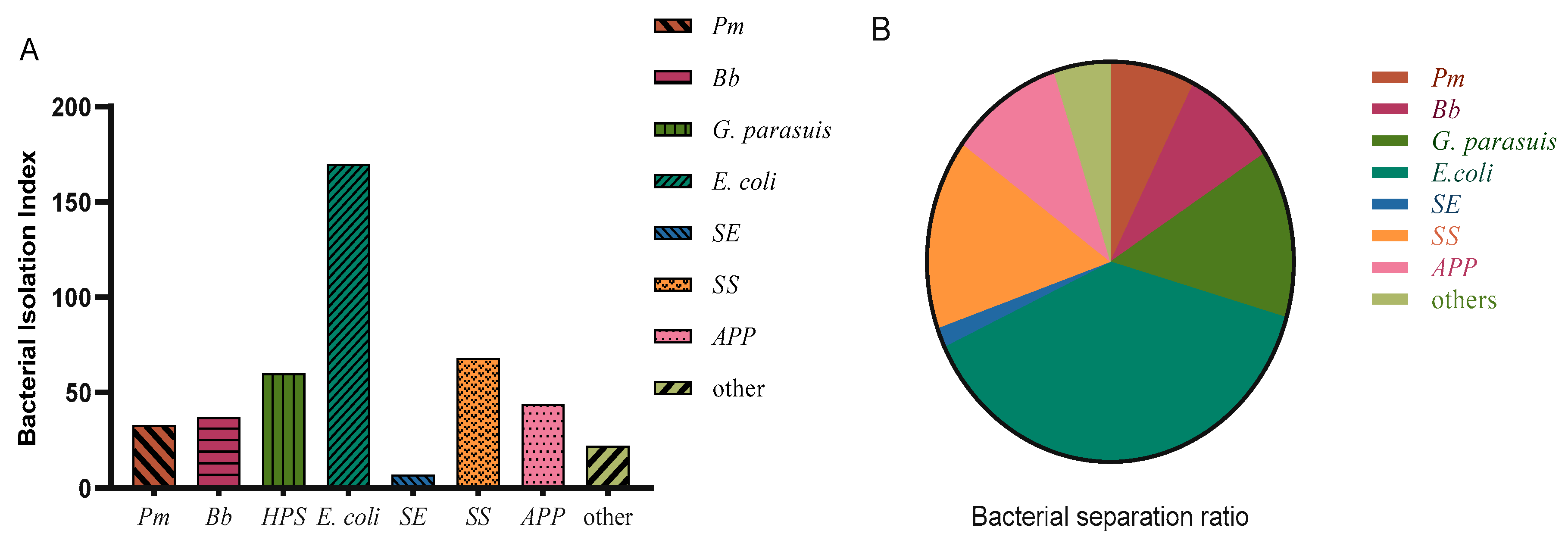
The 169 batches usually included samples from several pigs or samples from multiple parts of a pig, and 526 samples were actually tested. After separation, purification, Gram staining, and PCR amplification, 16 of the tested samples were contaminated and could not be separated and purified, and no bacteria was detected in 89 samples. In the remaining samples, 36 Pasteurella multocida (Pm) isolates were detected, with an isolation rate of 6.84%; 37 Bordetella bronchiseptica (Bb) isolates were detected, with an isolation rate of 7.03%; 60 Glasserella parasuis (G. parasuis) isolates were detected, with an isolation rate of 11.41%; 170 Escherichia coli (E. coli) isolates were detected, with an isolation rate of 32.32%, E. coli isolated in this investigation included ETEC, EPEC, STEC, among which ETEC accounted for the vast majority; 67 Streptococcus suis (SS) isolates were detected, with an isolation rate of 12.74%; 44 Actinobacillus pleuropneumoniae (APP) isolates were detected, with an isolation rate of 8.37%; and 7 Salmonella enteritis (SE) isolates were detected, with an isolation rate of 1.33%(Figure 1A,B). SE was not included in the subsequent statistical analysis due to the small number of isolates.

3.2. Results of Mixed Infections

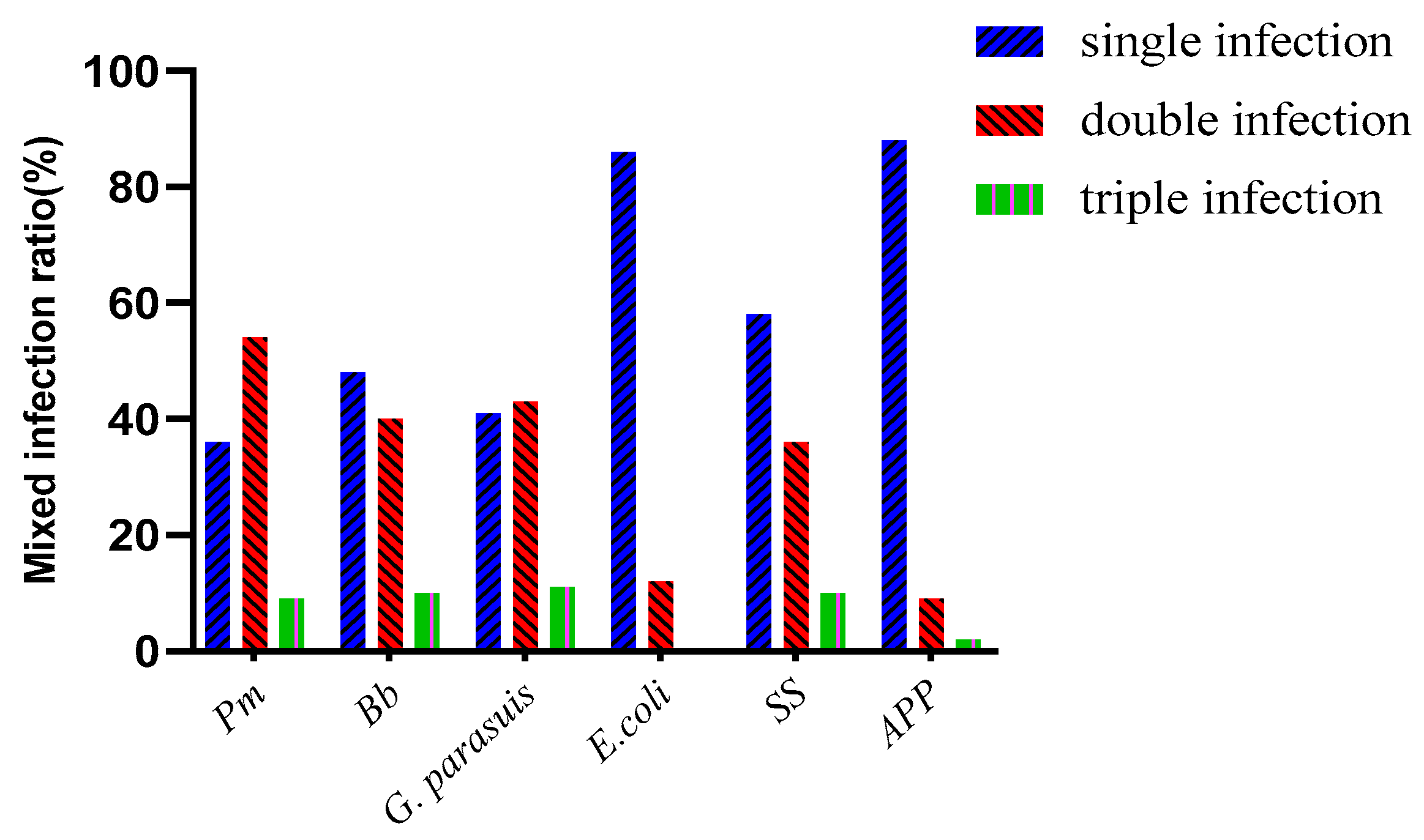
Among the various bacteria isolated, there were single infections, double infections, and triple infections. The numbers of single infections, double infections, and triple infections involving Pm were 12, 20, and 4, and the proportions of these infections were 33.33%, 55.56%, and 11.11%, respectively. The numbers of single infections, double infections and triple infections involving Bb were 18, 15, and 4, and the proportions of these infections were 48.65%, 40.54%, and 10.81%, respectively. The numbers of single infections, double infections and triple infections of G. parasuis were 26, 27 and 7, and the proportions of these infections were 43.33%, 46%, 11.67%, respectively. The numbers of single infections, double infections, and triple infections involving E. coli were 142, 27, and 1, and the percentages of these infections were 78.89%, 15%, and 0.56%, respectively. The numbers of single infections, double infections and triple infections involving SS were 36, 25, and 6, and these infections accounted for 53.73%, 37.31%, and 8.96%, respectively. The numbers of single infections, double infections and triple infections involving APP were 38, 5, and 1, and the percentages of these infections were 88.64%, 9.09%, and 2.27%, respectively. SS and G. parasuis accounted for the largest number of mixed infections, identified in 15 samples. The second largest number of mixed infections (10) involved Bb and G. parasuis (Table 3 and Table 4, Figure 2).

3.3. Results of Disc Diffusion Testing

Disc diffusion testing was performed on the detected strains. The same type of bacteria detected in the same batch in the same field was tested only once, and disc diffusion testing against 21 antibiotics was performed for 228 isolated strains, including 18 strains of Pm, 28 strains of Bb, 36 strains of G. parasuis, 94 strains of E. coli, 44 strains of SS, and 28 strains of APP.
Eighteen isolated Pm strains were tested against 21 antibacterial drugs (the numbers of resistant strains, the proportion of drug resistance to total isolates): ampicillin (3 16.67%), amoxicillin (2, 11.11%), penicillin (3, 16.67%), cephalothin (0, 0.00), ceftiofur (1, 5.56%), cefotaxime (0, 0.00), streptomycin (10, 55.56%), gentamicin (9, 50%), amikacin (5, 27.78%), kanamycin (8, 44.44%), spectinomycin (1, 5.56%), apramycin (0, 0.00), erythromycin (1, 5.56%), tilmicosin(14, 77.78%), doxycycline (0, 0.00), tetracycline (5, 27.78%), florfenicol (2, 11.11%), lincomycin (18, 100%), compound trimethoprim (11, 61.11%), enrofloxacin (2, 11.11%), and ciprofloxacin (2, 11.11%). The results reveal that Pm was most resistant to lincomycin, with a resistance rate of 100%, and it was sensitive to cephalothin, cefotaxime, apramycin, and doxycycline, with sensitivity rates of 100%. There were five antibiotics associated with a resistance rate of 50% or above: streptomycin (55.56%), gentamicin (50%), tilmicosin (77.78%), lincomycin (100%), and compound trimethoprim (61.11%). In terms of drug types, the highest sensitivity was observed for cephalosporins, the highest resistance rate was observed for ceftiofur, at only 5.56%, and the cephalothin and cefotaxime resistance rates were 0.00%. Among the aminoglycosides, the streptomycin and gentamicin resistance rates were 55.56% and 50%, respectively, and the apramycin resistance rate was 0%. Among the tetracyclines, the tilmicosin resistance rate was 77.78%, and the doxycycline sensitivity rate was 100%. (Table 5).
A total of 28 isolated Bb strains were tested against 21 antibacterial drugs (the numbers of resistant strains, the proportion of drug resistance to total isolates): ampicillin (27, 96.42%), amoxicillin (13, 46.43%), penicillin (27, 96.42%), cephalothin (13, 46.43%), ceftiofur (20, 71.42%), cefotaxime (17, 60.71%), streptomycin (26, 92.86%), gentamicin (9, 32.14%), amikacin (9, 32.14%), kanamycin (9, 32.14%), spectinomycin (21, 75%), apramycin (27, 96.42%), erythromycin (13, 21.43%), tilmicosin (26, 92.86%), doxycycline (6, 28.57%), tetracycline (8, 46.43%), florfenicol (13, 42.86%), lincomycin (28, 100%), compound trimethoprim (27, 96.42%), enrofloxacin (12, 46.43%), and ciprofloxacin (5, 17.86%). The results show that Bb was the most resistant to lincomycin, with a resistance rate of 100%. Ten antibiotics had associated resistance rates of 50% or higher: ampicillin (96.42%), penicillin (96.42%), ceftiofur (71.42%), cefotaxime (60.71%), streptomycin (92.86%), apramycin (96.42%), tilmicosin (92.86%), lincomycin (100%), and compound trimethoprim (96.42%). Among the quinolones, ciprofloxacin was associated with the highest sensitivity, with an associated resistance rate of 17.86% (Table 6).
A total of 36 isolated HPS strains were tested against 21 antibacterial drugs (the numbers of resistant strains, the proportion of drug resistance to total isolates): ampicillin (20, 55.56%), amoxicillin (14, 38.89%), penicillin (17, 47.22%), cephalothin (7, 19.44%), ceftiofur (8, 22.22%), cefotaxime (3, 8.33%), streptomycin (24, 66.67%), gentamicin (20, 55.56%), amikacin (22, 61.11%), kanamycin (13, 36.11%), spectinomycin (5, 13.89%), apramycin (33, 91.67%), erythromycin (16, 44.44%), tilmicosin (25, 69.44%), doxycycline (2, 5.56%), tetracycline (9, 25%), florfenicol (7, 19.44%), lincomycin (30, 83.33%), compound trimethoprim (34, 94.44%), enrofloxacin (18, 50%), and ciprofloxacin (11, 30.56%). The results reveal that HPS was the most resistant to compound trimethoprim, with a resistance rate of 94.44%, and most sensitive to doxycycline, with a resistance rate of 5.56%. Seven antibiotics were associated with resistance rates of 50% or higher: ampicillin (55.56%), penicillin (60.71%), streptomycin (66.67%), gentamicin (55.56%), amikacin (61.11%), apramycin (91.67%), and tilmicosin (69.44%). The following cephalosporin antibiotics were associated with the highest sensitivity, with drug resistance rates of less than 25%: cephalothin (19.44%), ceftiofur (22.22%), and cefotaxime (8.33%) (Table 7).
Ninety-four isolated E. coli strains were tested against 21 antibacterial drugs (the numbers of resistant strains, the proportion of drug resistance to total isolates): ampicillin (87, 92.55%), amoxicillin (84, 89.36%), penicillin (92, 97.87%), cephalothin (55, 58.51%), ceftiofur (30, 31.91%), cefotaxime (15, 15.96%), streptomycin (58, 61.70%), gentamicin (62, 65.96%), amikacin (25, 26.60%), kanamycin (52, 55.32%), spectinomycin (31, 32.98%), apramycin (53, 56.38%), erythromycin (79, 84.04%), tilmicosin (94, 100%), doxycycline (86, 91.49%), tetracycline (88, 93.62%), florfenicol (69, 73.40%), lincomycin (94, 100%), compound trimethoprim (86, 91.49%), enrofloxacin (71, 75.53%), and ciprofloxacin (44, 46.80%). The results reveal that E. coli was most resistant to tilmicosin and lincomycin, with resistance rates reaching 100%. Sensitivity to cefotaxime was the highest, and the resistance rate was 15.96%. There were 15 antibiotics associated with resistance rates of 50% or higher: ampicillin (92.55%), amoxicillin (89.36%), penicillin (97.87%), cephalothin (58.51%), streptomycin (61.70%), gentamicin (65.96%), kanamycin (55.32%), apramycin (56.38%), erythromycin (84.04%), tilmicosin (100%), doxycycline (91.49%), tetracycline (93.62%), florfenicol (73.40%), lincomycin (100%), and enrofloxacin (75.53%). Cephalosporins were associated with the highest sensitivity, with associated resistance rates of 58.51% against cephalothin, 31.91% against ceftiofur, and 15.96% against cefotaxime. In general, E. coli exhibited the highest level of antibiotic resistance (Table 8).
Forty-four isolated SS strains were tested against 21 antibacterial drugs (the numbers of resistant strains, the proportion of drug resistance to total isolates): ampicillin (10, 22.72%), amoxicillin (3, 6.82%), penicillin (6, 13.64%), cephalothin (1, 2.27%), ceftiofur (3, 6.82%), cefotaxime (4, 9.09%), streptomycin (26, 59.09%), gentamicin (40, 90.91%), amikacin (43, 97.73%), kanamycin (41, 93.18%), spectinomycin (16, 36.36%), apramycin (44, 100%), erythromycin (30, 68.18%), tilmicosin (43, 97.73%), doxycycline (14,31.82%), tetracycline (37, 84.09%), florfenicol (8, 18.08%), lincomycin (43, 97.73%), compound trimethoprim (33, 75%), enrofloxacin (19, 43.18%), and ciprofloxacin (14, 31.82%). The results show that SS was most resistant to apramycin, with a resistance rate of 100%, and had the highest sensitivity to cephalothin, with a resistance rate of 2.27%. There were nine antibiotics associated with resistance rates of 50% or higher: streptomycin (59.09%), gentamicin (90.91%), amikacin (97.73%), kanamycin (93.18%), apramycin (100%), erythromycin (68.18%), tilmicosin (97.73%), tetracycline (84.09%), and lincomycin (97.73%). SS had the highest sensitivity to cephalosporin antibiotics. The cephalothin resistance rate was 2.27%, the ceftiofur resistance rate was 6.82%, and the cefotaxime resistance rate was 9.09% (Table 9).
Twenty-eight isolated APP strains were tested against 21 antibacterial drugs (the numbers of resistant strains, the proportion of drug resistance to total isolates): ampicillin (11, 39.29%), amoxicillin (6, 21.42%), penicillin (13, 46.42%), cephalothin (1, 3.57%), ceftiofur (2, 7.14%), cefotaxime (0, 0.00), streptomycin (15, 53.57%), gentamicin (8, 28.57%), amikacin (15, 53.57%), kanamycin (15, 53.57%), spectinomycin (6, 21.42%), apramycin (24, 85.71%), erythromycin (3, 10.71%), tilmicosin(24, 85.71%), doxycycline (4, 14.28%), tetracycline (13, 46.42%), florfenicol (7, 25.00%), lincomycin (27, 96.43%), compound trimethoprim (16, 57.14%), enrofloxacin (11, 39.29%), and ciprofloxacin (0, 0). The results reveal that APP was most resistant to lincomycin, with a resistance rate of 96.43%, and most sensitive to cephalothin and ciprofloxacin, with resistance rates of 0. There were six antibiotics with associated resistance rates of 50% or higher: streptomycin (53.57%), amikacin (53.57%), kanamycin (53.57%), tilmicosin (85.71%), lincomycin (96.43%), and compound trimoxazole (57.14%). Cephalosporin antibiotics were associated with the highest sensitivity, with associated resistance rates of 0% for cephalothin, 7.14% for ceftiofur, and 3.57% for cefotaxime. Two values in Table 10 were ignored because they were zero (Table 10).

3.4. Multidrug Resistance of Isolated Strains

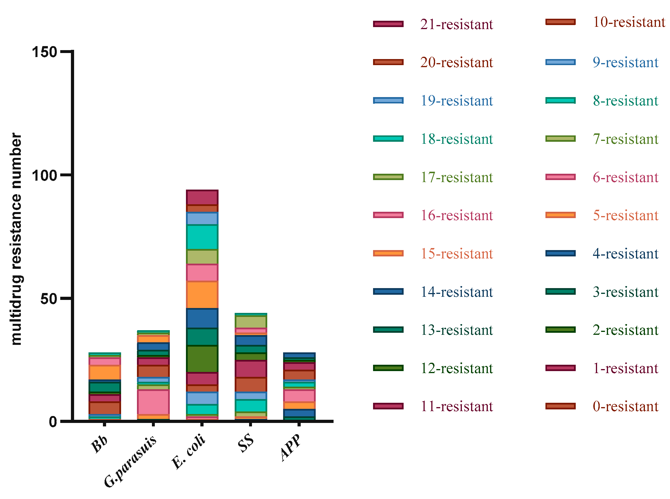
Among the 18 strains of Pm that were subjected to disc diffusion testing, 6 strains were resistant to 0–5 drugs, accounting for 33.33%; 11 strains were resistant to 6–10 drugs, accounting for 61.11%; 1 strain was resistant to 12 drugs, accounting for 5.56%; and no strains resistant to 16–20 or 21 drugs. Among the 28 Bb strains tested, 1 strain was resistant to 0–5 drugs, accounting for 3.57%; 7 strains were resistant to 6–10 drugs, accounting for 25%; 15 strains were resistant to 15 drugs, accounting for 53.57%; 5 strains resistant to 16–20 drugs, accounting for 17.86%; and no strains were resistant to 21 drugs. Among the 36 strains of G. parasuis tested, 3 strains were resistant to 0–5 drugs, accounting for 8.33%; 20 strains were resistant to 6–10 drugs, accounting for 55.56%; 12 strains were resistant to 11–15 drugs, accounting for 33.33%; 2 strains were resistant to 16–20 drugs, accounting for 5.56%; and no strains resistant to 21 drugs. Among the 94 E. coli strains tested, 1 strain was resistant to 0–5 drugs, accounting for 1.06%; 14 strains were resistant to 6–10 drugs, accounting for 38.89%; 11–15 strains were resistant to 42 drugs, accounting for 44.68%; 31 strains were resistant to 16–20 drugs, accounting for 32.98%; and 6 strains were resistant to 21 drugs, accounting for 6.38%. Among the 44 strains of SS tested, 2 strains were resistant to 0–5 drugs, accounting for 4.55%; 16 strains were resistant to 6–10 drugs, accounting for 36.36%; 18 strains were resistant to 11–15 drugs, accounting for 40.91%; 8 strains were resistant to 16–20 drugs, accounting for 18.18%; there were no strains resistant to 21 drugs. Among the 28 APP strains tested, 8 strains were resistant to 0–5 drugs, accounting for 28.57%; 13 strains were resistant to 6–10 drugs, accounting for 29.55%; 7 strains were resistant to 11–15 drugs, accounting for 15.91%; and there were no strains resistant to 16–20 or 21 drugs. The experimental results revealed that none of the isolated strains were resistant to 0 or 1 antibiotic. Pm had the best overall sensitivity, and E. coli had the highest rate of drug resistance. All the bacteria except for E. coli were resistant to 19 or more drugs; E. coli had resistance to 19 or more drugs, and 6 strains were resistant to all 21 drugs (Table 11, Figure 3).

3.5. Heatmap of Drug Resistance

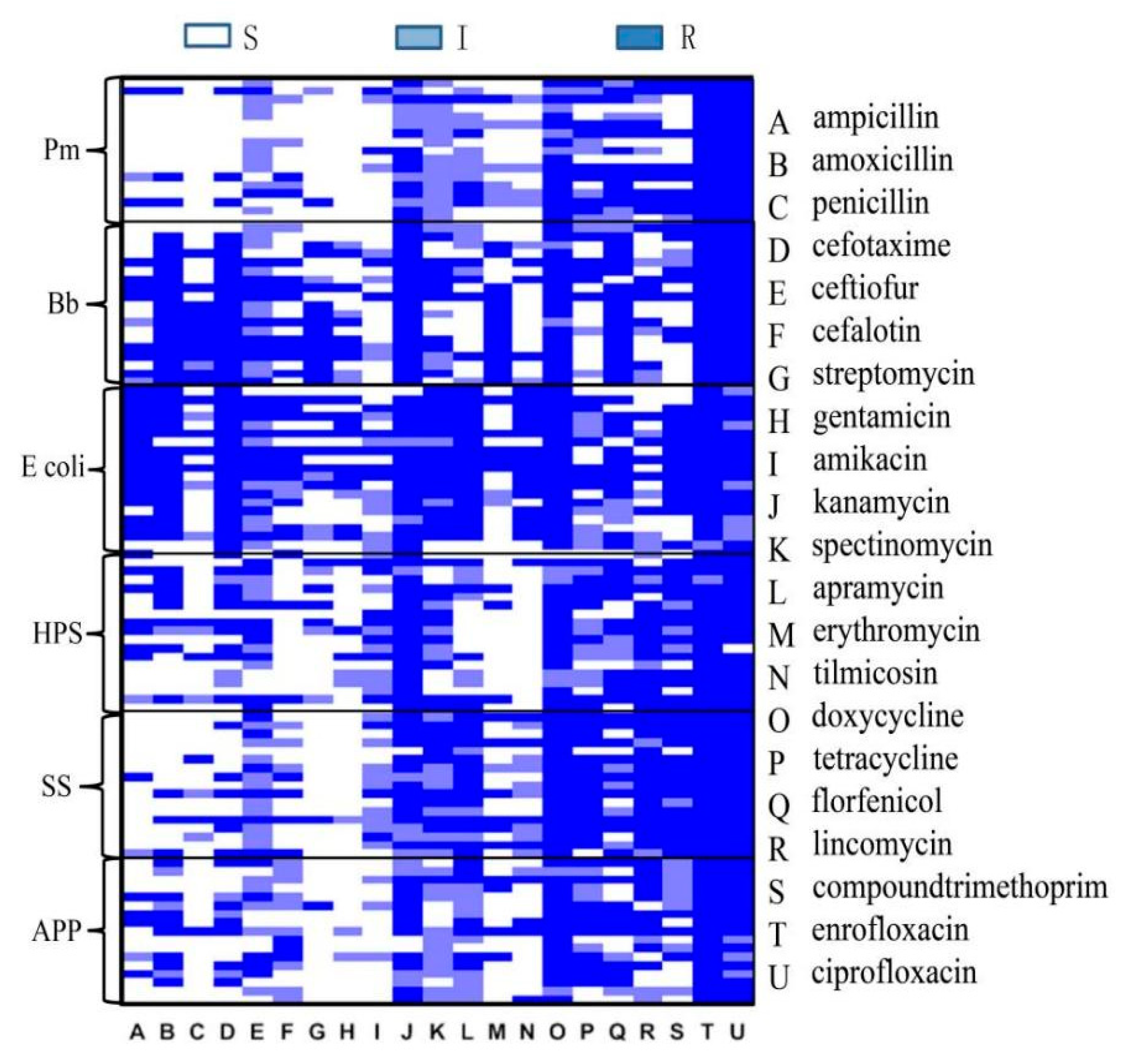
Among the Pm, Bb, G. parasuis, E. coli, SS, and APP isolates that were tested with the disc diffusion method, 18 were randomly selected for construction of a heatmap of resistance to 21 antibiotics (Figure 4). The results showed that E. coli was most resistant to several antibiotics. Pm and SS were similar in their sensitivity rates, and both were sensitive to antibiotics A, B, C, D, E, F, G, H, and I. Four antibiotics, trimethoprim, tilmicosin, apramycin, and lincomycin, had the highest associated resistance rates, which may be related to the high rates of use of these antibiotics on pig farms. Pm, G. parasuis, SS, and APP had similar drug resistance patterns. Most of these bacterial specimens were collected from the lungs, and the resistance patterns may be related to the sample collection site.

3.6. Three-Year Trend and Seasonal Trend of Bacteria

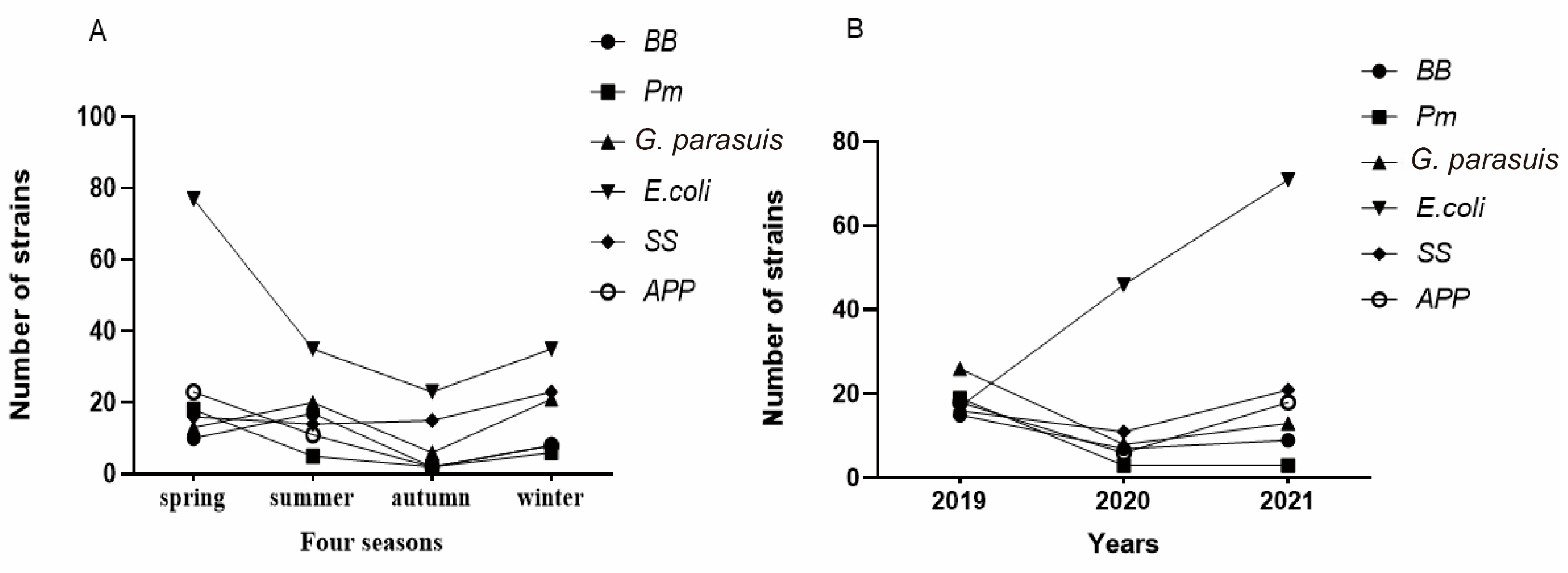
The year was divided into four seasons: spring, summer, autumn and winter. Spring was defined as February–April, summer was defined as May–July, autumn was defined as August–October, and winter was defined as November–January, and a line chart of bacterial prevalence rates in the four seasons was constructed (Figure 5A). According to the results, the numbers of isolates of the six bacteria were the lowest in autumn and highest in spring, with a decreasing trend from summer to autumn and an increasing trend in winter. Since this experiment was carried out only through September 2021, the three-year bacterial prevalence trend map was created using the isolates collected in the first 9 months of each year during 2019–2021 (Figure 5B). It can be seen in the figure that the number of E. coli isolates increased during the three-year period, while the numbers of the other five bacteria all decreased from 2019 to 2020, with upward trends in 2021.

4. Discussion

The experimental results revealed that Pm, Bb, G. parasuis, E coli, SS, and APP were widespread in Zhejiang, similar to previous reports [19] in China, though the bacterial isolation rates were different. For example, a previous report [20,21] showed that the isolation rate of SS in Thailand was very high, but the isolation rate of SS in this investigation was very low. This result may be caused by comprehensive factors, such as different regional characteristics, sample sources, and climates. The bacteria in this study can be divided into two categories, intestinal bacteria and respiratory bacteria, and infections caused by these bacteria can seriously affect the Chinese pig industry. Therefore, research on the isolation rates, drug resistance patterns, and seasonality of these bacteria is of great significance for the prevention and control of local bacterial diseases.
In this experiment, antimicrobial susceptibility testing against 21 antibiotics was carried out in the isolated strains. The results showed that in Pm, the resistance rate for lincomycin was the highest, at 100%. Sensitivity to cephalothin, cefotaxime, apramycin, and doxycycline was high, with sensitivity rates of 100%. A previous report [22] showed that Pm had the highest resistance to amoxicillin, with a resistance rate of 75.9%, in Vietnam. In this experiment, Bb had the highest resistance to lincomycin, with a resistance rate of 100%, and was relatively sensitive to ciprofloxacin, with a resistance rate of 17.86%. Another report [23] showed that Bb had the highest resistance to ampicillin, with a resistance rate of 83.98%, and there were 6 antibiotics with associated resistance rates of 0% in China. E. coli was had the highest resistance to tilmicosin and lincomycin, with resistance rates of 100%, and the highest sensitivity to cefotaxime, with a resistance rate of 15.96%. A previous report [24] showed that E. coli had highest resistance to trimethoprim, with a resistance rate of 100% in Poland. SS had the highest resistance to apramycin, with a resistance rate of 100%, and the highest sensitivity to cephalothin, with a resistance rate of 2.27%. Another previous report [25] showed that the highest rate of drug resistance in Streptococcus was associated with tetracycline, with a drug resistance rate of 84.2% in Ontario. There was a large gap in the resistance rates of different strains to different drug categories and even different antibiotics in similar drug categories. This is related to different treatment regimens of various countries and regions. This result demonstrates that for the treatment of bacterial diseases, more targeted medications based on drug resistance testing results are needed. In this study, the most common coinfection involved SS and G. parasuis. Coinfection is often more threatening to pigs than monoinfection [26,27]. None of the isolates of the six bacteria that were subjected to antimicrobial susceptibility testing were resistant to 0 or only 1 antibiotic. Six strains of E. coli showed multiresistance to 21 drugs simultaneously. On the basis of these investigation results, the drug resistance situation is severe. Additionally, the resistance patterns of each bacterial species showed certain differences according to antibiotic properties. Seasonal distributions of these bacteria were clearly observed in this study. These bacteria were generally prevalent in spring and began to decline in summer until autumn. In winter, the bacterial prevalence showed an increasing trend. This phenomenon may be related to the fact that hot temperatures in summer are not conducive to the growth of bacteria, while milder temperatures in spring are suitable for bacterial growth. Season affects prevalence of bacterial pathogenic genes [28,29]. Therefore, we cannot ignore the effects of temperature when implementing prevention and control measures for bacteria. The number of E. coli isolated during the three-year study period consistently increased, while the numbers of the other five bacteria declined from 2019 to 2020 and then increased in 2021. The monitoring of bacterial diseases in Zhejiang will be indispensable in the next few years. Future research will require long-term monitoring and bacterial typing to effectively guide prevention, control and treatment measures targeting bacteria.

5. Conclusions

This study focused on the isolation and identification of bacterial pathogens in 516 samples from 85 pig farms submitted for inspection from various cities in Zhejiang and parts of Anhui. The isolated bacteria were mainly as follows: Pm, Bb, G. parasuis, E. coli, SS, and APP. The numbers of bacterial isolates increased during the study period, and mixed infections were observed. Antimicrobial susceptibility testing showed that the drug resistance rates of the various bacteria were high, and the drug resistance spectra were broad. In this study, bacterial epidemiological surveys were conducted on pig farms in various cities in Zhejiang and parts of Anhui, and antimicrobial susceptibility testing was conducted in the isolated strains to provide a scientific basis for the epidemiological and scientific prevention of bacterial diseases, which could be influenced by drug resistance patterns. This study provides scientific guidance for the clinical treatment of bacterial diseases.

Author Contributions

Conceptualization, X.X., J.L., P.H. and X.C.; data curation, X.X., J.L. and Y.L.; formal analysis, Y.H.; funding acquisition, X.C. and Y.L.; investigation, Q.J.; methodology, X.X.; project administration, X.X.; resources, Y.H.; software, P.H. and X.C.; supervision, G.B.; validation, X.L., J.S., Q.J. and Q.W.; visualization, X.L. and J.S.; writing—original draft, X.X.; writing—review and editing, P.H. and Y.L. All authors have read and agreed to the published version of the manuscript.

Funding

The present research was supported by the Zhejiang Provincial Science and Technology Cooperation Program (No. 2023SNJF049) and supported by the Zhejiang Provincial Key Research and Development Program (No. 2021C02007), The China Agriculture Research System of MOF and MARA (No. CARS-43-C-2) and the National Natural Science Foundation of China (No. 31672598). “Jianbing” “lingyan” research and development project of Zhejiang Province (No. 2023C02047).

Institutional Review Board Statement

Not applicable.

Informed Consent Statement

Not applicable.

Conflicts of Interest

The authors declare no conflict of interest.

References

  1. Dayao, D.; Gibson, J.; Blackall, P.; Turni, C. Antimicrobial resistance in bacteria associated with porcine respiratory disease in Australia. Vet. Microbiol. 2014, 171, 232–235. [Google Scholar] [CrossRef] [PubMed] [Green Version]
  2. Sunaga, F.; Tsuchiaka, S.; Kishimoto, M.; Aoki, H.; Kakinoki, M.; Kure, K.; Okumura, H.; Okumura, M.; Okumura, A.; Nagai, M.; et al. Development of a one-run real-time PCR detection system for pathogens associated with porcine respiratory diseases. J. Vet. Med. Sci. 2020, 82, 217–223. [Google Scholar] [CrossRef] [Green Version]
  3. Hanchanachai, N.; Chumnanpuen, P. Interaction study of Pasteurella multocida with culturable aerobic bacteria isolated from porcine respiratory tracts using coculture in conditioned media. BMC Microbiol. 2021, 21, 19. [Google Scholar] [CrossRef]
  4. Zhao, Z.; Wang, C.; Xue, Y.; Tang, X.; Wu, B.; Cheng, X.; He, Q.; Chen, H. The occurrence of Bordetella bronchiseptica in pigs with clinical respiratory disease. Vet. J. 2011, 188, 337–340. [Google Scholar] [CrossRef]
  5. Moxley, R.; Duhamel, G. Comparative pathology of bacterial enteric diseases of swine. Adv. Exp. Med. Biol. 1999, 473, 83–101. [Google Scholar] [PubMed]
  6. Hong, S.; Rovira, A.; Davies, P.; Ahlstrom, C.; Muellner, P.; Rendahl, A.; Olsen, K.; Bender, J.B.; Wells, S.; Perez, A.; et al. Serotypes and Antimicrobial Resistance in Salmonella enterica Recovered from Clinical Samples from Cattle and Swine in Minnesota, 2006 to 2015. PLoS ONE 2016, 11, e0168016. [Google Scholar] [CrossRef] [Green Version]
  7. Hu, X.; Zhou, Q.; Luo, Y. Occurrence and source analysis of typical veterinary antibiotics in manure, soil, vegetables and groundwater from organic vegetable bases, northern China. Environ. Pollut. 2010, 158, 2992–2998. [Google Scholar] [CrossRef]
  8. Ling, Z.; Hua, D.Y.; Hui, W. Residues of veterinary antibiotics in manures from feedlot livestock in eight provinces of China. Sci. Total Environ. 2010, 408, 1069–1075. [Google Scholar]
  9. Barton, M.D. Impact of antibiotic use in the swine industry. Curr. Opin. Microbiol. 2014, 19, 9–15. [Google Scholar] [CrossRef]
  10. Lichtensteiger, C.A.; Steenbergen, S.M.; Lee, R.M.; Polson, D.D.; Vimr, E.R. Direct PCR analysis for toxigenic Pasteurella multocida. J. Clin. Microbiol. 1996, 34, 3035–3039. [Google Scholar] [CrossRef]
  11. Hozbor, D.; Fouque, F.; Guiso, N. Detection of Bordetella bronchiseptica by the polymerase chain reaction. Res. Microbiol. 1999, 150, 333–341. [Google Scholar] [CrossRef] [PubMed]
  12. Oliveira, S.; Galina, L.; Pijoan, C. Development of a PCR test to diagnose Haemophilus parasuis infections. J. Vet. Diagn. Investig. 2001, 13, 495–501. [Google Scholar] [CrossRef] [Green Version]
  13. Bej, A.; McCarty, S.; Atlas, R. Detection of Coliform bacteria and Escherichia coli by multiplex polymerase chain reaction: Comparison with defined substrate and plating methods for water quality monitoring. Appl. Environ. Microbiol. 1991, 57, 2429–2432. [Google Scholar] [CrossRef]
  14. Okwumabua, O.; O’Connor, M.; Shull, E. A polymerase chain reaction (PCR) assay specific for Streptococcus suis based on the gene encoding the glutamate dehydrogenase. FEMS Microbiol. Lett. 2003, 218, 79–84. [Google Scholar] [CrossRef] [PubMed]
  15. Gram, T.; Ahrens, P.; Andreasen, M.; Nielsen, J. An Actinobacillus pleuropneumoniae PCR typing system based on the apx and omlA genes—Evaluation of isolates from lungs and tonsils of pigs. Vet. Microbiol. 2000, 75, 43–57. [Google Scholar] [CrossRef] [PubMed]
  16. Rahn, K.; De Grandis, S.A.; Clarke, R.C.; McEwen, S.; Galán, J.; Ginocchio, C.; Curtiss, R.; Gyles, C. Amplification of an invA gene sequence of Salmonella typhimurium by polymerase chain reaction as a specific method of detection of Salmonella. Mol. Cell. Probes 1992, 6, 271–279. [Google Scholar] [CrossRef] [PubMed]
  17. DeLong, E.F. Archaea in coastal marine environments. Proc. Natl. Acad. Sci. USA 1992, 89, 5685–5689. [Google Scholar] [CrossRef]
  18. Soares, T.C.S.; Paes, A.C.; Megid, J.; Ribolla, P.E.M.; Paduan, K.D.S.; Gottschalk, M. Antimicrobial susceptibility of Streptococcus suis isolated from clinically healthy swine in Brazil. Can. J. Vet. Res. 2014, 78, 145–149. [Google Scholar]
  19. Zhang, B.; Ku, X.; Yu, X.; Sun, Q.; Wu, H.; Chen, F.; Zhang, X.; Guo, L.; Tang, X.; He, Q. Prevalence and antimicrobial susceptibilities of bacterial pathogens in Chinese pig farms from 2013 to 2017. Sci. Rep. 2019, 9, 9908. [Google Scholar] [CrossRef] [Green Version]
  20. Phongaran, D.; Khang-Air, S.; Angkititrakul, S. Molecular epidemiology and antimicrobial resistance of Salmonella isolates from broilers and pigs in Thailand. Vet. World 2019, 12, 1311–1318. [Google Scholar] [CrossRef] [Green Version]
  21. Katsumi, M.; Kataoka, Y.; Takahashi, T.; Kikuchi, N.; Hiramune, T. Bacterial Isolation from Slaughtered Pigs Associated with Endocarditis, Especially the Isolation of Streptococcus suis. J. Vet. Med. Sci. 1997, 59, 75–78. [Google Scholar] [CrossRef] [Green Version]
  22. Vu-Khac, H.; Trinh, T.T.H.; Nguyen, T.T.G.; Nguyen, X.T. Prevalence of virulence factor, antibiotic resistance, and serotype genes of Pasteurella multocida strains isolated from pigs in Vietnam. Vet. World 2020, 13, 896–904. [Google Scholar] [CrossRef]
  23. Zhang, Y.; Yang, H.; Guo, L.; Zhao, M.; Wang, F.; Song, W.; Hua, L.; Wang, L.; Liang, W.; Tang, X.; et al. Isolation, Antimicrobial Resistance Phenotypes, and Virulence Genes of Bordetella bronchiseptica from Pigs in China, 2018–2020. Front. Vet. Sci. 2021, 8, 672716. [Google Scholar] [CrossRef] [PubMed]
  24. Mazurek, J.; Bok, E.; Baldy-Chudzik, K. Complexity of Antibiotic Resistance in Commensal Escherichia coli Derived from Pigs from an Intensive-Production Farm. Microbes Environ. 2018, 33, 242–248. [Google Scholar] [CrossRef] [PubMed] [Green Version]
  25. Arndt, E.R.; Farzan, A.; MacInnes, J.I.; Friendship, R.M. Antimicrobial resistance of Streptococcus suis isolates recovered from clinically ill nursery pigs and from healthy pigs at different stages of production. Can. Vet. J. 2019, 60, 519–522. [Google Scholar] [PubMed]
  26. Zhu, H.; Chang, X.; Zhou, J.; Wang, D.; Zhou, J.; Fan, B.; Ni, Y.; Yin, J.; Lv, L.; Zhao, Y.; et al. Co-infection analysis of bacterial and viral respiratory pathogens from clinically healthy swine in Eastern China. Vet. Med. Sci. 2021, 7, 1815–1819. [Google Scholar] [CrossRef]
  27. Meng, F.; Wu, N.H.; Nerlich, A.; Herrler, G.; Valentin-Weigand, P.; Seitz, M. Dynamic Virus-Bacterium Interactions in a Porcine Precision-Cut Lung Slice Coinfection Model: Swine Influenza Virus Paves the Way for Streptococcus suis Infection in a Two-Step Process. Infect. Immun. 2015, 83, 2806–2815. [Google Scholar] [CrossRef] [Green Version]
  28. Li, Y.; Yang, Q.; Cao, C.; Cui, S.; Wu, Y.; Yang, H.; Xiao, Y.; Yang, B. Prevalence and characteristics of Salmonella isolates recovered from retail raw chickens in Shaanxi Province, China. Poult. Sci. 2020, 99, 6031–6044. [Google Scholar] [CrossRef]
  29. Fancher, C.A.; Thames, H.T.; Colvin, M.G.; Smith, M.; Easterling, A.; Nuthalapati, N.; Zhang, L.; Kiess, A.; Dinh, T.T.N.; Sukumaran, A.T. Prevalence and Molecular Characteristics of Avian Pathogenic Escherichia coli in “No Antibiotics Ever” Broiler Farms. Microbiol. Spectr. 2021, 9, e0083421. [Google Scholar] [CrossRef]
Figure 1. (A) Bacterial isolation results and (B) isolation rates.
Vetsci 10 00502 g001
Figure 2. Mixed infection results (%).
Vetsci 10 00502 g002
Figure 3. Multidrug resistance results of the six bacteria.
Vetsci 10 00502 g003
Figure 4. Heatmap of resistance.
Vetsci 10 00502 g004
Figure 5. (A) Seasonal trends of bacteria and (B) three-year trend of bacterial prevalence.
Vetsci 10 00502 g005
Table 1. Types of antibiotic susceptibility discs.
Antibiotic CategoryDrug Name
penicillinsampicillin, amoxicillin, penicillin
cephalosporinscefotaxime, ceftiofur, cephalothin
aminoglycosidesstreptomycin, gentamicin, amikacin, apramycin, kanamycin, spectinomycin
macrolidestilmicosin, erythromycin
tetracyclinesdoxycycline, tetracycline
chloramphenicolflorfenicol
lincomycinslincomycin
sulfonamidescompound trimethoprim
quinolonesenrofloxacin, ciprofloxacin
Table 2. Primers used in study.
StrainsGeneNameSequence(5′→3′)Amplicons SizeReferences
Pasteurella multocidaToxAToxA–1CTTAGATGAGCGACAAGG846 bp[10]
ToxA–2GAATGCCACACCTCTATAG
Bordetella bronchisepticaFlaFla4TGGCGCCTGCCCTATC237 bp[11]
Fla2AGGCTCCCAAGAGAGAAAGGCTT
Glasserella parasuis16SrRNAHPS–1GGCTTCGTCACCCTCTGT822 bp[12]
HPS–2GTGATGAGGAAGGGTGGTGT
Escherichia coliuidAEc–1AAAACGGCAAGAAAAAGCAG147 bp[13]
Ec–2GCGTGGTTACAGTCTTGCG
Streptococcus suisgdhJP4GCA GCGTATTCTGTCAAACG689 bp[14]
JP5CCATGGACAGATAAA GATGG
Actinobacillus pleuropneumoniaeapxIVAAPXIVA–1TGGCACTGACGGTGATGA442 bp[15]
APXIVA–2GGCCATCGACTCAACCAT
Salmonella enteritisinvA139GTGAAATTATCGCCACGTTCGGGCAA284 bp[16]
141TCATCGCACCGTCAAAGGAACC
Table 3. Mixed infection results.
Strain NameSingle InfectionDouble InfectionTriple Infection
Pm12204
Bb18154
G. parasuis26277
E. coli142271
SS36256
APP3941
Table 4. Number of mixed infections.
Double InfectionTriple Infection
TypesNumberTypesNumber
PmPm + Bb1Pm + APP + others1
Pm + G. parasuis5Pm + G. parasuis + SS3
Pm + E. coli1
Pm + SS3
Pm + APP2
Pm + others3
BbBb + Pm1Bb + G. parasuis + E.coli1
Bb + G. parasuis6Bb + G. parasuis + SS3
Bb + SS4
Bb + E.coli3
Bb + others3
G. parasuisG. parasuis + Pm5G. parasuis + SS + Bb3
G. parasuis + Bb6G. parasuis + SS + Pm3
G. parasuis + SS9G. parasuis + E.coli + Bb1
G. parasuis + E.coli3G. parasuis + Pm + others1
G. parasuis + APP1
G. parasuis + others4
E.coliE.coli + Pm1E.coli + G. parasuis + Bb1
E.coli + Bb3
E.coli + G. parasuis3
E.coli + SS8
E.coli + others1
SSSS + Bb4SS + Pm + G. parasuis3
SS + Pm3SS + Bb + G. parasuis3
SS + G. parasuis9
SS + E.coli8
SS + APP1
APPAPP + Pm2APP + Pm + others1
APP + G. parasuis1
APP + SS1
Table 5. Pm isolate disc diffusion testing results.
Types of AntibioticsAntibiotic NameNumber of StrainsResistance Rate (%)
SIR
penicillinsampicillin150316.67
amoxicillin151211.11
penicillin150316.67
cephalosporinscefotaxime18000.00
ceftiofur16115.56
cephalothin18000.00
aminoglycosidesstreptomycin351055.56
gentamicin18950
amikacin103527.78
kanamycin46844.44
spectinomycin12515.56
apramycin18000.00
macrolidestilmicosin041477.78
erythromycin21515.56
tetracyclinesdoxycycline13500.00
tetracycline58527.78
chloramphenicolflorfenicol124211.11
lincomycinslincomycin0018100
sulfonamidescompound trimethoprim161161.11
quinolonesenrofloxacin511211.11
ciprofloxacin142211.11
S: susceptible, I: intermediate, and R: resistance.
Table 6. Bb isolate disc diffusion test results.
Types of AntibioticsAntibiotic NameNumber of StrainsResistance Rate (%)
SIR
penicillinsampicillin012796.42
amoxicillin1141346.42
penicillin102796.42
cephalosporinscefotaxime1141346.42
ceftiofur622071.42
cephalothin831760.71
aminoglycosidesstreptomycin202692.86
gentamicin172932.14
amikacin154932.14
kanamycin154932.14
spectinomycin702175
apramycin102796.42
macrolideserythromycin1051346.42
tilmicosin022692.86
tetracyclinesdoxycycline175621.43
tetracycline146828.57
chloramphenicolflorfenicol1321346.43
lincomycinslincomycin0028100
sulfonamidescompound trimethoprim012796.42
quinolonesenrofloxacin4121242.86
ciprofloxacin158517.86
S: susceptible, I: intermediate, and R: resistance.
Table 7. G. parasuis isolate disc diffusion test results.
Types of AntibioticsAntibiotic NameNumber of StrainsResistance Rate (%)
SIR
penicillinsampicillin1152055.56
amoxicillin2201438.89
penicillin1721747.22
cephalosporinscefotaxime272719.44
ceftiofur253822.22
cephalothin26738.33
aminoglycosidesstreptomycin752466.67
gentamicin1152055.56
amikacin772261.11
kanamycin7161336.11
spectinomycin283513.89
apramycin123391.67
macrolideserythromycin1281644.44
tilmicosin3102569.44
tetracyclinesdoxycycline34025.56
tetracycline198925
chloramphenicolflorfenicol281719.44
lincomycinslincomycin153083.33
sulfonamidescompound trimethoprim023494.44
quinolonesenrofloxacin1081850
ciprofloxacin9161130.56
S: susceptible, I: intermediate, and R: resistance.
Table 8. Disc diffusion test results for E. coli isolates.
Types of AntibioticsAntibiotic NameNumber of StrainsResistance Rate (%)
SIR
penicillinsampicillin258792.55
amoxicillin918489.36
penicillin209297.87
cephalosporinscefotaxime3365558.51
ceftiofur53113031.91
cephalothin52271515.96
aminoglycosidesstreptomycin2795861.70
gentamicin2666265.96
amikacin53162526.60
kanamycin23195255.32
spectinomycin5583132.98
apramycin6355356.38
macrolideserythromycin0157984.04
tilmicosin0094100
tetracyclinesdoxycycline718691.49
tetracycline338893.62
chloramphenicolflorfenicol2326973.40
lincomycinslincomycin0094100
sulfonamidescompound trimethoprim178691.49
quinolonesenrofloxacin6177175.53
ciprofloxacin31154446.80
S: susceptible, I: intermediate, and R: resistance.
Table 9. SS isolate disc diffusion test results.
Types of AntibioticsAntibiotic NameNumber of StrainsResistance Rate (%)
SIR
penicillinsampicillin3041022.72
amoxicillin40136.82
penicillin353613.64
cephalosporinscefotaxime41212.27
ceftiofur39236.82
cephalothin34649.09
aminoglycosidesstreptomycin1442659.09
gentamicin314090.91
amikacin014397.73
kanamycin124193.18
spectinomycin2351636.36
apramycin0044100
macrolideserythromycin1133068.18
tilmicosin104397.73
tetracyclinesdoxycycline14161431.82
tetracycline343784.09
chloramphenicolflorfenicol279818.18
lincomycinslincomycin104397.73
sulfonamidescompound trimethoprim1103375
quinolonesenrofloxacin8171943.18
ciprofloxacin13171431.82
S: susceptible, I: intermediate, and R: resistance.
Table 10. APP isolate disc diffusion test results.
Types of AntibioticsAntibiotic NameNumber of StrainsResistance Rate (%)
SIR
penicillinsampicillin1341139.29
amoxicillin193621.42
penicillin1051346.42
cephalosporinscefotaxime26200.00
ceftiofur26027.14
cephalothin26113.57
aminoglycosidesstreptomycin941553.57
gentamicin1010828.57
amikacin761553.57
kanamycin671553.57
spectinomycin220621.42
apramycin022485.71
macrolideserythromycin817310.71
tilmicosin132485.71
tetracyclinesdoxycycline211414.28
tetracycline3121346.42
chloramphenicolflorfenicol1011725.00
lincomycinslincomycin102796.43
sulfonamidescompound trimethoprim481657.14
quinolonesenrofloxacin1161139.29
ciprofloxacin21700.00
S: susceptible, I: intermediate, and R: resistance.
Table 11. Multidrug resistance ratios of the six bacteria.
Number of Antibiotics
with Associated Resistance
PmBbG. parasuisE. coliSSAPP
0000000
1000000
25.88%00000
311.76%02.70%007.14%
411.76%0002.27%10.71%
55.88%3.57%5.40%1.06%2.27%10.71%
617.64%027.03%1.06%017.82%
711.76%05.40%1.06%4.54%3.57%
811.76%3.57%2.70%4.25%11.36%7.14%
95.88%3.57%5.40%5.31%6.81%3.57%
1017.64%17.86%13.51%3.19%13.63%14.29%
11010.71%8.11%5.31%15.90%10.71%
125.88%3.57%2.70%11.70%6.81%3.57%
13014.29%5.40%7.45%6.81%3.57%
1403.57%8.11%8.51%9.09%7.14%
15021.43%8.11%11.70%2.27%0
16010.71%07.45%4.54%0
1703.57%2.70%6.38%11.36%0
1803.57%2.70%10.64%2.27%0
190005.31%00
200003.19%00
210006.38%00
Disclaimer/Publisher’s Note: The statements, opinions and data contained in all publications are solely those of the individual author(s) and contributor(s) and not of MDPI and/or the editor(s). MDPI and/or the editor(s) disclaim responsibility for any injury to people or property resulting from any ideas, methods, instructions or products referred to in the content.