Communication

Identification of Prototheca from the Cerebrospinal Fluid of a Cat with Neurological Signs

1
Department of Veterinary Medicine, University of Bari Aldo Moro, Valenzano, 70010 Bari, Italy
2
Veterinary Orthopaedic Traumatologic Centre of Arenzano, Arenzano, 16011 Genova, Italy
3
Veterinary Institute of Novara, Granozzo con Monticello, 20060 Novara, Italy
4
Department of Animal Medicine, Production and Health, University of Padova, 35020 Legnaro, Italy
5
Clinic for Small Animal Internal Medicine, Vetsuisse Faculty, University of Zurich, CH-8057 Zurich, Switzerland
6
Veterinary Medical Research Institute, Hungarian Academy of Sciences, H-1143 Budapest, Hungary
7
Department of Pharmacology and Toxicology, University of Veterinary Medicine, H-1078 Budapest, Hungary
*
Author to whom correspondence should be addressed.
Vet. Sci. 2023, 10(12), 681; https://doi.org/10.3390/vetsci10120681
Submission received: 21 September 2023 / Revised: 10 November 2023 / Accepted: 27 November 2023 / Published: 30 November 2023

Abstract

:

Simple Summary

The genus Prototheca encompasses unicellular algae that are achlorophyllous and widespread in the environment. The genus is now included in the family Chlorellaceae, belonging to the order Chlorellales, which is included in the class, Trebouxiophyceae. Prototheca have repeatedly been reported to infect vertebrates. Cattle, dogs, and cats are the unique domestic animals in which Prototheca spp. have been reported, despite sporadic detection in goats, horses, and non-domesticated animals. Prototheca spp. have been reported to colonize different districts of the human body. Cats with protothecosis usually display a cutaneous disease, whereas dogs may develop both cutaneous and systemic forms. In this report, we identified molecularly Prototheca spp. in a cat with neurological signs. The animal presented a suspected diagnosis of multifocal lymphoma, and eventual immunological disorders/suppression likely triggered systemic diffusion of the achlorophyllic alga. Despite protothecosis not being regarded as a zoonosis, algal infections of animals should be recognized as indicators or sentinels of environmental risks for humans.

Abstract

Prototheca infections are rare in cats, and they are usually associated with cutaneous or subcutaneous infections by P. wickerhamii, with no evidence of neurological signs or systemic disease. In this study, we report the identification of prototheca in the cerebrospinal fluid (CSF) of a cat with neurological symptoms. Fourteen CSF samples were gathered from cats presented with neurological disease between 2012 and 2014. The inclusion criteria for the samples were an increase in CSF protein and cell number (pleocytosis), suggestive of an infectious inflammatory status of the central nervous system (CNS). Nine samples fulfilled the inclusion criteria (inflammatory samples), while five samples, used as control, did not (non-inflammatory samples). All the samples were screened molecularly for different pathogens associated with CNS disease in cats, including prototheca. Out of 14 CSF samples, only one inflammatory sample tested positive for prototheca. Upon sequence and phylogenetic analysis of the amplicon, the strain was characterized as P. bovis. This report is the first documented evidence of prototheca in the cerebrospinal fluid of a cat with neurological signs. Prototheca should be considered in the diagnostics procedures on the CNS of cats presented with infectious diseases.

1. Introduction

The Prototheca spp. consist of microscopical and unicellular organisms that are obligatory heterotrophs because they lack chloroplasts capable of photosynthesis [1,2,3]. Despite their yeast-like morphology, based on genetic features, Prototheca spp. have been classified as algae and included in the Prototheca genus closely related to Chlorella genus in the family, Chlorellaceae [4]. Prototheca spp. are ubiquitous, may also colonize animal and human gastrointestinal tracts, and have been occasionally reported in the skin and nail beds of asymptomatic human patients [1,3,5]. Prototheca spp. Are also able to infect animals, but their specific pathogenic mechanisms of infection are yet to be elucidated. Several Prototheca spp., i.e., P. cutis, P. miyajii, P. ciferrii, P. wickerhamii, P. bovis, and P. blaschkeae, are able to infect both humans and animals [6,7,8].
Protothecosis is a rare and occasional disease reported in humans and domesticated as well as wild animals. Human and canine infections have been described worldwide [9]. Mucosal contact, ingestion, or traumatic introduction from contaminated fonts are regarded as the most common sources of transmission of Prototheca spp. The algae penetrate the body via the respiratory or gastrointestinal tract and may then diffuse via ocular, cerebral, and renal routes [10,11]. Over 95% of infections in human patients are due to P. wickerhamii, with a small number of cases by P. bovis, P. miyajii, P. blaschkeae, P. ciferri, or P. cutis [12,13,14]. In dogs, most prototheca infections are caused by P. zopfii, with a few cases due to P. wickerhamii [11,15].
Feline protothecosis is quite infrequent, either due to natural resistance to infection or circumvention of environmental niches where algae commonly establish. The exiguous recorded cases have all been reported in clinically healthy adult cats with solid, non-ulcerated, cutaneous or subcutaneous masses located on the forehead, distal limbs, base of the tail, nose, or pinnae [16,17,18,19], and when the isolates have been speciated, they have all been characterized as P. wickerhamii [20]. Nasal localization of prototheca has also been reported in cats [15,21]. The lack of regional lymphadenomegaly and clinical signs associated with systemic infection/disease suggests that in cats, prototheca infection tends to be localized [10], although a unique cat displayed new distant nodules several months after excisional biopsy of an original solitary lesion [19]. Accordingly, unlike dogs, there is no evidence in the literature for neurological signs or systemic symptoms associated with prototheca infection in cats [10].

2. Materials and Methods

2.1. Collection of Samples

Fourteen cerebrospinal fluid (CSF) samples were gathered from cats with neurological disease at the veterinary clinics of Novara and Arma di Taggia, Imperia, Italy, between 2012 and 2014. The inclusion criteria for the samples were raised CSF protein and an increase in the CSF cell number (pleocytosis), parameters suggestive of an infectious inflammatory status of the central nervous system (CNS). Pleocytosis in a CFS sample (lumbar punctate) was categorized as positive with a protein fraction > 30 mg/dL or number of cells > 3 cells/µL, with a predominance of mononuclear cells. Nine samples fulfilled the inclusion criteria (inflammatory samples), while 5 samples did not and they were used as control.

2.2. Nucleic Acid Extraction

The nucleic acids were subjected to extraction from CSF samples employing the IndiSpin® Pathogen Kit (Indical Bioscience GmbH, Leipzig, Germany), following the manufacturer’s instructions. Nucleic acid templates were stored at –70 °C until use.

2.3. Screening for Prototheca spp.

Nucleic acid extracts were subjected to a PCR assay specific for the 18SrDNA of prototheca, using the forward primer Proto 18S-4F (5′-GACATGGCGAGGATTGACAGA-3′) and the reverse primer Proto 18S-4R-1 (5′-ATCACAGACCTGTTATC-3′) [22,23], which amplify a PCR product of approximately 250 bp (Table 1).
Amplification was conducted using the Accuprime PCR Kit (InvitrogenTM Thermo Fisher Scientific, Shanghai, China) and P. blaschkeae as a positive control. The bands were subjected to excision and purification by a QiaQuick Gel Extraction Kit (Qiagen GmbH, Hilden, Germany), and the sequence was determined. Sequencing was performed at Eurofins Genomics (Vimodrone, Milano) laboratories. As an internal control, primers targeting the 28S rRNA gene of the feline genome were used [33].

2.4. Quantitative Real Time PCR (qPCR), Specifically for P. bovis

A qPCR specific for P. bovis was performed on samples testing positive for Prototheca spp. Ten μL of sample DNA was combined with the 15-μL reaction master mix (IQ Supermix; Bio-Rad Laboratories SRL, Segrate, Italy), comprising 0.6 μmol/L of each primer and 0.2 μmol/L of the probe (Table 1). Thermal cycling was performed according to a previously described study [24].

2.5. Screening for Other Pathogens

Nucleic acid extracts were also screened for other feline pathogens, including feline infectious peritonitis, feline leukemia virus (FeLV), feline immunodeficiency virus (FIV), feline panleukopenia virus, rickettsia, neospora, toxoplasma, mycobacterium, and bacterial 16S rDNA (Table 1).

2.6. Sequence and Phylogenetic Analyses
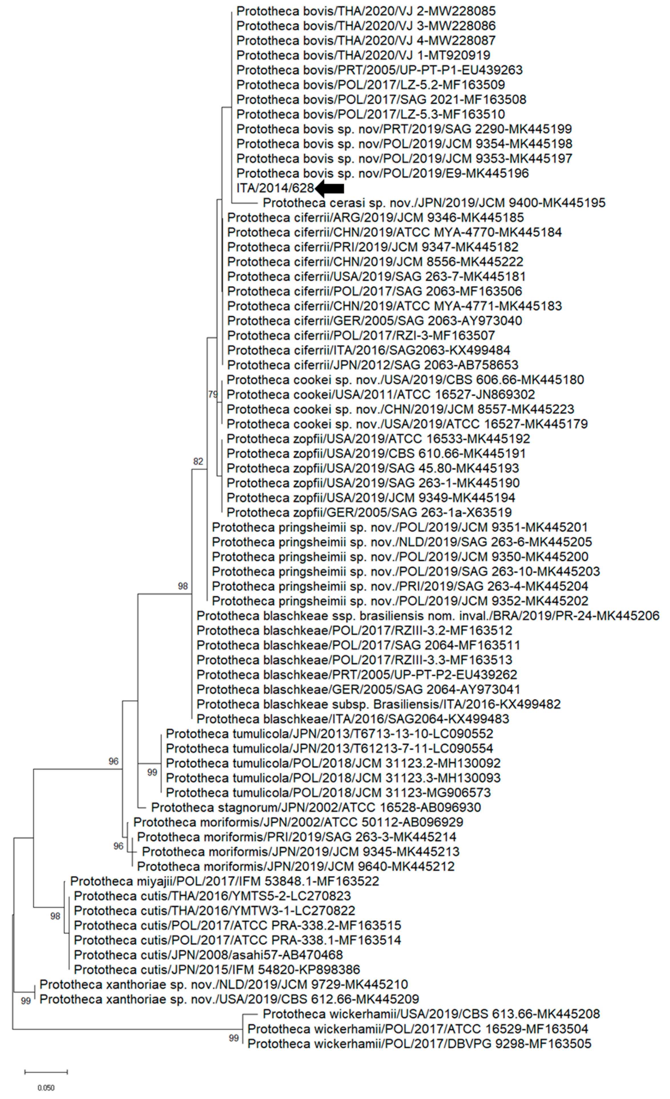
The online tool BLAST (https://blast.ncbi.nlm.nih.gov/Blast.cgi, accessed on 15 September 2023) was used to find the highest nt identity in the NBCI database. Sequence editing was performed by the software package Geneious Prime v. 2021.2 (Biomatters Ltd., Auckland, New Zealand). Sequence alignments were performed by the MAFFT [34] plugin implemented in Geneious Prime. The best-fitting substitution model settings for the phylogeny were explored by the tool “Find the best protein DNA/Protein Models” of the MEGA X v. 10.0.5 software [35]. The evolutionary history was deduced by using the maximum-likelihood method, the Kimura 2-parameter model, a discrete gamma distribution to model evolutionary rate differences among sites (6 categories), and supplying statistical support with 1000 replicates. Bayesian inference and neighbor-joining phylogenetic analyses were also performed to explore the phylogeny of Prototheca spp.

3. Results

Fourteen CSF samples collected in this study were subjected to molecular screening for Prototheca spp. and a panel of feline pathogens. Out of 14 CSF samples, 1 sample (#628/14) tested positive for Prototheca spp. by PCR, and the sequence was determined. By BlastN analysis performed on a 250 bp sequence of 18SrDNA of Prototheca spp., strain #628/14 shared the highest nucleotide (nt) identity (100%) with P. zopfii var. hydrocarbonea strain UP-PT-P1 (EU439263). All the samples tested negative for feline infectious peritonitis, feline leukemia virus, feline immunodeficiency virus, feline panleukopenia virus, rickettsia, neospora, toxoplasma, mycobacterium, and bacterial 16S rDNA.
Partial 18SrDNA sequence (250 nt)-based phylogenetic analysis was performed using the sequence of Prototheca spp. generated in the study and the cognate sequences of the closest relatives retrieved from the NCBI database. Different phylogenetic approaches were explored for Prototheca spp., and similar topologies with slight differences in bootstrap values at the nodes of the tree were noticed. Accordingly, the maximum-likelihood (ML) tree was used. Upon ML analysis, strain ITA/2014/628 segregated with strains belonging to the P. bovis clade (Figure 1). Upon qPCR specific for P. bovis, sample #628/14 yielded 27 Ct.
The animal that tested positive for Prototheca spp. was a 9-year-old male domestic European cat, presented at the veterinary clinic with a 24 h history of seizures, incoordination, circling, and disorientation. Clinical pathological evaluation included a complete blood count and clinical chemistry panel. Blood analysis showed a marked increase in creatine kinase (9827 U/L, reference interval [ref.]: 91–326 U/L), alanine aminotransferase (517 U/L, ref.: 22–45 U/L), and aspartate aminotransferase (98 U/L, ref.: 21–41 U/L). The other parameters were not altered. Complete blood count parameters were within the reference interval. In addition, at the time of clinical examination, the animal tested negative for FIV and FeLV, using a quick test (SNAP FIV/FeLV Combo Test—IDEXX Laboratories). Abdominal ultrasound examination revealed multiple spleen and liver nodules. Fine needle biopsy specimens taken from the spleen and liver nodules revealed many lymphoid cells, and a suspect of lymphoma was included in the differential diagnosis.
Within 24 h, the clinical condition of the animal worsened, and the owner opted for the gentle suppression of the animal. Extreme care was employed to guarantee that death had happened prior to discarding the animal remains [36]. Before euthanasia, a CSF sample (#628/14) was collected with the permission of the owner, exclusively for research purposes.
Examination of the CSF displayed a distinct increase in total protein (2432 mg/L, range <300 mg/L) and cytological features consistent with marked mixed-type pleocytosis (2448 cells/µL, range <3 cells/µL), composed mainly of small and medium-sized lymphocytes with no red blood cells. The owner did not give permission for further investigations (i.e., necropsy) in the animal.

4. Discussion

The taxonomic status of prototheca has been revised in the last few decades. Originally, seven species have been designated for the genus, namely P. stagnora, P. ulmea, P. wickerhamii, P. blaschkeae, P. zopfii, P. cutis, and P. miyajii [22,37,38]. P. blaschkeae, originally considered as biotype 3 of P. zopfii, has been re-classified as a new species, while P. zopfii biotypes 1 and 2 have been genetically correlated to distinct genotypes, I and II, respectively [22]. P. moriformis has been subsequently recognized as a distinct species [22,39]. However, in the late 2010s, a novel classification of the genus Prototheca was proposed [7], adding six new species (P. cookei sp. nov., P. cerasi sp. nov., P. pringsheimii sp. nov., P. xanthoriae sp. nov., P. tumulicula, and P. stagnora). Two main lineages were included in the genus: the triad composed of P. ciferrii sp. nov. (previously regarded as P. zopfii genotype I), P. zopfii (genetically distinct from P. ciferrii sp. nov.), P. bovis sp. nov. (previously named P. zopfii genotype II), and P. wickerhamii cluster. However, three novel species (P. cookei sp. nov., P. cerasi sp. nov., and P. pringsheimii sp. nov.) were related to the triad despite some important differences. Moreover, P. wickerhamii, P. cutis, P. miyajii, and the newly proposed species, P. xanthoriae sp. nov., have been regarded as closely related to the genera Chlorella and Auxenochlorella.
In this study, we identified prototheca in the CSF of a cat with neurological signs. Upon sequence and phylogenetic analysis, the feline strain 628/14 was characterized as P. bovis. P. bovis is responsible for most (i.e., 75 to 90%) of prototheca infections in dogs [10], of which skin tissue, digestive, nervous and ophthalmic systems [11,40,41,42,43,44,45,46], or localized changes in the nervous system or enteric tract have been reported [46,47]. Feline protothecosis seems to be very rare, and P. wickeramii has been mainly reported in cutaneous disease in cats [16,18,19,20,48], with only sporadic detection of P. bovis in a cat with nasal dermatitis [21].
In humans, protothecosis has been described in three main clinical forms, namely skin lesions, olecranon bursitis, and disseminated/systemic infections [5,49]. The cutaneous form seems more common form of protothecosis [50]. Additionally, an immunosuppressive factor has been identified in about half of human infections. P. bovis seems to be the predominant prototheca species in systemic infections, while P. wickerhamii is mostly involved in skin infections. Workers in animal productions or with food products of animal origin may be at a higher risk of exposure to protothecal infections [5]. Overall, protothecosis is considered a rare human disease with an increased incidence in patients with immunosuppression [50]. Epidemiological investigations based on a One Health approach, including humans, animals, and the environment, would be necessary to better understand the ecology of prototheca [51].
Algal invasion of the CNS is usually secondary to primary replication and lesions in other districts. The cat positive for prototheca in this study did not have a history of cutaneous disease, which is the common clinical picture associated with prototheca in cats. However, the animal had a suspected diagnosis of multifocal lymphoma, and therefore it was likely affected by immunological disorders/suppression, facilitating the infection and systemic spread of the algal pathogen.
Our study was affected by several limits. For instance, we failed to isolate the algal strain in cultural media. This was likely attributed to the fact that the sample was stored at −80° C for a long period before testing and isolation attempts. Sabouraud dextrose agar culture would have provided a firmer diagnosis [52], although molecular diagnostics have become standard in recent years [53,54]. Additionally, we could not further analyze the tissues/organs of the animal since the owners did not give permission for necropsy, and the retrospective nature of our investigation did not allow us to carry out other diagnostic investigations. CSF analysis is often inconclusive and should be used in combination with other approaches in the diagnostic pipeline for neurological diseases. The results obtained in this report should be further confirmed in other independent studies in order to understand the role of prototheca in neurological diseases of cats.
Prototheca was identified in this study through PCR targeting the 18SrDNA. When performing molecular screenings, we made several efforts to avoid laboratory contamination with DNA, enacting rigorous laboratory practices. The development of molecular assays for identification of prototheca allows us to expand the literature and records of algal infection in cats and dogs. Misdiagnosis with viral or bacterial pathogens based on the clinical/biochemical picture can be reduced by implementing diagnostic for these pathogens into microbiological laboratories.

5. Conclusions

Our findings indicate that prototheca should be considered in the diagnostic algorithms for infectious CNS diseases in cats. Moreover, while protothecosis is not considered a zoonosis, algal infections in animals should be considered as indicators or sentinels of environmental risks for humans.

Author Contributions

Conceptualization, V.M. and G.L.; methodology, M.C.; software G.L.; validation, M.C.; formal analysis, G.L.; investigation G.L., F.P. and G.P.; resources G.P., E.Z., E.M., P.Z.; data curation, F.P., K.B. and G.L.; writing—original draft preparation G.L.; writing—review and editing, V.M.; visualization, F.P.; supervision, M.C.; project administration V.M. All authors have read and agreed to the published version of the manuscript.

Funding

This research received no external funding.

Institutional Review Board Statement

The authors confirm that the ethical policies of the journal, as noted on the journal’s author guidelines page, have been adhered to. Ethical approval was not required, and an ethical statement is not applicable as all the samples were collected for diagnostic purposes. Informed consent was requested and then signed by the owner of the animals.

Informed Consent Statement

The owner of the animals involved in this study signed informed consent. All the samples were collected upon request of the attending veterinary practitioner. No additional requests were made by the researchers who performed the laboratory analyses for the purposes of drafting this study.

Data Availability Statement

Data are contained within the article.

Conflicts of Interest

The authors declare no conflict of interest.

References

  1. Shave, C.D.; Millyard, L.; May, R.C. Now for Something Completely Different: Prototheca, Pathogenic Algae. PLoS Pathog. 2021, 17, e1009362. [Google Scholar] [CrossRef] [PubMed]
  2. Jagielski, T.; Gawor, J.; Bakuła, Z.; Decewicz, P.; Maciszewski, K.; Karnkowska, A. Cytb as a New Genetic Marker for Differentiation of Prototheca Species. J. Clin. Microbiol. 2018, 56, 10–1128. [Google Scholar] [CrossRef] [PubMed]
  3. Kano, R. Emergence of Fungal-Like Organisms: Prototheca. Mycopathologia 2019, 185, 747–754. [Google Scholar] [CrossRef] [PubMed]
  4. Irrgang, A.; Murugaiyan, J.; Weise, C.; Azab, W.; Roesler, U. Well-Known Surface and Extracellular Antigens of Pathogenic Microorganisms among the Immunodominant Proteins of the Infectious Microalgae Prototheca Zopfii. Front. Cell. Infect. Microbiol. 2015, 5, 67. [Google Scholar] [CrossRef] [PubMed]
  5. Lass-Flörl, C.; Mayr, A. Human Protothecosis. Clin. Microbiol. Rev. 2007, 20, 230–242. [Google Scholar] [CrossRef] [PubMed]
  6. Jagielski, T.; Dyląg, M.; Roesler, U.; Murugaiyan, J. Isolation of Infectious Microalga Prototheca wickerhamii from a Carp (Cyprinus carpio)—A First Confirmed Case Report of Protothecosis in a Fish. J. Fish. Dis. 2017, 40, 1417–1421. [Google Scholar] [CrossRef]
  7. Jagielski, T.; Roeske, K.; Bakuła, Z.; Piech, T.; Wlazło, Ł.; Bochniarz, M.; Woch, P.; Krukowski, H. A Survey on the Incidence of Prototheca Mastitis in Dairy Herds in Lublin Province, Poland. J. Dairy Sci. 2019, 102, 619–628. [Google Scholar] [CrossRef]
  8. Masuda, M.; Jagielski, T.; Danesi, P.; Falcaro, C.; Bertola, M.; Krockenberger, M.; Malik, R.; Kano, R. Protothecosis in Dogs and Cats—New Research Directions. Mycopathologia 2021, 186, 143–152. [Google Scholar] [CrossRef]
  9. Jagielski, T.; Lagneau, P.-E. Protothecosis. A Pseudofungal Infection. J. Mycol. Med. 2007, 17, 261–270. [Google Scholar] [CrossRef]
  10. Pressler, B.M. Infectious Diseases of the Dog and Cat. In Protothecosis and Chlorellosis; Greene, C.E., Ed.; Saunders: St. Louis, MO, USA, 2012; Volume 1, pp. 696–701. [Google Scholar]
  11. Stenner, V.J.; MacKay, B.; King, T.; Barrs, V.R.D.; Irwin, P.; Abraham, L.; Swift, N.; Langer, N.; Bernays, M.; Hampson, E.; et al. Protothecosis in 17 Australian Dogs and a Review of the Canine Literature. Med. Mycol. 2007, 45, 249–266. [Google Scholar] [CrossRef]
  12. Hirose, N.; Hua, Z.; Kato, Y.; Zhang, Q.; Li, R.; Nishimura, K.; Masuda, M. Molecular Characterization of Prototheca Strains Isolated in China Revealed the First Cases of Protothecosis Associated with Prototheca Zopfii Genotype 1. Med. Mycol. 2018, 56, 279–287. [Google Scholar] [CrossRef] [PubMed]
  13. Masuda, M.; Hirose, N.; Ishikawa, T.; Ikawa, Y.; Nishimura, K. Prototheca miyajii sp. Nov., Isolated from a Patient with Systemic Protothecosis. Int. J. Syst. Evol. Microbiol. 2016, 66, 1510–1520. [Google Scholar] [CrossRef] [PubMed]
  14. Khan, I.D.; Sahni, A.K.; Sen, S.; Gupta, R.M.; Basu, A. Outbreak of Prototheca wickerhamii Algaemia and Sepsis in a Tertiary Care Chemotherapy Oncology Unit. Med. J. Armed Forces India 2018, 74, 358–364. [Google Scholar] [CrossRef] [PubMed]
  15. Wolf, A.M. Fungal Diseases of the Nasal Cavity of the Dog and Cat. Vet. Clin. N. Am. Small Anim. Pract. 1992, 22, 1119–1132. [Google Scholar] [CrossRef] [PubMed]
  16. Kaplan, W.; Chandler, F.W.; Holzinger, E.A.; Plue, R.E.; Dickinson, R.O. Protothecosis in a Cat: First Recorded Case. Med. Mycol. 1976, 14, 281–286. [Google Scholar] [CrossRef]
  17. Finnie, J.W.; Coloe, P.J. Cutaneous Protothecosis in a Cat. Aust. Vet. J. 1981, 57, 307–308. [Google Scholar] [CrossRef]
  18. Coloe, P.J.; Allison, J.F. Protothecosis in a Cat. J. Am. Vet. Med. Assoc. 1982, 180, 78–79. [Google Scholar]
  19. Dillberger, J.E.; Homer, B.; Daubert, D.; Altman, N.H. Protothecosis in Two Cats. J. Am. Vet. Med. Assoc. 1988, 192, 1557–1559. [Google Scholar]
  20. Endo, S.; Sekiguchi, M.; Kishimoto, Y.; Kano, R.; Aoki, S.; Sichinohe, T.; Hasegawa, A. The First Case of Feline Prototheca wickerhamii Infection in Japan. J. Vet. Med. Sci. 2010, 72, 1351–1353. [Google Scholar] [CrossRef]
  21. Huth, N.; Wenkel, R.F.; Roschanski, N.; Rösler, U.; Plagge, L.; Schöniger, S. Prototheca Zopfii Genotype 2-Induced Nasal Dermatitis in a Cat. J. Comp. Pathol. 2015, 152, 287–290. [Google Scholar] [CrossRef]
  22. Roesler, U.; Möller, A.; Hensel, A.; Baumann, D.; Truyen, U. Diversity within the Current Algal Species Prototheca Zopfii: A Proposal for Two Prototheca Zopfii Genotypes and Description of a Novel Species, Prototheca blaschkeae sp. Nov. Int. J. Syst. Evol. Microbiol. 2006, 56, 1419–1425. [Google Scholar] [CrossRef] [PubMed]
  23. Sobukawa, H.; Yamaguchi, S.; Kano, R.; Ito, T.; Suzuki, K.; Onozaki, M.; Hasegawa, A.; Kamata, H. Short Communication: Molecular Typing of Prototheca Zopfii from Bovine Mastitis in Japan. J. Dairy Sci. 2012, 95, 4442–4446. [Google Scholar] [CrossRef] [PubMed]
  24. Bacova, R.; Kralik, P.; Kucharovicova, I.; Seydlova, R.; Moravkova, M. A novel TaqMan qPCR assay for rapid detection and quantification of pro-inflammatory microalgae Prototheca spp. in milk samples. Med. Mycol. 2021, 59, 784–792. [Google Scholar] [CrossRef] [PubMed]
  25. Gut, M.; Leutenegger, C.M.; Huder, J.B.; Pedersen, N.C.; Lutz, H. One-Tube Fluorogenic Reverse Transcription-Polymerase Chain Reaction for the Quantitation of Feline Coronaviruses. J. Virol. Methods 1999, 77, 37–46. [Google Scholar] [CrossRef] [PubMed]
  26. Stiles, J.; Bienzle, D.; Render, J.A.; Buyukmihci, N.C.; Johnson, E.C. Use of Nested Polymerase Chain Reaction (PCR) for Detection of Retroviruses from Formalin-Fixed, Paraffin-Embedded Uveal Melanomas in Cats. Vet. Ophthalmol. 1999, 2, 113–116. [Google Scholar] [CrossRef]
  27. Endo, Y.; Cho, K.-W.; Nishigaki, K.; Momoi, Y.; Nishimura, Y.; Mizuno, T.; Goto, Y.; Watari, T.; Tsujimoto, H.; Hasegawa, A. Molecular Characteristics of Malignant Lymphomas in Cats Naturally Infected with Feline Immunodeficiency Virus. Vet. Immunol. Immunopathol. 1997, 57, 153–167. [Google Scholar] [CrossRef]
  28. Decaro, N.; Desario, C.; Lucente, M.S.; Amorisco, F.; Campolo, M.; Elia, G.; Cavalli, A.; Martella, V.; Buonavoglia, C. Specific Identification of Feline Panleukopenia Virus and Its Rapid Differentiation from Canine Parvoviruses Using Minor Groove Binder Probes. J. Virol. Methods 2008, 147, 67–71. [Google Scholar] [CrossRef]
  29. Claerebout, E.; Losson, B.; Cochez, C.; Casaert, S.; Dalemans, A.-C.; De Cat, A.; Madder, M.; Saegerman, C.; Heyman, P.; Lempereur, L. Ticks and Associated Pathogens Collected from Dogs and Cats in Belgium. Parasit. Vectors 2013, 6, 183. [Google Scholar] [CrossRef]
  30. Reitt, K.; Hilbe, M.; Voegtlin, A.; Corboz, L.; Haessig, M.; Pospischil, A. Aetiology of Bovine Abortion in Switzerland from 1986 to 1995? A Retrospective Study with Emphasis on Detection of Neospora Caninum and Toxoplasma Gondii by PCR. J. Vet. Med. Ser. A 2007, 54, 15–22. [Google Scholar] [CrossRef]
  31. Hashimoto, A.; Koga, H.; Kohno, S.; Miyazaki, Y.; Kaku, M.; Hara, K. Rapid Detection and Identification of Mycobacteria by Combined Method of Polymerase Chain Reaction and Hybridization Protection Assay. J. Infect. 1996, 33, 71–77. [Google Scholar] [CrossRef]
  32. Stecher, B.; Chaffron, S.; Käppeli, R.; Hapfelmeier, S.; Freedrich, S.; Weber, T.C.; Kirundi, J.; Suar, M.; McCoy, K.D.; von Mering, C.; et al. Like Will to Like: Abundances of Closely Related Species Can Predict Susceptibility to Intestinal Colonization by Pathogenic and Commensal Bacteria. PLoS Pathog. 2010, 6, e1000711. [Google Scholar] [CrossRef] [PubMed]
  33. Helps, C.; Reeves, N.; Egan, K.; Howard, P.; Harbour, D. Detection of Chlamydophila felis and feline herpesvirus by multi-plex real-time PCR analysis. J. Clin. Microbiol. 2003, 41, 2734–2736. [Google Scholar] [CrossRef] [PubMed]
  34. Katoh, K. MAFFT: A Novel Method for Rapid Multiple Sequence Alignment Based on Fast Fourier Transform. Nucleic Acids Res. 2002, 30, 3059–3066. [Google Scholar] [CrossRef] [PubMed]
  35. Kumar, S.; Stecher, G.; Li, M.; Knyaz, C.; Tamura, K. MEGA X: Molecular Evolutionary Genetics Analysis across Computing Platforms. Mol. Biol. Evol. 2018, 35, 1547–1549. [Google Scholar] [CrossRef] [PubMed]
  36. Meyer, R.E. Euthanasia and Humane Killing. In Veterinary Anesthesia and Analgesia, 5th ed.; Grimm, K.A., Lamont, L.A., Tranquilli, W.J., Greene, S.A., Robertson, S.A., Eds.; Wiley Blackwell: Chichester, UK, 2015; pp. 130–143. [Google Scholar]
  37. Pore, R.S.; Barnett, E.A.; Barnes, W.C.; Walker, J.D. Prototheca Ecology. Mycopathologia 1983, 81, 49–62. [Google Scholar] [CrossRef]
  38. Satoh, K.; Ooe, K.; Nagayama, H.; Makimura, K. Prototheca cutis sp. Nov., a Newly Discovered Pathogen of Protothecosis Isolated from Inflamed Human Skin. Int. J. Syst. Evol. Microbiol. 2010, 60, 1236–1240. [Google Scholar] [CrossRef]
  39. Severgnini, M.; Lazzari, B.; Capra, E.; Chessa, S.; Luini, M.; Bordoni, R.; Castiglioni, B.; Ricchi, M.; Cremonesi, P. Genome Sequencing of Prototheca Zopfii Genotypes 1 and 2 Provides Evidence of a Severe Reduction in Organellar Genomes. Sci. Rep. 2018, 8, 14637. [Google Scholar] [CrossRef]
  40. Thomas, J.; Preston, N. Generalised Protothecosis in a Collie Dog. Aust. Vet. J. 1990, 67, 25–27. [Google Scholar] [CrossRef]
  41. Schultze, A.E.; Ring, R.D.; Morgan, R.V.; Patton, C.S. Clinical, Cytologic and Histopathologic Manifestations of Protothecosis in Two Dogs. Vet. Ophthalmol. 1998, 1, 239–243. [Google Scholar] [CrossRef]
  42. Lane, L.V.; Meinkoth, J.H.; Brunker, J.; Smith, S.K.; Snider, T.A.; Thomas, J.; Bradway, D.; Love, B.C. Disseminated Protothecosis Diagnosed by Evaluation of CSF in a Dog. Vet. Clin. Pathol. 2012, 41, 147–152. [Google Scholar] [CrossRef]
  43. Vince, A.R.; Pinard, C.; Ogilvie, A.T.; Tan, E.O.; Abrams-Ogg, A.C.G. Protothecosis in a Dog. Can. Vet. J. 2014, 55, 950–954. [Google Scholar] [PubMed]
  44. Carfora, V.; Noris, G.; Caprioli, A.; Iurescia, M.; Stravino, F.; Franco, A. Evidence of a Prototheca Zopfii Genotype 2 Disseminated Infection in a Dog with Cutaneous Lesions. Mycopathologia 2017, 182, 603–608. [Google Scholar] [CrossRef] [PubMed]
  45. Sonne, L.; de Oliveira, E.C.; Argenta, F.F.; Monteggia, R.S.; Ferreiro, L.; Rösler, U.; Kershaw, O.; Driemeier, D. Prototheca Zopfii Genotype 2 Disseminated Infection in a Dog with Neurological Signs. Ciência Rural. 2017, 47, e20160877. [Google Scholar] [CrossRef]
  46. Ribeiro, M.G.; Rodrigues de Farias, M.; Roesler, U.; Roth, K.; Rodigheri, S.M.; Ostrowsky, M.A.; Salerno, T.; Siqueira, A.K.; Fernandes, M.C. Phenotypic and Genotypic Characterization of Prototheca Zopfii in a Dog with Enteric Signs. Res. Vet. Sci. 2009, 87, 479–481. [Google Scholar] [CrossRef] [PubMed]
  47. Walker, A.; MacEwan, I.; Fluen, T.; Hardcastle, M. Disseminated Protothecosis with Central Nervous System Involvement in a Dog in New Zealand. N. Z. Vet. J. 2022, 70, 238–243. [Google Scholar] [CrossRef] [PubMed]
  48. Kessell, A.E.; McNair, D.; Munday, J.S.; Savory, R.; Halliday, C.; Malik, R. Successful Treatment of Multifocal Pedal Prototheca wickerhamii Infection in a Feline Immunodeficiency Virus-Positive Cat with Multiple Bowenoid in Situ Carcinomas Containing Papillomaviral DNA Sequences. J. Feline Med. Surg. Open Rep. 2017, 3, 205511691668859. [Google Scholar] [CrossRef]
  49. Mayorga, J.; Barba-Gómez, J.F.; Verduzco-Martínez, A.P.; Munoz-Estrada, V.F.; Welsh, O. Protothecosis. Clin. Dermatol. 2012, 3, 432–436. [Google Scholar] [CrossRef] [PubMed]
  50. Todd, J.R.; Matsumoto, T.; Ueno, R.; Murugaiyan, J.; Britten, A.; King, J.W.; Odaka, Y.; Oberle, A.; Weise, C.; Roesler, U.; et al. Medical Phycology 2017. Med. Mycol. 2018, 56, S188–S204. [Google Scholar] [CrossRef]
  51. Jinatham, V.; Cantoni, D.M.; Brown, I.R.; Vichaslip, T.; Suwannahitatorn, P.; Popluechai, S.; Tsaousis, A.D.; Gentekaki, E. Prototheca bovis, a Unicellular Achlorophyllous Trebouxiophyte Green Alga in the Healthy Human Intestine. J. Med. Microbiol. 2021, 70, 001415. [Google Scholar] [CrossRef]
  52. Hillesheim, P.B.; Bahrami, S. Cutaneous Protothecosis. Arch. Pathol. Lab. Med. 2011, 135, 941–944. [Google Scholar] [CrossRef]
  53. Silveira, C.S.; Cesar, D.; Keating, M.K.; DeLeon-Carnes, M.; Armién, A.G.; Luhers, M.; Riet-Correa, F.; Giannitti, F. A Case of Prototheca zopfii Genotype 1 Infection in a Dog (Canis lupus Familiaris). Mycopathologia 2018, 183, 853–858. [Google Scholar] [CrossRef] [PubMed]
  54. Tsuji, H.; Kano, R.; Hirai, A.; Murakami, M.; Yanai, T.; Namihira, Y.; Chiba, J.; Hasegawa, A. An Isolate of Prototheca wickerhamii from Systemic Canine Protothecosis. Vet. Microbiol. 2006, 118, 305–311. [Google Scholar] [CrossRef] [PubMed]
Figure 1. Partial 18SrDNA sequence (250 nt)-based phylogenetic tree of Prototheca spp. strains reported in this study and reference strains retrieved from the NCBI database. The Maximum Likelihood method and Kimura model (two parameters), with a discrete gamma distribution, were applied for the phylogenic analysis. One thousand bootstrap replicates were employed to assess the robustness of individual nodes on the phylogenetic tree. Bootstrap values higher than 75% were displayed. Black arrows denote strains reported in this study. Numbers of nucleotide substitutions are connoted by the scale bar.
Vetsci 10 00681 g001
Table 1. Detailed list of protocols (pathogens, assays, primers, probes, and references) used for the molecular screening of samples included in the study.
PathogenAssayPrimers and ProbesOligonucleotide SequenceReference(s)
Prototheca spp.PCRProto 18S-4F
Proto 18S-4R-1
5′-GACATGGCGAGGATTGACAGA-3′
5′-ATCACAGACCTGTTATC-3′
[22]
[23]
Prototheca bovisqPCRPZg2F
SPZg2
PZg2R
5′-GACGATGATCCTAGTTATGGTGTAC-3′[24]
5′Fam-TGGTAGAAGACAAATAATGTACCAAAACCA-BHQ13′
5′-TATAAAAGCAAGTCCAGTTACAGCAC-3′
feline infectious peritonitisqPCRFCoV1128f
FCoV1200p
FCoV1229r
5′-GATTTGATTTGGCAATGCTAGATTT-3′
5′Fam-TCCATTGTTGGCTCGTCATAGCGGA-Tamra3′
5′-AACAATCACTAGATCCAGACGTTAGCT-3′
[25]
feline leukemia virusPCR118for
119rev
5′-TTACTCAAGTATGTTCCCATG-3′
5′-CTGGGGAGCCTG GAGACTGCT-3′
[26]
feline immunodeficiency virusPCR158for
159rev
5′-GAGTAGATACWTGGTTRCAAG-3′
5′-CATCCTAATTCTTGCATAGC-3′
5′-CAAAATGTGGATGGTGGAAY-3′
5′-ACCATTCCWATAGCAGTRGC-3′
[27]
nPCR160for
161rev
feline panleukopenia virusqPCRFPV/CPV-For
FPV-Pb
CPV-Pb
FPV/CPV-Rev
5′-ACAAGATAAAAGACGTGGTGTAACTCAA-3′
5′Vic-ATGGGAAATACAGACTATAT-MGB3′
5′Fam-ATGGGAAATACAAACTATAT-MGB3′
5′-CAACCTCAGCTGGTCTCATAATAGT-3′
[28]
rickettsiaPCRRSFG 877
RSFG1258
5′-GGGGGCCTGCTCACGGCGG-3′
5′-ATTGCAAAAAGTACAGTGAACA-3′
[29]
neosporaqPCRNeo For
Neo Probe
Neo Rev
5′-GCATCGGAGGACACTGCT-3′
5′Fam-CTGACTCTGAACACCGGAGGCACG-Tamra3′
5′-ATGTCGTAAATCGGAGTTGCTTC-3′
[30]
toxoplasmaqPCRTox For
Tox Probe
Tox Rev
5′-GTCCTATCGCAACGGAGTTCTT-3′
5′Fam-CCAGACGTGGATTTCCGTTGGTTCC-Tamra3′
5′-TTCGTCCGTCGTAATATCAGGC-3′
mycobacteriumPCRMyc For
Myc Rev
5′-CATGCAAGTCGAACGGAAAG-3′
5′-CGGTGCTTCTTCTCCACCTA-3′
5′-TACTCGAGTGGCGAACGGGT-3′
5′-CGGACCTTCGTCGATGGTGA-3′
[31]
nPCRMyc NFor
Myc NRev
bacterial 16S rDNAPCRB-V5
A-V6
5′-ATTAGATACCCYGGTAGTCC-3′
5′-ACGAGCTGACGACARCCATG-3′
[32]
Internal controlPCRfeline 28S rDNA Fw5’-AGCAGGAGG TGTTGGAAGAG-3′[33]
feline 28S rDNA Rv5′-AGGGAGAGCCTAAATCAAAGG-3′
Disclaimer/Publisher’s Note: The statements, opinions and data contained in all publications are solely those of the individual author(s) and contributor(s) and not of MDPI and/or the editor(s). MDPI and/or the editor(s) disclaim responsibility for any injury to people or property resulting from any ideas, methods, instructions or products referred to in the content.

