Article

Doxorubicin as a Potential Treatment Option in Canine Mammary Tumors

by
Madalina Luciana Gherman
1,2,†,
Oana Zanoaga
2,†,
Liviuta Budisan
2,
Lajos Raduly
2,* and
Ioana Berindan-Neagoe
2
1
Experimental Center, Iuliu Hatieganu University of Medicine and Pharmacy, 400349 Cluj-Napoca, Romania
2
Research Center for Functional Genomics, Biomedicine and Translational Medicine, Iuliu Hatieganu University of Medicine and Pharmacy, 400337 Cluj-Napoca, Romania
*
Author to whom correspondence should be addressed.
These authors contributed equally to this work.
Vet. Sci. 2023, 10(11), 654; https://doi.org/10.3390/vetsci10110654
Submission received: 19 September 2023 / Revised: 11 October 2023 / Accepted: 10 November 2023 / Published: 14 November 2023
(This article belongs to the Special Issue Comparative Oncology and Veterinary Cancer Surveillance)

Abstract

:

Simple Summary

Due to the limited options in mammary tumor treatments, the following study demonstrates the efficiency of Doxorubicin on canine mammary tumor therapy and in the alteration of expression of the key genes involved in the epithelial-to-mesenchymal transition processes.

Abstract

Canine mammary tumors represent one of the leading malignant pathologies in female dogs, displaying the importance of efficient therapeutic findings, besides the golden-standard surgery, able to limit the development of the disease. Studies in human cancers demonstrated that Doxorubicin presents a good effect in different biological processes like apoptosis, autophagy, the cell cycle, cell invasion, and the epithelial-to-mesenchymal transition. This study followed the effects of Doxorubicin on two canine mammary cancer cell lines P114 and CMT-U27. Doxorubicin treatment in both cell lines shows an inhibitory effect in cell proliferation and an alteration in expression of the EMT-related genes. The obtained results provide valuable information for revealing the link between Doxorubicin, phenotypic changes, and proliferation dynamics in canine mammary tumor models.

1. Introduction

Mammary tumors represent the most frequent neoplasia in female dogs. In epidemiological studies, the annual incidence of mammary tumors is 40–50% for benign and almost 50% for malignant tumors [1,2,3,4]. In the countries like the USA, mammary tumors in canines have decreased because of early neutering, but the most frequent mammary cancer forms in female dogs are still present in most of the European countries [1,5]. The golden-standard treatment procedure worldwide is represented by the surgical excision of the mammary chain but with unsatisfactory results. A high number of dogs with mammary cancer show lymphatic or vascular invasion and high rates of recurrence and metastasis [6,7,8,9]. There is limited information about chemotherapy and radiotherapy efficacy in treating canine mammary tumors (CMTs), probably because the procedures are expensive. Despite some case reports regarding the responses to chemotherapeutics such as Doxorubicin [10], mitoxantrone [11], paclitaxel [12,13], and carboplatin [4], it is not clear if chemotherapy improves the overall survival of the treated dogs [5,8,14].
One of the most used therapeutic agents in human cancer is the anthracycline Doxorubicin (DOXO) [15,16,17]. In recent years, a few studies have shown the efficiency of this drug in canine patients [7,18]. Doxorubicin has been reported by many studies to treat cancers including breast, lung, thyroid, and hematologic cancers [19,20]. Doxorubicin induces the production of free radicals, intercalation into DNA strands and inhibition of topoisomerases I and II, causing DNA damage and activating caspases, ultimately leading to apoptosis [16,21].
The epithelial-to-mesenchymal transition (EMT) is essential in embryonic development and tissue regeneration. The EMT is characterized by a progressive loss of the cells’ epithelial characteristics, which acquire mesenchymal features through a molecular reprogramming of the cells, promoting progression and invasion of the neighboring stroma [22,23,24]. The EMT has been demonstrated to play a key role in the development of the metastatic process in canine mammary carcinoma, promoting the idea that the EMT is a dynamic reversible process [25]. An aberrant reactivation of the EMT is associated with malignant properties of tumor cells during cancer progression and metastasis, including migration and invasiveness, increased tumor stemness, and enhanced resistance to therapy [26,27,28]. Studies performed in humans identified the EMT as involved in DOX-resistance to therapy, stimulating the migration of tumor cells and metastasis; furthermore, the EMT mediates the resistance of cancer cells to DOX-mediated apoptosis [28,29].
The present study aimed to analyze the inhibitory effect of Doxorubicin in vitro models of canine mammary cancer to evaluate the efficiency of this therapeutic agent and the expression levels of key genes involved in the EMT process.

2. Materials and Methods

2.1. Cell Lines and Cell Culture

This study was conducted in two canine mammary carcinoma cell lines (P114 and CMT-U27) kindly donated by Prof. Gerard Rutteman from the Netherlands and Prof. Eva Hellmen from Sweden. The two canine cell lines were cultured in RPMI supplemented with 10% Fetal Bovine Serum (FBS) and 1% Glutamine for P114, and in low-glucose DMEM supplemented with 10% Fetal Bovine Serum (FBS) and 1% Glutamine for CMT-U27. The culture medium and supplements were obtained from Sigma-Aldrich (St. Louis, MO, USA). Both cell lines were treated with Doxorubicin (2.5 µM) for 48 h and different in vitro functional tests were performed.
The P114 cell line is a canine anaplastic mammary carcinoma (AMC) cell line shown in dogs and felines. In dogs, AMC is considered the most aggressive type of mammary tumor, showing a low frequency, and is associated with a short survival time. The CMT-U27 cell line is a canine mammary carcinoma cell line [30,31,32].

2.2. MTT Cell Viability Assay

A total of 1 × 10⁴ cells/well were cultured in 96-well culture plates for 24 h at 37 °C in 5% CO2 atmosphere incubators. After 24 h incubation, cell cultures were treated with Doxorubicin (DOXO) doses (Sigma Aldrich, St. Louis, MO, USA). At 48 h after treatment, the medium was discarded, and 100 µL MTT solution was added to every well. After 2 h incubation at 37 °C, the MTT solution was removed, and the formazan crystal was solubilized with 100 µL DMSO (dimethyl sulfoxide) (Sigma-Aldrich). The absorbance was measured at 570/690 nm for the cell viability assay in a microplate reader (Synergy H1 Hybrid Reader Biotech).

2.3. Apoptosis Assay through Fluorescence Microscopy

The Multi-Parameter Apoptosis Kit (Cayman, Ann Arbor, MI, USA) was used for the fluorescence microscopy evaluation of apoptosis. Cell staining was performed according to the manufacturer’s protocol with tertamethylrhodamin ethyl ester (TMRE) for mitochondria and Hoechst for nuclei. The stained cells were analyzed at UV wavelengths for Hoechst and 560/595 nm for TMRE staining on an Olympus IX71 inverted microscope. Hoechst is a specific stain for nucleus morphology and counting, while TMRE indicates the mitochondrial membrane activity potential.
Apoptosis evaluation was also performed through triple-fluorescence staining using Actin-stain 488 Fluorescent Phalloidin (Cytoskeleton, Inc., Denver, CO, USA) for cytoskeleton, Mitotracker (Invitrogen) for mitochondria labeling, and DAPI (4′,6-diamidino-2-phenylindole) (Invitrogen) for nucleus staining.

2.4. Confluency Assay

Cell lines were cultured in a 96-well culture plate for 48 h and then treated with 2.5 µM DOXO. After 48 h treatment, cell confluency was analyzed using a Celigo Image Cytometer (Nexcelom, Lawrence, MA, USA).

2.5. Cell Cycle Assay using Celigo

Cell lines were cultured in a 96-well culture plate for 48 h and then treated with 2.5 μM DOXO. After 48 h treatment, cells were fixed in ice-cold methanol at 4 °C for 30 min. After fixation, the cells were marked with RNase A and Propidium Iodide (PI) solution for 45 min in the dark at 37 °C in 5% CO2 atmosphere incubators. The cell cycle analysis was performed using Celigo Image Cytometer (Nexcelom).

2.6. Colony Formation Assay

P114 and CMT-U27 cells were incubated with 2.5 µM Doxorubicin for 48 h to induce DNA damage. The cells were detached after 48 h, counted, and reseeded in the standard medium in triplicate for each condition and cellular type (500 cells/6 well plate). Cells returned to the incubator for 7 days to form colonies and then were fixed with ice-cold methanol for 15 min, dried, and stained with 0.5% crystal violet in 25% methanol for 20 min. After washing with water and drying, colonies were counted and visualized using the Celigo Image Cytometer (Nexcelom).

2.7. Scratch Assay

Canine mammary cancer cells were individually pre-treated with DOXO for 48 h in 24-well plates. After treatment, cell proliferation was blocked through a 30 min pretreatment with mitomycin C (40 μg/mL). A scratch was then made in each well using a 20 μL pipette tip, and cells were maintained in a medium with 1% serum. The gaps were scaled (µm2) at 0, 8, and 24 h and visualized using an inverted microscope Olympus IX71.

2.8. Cell Invasion Assay

Canine cells (15 × 103 cells/well) were cultured in the upper 8 µm Falcon cell culture inserts pre-coated with 100 µL Matrigel in 500 µL media without serum. In the lower chambers (24 well plates), 750 µL complete medium containing 10% FBS was added. After 48 h of incubation, the cells that had invaded through the pores and attached to the bottom surface of the membrane were stained with Calcein-AM (Invitrogen, Waltham, WA, USA) according to the manufacturer’s protocol. Images of the stained invaded cells were captured under an Olympus IX71 inverted fluorescent microscope.

2.9. Gene Expression Evaluation Using PCR Array

For the extraction of total RNA, the Trizol (TriReagent Sigma-Aldrich) protocol was used, and the quantitative and qualitative control was performed using the Nanodrop-1000 spectrophotometer (Thermo Scientific, Waltham, WA, USA) and the Agilent Bioanalyzer 2100. cDNA synthesis was performed using the RT2 First Strand Kit (Qiagen, Singapore) from 1000 ng of total RNA, according to the manufacturer’s recommendations. For the RT2 Profiler PCR Array (PAFD-090ZE-1 Dog Epithelial-to-Mesenchymal Transition—EMT-Qiagen), we used the SYBR Green Master Mix and the ViiA7 instrument from Applied Biosystems according to the manufacturer’s protocol. Alterations in gene expression levels for the studied genes were evaluated using the Qiagen RT2 Profiler PCR Array and Assays Data Analysis software (v3.5).

2.10. Statistical Analysis

Standard deviation (mean ± SD) and a t-test were used to study the difference between experimental and control conditions (p-value < 0.05 was considered statistically significant; ns/p > 0.05; */p ≤ 0.05; **/p ≤ 0.01; ***/p ≤ 0.001; ****/p ≤ 0.0001). Statistical analyses and graphic representation were performed using GraphPad Prism software (version 8.0; GraphPad Software, Inc., San Diego, CA, USA).

3. Results

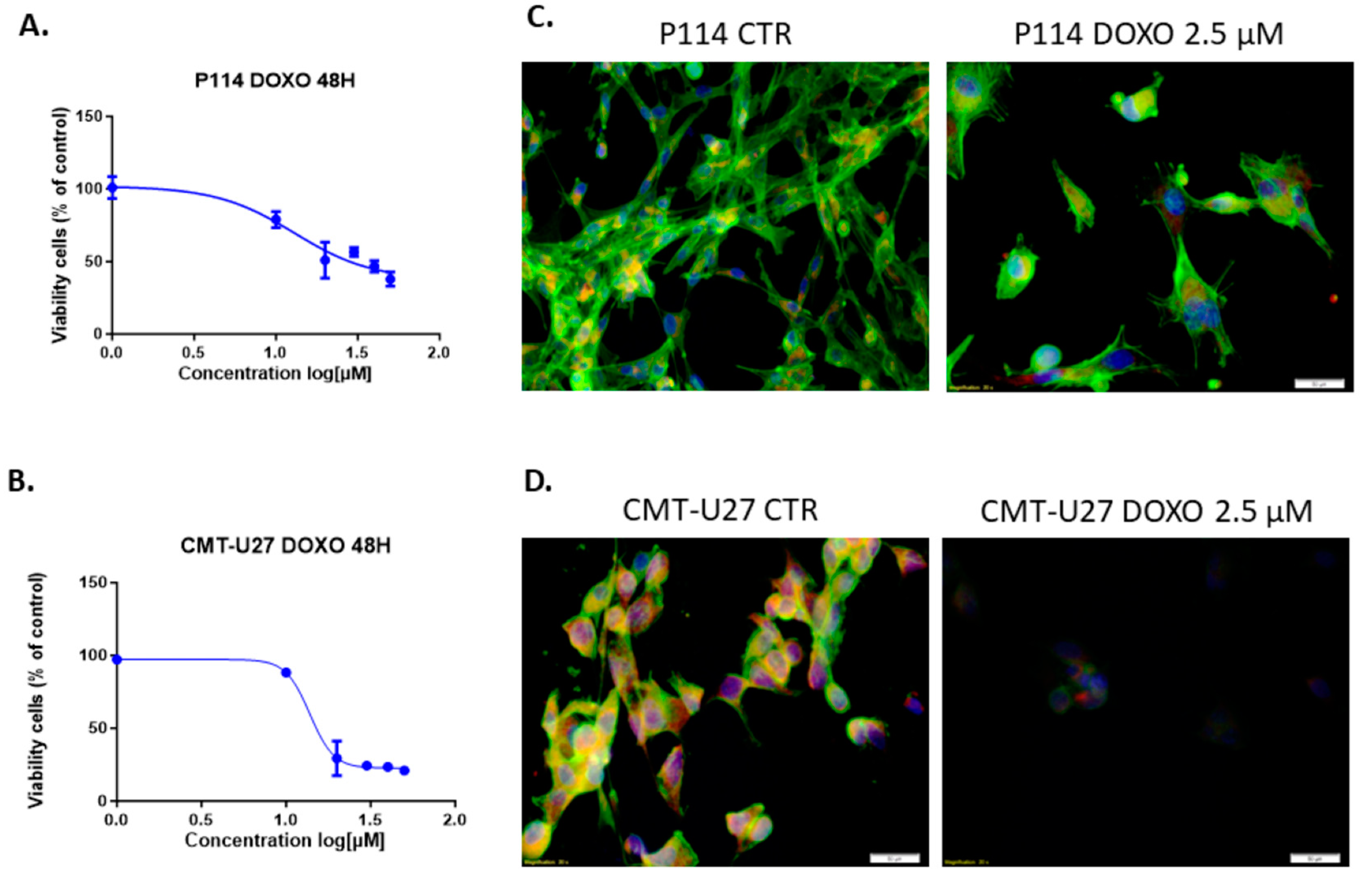
3.1. Doxorubicin Inhibits Cell Viability in Both In Vitro Canine Mammary Cancer Models

The treatment of P114 and CMT-U27 mammary cancer cell lines with different concentrations of DOXO for 48 h shows the inhibition of viability in a dose-dependent manner for both canine cell lines. Moreover, the two cell lines have similar values for the half maximal inhibitory concentration with an approximate dose of 13 µM Doxorubicin (Figure 1A,B). Because of the high toxicity of the DOXO, we chose to apply the smallest concentration that shows an inhibitory effect in the cell proliferation in the canine mammary tumor cell lines. Due to the relatively equal concentration of DOXO corresponding to the IC50 of each cell line, we decided to continue the experiments with 2.5 µM of therapeutic agent to compare the response of each cell line under standard conditions.

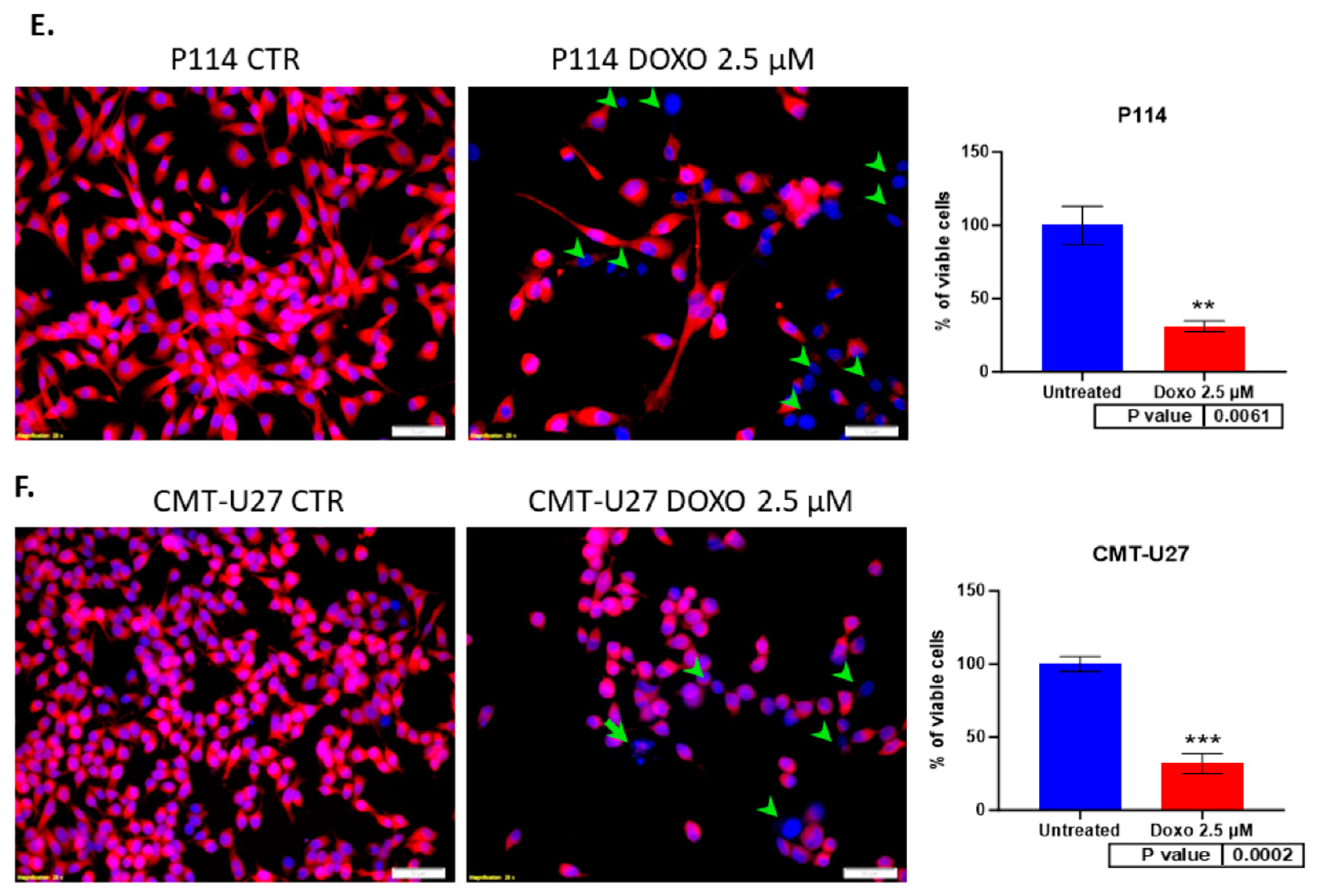
3.2. Doxorubicin Treatment Decreases Cell Viability and Induces Apoptosis in Cell Lines

The triple-fluorescence staining for the cytoskeleton, mitochondria, and nucleus shows damage to the cells through the treatment (Figure 1C,D). The fluorescence microscopy assay revealed that DOXO treatment after 48 h induced in P114 and CMT-U27 cell lines a decreased mitochondrial membrane potential activity and morphological changes, including nuclear DNA condensation, nuclear shrinkage, and fragmentation (marked with green arrows). We can observe a significant reduction in the viable cell’s percentage to 30% (p-value = 0.0061) in the P114-treated cells versus untreated cells and 32% for the CMT-U27 cells (p-value = 0.0002) (Figure 1E,F).

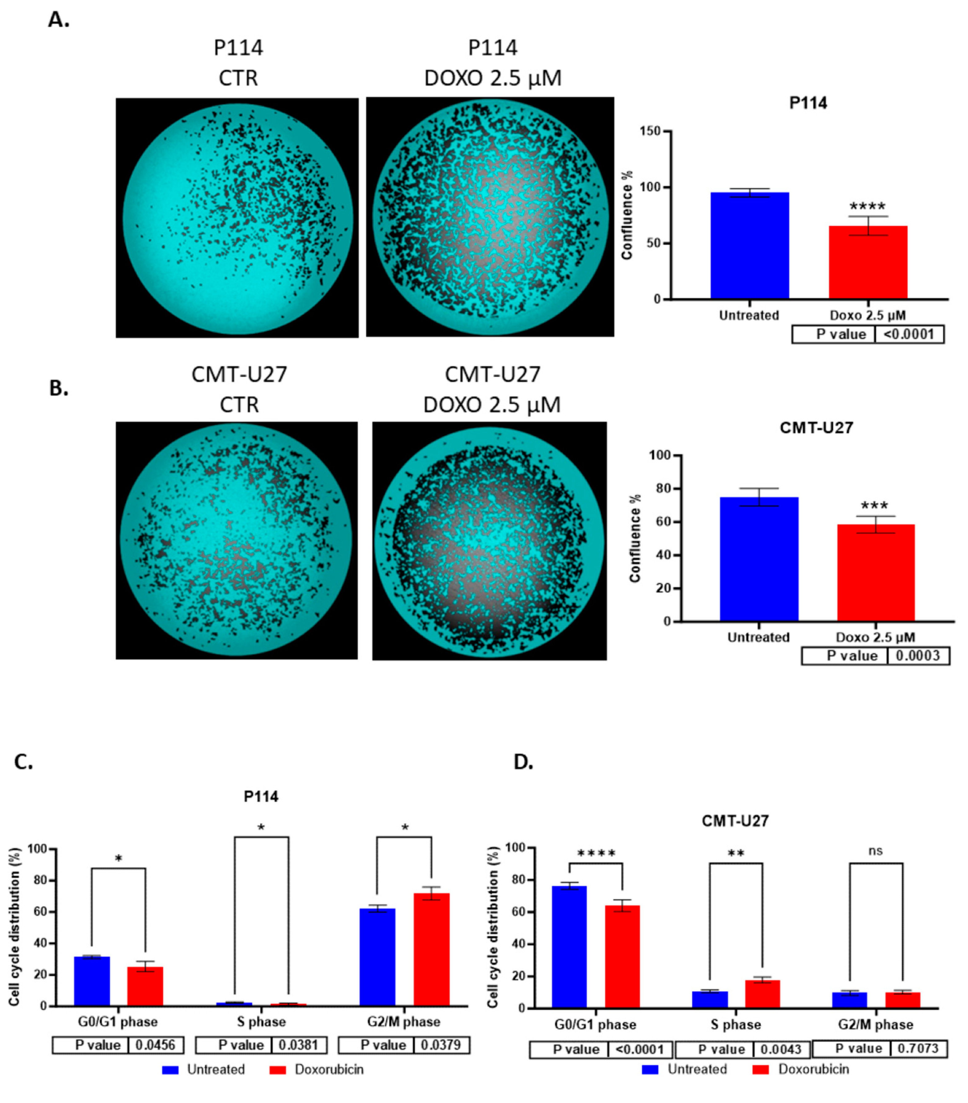
3.3. Reduced Cell Confluency in the Treated versus Untreated Cells

P114 and CMT-U27 treated with 2.5 µM of DOXO for 48 h present a lower confluency rate than untreated cells. We can observe that the treated cells show between a 30 and 40% reduced confluency versus the untreated cells (P114 65.62% and CMT-U27 58.44%) (Figure 2A,B).

3.4. Cell Cycle Arrest Induced by Doxorubicin

Doxorubicin induces cell cycle arrest in the G0/G1 phase for the CMT-U27 and in the G2/M for the P114. For the evaluation of cell growth inhibition induced by ABT-199, a cell cycle assay was performed using Celigo imaging flow cytometry. In both cell lines, a decreased percentage of cells was observed in the G0/G1 phase in treated cells versus control cells. In the P114-treated cell line, the S phase distribution was slightly decreased (p-value 0.0456), the G2/M phase distribution increased from 62% to 72% (p-value 0.0379), and the G0/G1 phase distribution decreased from 31% to 25% (p-value 0.0456). CMT-U27-treated cells showed an increased distribution in the S phase from 10% to 17% (p-value 0.0043), while the G0/G1 phase distribution decreased from 76% to 64% (p-value < 0.0001). In the G2/M phase, no statistically significant changes were observed (Table 1) (Figure 2C,D).

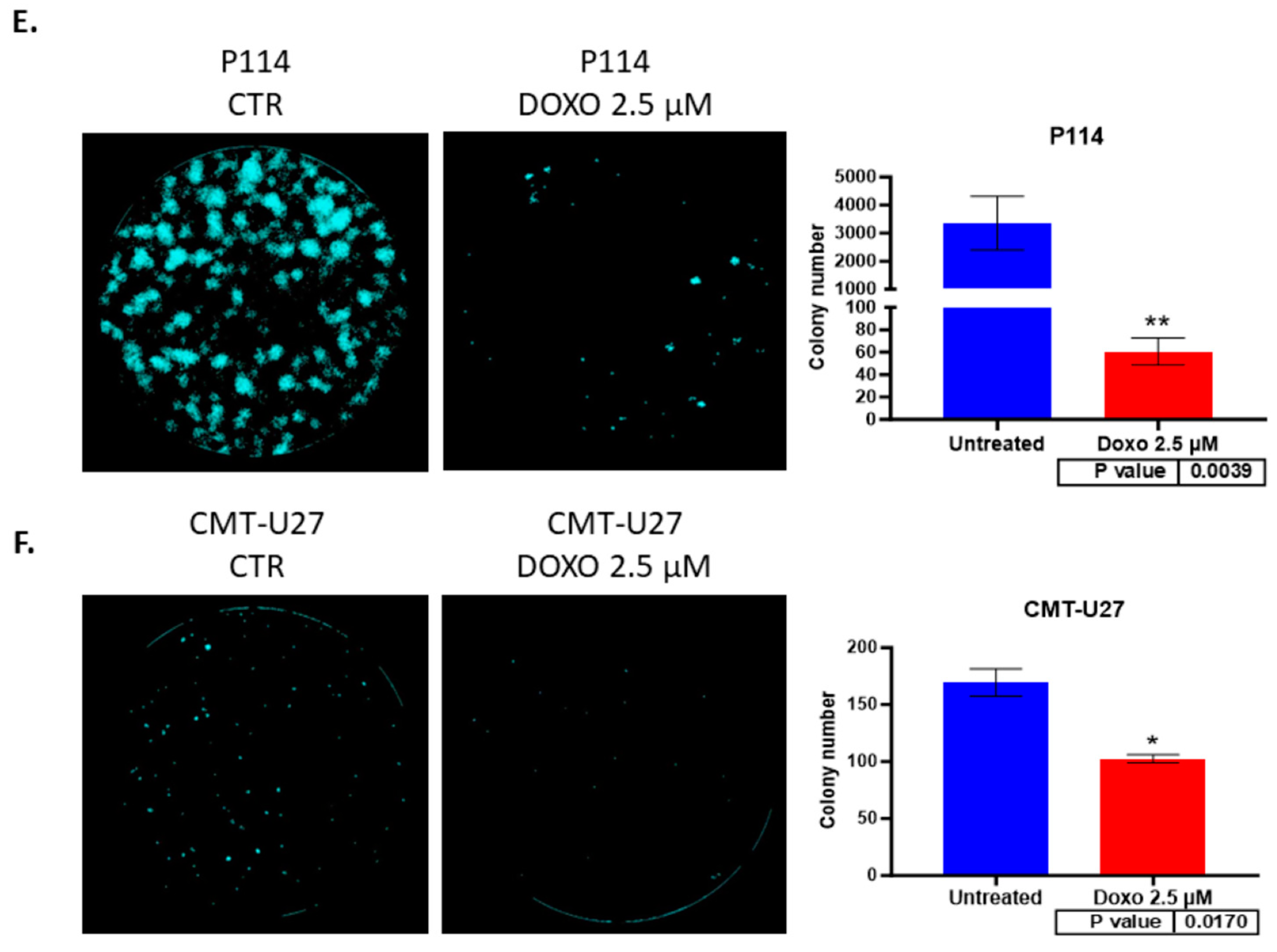
3.5. Doxorubicin Reduces Colony Formation in Both Canine Mammary Cancer Cell Lines

Both canine cell lines treated with 2.5 µM of DOXO for 48 h show a significantly reduced colony formation capacity demonstrated through the ability of untreated cells to form more colonies compared to the treated (P114 p-value 0.0039; CMT-U27 p-value 0.017) ones where we can observe a significantly low number of colonies after 7 days from the treatment (Figure 2E,F).

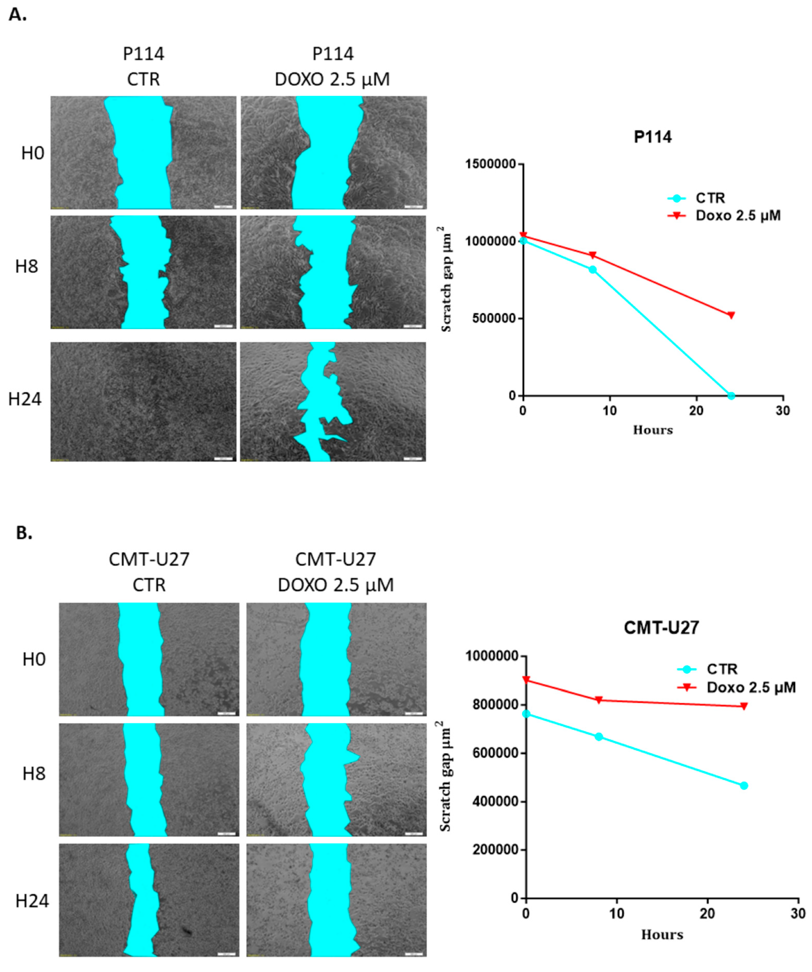
3.6. Doxorubicin Impairs Cell Migration in Both Canine In Vitro Mammary Cancer Models

CMT-U27 and P114 cell lines treated with 2.5 µM of DOXO for 48 h show a significantly reduced migratory capacity demonstrated through the ability of control cells to close the gap from the scratch assay compared to the treated ones where the gap is still significantly visible after 48 h (Figure 3A,B).

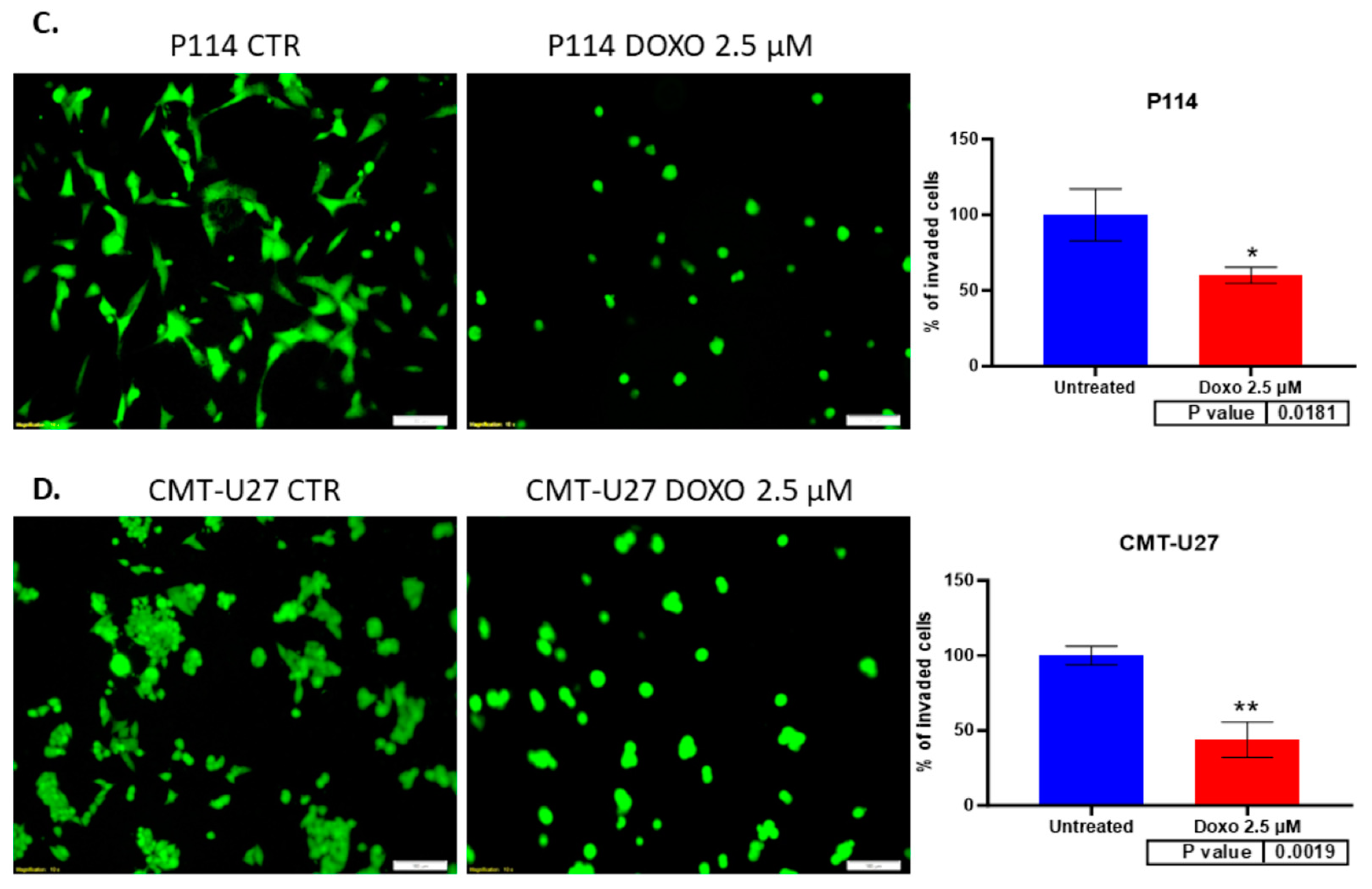
3.7. Doxorubicin Treatment Suppresses Cell Invasion

A significantly suppressed invasion ability was observed in both canine mammary cancer cell lines treated with DOXO. Canine cells treated with 2.5 µM DOXO show a reduced percentage of the invaded cells: a 60% p-value of 0.018 for P114 and a 43% p-value of 0.001 for the CMT-U27 cell line (Figure 3C,D).

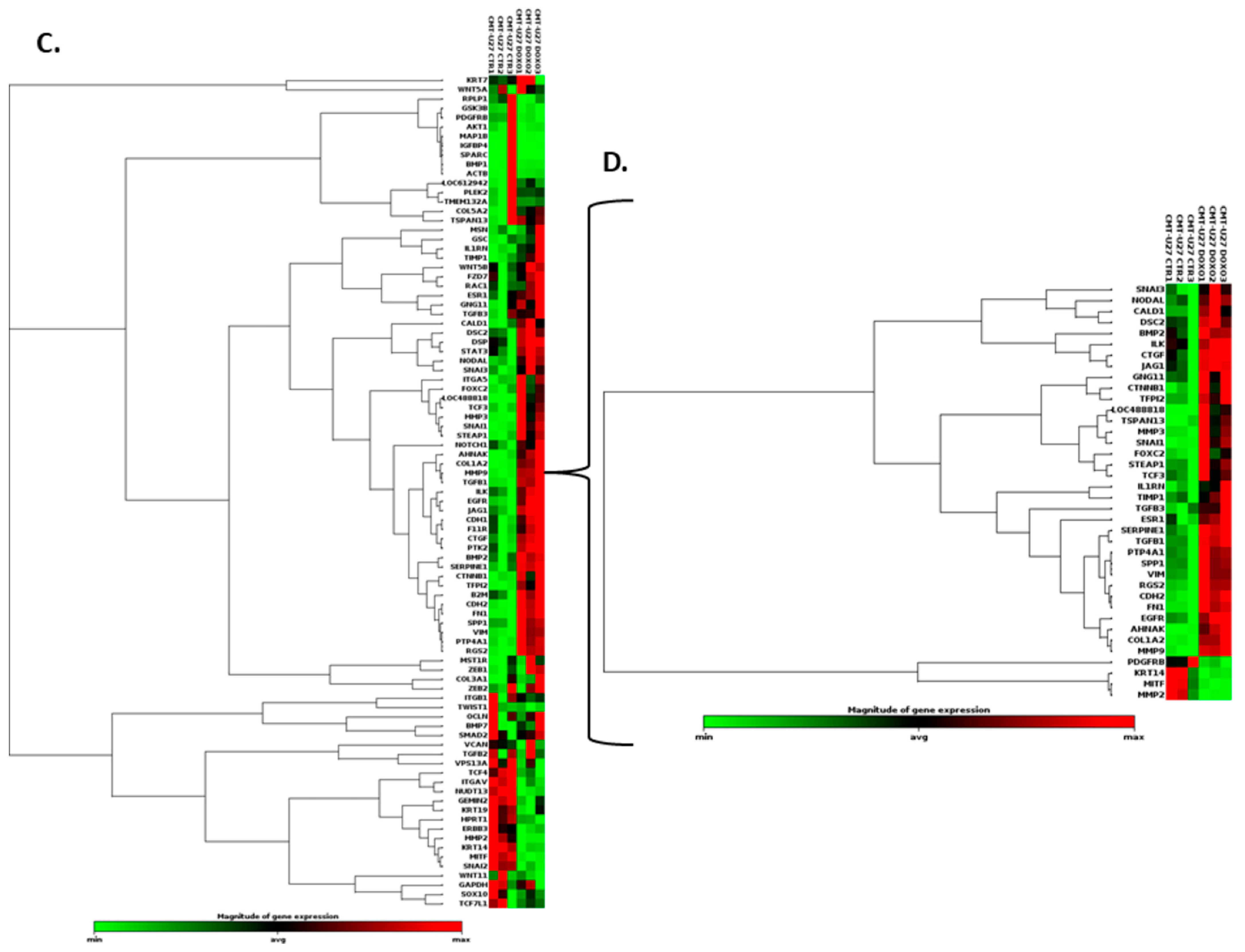
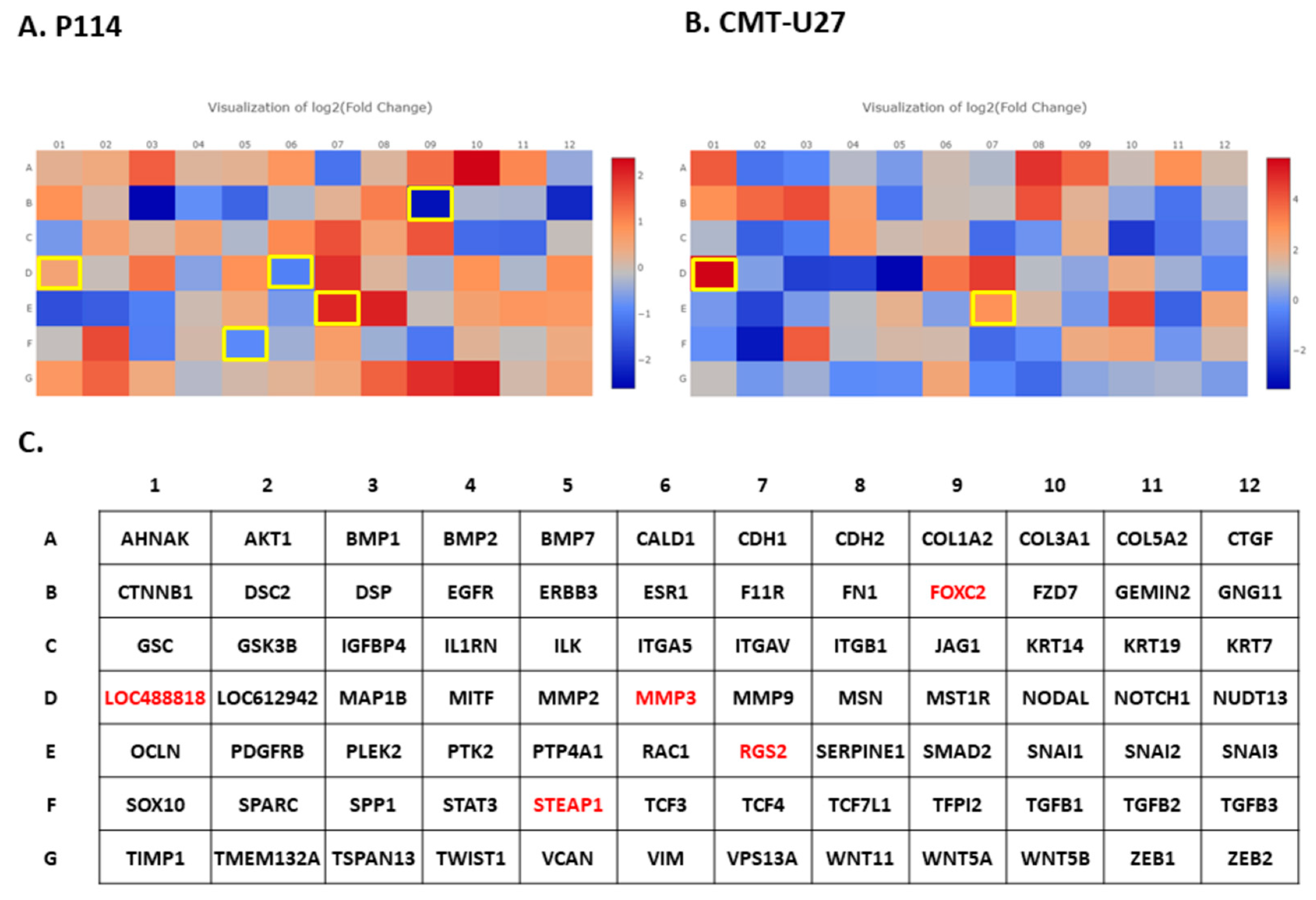
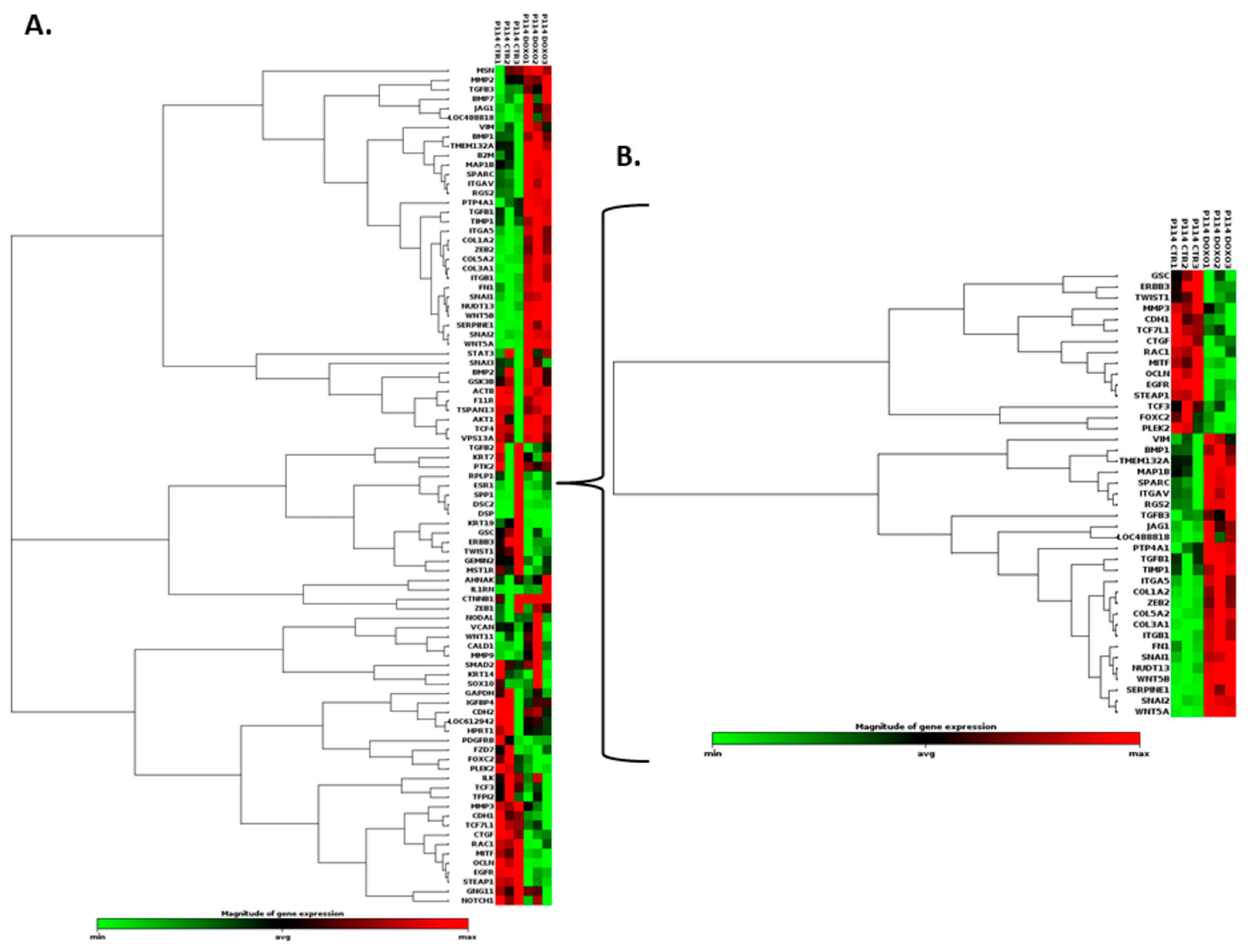
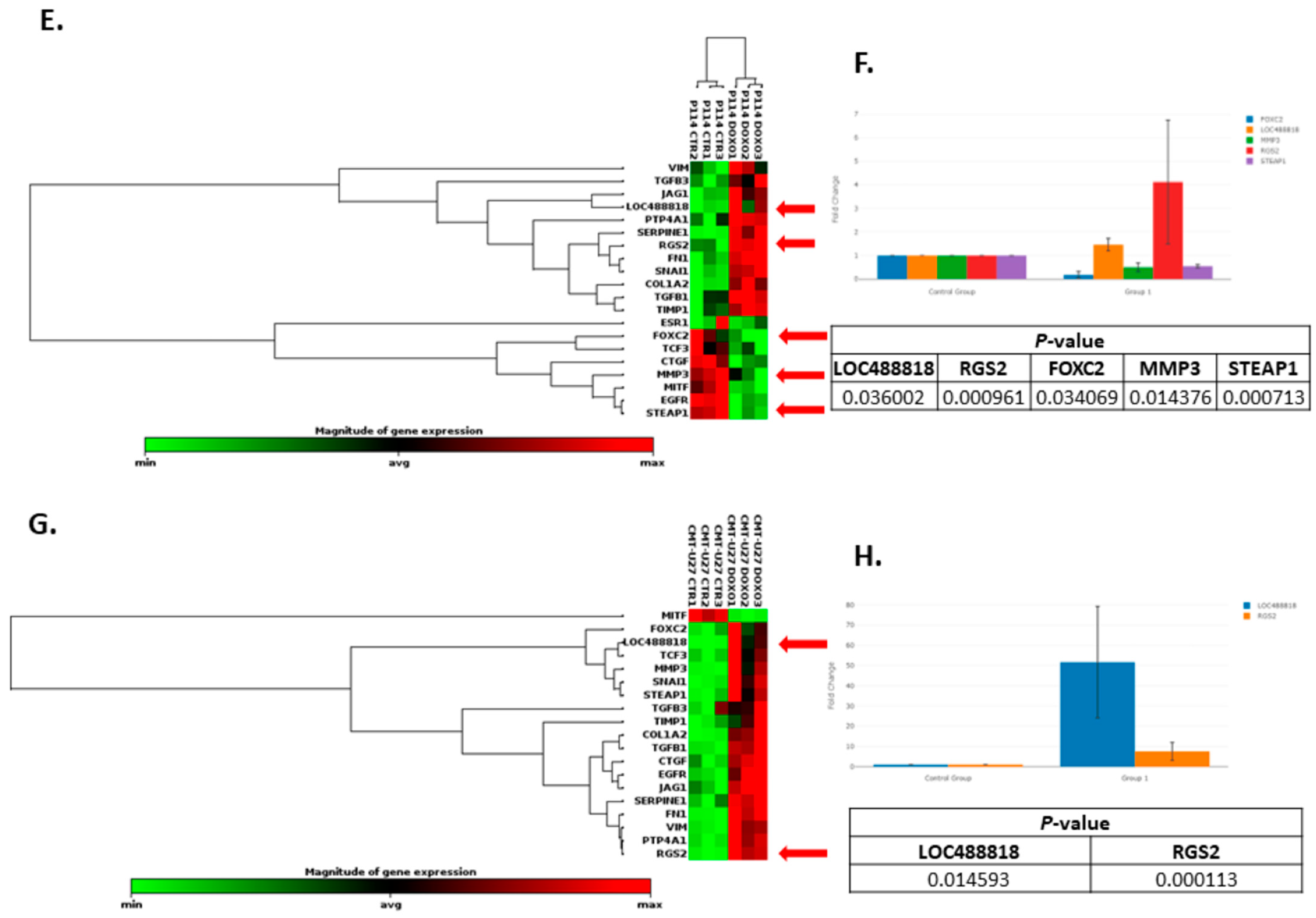
3.8. Doxorubicin Treatment Induces EMT Gene Expression Level Alteration

Based on the RT2 Profiler PCR Array analysis of Dog Epithelial-to-Mesenchymal-Transition genes, totals of 41 and 38 significantly (p-value < 0.05) differentially expressed EMT-related genes for the P114 and CMT-U27 cell lines, respectively, were identified (Figure 4A,B,C,D and Figure 5A,B,C). Table 2 and Table 3 present the fold changes and the list of genes involved in different biological processes including the EMT for both cell lines.
Nineteen genes were identified to be common in the two cell lines treated with DOXO. The P114 cell line treated with DOXO shows an altered expression level for five genes (LOC488818/FGF-BP1, RGS2, FOXC2, STEAP1, and MMP3) and CMT-U27 shows an altered expression level only for two genes (LOC488818 and RGS2) (Figure 4E–H).

4. Discussion

The present in vitro study aimed to investigate Doxorubicin’s efficacy in treating CMTs. The obtained results suggest that therapy with Doxorubicin has apoptotic effects on canine in vitro models of mammary cancer and can significantly reduce cell migration, invasion, confluency, and cell colony formation and can induce cell cycle arrest in treated versus untreated cells. Moreover, Doxorubicin treatment can alter EMT gene expression levels. Consistent with the obtained data, DOXO treatment inhibited the cell proliferation at 48 h in two CMT cell lines: one from a primary tumor and one from a lymph node metastasis; furthermore, a substantial effect on the cell cycle was observed, as well as an increase in the gene expression of P-glycoprotein (P-gp) and Breast Cancer Resistance Protein (BCRP) compared to the controls [18].
The epithelial-to-mesenchymal transition (EMT) is a biological process that occurs in various types of tumors, including canine mammary tumors [1]. In this complex cellular phenomenon, epithelial cells are tightly organized and present specific adhesion properties. They suffer molecular and structural characteristics to become more mesenchymal cells, losing their connectivity and acquiring migratory properties [2]. This phenomenon involves cancer progression, invasion, and metastasis [3]. The EMT plays a key role in cancer progression [33,34]. The results suggest that canine mammary tumors can benefit from the activity of Doxorubicin through inhibition of the EMT and different cancer-related biological processes, inducing tumor suppressor mechanisms. Regulator of G protein signaling 2 (RGS2) belongs to a family of proteins that serve as a GTPase-activating protein (GAP) for Gα subunits [35]. It has been described as a potential target in different types of human cancer, including breast cancer [35,36]. Thus, RGS2 was upregulated in MCF7 breast cancer cells, inhibiting cell proliferation [35]. In breast-invasive carcinoma of no special type (BIC-NST), the downregulation of RGS2 was associated with a significantly poorer overall survival rate [35]. LOC488818 is also named FGF-BP1 (Fibroblast Growth Factor Binding Protein 1) and plays an important role in cell proliferation, differentiation, and migration. FGF-BP1 is a protein that acts as a chaperone molecule and is found to be upregulated in breast cancers, squamous cell cancer, and colon cancer [37]. The overexpression of FGF-BP1 was correlated with better prognosis in human breast cancer patients [38]. A transcription factor known as FOXC2, which plays an important role in determining the fate of mesenchymal cells during embryonic development, has also been implicated in cancer cells’ ability to metastasize [39]. FOXC2 is a transcription factor that induces mesenchymal differentiation during the EMT [39,40] and has the potential to be a therapeutic target in the inhibition of metastasis [41]. FOXC2 overexpression was significantly associated with aggressive basal-like human breast cancers [39]. The matrix metalloproteinase (MMP) family of zinc-containing and calcium-dependent proteinases play a crucial role in various biological processes, including tissue remodeling, wound healing, and angiogenesis, and in diseases such as arthritis, cancer, and tissue ulceration. As they hydrolyze extracellular matrix components (ECM), they are also known as matrixins. MMP3 is a zinc-dependent proteolytic enzyme that regulates different biological processes by altering the extracellular matrix [42]. MMP3 over-expression was correlated with tumor growth and metastasis in different types of cancer, including breast cancer [42,43]. Several changes are induced by MMP-3 (increased cell proliferation, apoptosis, angiogenesis, changes in the stroma, etc.), which may result in the development of breast cancer in women. This protein may also affect mammary tumors in canines [44]. STEAP1 is a membrane-bound channel protein found overexpressed in various cancers [45,46,47,48] and was associated with a malignant phenotype and disease prognosis [49]. STEAP1 upregulation significantly inhibited the capacity of migration of the tumor cells and the invasion potential and downregulated the expression of EMT-related genes in human breast cancer [49].

5. Conclusions

The treatment and knowledge of canine mammary tumors (CMTs) have made significant progress in veterinary oncology as a spontaneous model for breast cancer research in the last few decades. Using Doxorubicin to target specific biological processes like apoptosis, the cell cycle, migration, invasion, and the EMT may result in better outcomes for patients with this disease. The canine model of breast cancer represents a good opportunity for new therapeutic strategies to be developed for human breast cancer. Doxorubicin is a widely used antineoplastic agent in humans and animals. Multiple mechanisms are involved in DOXO’s action, producing free radicals, intercalation into DNA strands, and inhibiting topoisomerases I and II, causing DNA damage, activating caspases, and ultimately causing apoptosis. The importance of the development of new therapeutic strategies in canine mammary cancer represents a major challenge worldwide. The results of present study provide valuable information for revealing the link between Doxorubicin, phenotypic changes, and proliferation dynamics in malignant cells that may contribute to chemotherapy treatment limitations in the canine model.

Author Contributions

Conceptualization, M.L.G., O.Z., L.R. and I.B.-N.; methodology, M.L.G., L.R., O.Z. and L.B.; data curation, O.Z., L.R. and I.B.-N.; writing—original draft preparation, M.L.G., O.Z. and L.R.; writing—review and editing, O.Z., L.R. and I.B.-N.; supervision, I.B.-N. All authors have read and agreed to the published version of the manuscript.

Funding

The authors acknowledge the support from the PCD 2461/28/17 January 2020, and PCD 1033/27/13 January 2021 research projects founded by “Iuliu Hatieganu” University of Medicine and Pharmacy, Cluj-Napoca, Romania, and the project PDI-PFE-CDI2021, entitled Increasing the Performance of Scientific Research, Supporting Excellence in Medical Research, and Innovation in Medicine, PROGRES, no. 40PFE/30 December 2021.

Institutional Review Board Statement

Not applicable.

Informed Consent Statement

Not applicable.

Data Availability Statement

Data are contained within the article.

Acknowledgments

This work was granted by project PDI-PFE-CDI2021, entitled Increasing the Performance of Scientific Research, Supporting Excellence in Medical Research, and Innovation in Medicine, PROGRES, no. 40PFE/30 December 2021 and projects PCD 2461/28/17 January 2020 research project founded by “Iuliu Hatieganu” University of Medicine and Pharmacy, Cluj-Napoca, Romania. The authors acknowledge the support from the research project —PCD 1033/27/13 January 2021, founded by “Iuliu Hatieganu” University of Medicine and Pharmacy, Cluj-Napoca, Romania.

Conflicts of Interest

The authors declare no conflict to interest.

References

  1. Salas, Y.; Marquez, A.; Diaz, D.; Romero, L. Epidemiological Study of Mammary Tumors in Female Dogs Diagnosed during the Period 2002-2012: A Growing Animal Health Problem. PLoS ONE 2015, 10, e0127381. [Google Scholar] [CrossRef]
  2. Goldschmidt, M.H.; Peña, L.; Zappulli, V. Tumors of the Mammary Gland. In Tumors in Domestic Animals; John Wiley & Sons, Inc.: Hoboken, NJ, USA, 2016; pp. 723–765. [Google Scholar] [CrossRef]
  3. Burrai, G.P.; Gabrieli, A.; Moccia, V.; Zappulli, V.; Porcellato, I.; Brachelente, C.; Pirino, S.; Polinas, M.; Antuofermo, E. A Statistical Analysis of Risk Factors and Biological Behavior in Canine Mammary Tumors: A Multicenter Study. Anim. Open Access J. 2020, 10, 1687. [Google Scholar] [CrossRef]
  4. Sorenmo, K. Canine mammary gland tumors. Vet. Clin. N. Am. Small Anim. Pract. 2003, 33, 573–596. [Google Scholar] [CrossRef] [PubMed]
  5. Raduly, L.; Cojocneanu, R.; Sarpataki, O.; Chira, S.; Atanasov, A.; Braicu, C.; Berindan-Neagoe, I.; Marcus, I. Canis lupus familiaris as relevant animal model for breast cancer—A comparative oncology review. Anim. Sci. Pap. Rep. 2018, 36, 119–148. [Google Scholar]
  6. Sorenmo, K.U.; Rasotto, R.; Zappulli, V.; Goldschmidt, M.H. Development, anatomy, histology, lymphatic drainage, clinical features, and cell differentiation markers of canine mammary gland neoplasms. Vet. Pathol. 2011, 48, 85–97. [Google Scholar] [CrossRef] [PubMed]
  7. Valdivia, G.; Alonso-Diez, A.; Perez-Alenza, D.; Pena, L. From Conventional to Precision Therapy in Canine Mammary Cancer: A Comprehensive Review. Front. Vet. Sci. 2021, 8, 623800. [Google Scholar] [CrossRef] [PubMed]
  8. Lavalle, G.E.; De Campos, C.B.; Bertagnolli, A.C.; Cassali, G.D. Canine malignant mammary gland neoplasms with advanced clinical staging treated with carboplatin and cyclooxygenase inhibitors. In Vivo 2012, 26, 375–379. [Google Scholar]
  9. Kim, J.-H.; Im, K.-S.; Kim, N.-H.; Chon, S.-K.; Doster, A.R.; Sur, J.-H. Inflammatory mammary carcinoma with metastasis to the brain and distant organs in a spayed Shih Tzu dog. J. Vet. Diagn. Investig. 2011, 23, 1079–1082. [Google Scholar] [CrossRef]
  10. Simon, D.; Schoenrock, D.; Baumgartner, W.; Nolte, I. Postoperative adjuvant treatment of invasive malignant mammary gland tumors in dogs with doxorubicin and docetaxel. J. Vet. Intern. Med. 2006, 20, 1184–1190. [Google Scholar] [CrossRef]
  11. Ogilvie, G.K.; Obradovich, J.E.; Elmslie, R.E.; Vail, D.M.; Moore, A.S.; Straw, R.C.; Dickinson, K.; Cooper, M.F.; Withrow, S.J. Efficacy of mitoxantrone against various neoplasms in dogs. J. Am. Vet. Med. Assoc. 1991, 198, 1618–1621. [Google Scholar]
  12. Poirier, V.J.; Hershey, A.E.; Burgess, K.E.; Phillips, B.; Turek, M.M.; Forrest, L.J.; Beaver, L.; Vail, D.M. Efficacy and toxicity of paclitaxel (Taxol) for the treatment of canine malignant tumors. J. Vet. Intern. Med. 2004, 18, 219–222. [Google Scholar] [CrossRef]
  13. von Euler, H.; Rivera, P.; Nyman, H.; Haggstrom, J.; Borga, O. A dose-finding study with a novel water-soluble formulation of paclitaxel for the treatment of malignant high-grade solid tumours in dogs. Vet. Comp. Oncol. 2013, 11, 243–255. [Google Scholar] [CrossRef]
  14. Raduly, L.; Gulei, D.; Jurj, A.; Moldovan, C.; Balint, E.; Marcus, I.; Sarpataki, O.; Korban, S.; Berindan-Neagoe, I. Comparative effects of BCL-2 inhibition in canine and human breast cancer in vitro models-a review. Anim. Sci. Pap. Rep. 2022, 40, 5–22. [Google Scholar]
  15. Mentoor, I.; Engelbrecht, A.M.; van de Vyver, M.; van Jaarsveld, P.J.; Nell, T. The paracrine effects of adipocytes on lipid metabolism in doxorubicin-treated triple negative breast cancer cells. Adipocyte 2021, 10, 505–523. [Google Scholar] [CrossRef]
  16. Walsh, E.M.; Smith, K.L.; Stearns, V. Management of hormone receptor-positive, HER2-negative early breast cancer. Semin. Oncol. 2020, 47, 187–200. [Google Scholar] [CrossRef]
  17. Ciocan-Cartita, C.A.; Jurj, A.; Zanoaga, O.; Cojocneanu, R.; Pop, L.A.; Moldovan, A.; Moldovan, C.; Zimta, A.A.; Raduly, L.; Pop-Bica, C.; et al. Correction to: New insights in gene expression alteration as effect of doxorubicin drug resistance in triple negative breast cancer cells. J. Exp. Clin. Cancer Res. 2020, 39, 287. [Google Scholar] [CrossRef] [PubMed]
  18. Levi, M.; Salaroli, R.; Parenti, F.; De Maria, R.; Zannoni, A.; Bernardini, C.; Gola, C.; Brocco, A.; Marangio, A.; Benazzi, C.; et al. Doxorubicin treatment modulates chemoresistance and affects the cell cycle in two canine mammary tumour cell lines. BMC Vet. Res. 2021, 17, 30. [Google Scholar] [CrossRef] [PubMed]
  19. Yun, U.-J.; Lee, J.-H.; Shim, J.; Yoon, K.; Goh, S.-H.; Yi, E.H.; Ye, S.-K.; Lee, J.-S.; Lee, H.; Park, J.; et al. Anti-cancer effect of doxorubicin is mediated by downregulation of HMG-Co A reductase via inhibition of EGFR/Src pathway. Lab. Investig. 2019, 99, 1157–1172. [Google Scholar] [CrossRef] [PubMed]
  20. Ramalingam, V.; Varunkumar, K.; Ravikumar, V.; Rajaram, R. Target delivery of doxorubicin tethered with PVP stabilized gold nanoparticles for effective treatment of lung cancer. Sci. Rep. 2018, 8, 3815. [Google Scholar] [CrossRef]
  21. Ciocan-Cartita, C.A.; Jurj, A.; Raduly, L.; Cojocneanu, R.; Moldovan, A.; Pileczki, V.; Pop, L.A.; Budisan, L.; Braicu, C.; Korban, S.S.; et al. New perspectives in triple-negative breast cancer therapy based on treatments with TGFbeta1 siRNA and doxorubicin. Mol. Cell Biochem. 2020, 475, 285–299. [Google Scholar] [CrossRef]
  22. Kalluri, R.; Weinberg, R.A. The basics of epithelial-mesenchymal transition. J. Clin. Investig. 2009, 119, 1420–1428. [Google Scholar] [CrossRef] [PubMed]
  23. Tiwari, N.; Gheldof, A.; Tatari, M.; Christofori, G. EMT as the ultimate survival mechanism of cancer cells. Semin. Cancer Biol. 2012, 22, 194–207. [Google Scholar] [CrossRef]
  24. Brabletz, T. To differentiate or not--routes towards metastasis. Nat. Rev. Cancer 2012, 12, 425–436. [Google Scholar] [CrossRef]
  25. Raposo-Ferreira, T.M.M.; Brisson, B.K.; Durham, A.C.; Laufer-Amorim, R.; Kristiansen, V.; Pure, E.; Volk, S.W.; Sorenmo, K. Characteristics of the Epithelial-Mesenchymal Transition in Primary and Paired Metastatic Canine Mammary Carcinomas. Vet. Pathol. 2018, 55, 622–633. [Google Scholar] [CrossRef] [PubMed]
  26. Huang, Y.; Hong, W.; Wei, X. The molecular mechanisms and therapeutic strategies of EMT in tumor progression and metastasis. J. Hematol. Oncol. 2022, 15, 129. [Google Scholar] [CrossRef]
  27. Pirlog, R.; Chiroi, P.; Rusu, I.; Jurj, A.M.; Budisan, L.; Pop-Bica, C.; Braicu, C.; Crisan, D.; Sabourin, J.C.; Berindan-Neagoe, I. Cellular and Molecular Profiling of Tumor Microenvironment and Early-Stage Lung Cancer. Int. J. Mol. Sci. 2022, 23, 5346. [Google Scholar] [CrossRef] [PubMed]
  28. Groza, I.M.; Braicu, C.; Jurj, A.; Zanoaga, O.; Lajos, R.; Chiroi, P.; Cojocneanu, R.; Paun, D.; Irimie, A.; Korban, S.S.; et al. Cancer-Associated Stemness and Epithelial-to-Mesenchymal Transition Signatures Related to Breast Invasive Carcinoma Prognostic. Cancers 2020, 12, 3053. [Google Scholar] [CrossRef]
  29. Mirzaei, S.; Abadi, A.J.; Gholami, M.H.; Hashemi, F.; Zabolian, A.; Hushmandi, K.; Zarrabi, A.; Entezari, M.; Aref, A.R.; Khan, H.; et al. The involvement of epithelial-to-mesenchymal transition in doxorubicin resistance: Possible molecular targets. Eur. J. Pharmacol. 2021, 908, 174344. [Google Scholar] [CrossRef]
  30. Hellmen, E. Characterization of four in vitro established canine mammary carcinoma and one atypical benign mixed tumor cell lines. Vitr. Cell. Dev. Biol. J. Tissue Cult. Assoc. 1992, 28A, 309–319. [Google Scholar] [CrossRef]
  31. Rao, N.A.; van Wolferen, M.E.; Gracanin, A.; Bhatti, S.F.; Krol, M.; Holstege, F.C.; Mol, J.A. Gene expression profiles of progestin-induced canine mammary hyperplasia and spontaneous mammary tumors. J. Physiol. Pharmacol. Off. J. Pol. Physiol. Soc. 2009, 60 (Suppl. S1), 73–84. [Google Scholar]
  32. Krol, M.; Pawlowski, K.M.; Skierski, J.; Rao, N.A.; Hellmen, E.; Mol, J.A.; Motyl, T. Transcriptomic profile of two canine mammary cancer cell lines with different proliferative and anti-apoptotic potential. J. Physiol. Pharmacol. Off. J. Pol. Physiol. Soc. 2009, 60 (Suppl. S1), 95–106. [Google Scholar]
  33. Hong, D.; Fritz, A.J.; Zaidi, S.K.; van Wijnen, A.J.; Nickerson, J.A.; Imbalzano, A.N.; Lian, J.B.; Stein, J.L.; Stein, G.S. Epithelial-to-mesenchymal transition and cancer stem cells contribute to breast cancer heterogeneity. J. Cell Physiol. 2018, 233, 9136–9144. [Google Scholar] [CrossRef] [PubMed]
  34. Thiery, J.P.; Acloque, H.; Huang, R.Y.; Nieto, M.A. Epithelial-mesenchymal transitions in development and disease. Cell 2009, 139, 871–890. [Google Scholar] [CrossRef] [PubMed]
  35. Lyu, J.H.; Park, D.W.; Huang, B.; Kang, S.H.; Lee, S.J.; Lee, C.; Bae, Y.S.; Lee, J.G.; Baek, S.H. RGS2 suppresses breast cancer cell growth via a MCPIP1-dependent pathway. J. Cell Biochem. 2015, 116, 260–267. [Google Scholar] [CrossRef] [PubMed]
  36. Wang, C.; Ye, Q.; Cao, Y.; Tan, J.; Wang, F.; Jiang, J.; Cao, Y. Downregulation of regulator of G protein signaling 2 expression in breast invasive carcinoma of no special type: Clinicopathological associations and prognostic relevance. Oncol. Lett. 2018, 15, 213–220. [Google Scholar] [CrossRef]
  37. Tassi, E.; Al-Attar, A.; Aigner, A.; Swift, M.R.; McDonnell, K.; Karavanov, A.; Wellstein, A. Enhancement of fibroblast growth factor (FGF) activity by an FGF-binding protein. J. Biol. Chem. 2001, 276, 40247–40253. [Google Scholar] [CrossRef]
  38. Zhang, Y.; Di, X.; Chen, G.; Liu, J.; Zhang, B.; Feng, L.; Cheng, S.; Wang, Y. An immune-related signature that to improve prognosis prediction of breast cancer. Am. J. Cancer Res. 2021, 11, 1267–1285. [Google Scholar]
  39. Mani, S.A.; Yang, J.; Brooks, M.; Schwaninger, G.; Zhou, A.; Miura, N.; Kutok, J.L.; Hartwell, K.; Richardson, A.L.; Weinberg, R.A. Mesenchyme Forkhead 1 (FOXC2) plays a key role in metastasis and is associated with aggressive basal-like breast cancers. Proc. Natl. Acad. Sci. USA 2007, 104, 10069–10074. [Google Scholar] [CrossRef]
  40. Hollier, B.G.; Tinnirello, A.A.; Werden, S.J.; Evans, K.W.; Taube, J.H.; Sarkar, T.R.; Sphyris, N.; Shariati, M.; Kumar, S.V.; Battula, V.L.; et al. FOXC2 expression links epithelial-mesenchymal transition and stem cell properties in breast cancer. Cancer Res. 2013, 73, 1981–1992. [Google Scholar] [CrossRef]
  41. Werden, S.J.; Sphyris, N.; Sarkar, T.R.; Paranjape, A.N.; LaBaff, A.M.; Taube, J.H.; Hollier, B.G.; Ramirez-Pena, E.Q.; Soundararajan, R.; den Hollander, P.; et al. Phosphorylation of serine 367 of FOXC2 by p38 regulates ZEB1 and breast cancer metastasis, without impacting primary tumor growth. Oncogene 2016, 35, 5977–5988. [Google Scholar] [CrossRef]
  42. Suhaimi, S.A.; Chan, S.C.; Rosli, R. Matrix Metallopeptidase 3 Polymorphisms: Emerging genetic Markers in Human Breast Cancer Metastasis. J. Breast Cancer 2020, 23, 1–9. [Google Scholar] [CrossRef]
  43. Argote Camacho, A.X.; Gonzalez Ramirez, A.R.; Perez Alonso, A.J.; Rejon Garcia, J.D.; Olivares Urbano, M.A.; Torne Poyatos, P.; Rios Arrabal, S.; Nunez, M.I. Metalloproteinases 1 and 3 as Potential Biomarkers in Breast Cancer Development. Int. J. Mol. Sci. 2021, 22, 9012. [Google Scholar] [CrossRef] [PubMed]
  44. Pandey, M.; Kumar, B.V.; Singh, S.; Verma, R. Development of recombinant matrix metalloproteinase-3 based sandwich ELISA for sero-diagnosis of canine mammary carcinomas. J. Immunoass. Immunochem. 2017, 38, 34–44. [Google Scholar] [CrossRef] [PubMed]
  45. Grunewald, T.G.P.; Ranft, A.; Esposito, I.; da Silva-Buttkus, P.; Aichler, M.; Baumhoer, D.; Schaefer, K.L.; Ottaviano, L.; Poremba, C.; Jundt, G.; et al. High STEAP1 expression is associated with improved outcome of Ewing’s sarcoma patients. Ann. Oncol. Off. J. Eur. Soc. Med. Oncol. 2012, 23, 2185–2190. [Google Scholar] [CrossRef] [PubMed]
  46. Gomes, I.M.; Arinto, P.; Lopes, C.; Santos, C.R.; Maia, C.J. STEAP1 is overexpressed in prostate cancer and prostatic intraepithelial neoplasia lesions, and it is positively associated with Gleason score. Urol. Oncol. 2014, 32, 53.e23–53.e29. [Google Scholar] [CrossRef] [PubMed]
  47. Lee, C.H.; Chen, S.L.; Sung, W.W.; Lai, H.W.; Hsieh, M.J.; Yen, H.H.; Su, T.C.; Chiou, Y.H.; Chen, C.Y.; Lin, C.Y.; et al. The Prognostic Role of STEAP1 Expression Determined via Immunohistochemistry Staining in Predicting Prognosis of Primary Colorectal Cancer: A Survival Analysis. Int. J. Mol. Sci. 2016, 17, 592. [Google Scholar] [CrossRef]
  48. Grunewald, T.G.; Diebold, I.; Esposito, I.; Plehm, S.; Hauer, K.; Thiel, U.; da Silva-Buttkus, P.; Neff, F.; Unland, R.; Muller-Tidow, C.; et al. STEAP1 is associated with the invasive and oxidative stress phenotype of Ewing tumors. Mol. Cancer Res. 2012, 10, 52–65. [Google Scholar] [CrossRef]
  49. Xie, J.; Yang, Y.; Sun, J.; Jiao, Z.; Zhang, H.; Chen, J. STEAP1 Inhibits Breast Cancer Metastasis and Is Associated with Epithelial-Mesenchymal Transition Procession. Clin. Breast Cancer 2019, 19, e195–e207. [Google Scholar] [CrossRef] [PubMed]
Figure 1. MTT cell viability assay and IC50 value identification after treatment with gradual doses of Doxorubicin in P114 and CMT-U27 canine mammary cancer cell lines for 48 h. Doxorubicin inhibits cell viability within all cell lines with an IC50 of 13 µM for P114 and 13.80 µM for CMT-U27 cell line (A,B). Actin damage and mitochondrial membrane potential activity and nuclear morphology evaluation. Observation of the Phalloidin-FITC/Mitotracker/DAPI staining using Olympus IX71 inverted fluorescence microscope after 48 h of treatment with 2.5 µM of Doxorubicin (image magnification 20×) (C,D), and the mitochondrial potential activity inhibition, cytoskeleton damage, and nuclear morphology changes using TMRE/Hoechst (E,F), marked with green arrows.
Vetsci 10 00654 g001aVetsci 10 00654 g001b
Figure 2. The confluency quantification after 2.5 µM DOXO treatment for 48 h. P114 and CMT-U27 cell lines treated with Doxorubicin show decreased confluency evaluated using Celigo Image Cytometer (A,B). Cell cycle distribution after 2.5 µM DOXO treatment for 48 h. P114 and CMT-U27 cell lines treated with Doxorubicin show a cell cycle arrest in G0/G1 phase (C,D). Colony formation after 2.5 µM DOXO treatment for 48 h. P114 and CMT-U27 cell lines treated with Doxorubicin show decreased colony formation capacity. Colony quantification in treated cells versus untreated cells after 7 days using Celigo Image Cytometer (E,F).
Vetsci 10 00654 g002aVetsci 10 00654 g002b
Figure 3. Scratch assay and quantification of cell migration after 2.5 µM DOXO treatment for 48 h. P114 and CMT-U27 cell lines treated with Doxorubicin show decreased migratory capacity after 48 h. Wound area quantification in control cells versus treated ones was evaluated after 0 h, 8 h, and 48 h using an Olympus IX71 microscope, objective 4×, and CellSens Imaging software (A,B). Invasion capacity evaluation using Matrigel (Invitrogen) and fluorescence microscopy. In both canine cell lines treated with DOXO, we can observe a reduced percentage of invaded cells versus the untreated cells. The fluorescence microscopy evaluation was performed by marking the cells with Calcein-AM and observed using Olympus IX71 inverted fluorescence microscope (image magnification 20×) (C,D).
Vetsci 10 00654 g003aVetsci 10 00654 g003b
Figure 4. EMT PCR Array gene expression analysis. In both canine cell lines treated with DOXO, we can observe a significantly (p-value < 0.05) EMT-related gene expression alteration represented by clustergrams generated using Qiagen analysis software for P114 (A,B) and CMT-U27 (C,D). Clustergrams, multigroup plots, and p-values for the common altered genes for the treated P114 (E,F) and CMT-U27 cell lines (G,H).
Vetsci 10 00654 g004aVetsci 10 00654 g004bVetsci 10 00654 g004c
Figure 5. EMT PCR Array gene expression analysis. In both canine cell lines treated with DOXO, we can observe a significant (p-value < 0.05) EMT-related gene expression alteration (marked with yellow) represented by heat maps generated using Qiagen analysis software for P114 (A) and CMT-U27 (B). (C) List of the genes from the PCR array and the significantly (p-value < 0.05) expressed genes marked with red.
Vetsci 10 00654 g005
Table 1. Cell cycle distribution in both canine mammary cancer cell lines.
Cell LineMean Number of Cells (%)
G0/G1SG2/MG0/G1SG2/M
CTRDOXO
P11431.342.4762.1725.281.3771.84
CMT-U2776.3510.869.6963.9517.7910.20
Table 2. The list of genes for the P114 cell line treated with Doxorubicin and biological processes.
P114
GenesFold Change (Compared to Control Group)Biological Processes
BMP12.73EMT, Cell growth and proliferation
CDH10.44EMT
COL1A22.43EMT
COL3A15.24EMT
COL5A22.00EMT
CTGF0.73fJAG
EGFR0.57Cell growth and proliferation
ERBB30.38Cell growth and proliferation
FN12.16EMT
FOXC20.18EMT, Differentiation and development, Cell growth and proliferation, fJAG, Transcription factors
GSC0.63EMT
ITGA51.96EMT
ITGAV3.04EMT
ITGB11.45fJAG
JAG12.93Differentiation and development, Cell growth and proliferation, Cell migration and motility
LOC4888181.46EMT, Cell growth and proliferation
MAP1B2.33Cytoskeleton regulation
MITF0.68EMT, Differentiation and development, Transcription factors
MMP30.50EMT, fJAG
NUDT131.91EMT
OCLN0.32EMT
PLEK20.50Cytoskeleton regulation
PTP4A11.35Differentiation and development
RAC10.64Cell morphogenesis, Cell migration and motility, Cytoskeleton regulation, fJAG
RGS24.12EMT
SERPINE14.26EMT, fJAG
SNAI11.52EMT, Differentiation and development, Cell morphogenesis
SNAI21.75EMT, Transcription factors
SPARC3.18EMT
STEAP10.55EMT
TCF30.77Transcription factors
TCF7L10.77Transcription factors
TGFB11.17Cell morphogenesis, Cell migration and motility, fJAG
TGFB31.32Differentiation and development, Cell growth and proliferation, Cell morphogenesis
TIMP11.69EMT, Cell growth and proliferation, fJAG
TMEM132A2.66EMT
TWIST10.87EMT, Differentiation and development, Cell morphogenesis, Transcription factors
VIM1.18EMT, Cell migration and motility, Cytoskeleton regulation
WNT5A3.87EMT, Differentiation and development, Cell morphogenesis, Signal transduction
WNT5B4.46EMT, Differentiation and development, Signal transduction
ZEB21.46Transcription factors
Table 3. The list of genes for the CMT-U27 cell line treated with Doxorubicin and biological processes.
CMT-U27
GenesFold Change (Compared to Control Group)Biological Processes
AHNAK16.47EMT
BMP21.93Differentiation and development
CALD12.51EMT, Cell migration and motility
CDH228.15EMT, fJAG
COL1A214.38EMT
CTGF2.61fJAG
CTNNB17.76Differentiation and development, Cell morphogenesis, Cell growth and proliferation, fJAG, Transcription factors
DSC213.75fJAG
EGFR5.92Cell growth and proliferation
ESR12.46Transcription factors
FN118.71EMT, Cell migration and motility, fJAG
FOXC23.41EMT, Differentiation and development, Cell growth and proliferation, fJAG, Transcription factors
GNG111.76EMT, Signal transduction
IL1RN6.20EMT
ILK2.57Cell growth and proliferation, fJAG
JAG13.76Differentiation and development, Cell growth and proliferation, Cell migration and motility
KRT140.21Differentiation and development
LOC48881851.71EMT, Cell growth and proliferation
MITF0.25EMT, Differentiation and development, Transcription Factors
MMP20.09EMT, fJAG
MMP311.63EMT, fJAG
MMP924.81EMT, fJAG
NODAL4.15Differentiation and development, Cell growth and proliferation, Cell migration and motility
PDGFRB0.25Cell growth and proliferation, Cell migration and motility, Signal transduction
PTP4A13.61Differentiation and development
RGS27.56EMT
SERPINE12.90EMT, fJAG
SNAI122.24EMT, Differentiation and development, Cell morphogenesis
SPP116.28EMT, fJAG
STEAP12.83EMT
TCF32.67Transcription factors
TFPI24.10EMT
TGFB14.92Cell morphogenesis, Cell migration and motility, fJAG
TGFB32.79Differentiation and development, Cell growth and proliferation, Cell morphogenesis
TIMP12.23EMT, Cell growth and proliferation, fJAG
TSPAN131.55EMT
VIM4.84EMT, Cell migration and motility, Cytoskeleton regulation
Disclaimer/Publisher’s Note: The statements, opinions and data contained in all publications are solely those of the individual author(s) and contributor(s) and not of MDPI and/or the editor(s). MDPI and/or the editor(s) disclaim responsibility for any injury to people or property resulting from any ideas, methods, instructions or products referred to in the content.

