Article

Subunit Vaccine Targeting Phosphate ABC Transporter ATP-Binding Protein, PstB, Provides Cross-Protection against Streptococcus suis Serotype 2, 7, and 9 in Mice

1
Shanghai Veterinary Research Institute, Chinese Academy of Agricultural Science, Shanghai 200241, China
2
College of Veterinary Medicine, Hebei Agricultural University, Baoding 071000, China
3
School of Biomedical Sciences, University of Plymouth, Plymouth, Devon PL4 8AA, UK
4
The Vaccine Group Ltd., Plymouth, Derriford Research Facility, Devon PL6 8BX, UK
5
College of Veterinary Medicine, Yangzhou University, Yangzhou 225009, China
*
Authors to whom correspondence should be addressed.
These authors contribute equally to this work.
Vet. Sci. 2023, 10(1), 48; https://doi.org/10.3390/vetsci10010048
Submission received: 2 December 2022 / Revised: 3 January 2023 / Accepted: 5 January 2023 / Published: 9 January 2023
(This article belongs to the Special Issue Research on Streptococcus in Veterinary Medicine)

Abstract

:

Simple Summary

Streptococcus suis is an important bacterial pathogen, causing meningitis, arthritis, and endocarditis in pigs. Infections caused by S. suis lead to significant economic losses in the pig industry worldwide. The increasing prevalence of antimicrobial resistance (AMR), resulting in heightened global regulations on antibiotic use in livestock, has highlighted the critical need for alternative S. suis control strategies, such as vaccination. In the present study, we evaluated the protective effect of a subunit protein-based vaccine, targeting the phosphate ABC transporter ATP-binding protein (PstB) of S. suis serotype 2. PstB was shown to be highly conserved across various S. suis isolates. In mice, a candidate vaccine targeting the PstB protein induced the production of high levels of cytokines IFN-γ and IL-4, both considered to be important for protection against S. suis. Furthermore, the PstB-based vaccine was shown to provide a high level of (87.5%) protection against S. suis serotypes 2 and 9, with lower protection (62.5%) against S. suis serotype 7. These data indicate that PstB is a promising target antigen for development as a universal subunit vaccine against different S. suis serotypes.

Abstract

Streptococcus suis is a significant pathogen in pigs and a newly emerging zoonotic agent in humans. The presence of multiple serotypes and strains with diversified sequence types in pig herds highlights the need for the identification of broadly cross-reactive universal vaccine antigen targets, capable of providing cross-protection against S. suis infection. Subunit vaccines based on the conserved proteins shared between different S. suis serotypes are potential candidates for such a universally protective vaccine. In the present study, phosphate ABC transporter ATP-binding protein PstB (PstB), an immunogenic protein of the S. suis bacterium, was expressed and purified, and then subjected to cross-protection evaluation in mice. The PstB protein showed nearly 100% amino acid similarity across a panel of 31 S. suis isolates representing different serotypes, which were collected from different countries. A recombinant PstB (rPstB) protein (S. suis serotype 2) was recognized by rabbit sera specific to this serotype, and induced high levels of IFN-γ and IL-4 in mice immunized with the recombinant protein. These cytokines are considered important for protection against S. suis infection. Immunization of mice with rPstB resulted in an 87.5% protection against challenge with S. suis serotype 2 and 9 strains, suggesting a high level of cross-protection for S. suis serotypes 2 and 9. A lower protection rate (62.5%) was observed in mice challenged with the S. suis serotype 7 strain. These data demonstrate that PstB is a promising target antigen for development as a component of a universal subunit vaccine against multiple S. suis serotypes.

1. Introduction

Streptococcus suis (S. suis) is a major swine pathogen, causing meningitis, arthritis, endocarditis, and many other diseases and resulting in significant economic losses in the pig industry [1,2,3]. It has been reported that nearly 100% of pigs are carriers of S. suis, with the upper respiratory tract being the primary site of persistence [4]. S. suis is also an important zoonotic agent, with sporadic outbreaks of human infection reported following contact with S. suis-infected pigs or pork-derived products [5,6]. To date, 29 serotypes have been described, based on the antigenicity of their capsular polysaccharides (CPS) [7,8]. Serotype 2 is the predominant serotype associated with pig and human infection, with serotypes 9, 3, 1/2, and 7 also being frequently isolated from diseased pigs [3].
Antibiotics are the main strategy for prevention and control of S. suis infection in pig farms. However, commensurate with the long duration and high levels of antibiotics being used, antimicrobial resistance (AMR) to the most commonly used antibiotics is increasingly being reported. Together with increasing restrictions on the prophylactic use of antibiotics in livestock, the emergence of resistant strains highlights the need for alternative control strategies such as vaccination for S. suis infection. To date, the main commercial S. suis vaccine is inactivated whole-cell bacterin of pathogenic serotype 2 strains [9]. However, this vaccine has shown positive outcomes restricted to the same serotype or closely related strains [10]. Due to the large number of different S. suis serotypes and differences in geographical serotype prevalence, such serotype- or strain-dependent vaccines lack the ability to confer cross-protection against infection by heterologous S. suis strains [9,10,11]. Since multiple serotypes and multiple strains with diversified sequence types are present in pig herds, this diversity represents a common source of vaccine failure in pig farms [12].
Highly conserved immunogenic proteins in different S. suis serotypes and strains are attractive candidates for development as universal subunit vaccines. Over the last ten years, multiple surface and extracellular immunogenic proteins of S. suis have been identified and evaluated for their ability to provide broad cross-protection [9,13,14,15]. Many of these proteins were shown to trigger the host immune response, to induce specific antibodies and stimulate phagocytosis. Antibodies specific to these conserved protein components also increased the recognition of S. suis by the immune system and stimulated phagocytosis [16,17].
In a previous study, an immunogenic protein of S. suis, phosphate ABC transporter ATP-binding protein PstB was identified from the S. suis serotype 2 ZYS strain by immunoproteomic analysis [18]. It seems that, as in S. pneumonia and other bacteria, the protein PstB of S. suis is a ATPase component of a ABC-type phosphate transport system, which is an essential system for the uptake of inorganic orthophosphate from the environment [19]. However, the detailed biological function of PstB in S. suis has not been demonstrated. In the present study, we analyzed the conservation and prevalence of PstB across S. suis strains from different serotypes and different geographic locations. The protective capacity of a recombinant PstB (rPstB) against challenge with S. suis serotypes 2, 7, and 9 was evaluated in mice.

2. Materials and Methods

2.1. Analysis of Sequence Conservation of PstB

The presence of the PstB gene in S. suis field strains was analyzed using polymerase chain reaction (PCR) with specific primers (5′-TTATCGCAACATCACATTCG-3′ and 5′-TCAAGTCACCCAGATAGAAGAA-3′), chosen based on the sequence of S. suis 05ZYH33 strain (GenBank: CP000407.1) deposited in GenBank. The genome data of 31 S. suis isolates of different serotypes from different countries were collected from GenBank. Amino acid sequences of PstB from these isolates were determined from the genomes and aligned using ClustalW software (version 2.1. https://www.genome.jp/tools-bin/clustalw (accessed on 6 July 2021)), followed by analysis using ESPript software (version 3.0. https://espript.ibcp.fr/ESPript/cgi-bin/ESPript.cgi (accessed on 6 July 2021)).

2.2. Prediction of Linar B Cell Epitopes and Enzyme Linked Immunosorbent Assay (ELISA) Analysis

ABCpred (http://crdd.osdd.net/raghava/abcpred/ABC_submission.html (accessed on 10 July 2021)) and IEDB (http://tools.iedb.org/main/bcell/ (accessed on 11 July 2021)) were used to predict linear B cell epitopes on PstB. The peptide fragments identified in this analysis were selected for further study. Briefly, the corresponding peptides were chemically synthesized (Shanghai Apeptide Co., Ltd., Shanghai, China), and immunogenicity was assessed by ELISA. Peptides were coated on ELISA plates at a concentration of 1 μg/mL, 5 μg/mL, and 10 μg/mL at 4 °C overnight. After blocking with 10% bovine serum albumin for 10 min, plates were incubated at 37 °C for 1 h with positive sera (1:500 dilution) collected from rabbits infected with an S. suis serotype 2 strain. After the washing steps, the plates were incubated with horse radish peroxidase (HRP)-conjugated goat anti-rabbit secondary antibodies and then washed again. Finally, tetramethylbenzidine-hydrogen peroxide (TMB-H2O2) solution was added, and the color signal at an absorbance of 450 nm (OD450) was determined. A relative OD450 value of >2.1 was considered positive.

2.3. Expression and Purification of Recombinant Proteins

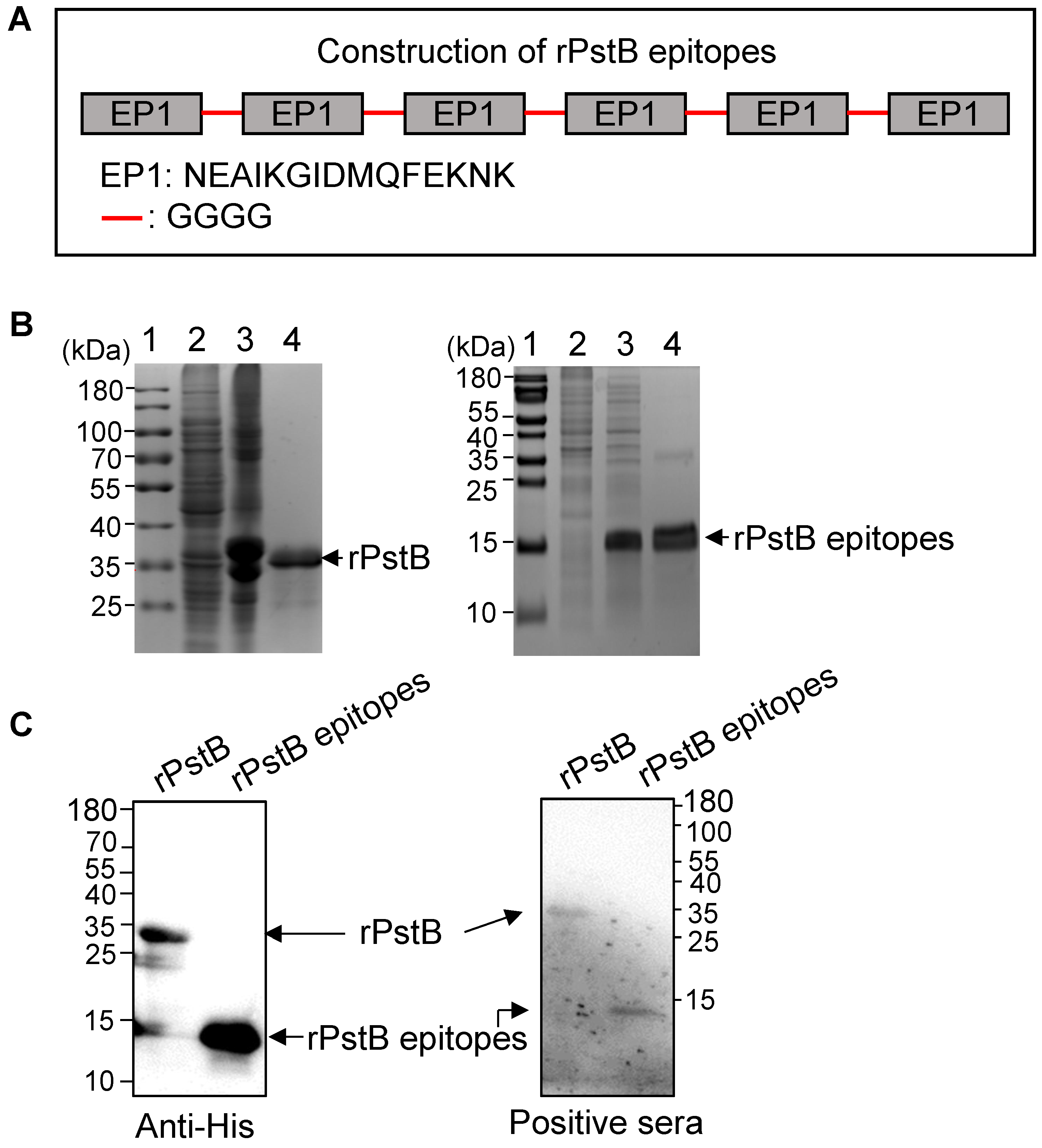
The full-length PstB gene or a synthetic nucleotide sequence encoding tandem B cell epitope EP1 (NEAIKGIDMQFEKNK) linked with a GGGG linker were cloned into a pET28a vector for expression of recombinant PstB (rPstB) and recombinant PstB epitopes (rPstB-epitope), respectively. The constructed plasmids were transformed into E. coli BL21 (DE3) cells and single colonies were selected on antibiotic-supplemented agar plates. Confirmed positive colonies were inoculated into LB broth and grown overnight. Overnight cultures were diluted 1:100 and grown to OD600 = 0.6–0.8. The rPstB and rPstB-epitope were expressed using isopropyl-β-D-thiogalactopyranoside (IPTG) induction and purified using a Ni–NTA column, according to the manufacturer’s instructions (SMART, Changzhou, China). The PstB proteins were evaluated on SDS-PAGE and subsequently concentrated using 10-kDa and 3-kDa Amicon Ultra Centrifugal Filter Units (EMD Millipore Billerica, MA, USA), respectively. Moreover, to improve the safety of the expressed proteins, an affinity matrix of modified polymyxin B (GenScript, Nanjing, China) was used to remove protein endotoxins. The endotoxin levels of the proteins were determined with a Limulus assay to be <0.01 EU/μg protein.

2.4. Western Blot Analysis

Western blots for detection of rPstB and rPstB-epitope were performed with anti-His antibodies (Abcam, Shanghai, China) and rabbit sera specific to S. suis serotype 2, as described previously [20].

2.5. Immunization and Challenge

Three to four week-old female BALB/c mice were used for all experiments. All animal experiments were approved by the Institutional Animal Care and Use Committee of Shanghai Veterinary Research Institute, China (IACUC No: SHVRI-SZ-2019070603) and performed in compliance with the Guidelines on the Humane Treatment of Laboratory Animals (Ministry of Science and Technology of the People’s Republic of China, Policy No. 2006 398). BALB/c mice were randomly assigned to three groups (8 per group), representing rPstB, rPstB-epitope, and control groups. In the rPstB and rPstB-epitope groups, each mouse was immunized subcutaneously with 50 μg of purified rPstB or rPstB-epitope, respectively, in combination with Freund’s complete adjuvant. Mice were boosted with 50 μg of the same purified rPstB or rPstB-epitope proteins emulsified with Freund’s incomplete adjuvant at 14 days post-primary immunization. Control group mice received the same strategy of immunization with phosphate buffered saline (PBS) with Freund’s complete and incomplete adjuvants. A challenge experiment was conducted with S. suis serotype 2 (ZY05719 strain), serotype 7 (SH04815 strains), or serotype 9 (SH26 strain). The serotype 2 strain ZY05719 was isolated from a diseased pig in China, in 2005 [21]. The serotype 7 SH04815 and serotype 9 SH26 strains were both clinical isolates from China, which were kindly gifted from Dr. Huochun Yao (College of Veterinary Medicine, Nanjing Agricultural University). The LD50 values of the serotype 2, 7, and 9 strains used in this study were 3.56 × 10⁷ CFU/mL, 4.02 × 10⁷ CFU/mL, and 7.88 × 10⁷ CFU/mL for BALB/c mice, respectively. Mice were challenged intraperitoneally at 10 days post-booster immunization with 8 × LD50 of the three S. suis strains. Mice were closely monitored for 7 days, with the survival time of each mouse being recorded. Infected mice exhibiting extreme lethargy were humanely euthanized, in accordance with the Guidelines on the Humane Treatment of Laboratory Animals (Ministry of Science and Technology of the People’s Republic of China, Policy No. 2006 398).

2.6. ELISA for Detection of Antibody

Blood samples were collected from individual mice by retro-orbital bleeding at 0, 14, and 24 days post-primary immunization for detection of antibodies. Aliquots of sera from individual mice were subjected to analysis of protein-specific antibodies by ELISA, using 96 well polystyrene microtiter trays coated with purified rPstB or rPstB-epitope, as described previously [22].

2.7. Detection of IFN-γ and IL-4

Blood, lung, and spleen samples were collected from the immunized mice at 24 days post primary immunization (10 days post-booster immunization), for analysis of levels of IFN-γ and IL-4 by ELISA, according to the manufacturer’s instructions (Boster, Wuhan, China).

2.8. Statistical Analysis

The measured values are presented as mean ± standard deviations (SD). Significance was determined using Student’s t-test. A value of p < 0.05 was considered statistically significant.

3. Results

3.1. Prevalence and Conservation of PstB among S. suis Strains

The purpose of this study was to evaluate whether PstB showed promise as a universal vaccine antigen for different S. suis serotypes. We first investigated the prevalence and conservation of PstB among S. suis strains. We randomly selected 15 field S. suis strains and reference serotype 2, 7, and 9 strains and tested for the prevalence of the PstB gene. The PCR results showed that all these strains were positive for the pstB gene (Figure 1). Furthermore, we collected the amino acid sequences of PstB proteins from the whole genome data of 31 S. suis strains deposited in GenBank, including 24 serotype 2, 2 serotype 7, and 5 serotype 9 strains. These serotypes are frequently observed in diseased pigs in pig farms globally [8,9,12]. Multiple-sequence alignment showed that there was only one amino acid variation in the 267-aa PstB proteins, showing an almost 100% conservation in these strains (Figure 1). Taken together, these results demonstrated that PstB is a highly prevalent and conserved protein in S. suis.

3.2. Linear Immunodominant B Cell Epitopes of PstB Screened by ELISA

A previous study showed that PstB could be recognized by porcine sera against S. suis serotype 2 [18]. We next used a series of bioinformatic tools to characterize this protein and analyze for the presence of potential B cell epitopes. PstB is a hydrophilic and antigenic protein, without a transmembrane domain (Supplementary Figure S1). Prediction using ABCpred and IEDB software identified three potential B cell epitopes (Figure 2A and Table 1). The three peptides were chemically synthesized and subjected to ELISA analysis. Of these, only peptide EP1 (NEAIKGIDMQFEKNK) reacted with rabbit sera specific to the S. suis serotype 2 strain (Table 1 and Figure 2B).

3.3. Expression and Purification of rPstB and rPstB-Epitope

The rPstB-epitope was constructed by tandemly splicing the B cell epitope EP1 (NEAIKGIDMQFEKNK) with GGGG linkers (Figure 3A). His-tagged rPstB and the rPstB-epitope construct were then expressed in E. coli and purified using Ni–NTA columns (Figure 3B). Western blot analysis with His-tag antibody confirmed the correct expression and purification of rPstB and rPstB-epitope (Figure 3C, Anti-His panel, Supplementary Figure S2). Moreover, both purified proteins were reactive against the rabbit sera specific to the S. suis serotype 2 strain (Figure 3C, Positive sera panel, Supplementary Figure S3). Together, these data indicated that rPstB and rPstB-epitope have potential as subunit vaccine candidates and elicit a specific immune response against S. suis infection.

3.4. Antibodies and Cytokines in Immunized Mice

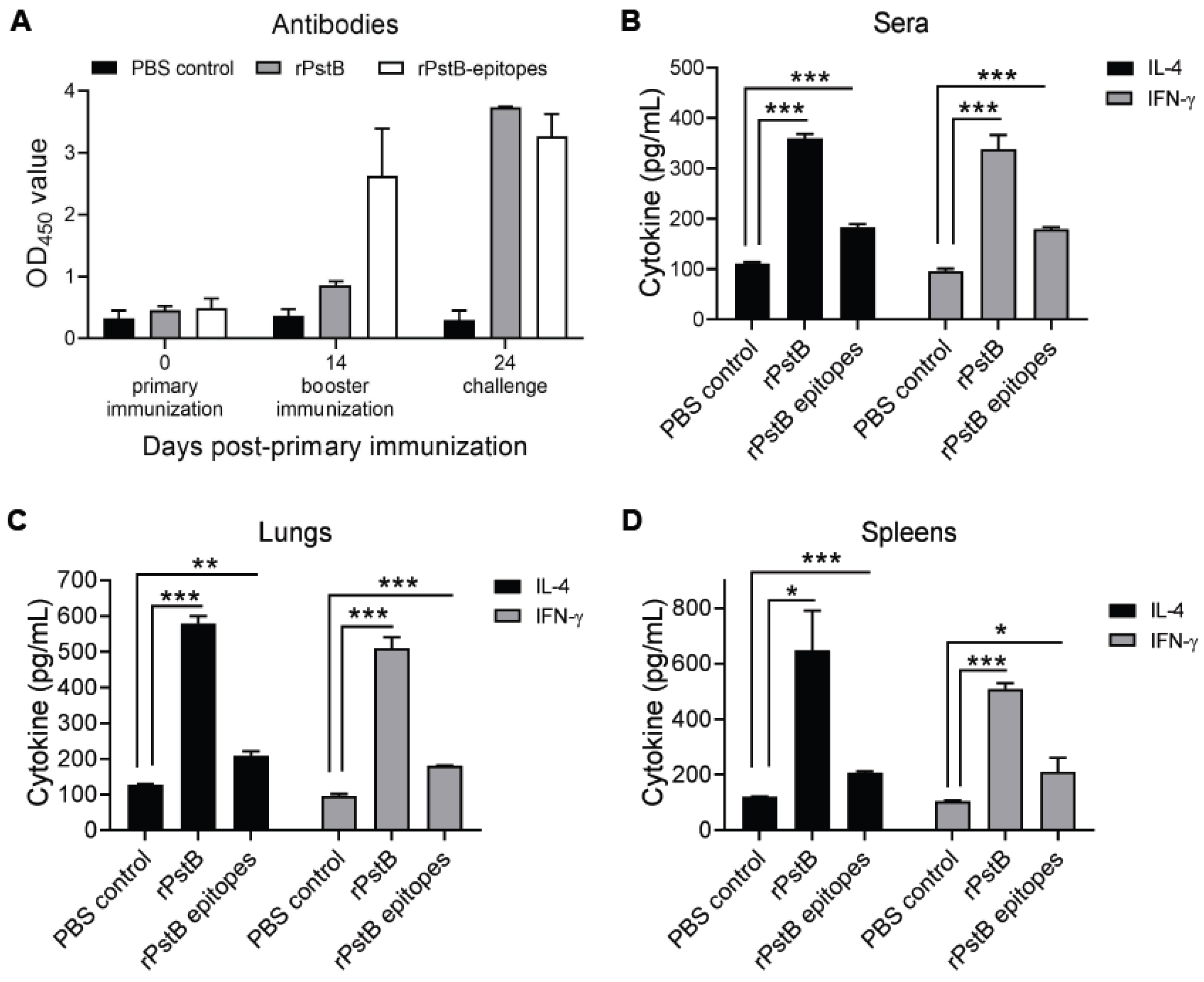
Next, the immunogenicity of rPstB and rPstB-epitope were assessed in mice. The dynamics of antibodies in the immunized mice were monitored using ELISA. A low level of rPstB antibodies was observed at 14 days post-primary immunization, which increased substantially after booster immunization. In contrast, a relatively higher level of antibodies specific to rPstB-epitope was detected in the mice at 14 days post-primary immunization, which only increased slightly after booster immunization (Figure 4A).
Th1-type and Th2-type immune responses are critical for resistance to S. suis infection [9]. Sera, lung, and spleen samples of immunized mice at 10 days post-booster immunization were collected, and ELISA was used to analyze these samples for the induction level of IFN-γ and IL-4, as indicator cytokines for Th1-type and Th2-type immune responses in S. suis infection, respectively [23,24,25,26,27]. For sera samples, compared with the PBS control group, the immunization of mice with rPstB or rPstB-epitope generated significantly higher levels of both IFN-γ and IL-4. However, IFN-γ and IL-4 levels in the rPstB group were considerably higher than those in the group immunized with rPstB-epitope (Figure 4B). A similar tendency of higher IFN-γ and IL-4 levels for the rPstB compared to rPstB-epitope was also observed in the lungs (Figure 4C) and spleens (Figure 4D).
In summary, compared to rPstB-epitope, rPstB showed stronger antigen-specific antibody responses and generated higher levels of IFN-γ and IL-4, indicating that the full-length version of the antigen is more immunogenic and may elicit better levels of protection against S. suis infection.

3.5. Protection of rPstB and rPstB-Epitope against S. suis Infection in Mice

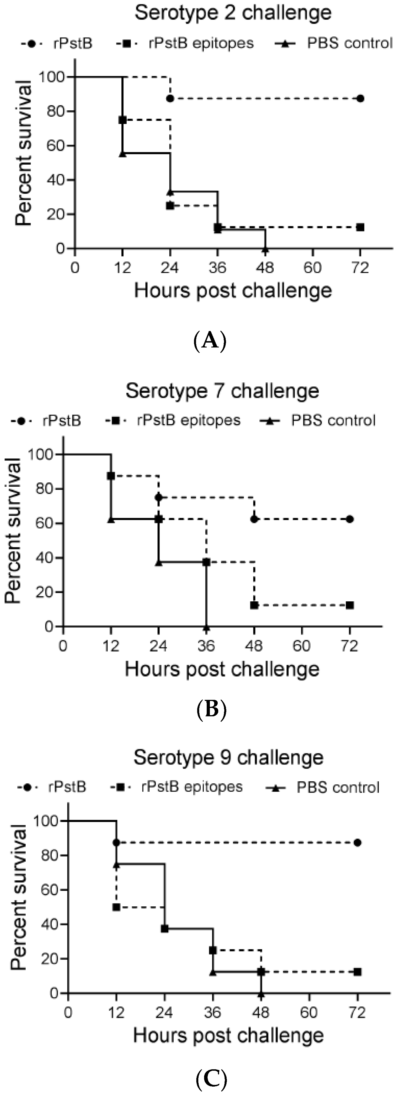
To test the respective efficacy against lethal S. suis challenge, groups of BALB/c mice were immunized with rPstB and rPstB-epitope, followed by challenge at 10 days post-booster immunization with a lethal dose of S. suis serotype 2, 7, or 9. In the serotype 2 challenge experiment, the rPstB provided 87.5% protection, while only 12.5% mice of the rPstB-epitope group survived the challenge (Figure 5A). As predicted, and consistent with the higher immunogenicity, rPstB provided greater levels of protection compared to rPstB-epitope for all serotypes. In the serotype 7 challenge experiment, survival rates of 62.5% and 12.5% were observed for the rPstB and rPstB-epitope groups, respectively (Figure 5B). In the serotype 9 challenge experiment, 87.5% of mice in the rPstB group survived, while rPstB-epitope yielded only 12.5% protection (Figure 5C).

4. Discussion

The complexity of S. suis epidemiology, as characterized by the presence of multiple serotypes and multiple strains with diversified sequence types in pig herds, suggests the need for a universal vaccine, to confer cross-protection against infection with different S. suis serotypes. Well-conserved proteins of S. suis are attractive vaccine candidates, because these proteins may allow effective immune responses against infection by various divergent S. suis strains [28,29]. In recent years, several surface/extracellular proteins of S. suis have been identified, based on immunoproteomic analysis, such as muramidase-released protein (MRP) suilysin, L-lactate dehydrogenase (Ldh), dihy-drolipoamide dehydrogenase (Dldh), pyruvate dehydrogenase E1component, ɑ subunit of (Pec), and amino acid ABC substrate binding protein (Sbp) [18,21,30]. PstB has been identified as a novel immunogenic protein of S. suis serotype 2 using immunoproteomic analysis. It is recognized by swine sera against formaldehyde-inactivated S. suis ZYS strain, as well as by the field convalescent sera from S. suis serotype 2 infected pigs [18]. However, the immunogenicity and protection capacity against S. suis infection of this protein has not been further studied. In the present study, we evaluated if PstB could be developed as a universal vaccine for different S. suis serotypes.
Sequence alignment and PCR screening revealed that PstB was highly prevalent in clinical isolates and very highly conserved, with nearly 100% identity among serotype 2, 7, and 9 strains from different regions of the world. Moreover, bioinformatics analysis showed that PstB did not consist of transmembrane domains. Together with the fact that this protein could be recognized by convalescent or hyperimmune sera [18], our results demonstrated that PstB is a well-conserved protein of S. suis, suggesting that it has the potential to be developed as a universal vaccine, to provide cross-protection against different serotypes and strains. The immune responses generated by PstB immunization were further studied, and high levels of cytokines IFN-γ and IL-4 were observed in the immunized mice. These two cytokines are generally considered indicators for Th1- and Th2-type immune responses, respectively, which are important assessment indices of vaccine development for S. suis [15,23,25,26,27]. Our results suggest that immunization with PstB could stimulate mixed Th1/Th2 responses that resist S. suis infection.
Several studies have evaluated the protection capability of surface/extracellular proteins of S. suis [9]. For example, elongation factor Tu(EF-Tu), a cytosolic GTP binding protein, provided 50% protection against S. suis serotype 2 infection, while FtsZ could protect 60% of mice against lethal infection [25]. HtpS is highly conserved in S. suis serotype 2 strains and shows 80% protection against challenge with S. suis serotype 2 infection in mice [31]. HP0197 provided nearly 100% protection in mice, but only 33% protection in pigs, after lethal infection of S. suis serotype 2 [32]. SsPepO is well-conserved and confers 100% and 33% protection in mice and pigs, respectively [33]. It should be noted that most of these studied only evaluated the protective capability with a challenge of S. suis serotype 2, which is the most virulent and prevalent serotype in pig and human infections worldwide [9]. However, serotypes 7 and 9 are frequently observed in diseased pigs, and are rarely included in these subunit vaccine evaluations. A recent study revealed that enolase protein provides 100%, 80%, and 100% protection against challenges with S. suis serotypes 2, 7, and 9, respectively, in mice [15]. In the present study, we evaluated the protective ability of rPstB in mice against challenges with S. suis serotype 2, 7, and 9 strains. We observed 87.5%, 62.5%, and 87.5% protection rates against challenge with S. suis serotypes 2, 7, and 9, respectively, in mice. The physiological role of PstB of S. suis has not been fully elucidated. It could be recognized by both hyperimmune sera and convalescent sera in an immunoproteomic analysis, which indicated that this protein is an immunogenic cell wall-associated protein in S. suis [18]. The protection effect of PstB against challenge with S. suis may be due to opsonin-dependent phagocytosis and stimulation of mixed Th1/Th2 responses, both of which are important defense mechanisms against S. suis in vivo [9]. The detailed protective mechanism and biological features should be further investigated. Moreover, there are several factors that could influence the protection efficacy, such as the immune dose, route, and adjuvant. Further improvement of the protection efficacy and evaluation in a pig models are needed.
Besides rPstB, we also identified one B cell epitope of PstB (EP1) and evaluated the immunogenicity and protection capacity of rPstB-epitope, which consisted of tandem EP1 epitopes. Experimental immunoassays are mostly used to identify B cell epitopes. However, this approach is labor-intensive and time-consuming. Immunoinformatics is a powerful tool for the identification of potential B cell epitopes and is becoming more frequently applied to the initial screening process [34]. The candidate peptides were further confirmed using ELISA with rabbit sera specific to S. suis serotype 2. With this strategy, we identified one B-cell epitope of PstB and expressed and purified the rPstB-epitope. However, compared to rPstB, the rPstB-epitope induced low-level IFN-γ and IL-4 production and provided poor protection (12.5%) to challenges with S. suis serotypes 2, 7, and 9. The development of vaccines based on protective B cell epitopes has been investigated for many pathogens [35]. In S. suis, a subunit vaccine named GMD was designed and constructed containing the B cell dominant epitopes of three important protective antigens (GAPDH, MRP, and DLDH). Ninety percent protection rates were observed after GMD immunization with challenge with S. suis serotype 2 [36]. The poor protection of rPstB-epitopes in this study may be due to the EP1 epitope not being the key immunodominant epitope for the PstB protein.

5. Conclusions

In summary, the protective efficacy of the PstB protein, identified using proteomics analysis, was evaluated in this study. Database searching showed that PstB is a highly conserved protein in S. suis. Immunization with PstB could induce high-level IFN-γ and IL-4 production and confer significant protection against S. suis serotypes 2, 7, and 9 in mice. These results demonstrated that PstB is a promising novel subunit vaccine candidate, which could provide cross-serotype protection against infection with S. suis for use individually or as a component of a universal S. suis vaccine.

Supplementary Materials

The following supporting information can be downloaded at: https://www.mdpi.com/article/10.3390/vetsci10010048/s1, Figure S1: Bioinformatic analysis of PstB; Figure S2: The original western blot of Figure 3C-left; Figure S3: The original western blot of Figure 3C-right.

Author Contributions

Conceptualization and methodology, Z.M., K.S., M.U. and J.L.; software, X.L. and J.L.; validation and formal analysis, Z.Y., R.P., J.Z. and N.D.; investigation, Z.M. and X.M.; resources, J.W., K.L., Y.Q. and X.Y.; data curation, J.L.; writing—original draft preparation, H.N., M.A.J. and B.L; writing—review and editing, Z.M. and B.L. All authors have read and agreed to the published version of the manuscript.

Funding

The study was supported by the National Key Research and Development Program of China (No. 2018YFE0101300) and the Department of Health and Social Care, UK (No. IUK 104985).

Institutional Review Board Statement

All of the animals were handled in strict accordance with the Animal Ethics Procedures and Guidelines of the People’s Republic of China, and the current study was approved by the Animal Ethics Committee of Shanghai Veterinary Research Institute, Chinese Academy of Agricultural Sciences (IACUC No: SHVRI-SZ-2019070603).

Informed Consent Statement

Informed consent was obtained from all subjects involved in the study.

Data Availability Statement

The data presented in this study are available on request from the corresponding author. The data are not publicly available due to privacy.

Conflicts of Interest

The authors declare no conflict of interest.

References

  1. Staats, J.J.; Feder, I.; Okwumabua, O.; Chengappa, M.M. Streptococcus suis: Past and Present. Vet. Res. Commun. 1997, 21, 381–407. [Google Scholar] [CrossRef] [PubMed]
  2. Fittipaldi, N.; Segura, M.; Grenier, D.; Gottschalk, M. Virulence Factors Involved in the Pathogenesis of the Infection Caused by the Swine Pathogen and Zoonotic Agent Streptococcus suis. Future Microbiol. 2012, 7, 259–279. [Google Scholar] [CrossRef] [PubMed]
  3. Goyette-Desjardins, G.; Auger, J.P.; Xu, J.; Segura, M.; Gottschalk, M. Streptococcus suis, an Important Pig Pathogen and Emerging Zoonotic Agent-an Update on the Worldwide Distribution Based on Serotyping and Sequence Typing. Emerg. Microbes Infect. 2014, 3, e45. [Google Scholar] [CrossRef]
  4. Désirée, V.; Maren, W.; Weldearegay, Y.B.; Peter, V.W. Streptococcus suis—The “Two Faces” of a Pathobiont in the Porcine Respiratory Tract. Front. Microbiol. 2018, 9, 480. [Google Scholar]
  5. Gottschalk, M.; Segura, M.; Xu, J. Streptococcus suis Infections in Humans: The Chinese Experience and the Situation in North America. Anim. Health Res. Rev. 2007, 8, 29–45. [Google Scholar] [CrossRef]
  6. Ye, C.; Zhu, X.; Jing, H.; Du, H.; Segura, M.; Zheng, H.; Kan, B.; Wang, L.; Bai, X.; Zhou, Y.; et al. Streptococcus suis Sequence Type 7 Outbreak, Sichuan, China. Emerg. Infect. Dis. 2006, 12, 1203–1208. [Google Scholar] [CrossRef]
  7. Bailey, A.M.; Alberti, F.; Kilaru, S.; Collins, C.M.; de Mattos-Shipley, K.; Hartley, A.J.; Hayes, P.; Griffin, A.; Lazarus, C.M.; Cox, R.J.; et al. Identification and Manipulation of the Pleuromutilin Gene Cluster from Clitopilus Passeckerianus for Increased Rapid Antibiotic Production. Sci. Rep. 2016, 6, 25202. [Google Scholar] [CrossRef]
  8. Okura, M.; Auger, J.P.; Shibahara, T.; Goyette-Desjardins, G.; Calsteren, M.; Maruyama, F.; Kawai, M.; Osaki, M.; Segura, M.; Gottschalk, M. Capsular Polysaccharide Switching in Streptococcus suis Modulates Host Cell Interactions and Virulence. Cold Spring Harb. Lab. 2020, 11, 6513. [Google Scholar] [CrossRef]
  9. Segura, M. Streptococcus suis Vaccines: Candidate Antigens and Progress. Expert Rev. Vaccines 2015, 14, 1587–1608. [Google Scholar] [CrossRef]
  10. Tan, C.; Zhang, A.; Chen, H.; Zhou, R. Recent Proceedings on Prevalence and Pathogenesis of Streptococcus suis. Curr. Issues Mol. Biol. 2019, 32, 473–520. [Google Scholar] [CrossRef]
  11. Pallarés, F.J.; Schmitt, C.S.; Roth, J.A.; Evans, R.B.; Kinyon, J.M.; Halbur, P.G. Evaluation of a Ceftiofur-Washed Whole Cell Streptococcus suis Bacterin in Pigs. Can. J. Vet. Res. 2004, 68, 236–240. [Google Scholar]
  12. Wei, Z.; Li, R.; Zhang, A.; He, H.; Hua, Y.; Xia, J.; Cai, X.; Chen, H.; Jin, M. Characterization of Streptococcus suis Isolates from the Diseased Pigs in China between 2003 and 2007. Vet. Microbiol. 2009, 137, 196–201. [Google Scholar] [CrossRef]
  13. Wisselink, H.J.; Vecht, U.; Stockhofe-Zurwieden, N.; Smith, H.E. Protection of Pigs against Challenge with Virulent Streptococcus suis Serotype 2 Strains by a Muramidase-Released Protein and Extracellular Factor Vaccine. Vet. Rec. 2001, 148, 473–477. [Google Scholar] [CrossRef]
  14. Jiang, X.; Yang, Y.; Zhou, J.; Liu, H.; Liao, X.; Luo, J.; Li, X.; Fang, W. Peptidyl Isomerase PrsA Is Surface-Associated on Streptococcus suis and Offers Cross-Protection against Serotype 9 Strain. FEMS Microbiol. Lett. 2019, 366. [Google Scholar] [CrossRef]
  15. Li, Q.; Lv, Y.; Li, Y.-A.; Du, Y.; Guo, W.; Chu, D.; Wang, X.; Wang, S.; Shi, H. Live Attenuated Salmonella Enterica Serovar Choleraesuis Vector Delivering a Conserved Surface Protein Enolase Induces High and Broad Protection against Streptococcus suis Serotypes 2, 7, and 9 in Mice. Vaccine 2020, 38, 6904–6913. [Google Scholar] [CrossRef]
  16. Charland, N.; Jacques, M.; Lacouture, S.; Gottschalk, M. Characterization and Protective Activity of a Monoclonal Antibody against a Capsular Epitope Shared by Streptococcus suis Serotypes 1, 2 and 1/2. Microbiol. Read. Engl. 1997, 143 Pt 11, 3607–3614. [Google Scholar] [CrossRef]
  17. Goyette-Desjardins, G.; Lacouture, S.; Auger, J.-P.; Roy, R.; Gottschalk, M.; Segura, M. Characterization and Protective Activity of Monoclonal Antibodies Directed against Streptococcus suis Serotype 2 Capsular Polysaccharide Obtained Using a Glycoconjugate. Pathogens 2019, 8, 139. [Google Scholar] [CrossRef]
  18. Zhang, A.; Xie, C.; Chen, H.; Jin, M. Identification of Immunogenic Cell Wall-Associated Proteins of Streptococcus suis Serotype 2. Proteomics 2008, 8, 3506–3515. [Google Scholar] [CrossRef]
  19. Zheng, J.J.; Sinha, D.; Wayne, K.J.; Winkler, M.E. Physiological Roles of the Dual Phosphate Transporter Systems in Low and High Phosphate Conditions and in Capsule Maintenance of Streptococcus pneumoniae D39. Front. Cell. Infect. Microbiol. 2016, 6, 63. [Google Scholar] [CrossRef]
  20. Zhu, Z.; Shi, Z.; Yan, W.; Wei, J.; Shao, D.; Deng, X.; Wang, S.; Li, B.; Tong, G.; Ma, Z. Nonstructural Protein 1 of Influenza A Virus Interacts with Human Guanylate-Binding Protein 1 to Antagonize Antiviral Activity. PLoS ONE 2013, 8, e55920. [Google Scholar] [CrossRef]
  21. Zhang, W.; Lu, C.P. Immunoproteomics of Extracellular Proteins of Chinese Virulent Strains of Streptococcus suis Type 2. Proteomics 2007, 7, 4468–4476. [Google Scholar] [CrossRef] [PubMed]
  22. Ogunniyi, A.D.; Folland, R.L.; Briles, D.E.; Hollingshead, S.K.; Paton, J.C. Immunization of Mice with Combinations of Pneumococcal Virulence Proteins Elicits Enhanced Protection against Challenge with Streptococcus pneumoniae. Infect. Immun. 2000, 68, 3028–3033. [Google Scholar] [CrossRef] [PubMed]
  23. Hsueh, K.-J.; Cheng, L.-T.; Lee, J.-W.; Chung, Y.-C.; Chung, W.-B.; Chu, C.-Y. Immunization with Streptococcus suis Bacterin plus Recombinant Sao Protein in Sows Conveys Passive Immunity to Their Piglets. BMC Vet. Res. 2017, 13, 15. [Google Scholar] [CrossRef] [PubMed]
  24. Gunjan, K.; Mohsin, R.; Brijendra, K.T. Interferon-Gamma (IFN-γ): Exploring Its Implications in Infectious Diseases. Biomol. Concepts 2018, 9, 64–79. [Google Scholar]
  25. Feng, L.; Niu, X.; Mei, W.; Li, W.; Liu, Y.; Willias, S.P.; Yuan, C.; Bei, W.; Wang, X.; Li, J. Immunogenicity and Protective Capacity of EF-Tu and FtsZ of Streptococcus suis Serotype 2 against Lethal Infection. Vaccine 2018, 36, 2581–2588. [Google Scholar] [CrossRef] [PubMed]
  26. Guo, G.; Kong, X.; Wang, Z.; Li, M.; Tan, Z.; Zhang, W. Evaluation of the Immunogenicity and Protective Ability of a Pili Subunit, SBP2′, of Streptococcus suis Serotype 2. Res. Vet. Sci. 2021, 137, 201–207. [Google Scholar] [CrossRef]
  27. Yi, L.; Du, Y.; Mao, C.; Li, J.; Jin, M.; Sun, L.; Wang, Y. Immunogenicity and Protective Ability of RpoE against Streptococcus suis Serotype 2. J. Appl. Microbiol. 2021, 130, 1075–1083. [Google Scholar] [CrossRef]
  28. Olaya-Abril, A.; Jiménez-Munguía, I.; Gómez-Gascón, L.; Rodríguez-Ortega, M.J. Surfomics: Shaving Live Organisms for a Fast Proteomic Identification of Surface Proteins. J. Proteom. 2014, 97, 164–176. [Google Scholar] [CrossRef]
  29. Zhang, A.; Chen, B.; Mu, X.; Li, R.; Zheng, P.; Zhao, Y.; Chen, H.; Jin, M. Identification and Characterization of a Novel Protective Antigen, Enolase of Streptococcus suis Serotype 2. Vaccine 2009, 27, 1348–1353. [Google Scholar] [CrossRef]
  30. Zhang, W.; Lu, C.P. Immunoproteomic Assay of Membrane-Associated Proteins of Streptococcus suis Type 2 China Vaccine Strain HA9801. Zoonoses Public Health 2007, 54, 253–259. [Google Scholar] [CrossRef]
  31. Shao, Z.; Pan, X.; Li, X.; Liu, W.; Han, M.; Wang, C.; Wang, J.; Zheng, F.; Cao, M.; Tang, J. HtpS, a Novel Immunogenic Cell Surface-Exposed Protein of Streptococcus suis, Confers Protection in Mice. FEMS Microbiol. Lett. 2011, 314, 174–182. [Google Scholar] [CrossRef]
  32. Zhang, A.; Chen, B.; Li, R.; Mu, X.; Han, L.; Zhou, H.; Chen, H.; Meilin, J. Identification of a Surface Protective Antigen, HP0197 of Streptococcus suis Serotype 2. Vaccine 2009, 27, 5209–5213. [Google Scholar] [CrossRef]
  33. Li, J.; Xia, J.; Tan, C.; Zhou, Y.; Wang, Y.; Zheng, C.; Chen, H.; Bei, W. Evaluation of the Immunogenicity and the Protective Efficacy of a Novel Identified Immunogenic Protein, SsPepO, of Streptococcus suis Serotype 2. Vaccine 2011, 29, 6514–6519. [Google Scholar] [CrossRef]
  34. Li, W.; Li, L.; Sun, T.; He, Y.; Liu, G.; Xiao, Z.; Fan, Y.; Zhang, J. Spike Protein-Based Epitopes Predicted against SARS-CoV-2 through Literature Mining. Med. Nov. Technol. Devices 2020, 8, 100048. [Google Scholar] [CrossRef]
  35. Sharon, J.; Rynkiewicz, M.J.; Lu, Z.; Yang, C.-Y. Discovery of Protective B-Cell Epitopes for Development of Antimicrobial Vaccines and Antibody Therapeutics. Immunology 2014, 142, 1–23. [Google Scholar] [CrossRef]
  36. Kong, L.C.; Wang, Z.F.; Sun, J.H. Tandem Protective Epitope of Streptococcus suis and the Immunoprotective Efficacy in Mice. J. Shanghai Jiaotong Univ. Sci. 2019, 37, 1–8. [Google Scholar]
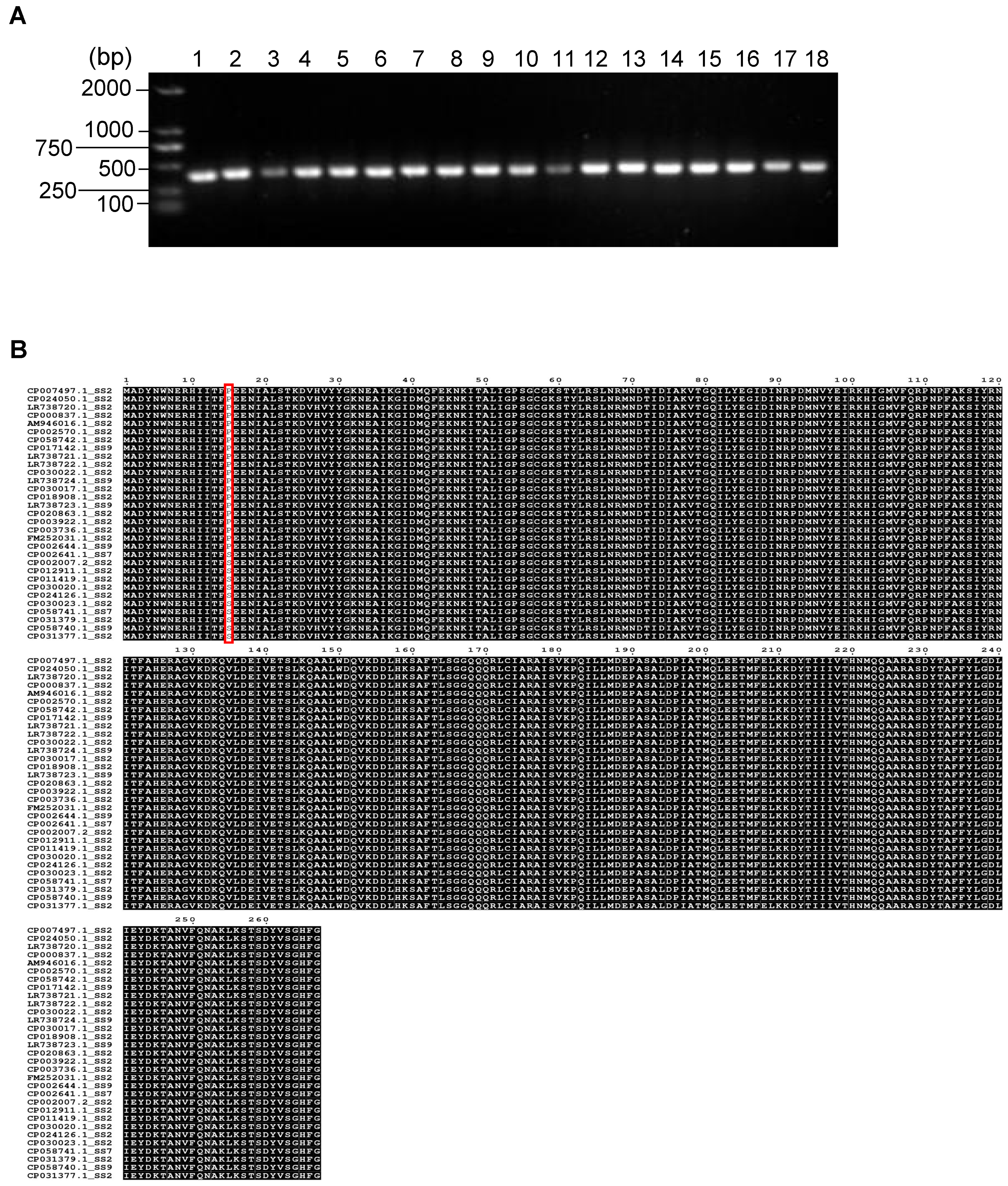
Figure 1. Sequence alignment and PCR screening of PstB in different S. suis strains. (A) The PCR results of pstB gene screening in 18 S. suis isolates. Lane 1, Serotype 2. Lane 2, Serotype 7. Lane 3, Serotype 9. Lane 4–18, S. suis strains randomly selected from clinical isolates. (B) Amino acid sequence alignment of PstB from S. suis strains collected from the GenBank database. The amino acid mutation position is marked with a red box.
Vetsci 10 00048 g001
Figure 2. Identification of linear B cell epitopes of PstB. (A) Prediction of linear B cell epitopes of PstB using ABCpred and IEDB. The epitope sequences identified by both software tools are boxed. (B) Confirmation of linear B cell epitope peptides (EP) with ELISA immunoassay. The OD450 nm value of each peptide was normalized to the control value and plotted. The relative OD450 nm value of >2.1 was considered positive.
Vetsci 10 00048 g002
Figure 3. Expression and purification of rPstB and rPstB epitopes. (A) Designation of the rPstB epitopes. The EP1 was tandemly strung with a linker of GGGG for expression in E. coli. (B) SDS-PAGE analysis of recombinant proteins. The rPstB and rPstB-epitopes were expressed in E. coli and purified using a Ni–NTA column. Lane 1, protein marker. Lane 2, uninduced bacterial cells. Lane 3, bacterial cells induced with isopropyl-β-D-thiogalactopyranoside (IPTG). Lane 4, purified recombinant proteins. (C) Western blot analysis of rPstB and rPstB-epitopes. The purified rPstB and rPstB-epitopes were detected by with anti-His antibodies and rabbit sera specific to S. suis serotype 2.
Vetsci 10 00048 g003
Figure 4. Antibodies and cytokines in immunized mice. (A) The levels of antibodies specific to rPstB or rPstB epitopes in sera collected before primary immunization (0 days post-primary immunization), after booster immunization (14 days post-primary immunization), and before challenge (24 days post-primary immunization). The levels of IL-4 and IFN-γ in sera (B), lungs (C), and spleens (D) collected at 24 days post-primary immunization were examined by ELISA. Statistically significant differences compared with the PBS control group are indicated by asterisks (* p < 0.05; ** p < 0.01; *** p < 0.001).
Vetsci 10 00048 g004
Figure 5. Survival curve of mice immunized with rPstB and rPstB-epitopes. The immunized mice were challenged with a lethal dose of S. suis serotype 2 (ZY05719), 7 (SH04815) or 9 (SH26) strains, respectively, after 10 days post-booster immunization. Death was observed between 12–48 h post-challenge and survival curves were plotted.
Vetsci 10 00048 g005
Table 1. Predicted linear B cell epitopes of PstB.
No.PositionAmino Acid SequenceLengthRecognized by
Positive Sera
EP133–47NEAIKGIDMQFEKNK15Yes
EP258–73GKSTYLRSLNRMNDTID17No
EP388–104DINRPDMNVYEIRKHGM17No
Disclaimer/Publisher’s Note: The statements, opinions and data contained in all publications are solely those of the individual author(s) and contributor(s) and not of MDPI and/or the editor(s). MDPI and/or the editor(s) disclaim responsibility for any injury to people or property resulting from any ideas, methods, instructions or products referred to in the content.