
Expression of Genes Associated with Epithelial to Mesenchymal Transition in MCF-7 Breast Cancer Cells Treated with Monocarbonyl Analogs of Curcumin C66 and B2BrBC—RT-qPCR Array Dataset

 ,
,  , , , , and
Abstract
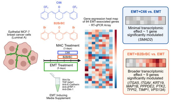
1. Summary
1.1. Background and Rationale
1.2. Scope, Aims, and Objectives
2. Data Description
3. Methods
3.1. Materials
3.2. Experimental Design
3.3. RNA Extraction
3.4. RNA Quantification and Quality Control
3.5. cDNA Synthesis
3.6. Quantitative RT2 Profiler PCR Array
3.7. Data Analysis
4. Limitations
Author Contributions
Funding
Institutional Review Board Statement
Informed Consent Statement
Data Availability Statement
Conflicts of Interest
Abbreviations
| MACs | Monocarbonyl Analogs of Curcumin |
| EMT | Epithelial-to-Mesenchymal Transition |
| C66 | ((2E,6E)-2,6-bis[(2-trifluoromethyl)benzylidene]cyclohexanone) |
| B2BrBC | ((2E,6E)-2,6-bis(2-bromobenzylidene)cyclohexanone) |
| DMSO | Dimethyl Sulfoxide |
| PBS | Phosphate-Buffered Saline |
| Ct | Cycle threshold |
| RNA | Ribonucleic Acid |
| cDNA | Complementary Deoxyribonucleic Acid |
| RT-qPCR | Reverse Transcription–Quantitative Polymerase Chain Reaction |
References
- Sazdova, I.; Keremidarska-Markova, M.; Dimitrova, D.; Mitrokhin, V.; Kamkin, A.; Hadzi-Petrushev, N.; Bogdanov, J.; Schubert, R.; Gagov, H.; Avtanski, D.; et al. Anticarcinogenic Potency of EF24: An Overview of Its Pharmacokinetics, Efficacy, Mechanism of Action, and Nanoformulation for Drug Delivery. Cancers 2023, 15, 5478. [Google Scholar] [CrossRef]
- Stojchevski, R.; Velichkovikj, S.; Bogdanov, J.; Hadzi-Petrushev, N.; Mladenov, M.; Poretsky, L.; Avtanski, D. Monocarbonyl Analogs of Curcumin C66 and B2BrBC Modulate Oxidative Stress, JNK Activity, and Pancreatic Gene Expression in Rats with Streptozotocin-Induced Diabetes. Biochem. Pharmacol. 2024, 229, 116491. [Google Scholar] [CrossRef]
- Stojchevski, R.; Hadzi-Petrushev, N.; Mladenov, M.; Bogdanov, J.; Velichkovikj, S.; Poretsky, L.; Avtanski, D.B. THU557 Effects of Two Experimental Monocarbonyl Analogs of Curcumin (MACs) on Breast Cancer Growth, Migration, and Epithelial-To-Mesenchymal Transition (EMT). J. Endocr. Soc. 2023, 7, bvad114.2183. [Google Scholar] [CrossRef]
- Stojchevski, R.; Sutanto, E.; Dragarska, K.; Todorovska, I.; Velichkovikj, S.; Hadzi-Petrushev, N.; Bogdanov, J.; Mladenov, M.; Poretsky, L.; Avtanski, D.B. SAT-307 Drug-like ADME Profiles and Antimetastatic Properties of Monocarbonyl Curcumin Analogs C66 and B2BrBC with Improved Stability. J. Endocr. Soc. 2025, 9, bvaf149.2449. [Google Scholar] [CrossRef]
- Brown, M.S.; Muller, K.E.; Pattabiraman, D.R. Quantifying the Epithelial-to-Mesenchymal Transition (EMT) from Bench to Bedside. Cancers 2022, 14, 1138. [Google Scholar] [CrossRef] [PubMed]
- Błaszczak, E.; Miziak, P.; Odrzywolski, A.; Baran, M.; Gumbarewicz, E.; Stepulak, A. Triple-Negative Breast Cancer Progression and Drug Resistance in the Context of Epithelial–Mesenchymal Transition. Cancers 2025, 17, 228. [Google Scholar] [CrossRef] [PubMed]
- Liu, D.-K.; Dong, H.-F.; Liu, R.-F.; Xiao, X.-L. Baicalin Inhibits the TGF-Β1/p-Smad3 Pathway to Suppress Epithelial-Mesenchymal Transition-Induced Metastasis in Breast Cancer. Oncotarget 2020, 11, 2863–2872. [Google Scholar] [CrossRef] [PubMed]
- Stojchevski, R.; Velichkovikj, S.; Bogdanov, J.; Dragarska, K.; Todorovska, I.; Hadzi-Petrushev, N.; Mladenov, M.; Poretsky, L.; Avtanski, D. Assessment of the Anticancer and Antimetastatic Effects of Monocarbonyl Analogs of Curcumin, C66 and B2BrBC, in Breast Cancer Cells. Cancer Cell Int. 2026, 26, 90. [Google Scholar] [CrossRef] [PubMed]
- Stojchevski, R.; Velichkovikj, S.; Bogdanov, J.; Dragarska, K.; Todorovska, I.; Hadzi-Petrushev, N.; Mladenov, M.; Poretsky, L.; Avtanski, D. EMT-Related Gene Expression in MCF-7 Cells Treated with Monocarbonyl Analogs of Curcumin C66 and B2BrBC Following EMT Induction—RT-QPCR Array Dataset. Mendeley Data. 2026. Available online: https://data.mendeley.com/datasets/zn8rpyh9v4/2 (accessed on 17 May 2026).
- Qiagen GeneGlobe Data Analysis Center. Available online: https://geneglobe.qiagen.com/us/analyze (accessed on 21 January 2026).



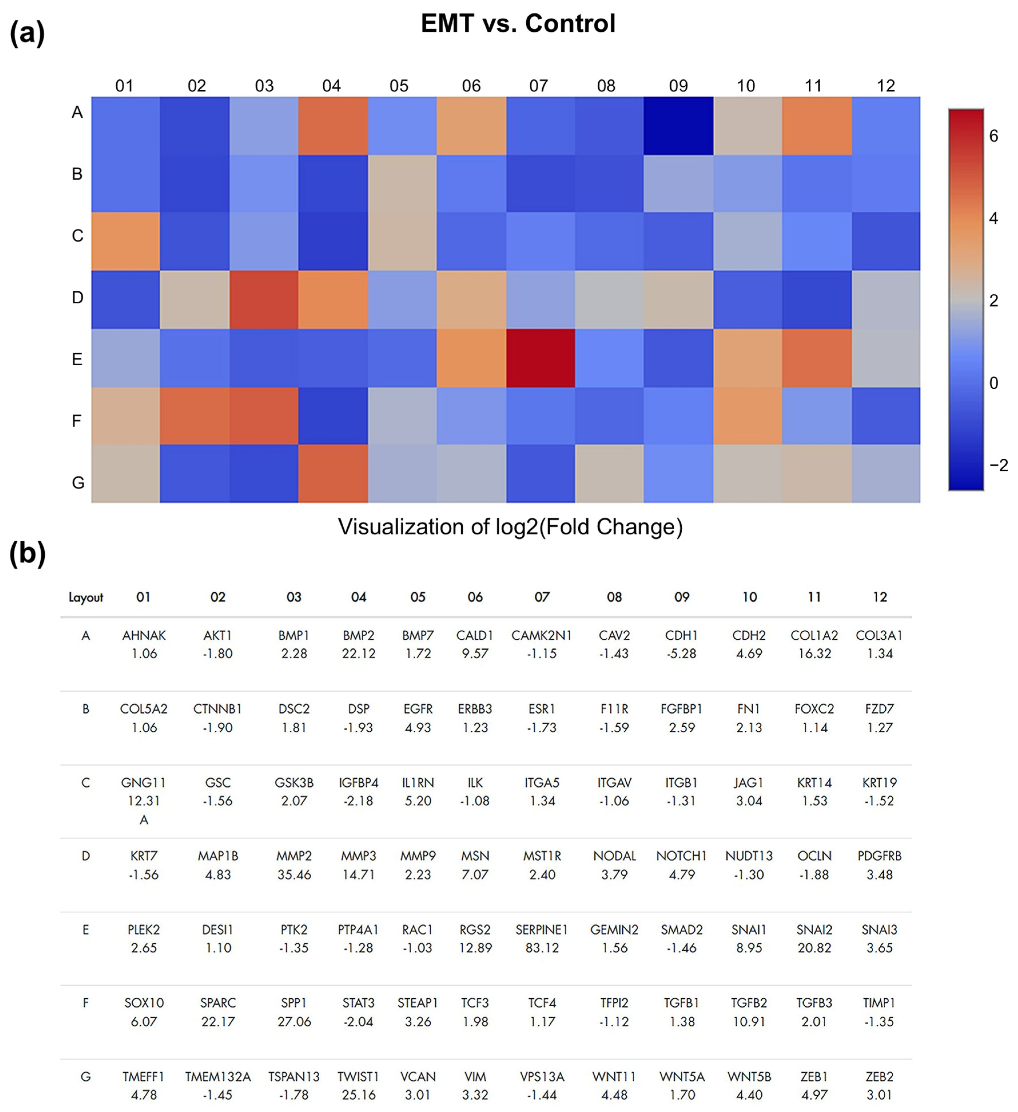
| Gene Symbol | Fold Regulation | Fold Change | p-Value | Significance |
|---|---|---|---|---|
| AHNAK | 1.06 | 1.06 | 0.8576 | ns |
| AKT1 | −1.80 | 0.55 | 0.0852 | ns |
| BMP1 | 2.28 | 2.28 | 0.1856 | ns |
| BMP2 | 22.12 | 22.12 | 0.3539 | ns |
| BMP7 | 1.72 | 1.72 | 0.1557 | ns |
| CALD1 | 9.57 | 9.57 | 0.3575 | ns |
| CAMK2N1 | −1.15 | 0.87 | 0.5695 | ns |
| CAV2 | −1.43 | 0.70 | 0.4896 | ns |
| CDH1 | −5.28 | 0.19 | 0.0057 | ** |
| CDH2 | 4.69 | 4.69 | 0.3745 | ns |
| COL1A2 | 16.32 | 16.32 | 0.3615 | ns |
| COL3A1 | 1.34 | 1.34 | 0.4603 | ns |
| COL5A2 | 1.06 | 1.06 | 0.5048 | ns |
| CTNNB1 | −1.90 | 0.53 | 0.0198 | * |
| DSC2 | 1.81 | 1.81 | 0.3756 | ns |
| DSP | −1.93 | 0.52 | 0.0812 | ns |
| EGFR | 4.93 | 4.93 | 0.3141 | ns |
| ERBB3 | 1.23 | 1.23 | 0.4232 | ns |
| ESR1 | −1.73 | 0.58 | 0.2850 | ns |
| F11R | −1.59 | 0.63 | 0.4123 | ns |
| FGFBP1 | 2.59 | 2.59 | 0.3787 | ns |
| FN1 | 2.13 | 2.13 | 0.2089 | ns |
| FOXC2 | 1.14 | 1.14 | 0.4957 | ns |
| FZD7 | 1.27 | 1.27 | 0.4761 | ns |
| GNG11 | 12.31 | 12.31 | 0.3595 | ns |
| GSC | −1.56 | 0.64 | 0.1755 | ns |
| GSK3B | 2.07 | 2.07 | 0.0001 | **** |
| IGFBP4 | −2.18 | 0.46 | 0.1641 | ns |
| IL1RN | 5.20 | 5.20 | 0.3642 | ns |
| ILK | −1.08 | 0.92 | 0.7568 | ns |
| ITGA5 | 1.34 | 1.34 | 0.4802 | ns |
| ITGAV | −1.06 | 0.94 | 0.9873 | ns |
| ITGB1 | −1.31 | 0.76 | 0.3158 | ns |
| JAG1 | 3.04 | 3.04 | 0.0846 | ns |
| KRT14 | 1.53 | 1.53 | 0.4307 | ns |
| KRT19 | −1.52 | 0.66 | 0.0325 | * |
| KRT7 | −1.56 | 0.64 | 0.2319 | ns |
| MAP1B | 4.83 | 4.83 | 0.2354 | ns |
| MMP2 | 35.46 | 35.46 | 0.3626 | ns |
| MMP3 | 14.71 | 14.71 | 0.3572 | ns |
| MMP9 | 2.23 | 2.23 | 0.3753 | ns |
| MSN | 7.07 | 7.07 | 0.3617 | ns |
| MST1R | 2.40 | 2.40 | 0.3853 | ns |
| NODAL | 3.79 | 3.79 | 0.3732 | ns |
| NOTCH1 | 4.79 | 4.79 | 0.0162 | * |
| NUDT13 | −1.30 | 0.77 | 0.3397 | ns |
| OCLN | −1.88 | 0.53 | 0.0056 | ** |
| PDGFRB | 3.48 | 3.48 | 0.3771 | ns |
| PLEK2 | 2.65 | 2.65 | 0.3199 | ns |
| PPPDE2 | 1.10 | 1.10 | 0.6134 | ns |
| PTK2 | −1.35 | 0.74 | 0.3887 | ns |
| PTP4A1 | −1.28 | 0.78 | 0.3534 | ns |
| RAC1 | −1.03 | 0.97 | 0.7411 | ns |
| RGS2 | 12.89 | 12.89 | 0.2174 | ns |
| SERPINE1 | 83.12 | 83.12 | 0.1003 | ns |
| SIP1 | 1.56 | 1.56 | 0.3275 | ns |
| SMAD2 | −1.46 | 0.68 | 0.0063 | ** |
| SNAI1 | 8.95 | 8.95 | 0.0872 | ns |
| SNAI2 | 20.82 | 20.82 | 0.1840 | ns |
| SNAI3 | 3.65 | 3.65 | 0.3356 | ns |
| SOX10 | 6.07 | 6.07 | 0.3583 | ns |
| SPARC | 22.17 | 22.17 | 0.3586 | ns |
| SPP1 | 27.06 | 27.06 | 0.3586 | ns |
| STAT3 | −2.04 | 0.49 | 0.0554 | ns |
| STEAP1 | 3.26 | 3.26 | 0.3588 | ns |
| TCF3 | 1.98 | 1.98 | 0.2513 | ns |
| TCF4 | 1.17 | 1.17 | 0.5018 | ns |
| TFPI2 | −1.12 | 0.89 | 0.7700 | ns |
| TGFB1 | 1.38 | 1.38 | 0.1791 | ns |
| TGFB2 | 10.91 | 10.91 | 0.2963 | ns |
| TGFB3 | 2.01 | 2.01 | 0.3591 | ns |
| TIMP1 | −1.35 | 0.74 | 0.0073 | ** |
| TMEFF1 | 4.78 | 4.78 | 0.0147 | * |
| TMEM132A | −1.45 | 0.69 | 0.3363 | ns |
| TSPAN13 | −1.78 | 0.56 | 0.0005 | *** |
| TWIST1 | 25.16 | 25.16 | 0.3391 | ns |
| VCAN | 3.01 | 3.01 | 0.3816 | ns |
| VIM | 3.32 | 3.32 | 0.3030 | ns |
| VPS13A | −1.44 | 0.69 | 0.3412 | ns |
| WNT11 | 4.48 | 4.48 | 0.2793 | ns |
| WNT5A | 1.70 | 1.70 | 0.2516 | ns |
| WNT5B | 4.40 | 4.40 | 0.2849 | ns |
| ZEB1 | 4.97 | 4.97 | 0.3683 | ns |
| ZEB2 | 3.01 | 3.01 | 0.3767 | ns |
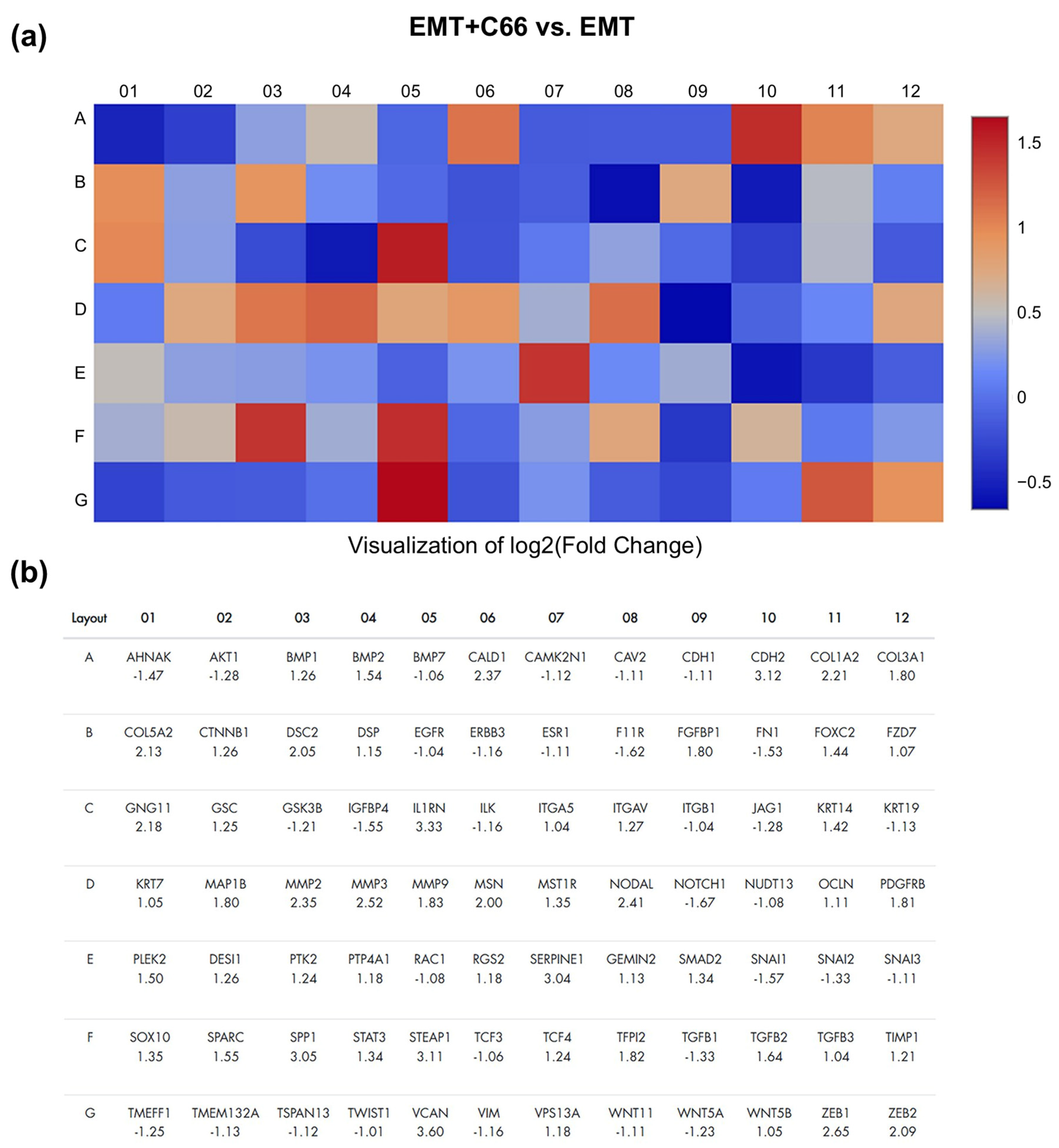
| Gene Symbol | Fold Regulation | Fold Change | p-Value | Significance |
|---|---|---|---|---|
| AHNAK | −1.47 | 0.68 | 0.6327 | ns |
| AKT1 | −1.28 | 0.78 | 0.4814 | ns |
| BMP1 | 1.26 | 1.26 | 0.7370 | ns |
| BMP2 | 1.54 | 1.54 | 0.6570 | ns |
| BMP7 | −1.06 | 0.95 | 0.9637 | ns |
| CALD1 | 2.37 | 2.37 | 0.8817 | ns |
| CAMK2N1 | −1.12 | 0.90 | 0.8106 | ns |
| CAV2 | −1.11 | 0.90 | 0.7174 | ns |
| CDH1 | −1.11 | 0.90 | 0.6614 | ns |
| CDH2 | 3.12 | 3.12 | 0.8044 | ns |
| COL1A2 | 2.21 | 2.21 | 0.9410 | ns |
| COL3A1 | 1.80 | 1.80 | 0.8155 | ns |
| COL5A2 | 2.13 | 2.13 | 0.8730 | ns |
| CTNNB1 | 1.26 | 1.26 | 0.4807 | ns |
| DSC2 | 2.05 | 2.05 | 0.5331 | ns |
| DSP | 1.15 | 1.15 | 0.8258 | ns |
| EGFR | −1.04 | 0.96 | 0.8817 | ns |
| ERBB3 | −1.16 | 0.86 | 0.8595 | ns |
| ESR1 | −1.11 | 0.90 | 0.8632 | ns |
| F11R | −1.62 | 0.62 | 0.2545 | ns |
| FGFBP1 | 1.80 | 1.80 | 0.9261 | ns |
| FN1 | −1.53 | 0.65 | 0.5221 | ns |
| FOXC2 | 1.44 | 1.44 | 0.8787 | ns |
| FZD7 | 1.07 | 1.07 | 0.9870 | ns |
| GNG11 | 2.18 | 2.18 | 0.9295 | ns |
| GSC | 1.25 | 1.25 | 0.5380 | ns |
| GSK3B | −1.21 | 0.83 | 0.5408 | ns |
| IGFBP4 | −1.55 | 0.65 | 0.2983 | ns |
| IL1RN | 3.33 | 3.33 | 0.8836 | ns |
| ILK | −1.16 | 0.86 | 0.4519 | ns |
| ITGA5 | 1.04 | 1.04 | 0.9171 | ns |
| ITGAV | 1.27 | 1.27 | 0.5315 | ns |
| ITGB1 | −1.04 | 0.96 | 0.8324 | ns |
| JAG1 | −1.28 | 0.78 | 0.6642 | ns |
| KRT14 | 1.42 | 1.42 | 0.9099 | ns |
| KRT19 | −1.13 | 0.89 | 0.8666 | ns |
| KRT7 | 1.05 | 1.05 | 0.9588 | ns |
| MAP1B | 1.80 | 1.80 | 0.6635 | ns |
| MMP2 | 2.35 | 2.35 | 0.9653 | ns |
| MMP3 | 2.52 | 2.52 | 0.9135 | ns |
| MMP9 | 1.83 | 1.83 | 0.8243 | ns |
| MSN | 2.00 | 2.00 | 0.9943 | ns |
| MST1R | 1.35 | 1.35 | 0.8274 | ns |
| NODAL | 2.41 | 2.41 | 0.8567 | ns |
| NOTCH1 | −1.67 | 0.60 | 0.2964 | ns |
| NUDT13 | −1.08 | 0.93 | 0.6407 | ns |
| OCLN | 1.11 | 1.11 | 0.5386 | ns |
| PDGFRB | 1.81 | 1.81 | 0.8965 | ns |
| PLEK2 | 1.50 | 1.50 | 0.8772 | ns |
| PPPDE2 | 1.26 | 1.26 | 0.6253 | ns |
| PTK2 | 1.24 | 1.24 | 0.5494 | ns |
| PTP4A1 | 1.18 | 1.18 | 0.4682 | ns |
| RAC1 | −1.08 | 0.92 | 0.5616 | ns |
| RGS2 | 1.18 | 1.18 | 0.9894 | ns |
| SERPINE1 | 3.04 | 3.04 | 0.1949 | ns |
| SIP1 | 1.13 | 1.13 | 0.8783 | ns |
| SMAD2 | 1.34 | 1.34 | 0.0320 | * |
| SNAI1 | −1.57 | 0.63 | 0.6722 | ns |
| SNAI2 | −1.33 | 0.75 | 0.7115 | ns |
| SNAI3 | −1.11 | 0.90 | 0.9671 | ns |
| SOX10 | 1.35 | 1.35 | 0.7800 | ns |
| SPARC | 1.55 | 1.55 | 0.8652 | ns |
| SPP1 | 3.05 | 3.05 | 0.8769 | ns |
| STAT3 | 1.34 | 1.34 | 0.4180 | ns |
| STEAP1 | 3.11 | 3.11 | 0.6782 | ns |
| TCF3 | −1.06 | 0.95 | 0.7917 | ns |
| TCF4 | 1.24 | 1.24 | 0.9708 | ns |
| TFPI2 | 1.82 | 1.82 | 0.5579 | ns |
| TGFB1 | −1.33 | 0.75 | 0.1690 | ns |
| TGFB2 | 1.64 | 1.64 | 0.9271 | ns |
| TGFB3 | 1.04 | 1.04 | 0.9694 | ns |
| TIMP1 | 1.21 | 1.21 | 0.0780 | ns |
| TMEFF1 | −1.25 | 0.80 | 0.6229 | ns |
| TMEM132A | −1.13 | 0.89 | 0.7289 | ns |
| TSPAN13 | −1.12 | 0.90 | 0.5709 | ns |
| TWIST1 | −1.01 | 0.99 | 0.8922 | ns |
| VCAN | 3.60 | 3.60 | 0.7976 | ns |
| VIM | −1.16 | 0.86 | 0.9312 | ns |
| VPS13A | 1.18 | 1.18 | 0.8261 | ns |
| WNT11 | −1.11 | 0.90 | 0.8706 | ns |
| WNT5A | −1.23 | 0.81 | 0.8925 | ns |
| WNT5B | 1.05 | 1.05 | 0.9676 | ns |
| ZEB1 | 2.65 | 2.65 | 0.9846 | ns |
| ZEB2 | 2.09 | 2.09 | 0.9057 | ns |
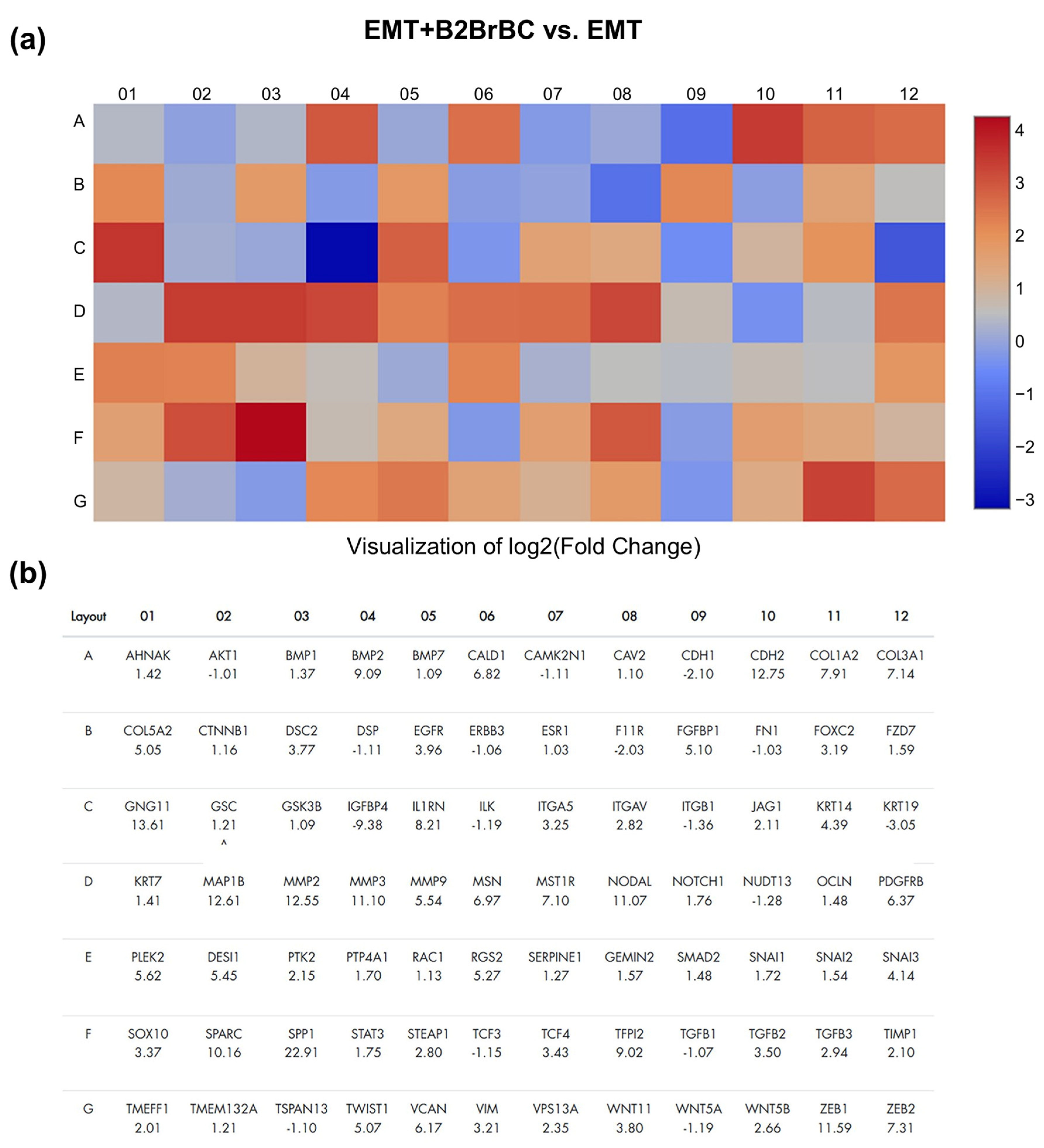
| Gene Symbol | Fold Regulation | Fold Change | p-Value | Significance |
|---|---|---|---|---|
| AHNAK | 1.42 | 1.42 | 0.3682 | ns |
| AKT1 | −1.01 | 0.99 | 0.7589 | ns |
| BMP1 | 1.37 | 1.37 | 0.4372 | ns |
| BMP2 | 9.09 | 9.09 | 0.3455 | ns |
| BMP7 | 1.09 | 1.09 | 0.5505 | ns |
| CALD1 | 6.82 | 6.82 | 0.3162 | ns |
| CAMK2N1 | −1.11 | 0.90 | 0.5801 | ns |
| CAV2 | 1.10 | 1.10 | 0.9019 | ns |
| CDH1 | −2.10 | 0.48 | 0.2298 | ns |
| CDH2 | 12.75 | 12.75 | 0.2792 | ns |
| COL1A2 | 7.91 | 7.91 | 0.3349 | ns |
| COL3A1 | 7.14 | 7.14 | 0.1721 | ns |
| COL5A2 | 5.05 | 5.05 | 0.2761 | ns |
| CTNNB1 | 1.16 | 1.16 | 0.5810 | ns |
| DSC2 | 3.77 | 3.77 | 0.1895 | ns |
| DSP | −1.11 | 0.90 | 0.8669 | ns |
| EGFR | 3.96 | 3.96 | 0.2732 | ns |
| ERBB3 | −1.06 | 0.94 | 0.9219 | ns |
| ESR1 | 1.03 | 1.03 | 0.7765 | ns |
| F11R | −2.03 | 0.49 | 0.1831 | ns |
| FGFBP1 | 5.10 | 5.10 | 0.2991 | ns |
| FN1 | −1.03 | 0.97 | 0.6603 | ns |
| FOXC2 | 3.19 | 3.19 | 0.3804 | ns |
| FZD7 | 1.59 | 1.59 | 0.3917 | ns |
| GNG11 | 13.61 | 13.61 | 0.2704 | ns |
| GSC | 1.21 | 1.21 | 0.4862 | ns |
| GSK3B | 1.09 | 1.09 | 0.4016 | ns |
| IGFBP4 | −9.38 | 0.11 | 0.0702 | ns |
| IL1RN | 8.21 | 8.21 | 0.3060 | ns |
| ILK | −1.19 | 0.84 | 0.5006 | ns |
| ITGA5 | 3.25 | 3.25 | 0.0240 | * |
| ITGAV | 2.82 | 2.82 | 0.0044 | ** |
| ITGB1 | −1.36 | 0.74 | 0.2466 | ns |
| JAG1 | 2.11 | 2.11 | 0.2297 | ns |
| KRT14 | 4.39 | 4.39 | 0.3209 | ns |
| KRT19 | −3.05 | 0.33 | 0.0106 | * |
| KRT7 | 1.41 | 1.41 | 0.3650 | ns |
| MAP1B | 12.61 | 12.61 | 0.0064 | ** |
| MMP2 | 12.55 | 12.55 | 0.3182 | ns |
| MMP3 | 11.10 | 11.10 | 0.2638 | ns |
| MMP9 | 5.54 | 5.54 | 0.1943 | ns |
| MSN | 6.97 | 6.97 | 0.3459 | ns |
| MST1R | 7.10 | 7.10 | 0.2724 | ns |
| NODAL | 11.07 | 11.07 | 0.2393 | ns |
| NOTCH1 | 1.76 | 1.76 | 0.0671 | ns |
| NUDT13 | −1.28 | 0.78 | 0.3323 | ns |
| OCLN | 1.48 | 1.48 | 0.0909 | ns |
| PDGFRB | 6.37 | 6.37 | 0.3969 | ns |
| PLEK2 | 5.62 | 5.62 | 0.0992 | ns |
| PPPDE2 | 5.45 | 5.45 | 0.0004 | *** |
| PTK2 | 2.15 | 2.15 | 0.0396 | * |
| PTP4A1 | 1.70 | 1.70 | 0.0715 | ns |
| RAC1 | 1.13 | 1.13 | 0.1019 | ns |
| RGS2 | 5.27 | 5.27 | 0.1012 | ns |
| SERPINE1 | 1.27 | 1.27 | 0.5273 | ns |
| SIP1 | 1.57 | 1.57 | 0.3798 | ns |
| SMAD2 | 1.48 | 1.48 | 0.0786 | ns |
| SNAI1 | 1.72 | 1.72 | 0.3531 | ns |
| SNAI2 | 1.54 | 1.54 | 0.5394 | ns |
| SNAI3 | 4.14 | 4.14 | 0.2584 | ns |
| SOX10 | 3.37 | 3.37 | 0.6731 | ns |
| SPARC | 10.16 | 10.16 | 0.3316 | ns |
| SPP1 | 22.91 | 22.91 | 0.1439 | ns |
| STAT3 | 1.75 | 1.75 | 0.0977 | ns |
| STEAP1 | 2.80 | 2.80 | 0.5374 | ns |
| TCF3 | −1.15 | 0.87 | 0.6873 | ns |
| TCF4 | 3.43 | 3.43 | 0.3132 | ns |
| TFPI2 | 9.02 | 9.02 | 0.0067 | ** |
| TGFB1 | −1.07 | 0.93 | 0.8974 | ns |
| TGFB2 | 3.50 | 3.50 | 0.2845 | ns |
| TGFB3 | 2.94 | 2.94 | 0.2623 | ns |
| TIMP1 | 2.10 | 2.10 | 0.0119 | * |
| TMEFF1 | 2.01 | 2.01 | 0.0694 | ns |
| TMEM132A | 1.21 | 1.21 | 0.6962 | ns |
| TSPAN13 | −1.10 | 0.91 | 0.3211 | ns |
| TWIST1 | 5.07 | 5.07 | 0.4240 | ns |
| VCAN | 6.17 | 6.17 | 0.2604 | ns |
| VIM | 3.21 | 3.21 | 0.2349 | ns |
| VPS13A | 2.35 | 2.35 | 0.0244 | * |
| WNT11 | 3.80 | 3.80 | 0.2421 | ns |
| WNT5A | −1.19 | 0.84 | 0.8780 | ns |
| WNT5B | 2.66 | 2.66 | 0.3527 | ns |
| ZEB1 | 11.59 | 11.59 | 0.2470 | ns |
| ZEB2 | 7.31 | 7.31 | 0.3052 | ns |
| Material/Equipment | Company/Source | Catalog # |
|---|---|---|
| MCF-7 cells | ATCC (Manassas, VA, USA) | HTB-22 |
| DMEM/F12 (50:50) | ATCC (Manassas, VA, USA) | 10-090-CM |
| Antibiotic/Antimycotic Mixture | Corning Life Sciences (Corning, NY, USA) | 30-004-Cl |
| Tissue Culture 6-Well Plates | Corning Life Sciences (Corning, NY, USA) | 07-200-83 |
| StemXVivo EMT Inducing Media Supplement * | Bio-Techne, (Minneapolis, MN, USA) | CCCM017 |
| C66 ((2E,6E)-2,6-bis[(2-trifluoromethyl)benzylidene]cyclohexanone) | Institute of Chemistry, Ss. Cyril and Methodius University in Skopje, North Macedonia | N/A (self-synthesized) |
| B2BrBC ((2E,6E)-2,6-bis(2-bromobenzylidene)cyclohexanone) | Institute of Chemistry, Ss. Cyril and Methodius University in Skopje, North Macedonia | N/A (self-synthesized) |
| TRIzol Reagent | Invitrogen, Thermo Fisher Scientific (Waltham, MA, USA) | 15596026 |
| qScript cDNA SuperMix | Quantabio (Beverly, MA, USA) | 95048 |
| PerfeCTa SYBR Green FastMix | Quantabio (Beverly, MA, USA) | 95072 |
| RT2 Profiler PCR Array Human Epithelial to Mesenchymal Transition (EMT) | QIAGEN (Hilden, Germany) | 330231, PAHS-090ZA |
| NanoDrop One Spectrophotometer | Thermo Fisher Scientific (Wilmington, DE, USA) | ND-ONE-W |
| SimpliAmp Thermal Cycler | Applied Biosystems, Thermo Fisher Scientific (Wilmington, DE, USA) | A24811 |
| QuantStudio 3 Real-Time PCR System | Applied Biosystems, Thermo Fisher Scientific (Wilmington, DE, USA) | A28567 |
| QuantStudio Design & Analysis Software (v1.5.1) | Applied Biosystems, Thermo Fisher Scientific (Wilmington, DE, USA) | N/A |
Disclaimer/Publisher’s Note: The statements, opinions and data contained in all publications are solely those of the individual author(s) and contributor(s) and not of MDPI and/or the editor(s). MDPI and/or the editor(s) disclaim responsibility for any injury to people or property resulting from any ideas, methods, instructions or products referred to in the content. |
© 2026 by the authors. Licensee MDPI, Basel, Switzerland. This article is an open access article distributed under the terms and conditions of the Creative Commons Attribution (CC BY) license.
Share and Cite
Stojchevski, R.; Velichkovikj, S.; Bogdanov, J.; Dragarska, K.; Todorovska, I.; Hadzi-Petrushev, N.; Mladenov, M.; Poretsky, L.; Avtanski, D. Expression of Genes Associated with Epithelial to Mesenchymal Transition in MCF-7 Breast Cancer Cells Treated with Monocarbonyl Analogs of Curcumin C66 and B2BrBC—RT-qPCR Array Dataset. Data 2026, 11, 125. https://doi.org/10.3390/data11050125
Stojchevski R, Velichkovikj S, Bogdanov J, Dragarska K, Todorovska I, Hadzi-Petrushev N, Mladenov M, Poretsky L, Avtanski D. Expression of Genes Associated with Epithelial to Mesenchymal Transition in MCF-7 Breast Cancer Cells Treated with Monocarbonyl Analogs of Curcumin C66 and B2BrBC—RT-qPCR Array Dataset. Data. 2026; 11(5):125. https://doi.org/10.3390/data11050125
Chicago/Turabian StyleStojchevski, Radoslav, Sara Velichkovikj, Jane Bogdanov, Katerina Dragarska, Ivana Todorovska, Nikola Hadzi-Petrushev, Mitko Mladenov, Leonid Poretsky, and Dimiter Avtanski. 2026. "Expression of Genes Associated with Epithelial to Mesenchymal Transition in MCF-7 Breast Cancer Cells Treated with Monocarbonyl Analogs of Curcumin C66 and B2BrBC—RT-qPCR Array Dataset" Data 11, no. 5: 125. https://doi.org/10.3390/data11050125
APA StyleStojchevski, R., Velichkovikj, S., Bogdanov, J., Dragarska, K., Todorovska, I., Hadzi-Petrushev, N., Mladenov, M., Poretsky, L., & Avtanski, D. (2026). Expression of Genes Associated with Epithelial to Mesenchymal Transition in MCF-7 Breast Cancer Cells Treated with Monocarbonyl Analogs of Curcumin C66 and B2BrBC—RT-qPCR Array Dataset. Data, 11(5), 125. https://doi.org/10.3390/data11050125