Article

Transcriptomic Response of L. monocytogenes to Co-Culture with S. cerevisiae

by
Spiros Paramithiotis
*,
Alexandra Katidi
and
Eleftherios H. Drosinos
Laboratory of Food Quality Control and Hygiene, Department of Food Science and Human Nutrition, Agricultural University of Athens, Iera Odos 75, 11855 Athens, Greece
*
Author to whom correspondence should be addressed.
Beverages 2021, 7(3), 55; https://doi.org/10.3390/beverages7030055
Submission received: 16 June 2021 / Revised: 28 July 2021 / Accepted: 2 August 2021 / Published: 5 August 2021
(This article belongs to the Special Issue Fermentation Process and Microbial Safety of Beverages)

Abstract

:
The aim of the present study was to assess the transcriptomic response of L. monocytogenes during co-culture with three S. cerevisiae strains. For this purpose, BHI broth was inoculated with 7 log CFU·mL−1 L. monocytogenes serotype 4b strain LQC 15257, isolated from a strawberry sample and 4 log CFU·mL−1 S. cerevisiae strains Y32, Y34 and Y37, isolated from spontaneous olive fermentation. Sampling took place after 24 and 48 h incubation at 5 and 20 °C. RNA was extracted, stabilized and the transcription of virulence associated genes prfA, sigB, hly, plcA, plcB, inlA, inlB, inlC and inlJ, was assessed by RT-qPCR. Co-culture with the yeast strains mostly affected the transcription of sigB and inlJ, the upregulation of which during growth at 5 °C for 24 h, reached 10.13 and 9.76 log2(fold change), respectively. Similarly, the effect that incubation time had on the relative transcription of the genes under study was dependent on the co-cultivating yeast strain. On the other hand, the effect of the yeast strain was less pronounced when the relative transcription of the genes under study was assessed between 20 °C and 5 °C. In that case, incubation temperature seemed to have an important effect since, in the 79.2% of the samples analyzed, upregulation was evident, irrespective of yeast strain presence. These results highlight the complex trophic relationships that take place during co-existence between L. monocytogenes and S. cerevisiae.

1. Introduction

Listeriosis is a serious infection that is caused by ingestion of food contaminated with the Gram-positive bacterium Listeria monocytogenes; it is characterized by high hospitalization and mortality rates on annual basis in the U.S. and the E.U. [1,2].
Control of this pathogen currently relies on implementation of good hygiene practices and, if applicable, the use of antimicrobial compounds and procedures; the effectiveness of which, is verified by frequent microbiological testing. However, based on the annual number of incidents and outbreaks, additional strategies that would assist in eliminating this bacterium from the food supply chain are still necessary. Science-based, targeted approaches that may be tailored according to the food matrix, may be enabled through the thorough assessment of the physiology of the pathogen under conditions encountered in food processing or storage.
Currently, research is focused in establishing a link between prior exposure to food-related abiotic and biotic stimuli and virulence potential through the effect on the transcription of virulence-associated genes. L. monocytogenes invasiveness is largely affected by exposure to environmental stresses, such as cold, heat, osmotic and sub-lethal acidic, as well as the type of food, in a strain-dependent manner [3,4,5,6,7,8,9,10,11,12]. The biotic stimuli that the pathogen may encounter refer to the co-existence with other microorganisms. Such co-existence has been reported in fresh produce, dairy and meat products [13,14,15,16,17]. The outcome of such a co-existence depends on the relative physiological and metabolic attributes as well as the environmental conditions, which concomitantly define the relative population dynamics of the members of the microecosystem. The effect that such co-existence may have on the transcription of virulence-associated genes and invasiveness of the pathogen, has been only marginally studied [18,19,20,21,22,23,24,25]; in some cases, a correlation between the transcriptomic response of the pathogen and its invasiveness has been suggested [6,25].
Saccharomyces cerevisiae is a key microorganism for the beverages sector. Apart from the central role in the production of alcoholic beverages, its contribution in the production of fermented milks such as kefir [26,27], Nunu [28] and Lait caillé [29], has been recognized. The ability of L. monocytogenes to survive the manufacturing process of some of the above products, has already been documented [30,31,32]. The aim of the present study was to assess the transcriptomic response of L. monocytogenes during co-culture with three S. cerevisiae strains during growth in BHI broth incubated at 5 °C and 20 °C. More specifically, the relative transcription of the key transcriptional regulators sigB and prfA as well as the virulence associated genes plcA, plcB, hly, inlA, inlB, inlC and inlJ were assessed.

2. Materials and Methods

2.1. Bacterial Strains and Sample Preparation

L. monocytogenes strain LQC 15257, serotype 4b, isolated from a strawberry sample [33,34] and Saccharomyces cerevisiae strains Y32, Y34 and Y37, isolated from fermented olives [35] were used throughout this study. The strains were stored at −20 °C in Nutrient broth (Lab M, Lancashire, UK) supplemented with 50% glycerol (Applichem, Darmstadt, Germany). Before experimental use, the strains were grown twice in Brain Heart Infusion (BHI) broth (Lab M) at 37 °C for 24 h and 25 °C for 3 days, for L. monocytogenes and S. cere-visiae, respectively.
Twenty-four hours culture of the pathogen and the yeast strains, incubated at 37 and 25 °C, respectively, were centrifuged (12,000× g; 15 min; 4 °C), washed twice with sterile saline, resuspended in the same diluent and used to inoculate BHI broth at ~7.00 log CFU/mL and ~4.00 log CFU/mL, respectively. Mono-and co-cultures of the pathogen with each of the yeast strains were incubated at 5 and 20 °C for 48 h.

2.2. Sampling and Microbiological Analyses

Sampling was performed after 24 and 48 h of incubation at 5 and 20 °C. L. monocytogenes and yeast populations were enumerated by plating serial dilutions on the surface of Polymyxin Acriflavine Lithium chloride Ceftazidine Aesculin Mannitol (PALCAM) agar (Lab M) and Rose Bengal Chloramphenicol (RBC) agar (Lab M) and incubation at 37 °C for 48 h and 25 °C for 3 days, respectively.

2.3. Gene Transcription Assay

Samples (10 mL) were centrifuged (12,000× g; 1 min, incubation temperature), the biomass was mixed with 200 μL of RNAlater ® solution (Ambion, Whaltham, MA, USA) and stored at −20 °C until further analysis. Extraction of RNA was performed using the Nucleo-Spin® RNA Kit (Macherey-Nagel, Duren, Germany), synthesis of cDNA took place using random hexamers and the PrimeScriptTM One Step RT-PCR Kit (Takara Bio, Shiga, Japan). For each sample, two reverse transcription reactions containing ca. 0.5 μg RNA each, were performed. Relative transcription of the key transcriptional regulators sigB and prfA, the virulence associated genes plcA, plcB, hly, inlA, inlB, inlC and inlJ, along with the house-keeping genes IGS, 16S-rDNA and rpob that were employed as references, was performed using the primers, PCR and melting curve conditions mentioned in Hadjilouka et al. [36] and presented in Table S1. Real-Time quantitative PCR was performed using KAPA SYBR qPCR Kit Master Mix (2X) for ABI Prism (KapaBiosystems, Boston, MA, USA).

2.4. Statistical Analysis

The Ct values of all genes were processed according to Hadjilouka et al. [37]; the fold changes that were calculated were converted to their log2 values for further processing. NormFinder v0.953 [38] was used to calculate the stability values of the genes employed as references. IGS was the most stably transcribed and used for normalization. In each case, the condition used as control is mentioned. Up- and down-regulation of a gene was only considered when the log2 value of the fold change (log2FC) was below −1 or above 1, respectively, assessed through one-sample t-test (p < 0.05). The Pearson coefficient was used to assess the correlation between the log2FC values of the genes. The differences between the population of the pathogen and the yeast strains in mono-or co-cultures was assessed by one-way analysis of variance (ANOVA). All calculations were performed in Statgraphics Centurion XVII (Statgraphics Technologies, Inc., The Plains, VA, USA).

3. Results

The S. cerevisiae strains employed in the present study exhibited no antilisterial activity, assessed through a well diffusion assay performed according to Syrokou et al. [39]. In Table 1, L. monocytogenes population after incubation in BHI broth, at 5 °C and 20 °C, for 24 and 48 h, in mono-or co-culture with the yeast strains, is presented. L. monocytogenes population after incubation as monoculture for 24 h at 5 °C and 20 °C reached 7.61 and 9.29 log CFU/mL, respectively and no statistically significant change was observed in the population of the pathogen, after 48 h incubation at both temperatures. Co-culture with a yeast strain had no statistically significant effect on the population of the pathogen at both temperatures and times assessed. The population of the yeast strains after incubation as monoculture for 24 h at 5 °C ranged from 4.12 to 4.27 log CFU/mL and at 20 °C from 5.49 to 5.68 log CFU/mL. No statistically significant change was observed in yeast strains population after 48 h incubation at 5 °C. On the contrary, after 48 h incubation at 20 °C, statistically significant increase in the population of all yeast strains was observed. In all cases, yeast population was not affected by the presence of the pathogen at both temperatures and times assessed (Table 1).
Co-culture with yeast strains had no effect on the transcription of the genes under study in the 82.5% of the samples analyzed. In these cases, the relative transcription of the genes, expressed in log2(FC), was between −1 and 1, which in the present study was not considered as regulation (Figure 1; Table 2). This was particularly visible in the case of inlA and inlB, the relative transcription of which was not affected by the presence of any of the three yeast strains examined. In addition, co-culture at 5 °C for 48 h had no effect on relative gene transcription compared to monoculture at the same conditions. However, notable exceptions did occur, the majority of which were observed during growth at 5 °C for 24 h and 20 °C for 48 h. More specifically, co-culture with any of the three yeast strains resulted in upregulation of sigB and inlJ during growth at 5 °C for 24 h. Upregulation of sigB ranged from 2.65 to 10.13 log2(FC) while upregulation of inlJ from 6.49 to 9.76 log2(FC), depending on the yeast strain. In both cases, the most pronounced upregulation was observed during co-culture with yeast strain Y32. Moreover, co-culture with yeast strains Y34 and Y37 resulted in downregulation of prfA, sigB and plcA during growth at 20 °C for 48 h. In addition, co-culture with yeast strains Y32 and Y34 resulted in upregulation of plcB during growth at 5 °C for 24 h. Finally, co-culture with yeast strain Y37 at 5 °C for 24 h resulted in downregulation of inlC. Co-culture with yeast strain Y34 at 20 °C for 24 h resulted in upregulation of inlC and inlJ.
In order to assess the effect of incubation time on the relative gene transcription, the transcript levels of the genes under study after growth at 5 °C for 48 h were compared with the respective after growth at 5 °C for 24 h. Similarly, the transcript levels of the genes under study after growth at 20 °C for 48 h were compared with the respective after growth at 20 °C for 24 h (Figure 2; Table 2). Incubation time seemed to have a rather restricted effect on the transcription of the genes under study, since in the 75% of the samples analyzed, the relative transcription of the genes, expressed in log2(FC), was above −1 and below 1. This was particularly visible in the relative transcription of inlB and inlC, which remained without any statistically significant change. However, notable exceptions were also observed, the majority of which occurred when the transcript levels after growth at 20 °C for 48 h were compared with the respective after 24 h. When L. monocytogenes was grown as monoculture, upregulation of sigB and inlJ was observed when the respective transcript levels after growth at 5 °C for 48 h were compared with the ones after growth for 24 h. Similarly, upregulation of prfA and plcA but downregulation of hly and inlA were observed when the respective transcript levels after growth at 20 °C for 48 h were compared with the ones after growth for 24 h. The effect that co-cultivation had on relative transcription was dependent on the yeast strain. More accurately, co-cultivation with the yeast strain Y32 at 5 °C, resulted in downregulation of sigB and inlJ and upregulation of plcA and plcB. Co-cultivation with yeast strain Y34 for 48 h at 20 °C resulted in upregulation of plcB and downregulation of sigB, inlA and inlJ. Finally, the co-culture with yeast strain Y37 for 48 h at 5 °C resulted in upregulation of sigB, whereas after 48 h growth at 20 °C, upregulation of inlJ and downregulation of sigB and inlA were observed.
In order to assess the effect of incubation temperature on the relative gene transcription, the transcript levels of the genes under study after growth for 24 h at 20 °C were compared with the respective after growth for 24 h at 5 °C. Similarly, the transcript levels of the genes under study after growth for 48 h at 20 °C were compared with the respective after growth for 48 h at 5 °C (Figure 3; Table 2). Incubation temperature seemed to have an important effect on the relative transcription of the genes under study, which was also affected by co-cultivation as well as the respective yeast strain. More specifically, upregulation was observed in the 79.2% of the samples analyzed and no statistically significant change in the remaining 20.8%. When L. monocytogenes was grown as monoculture, upregulation of all genes but prfA and plcA was observed when the respective transcript levels after growth for 24 h at 20 °C were compared with the ones after growth for 24 h at 5 °C. Similarly, upregulation of all genes but hly and inlA was observed when the respective transcript levels after growth for 48 h at 20 °C were compared with the ones after growth for 48 h at 5 °C. Co-cultivation with yeast strain Y32 resulted in upregulation of prfA, hly, inlA, inlB and inlC at both cultivation times and upregulation of plcA, plcB and inlJ after 48 h. Co-cultivation with yeast strain Y34 resulted in upregulation of prfA, sigB, hly, inlA, inlB, inlC and inlJ at both cultivation times and upregulation of plcB after 48 h. Finally, co-culture with yeast strain Y37 resulted in upregulation of prfA, plcB, hly, inlA, inlB, inlC and inlJ at both incubation times and upregulation of sigB after 24 h.
The correlation between the transcriptomic responses of the genes under study were collectively assessed by the Pearson correlation coefficient (Table 3). Out of a total of 36 correlations that were assessed, only in 10 cases, no statistically significant correlation was observed. Moderate correlation (0.30 < r < 0.49) was indicated in 9 cases and strong correlation (0.50 < r < 0.99) in 17 cases. The strongest correlation (r > 0.90) was observed between prfA and inlC.

4. Discussion

The effect that co-culture with another microorganism may have on L. monocytogenes virulence gene transcription and virulence capacity has been assessed by a limited number of studies [18,19,23,24,25,40]. Although strain diversity may be expected to play an important role, this has not yet been experimentally verified. In this study, the effect that co-culture with three different S. cerevisiae strains may have on the relative transcription of L. monocytogenes key virulence genes was assessed.
Co-culture had no effect on the population of the L. monocytogenes or the S. cerevisiae strains at any temperature or time assessed. This may be attributed to the absence of any antagonistic or mutualistic relationship between the strains employed under the specific conditions of the present study. The ability of several food borne yeasts to inhibit growth of L. monocytogenes has been reported, including two S. cerevisiae strains [41,42,43,44]. In addition, the L. monocytogenes anti-biofilm capacity of five S. cerevisiae strains was reported by Kim et al. [45] and attributed to the decrease of auto-aggregation, cell surface hydrophobicity and exopolysaccharide production by the pathogen.
Addressing the relative transcription of the genes under study collectively, only one trend could be identified. This was the relative upregulation of all genes, under mono- or co-culture, which was observed at 20 °C compared to 5 °C. This finding deserves further attention and the physiological basis of this response as well as the effect that such upregulation may have on the invasion efficiency of the pathogen, must be elucidated.
The genes that have a profound effect on homeostasis under stress conditions and virulence of L. monocytogenes are sigB and prfA, respectively [46]. A cross talk between the two regulatory elements σB and PrfA is evident [47], allowing successful colonization of diverse niches. In the present study, a moderate degree of correlation between the transcription of these genes was revealed, possibly indicating that both σB-dependent (P2prfA) and σB-independent (P1prfA) promoters were implicated in the transcription of prfA. The former seems to be the case after 48 h at 20 °C, in which both sigB and prfA were upregulated at comparable levels during co-culture of the pathogen with two of the three yeast strains assessed. On the contrary, P2prfA was not activated after 24 h at 5 °C, in which the upregulation of sigB observed during co-culture of the pathogen with the three yeast strains employed, was not accompanied by upregulation of prfA. Τhe contribution of the bicistronic plcA-prfA mRNA seems unlike since it has been reported to be absent at temperatures below 37 °C [48]. However, regulation of plcA was observed in many cases, which was accompanied by a similar regulation of prfA, with only one exception, in which the high standard deviation of prfA transcription level prevented statistical significance. This was also revealed by the high degree of correlation indicated by the Pearson correlation coefficient. Thus, a possible contribution of this bicitronic message is indicated.
hly possesses a very important role in virulence potential of L. monocytogenes strains; it encodes for listeriolysin O, absence of which results in tissue-dependent avirulence [49]. Two prfA-dependent and one prfA-independent promoter sites control transcription of hly. In the present study, the moderate correlation between prfA and hly, as indicated by Pearson correlation coefficient, suggested that the prfA-independent promoter was also activated. This was particularly evident during co-culture of the pathogen with Y34 and Y37 for 48 h at 20 °C, in which downregulation of prfA was accompanied by upregulation of hly.
Internalins are proteins that contain leucin-rich repeats and facilitate internalization of the pathogen into non-phagocytic cells. Thus, they are necessary for effective invasion, at least as far as InlA, InlB, InlC and InlJ are concerned [49,50,51,52]. The genes encoding for InlA and InlB form the inlAB operon, the transcription of which is controlled by both σB and PrfA. The latter also initiates transcription of inlC, whereas transcription of inlJ is not affected, at least directly, by none of the above. The high degree of correlation between the transcription of prfA, inlA, inlB and inlC, which was evident in the present study, is in accordance with the literature. In addition, the high degree of correlation between the transcription of sigB and inlA and the moderate between sigB and inlB as well as between sigB and inlC are also in accordance with the literature. Interestingly, in the present study, high degree of correlation was also revealed between the transcription of sigB and inlJ, which was also reported by Hatzilouka et al. [37] and may indicate that inlJ transcription may be at some degree σB-dependent. Another interesting result was the large upregulation of inlJ during co-culture of the pathogen with all yeast strains after 24 h at 5 °C. Such upregulation was also observed after co-culture of the pathogen with Y37 for 24 h at 20 °C. These are also findings that deserve further attention.
plcB is another very important gene for the intracellular lifestyle of L. monocytogenes. In the present study, high degree of correlation between plcB and prfA as well as plcB and sigB were revealed, which is in accordance with the literature [53].

5. Conclusions

The strain dependent character of L. monocytogenes transcriptomic responses to biotic and abiotic stimuli has been adequately documented. This strain dependent character refers not only to the microorganism under study, L. monocytogenes in this case, but to the co-cultivating microorganisms as well. Thus, the transcriptomic response of L. monocytogenes was in the majority of the cases dependent on the yeast strain. The major findings of this study include the increased transcript levels of all genes under study at 20 °C compared to 5 °C and the contribution of the bicitronic plcA-prfA message to the prfA transcript levels, even at temperature below 37 °C. In addition, the moderate correlation between the transcription of prfA and hly indicated the contribution of the prfA-independent promoter to hly transcription. Finally, the σΒ-dependence of inlJ transcription that was indicated by the high degree of correlation between the transcription of inlJ and sigB, needs to be experimentally verified.

Supplementary Materials

The following are available online at https://www.mdpi.com/article/10.3390/beverages7030055/s1, Table S1: Primer sequences, amplicon sizes, and PCR conditions used for the gene transcription assay.

Author Contributions

Conceptualization, S.P. and E.H.D.; methodology, S.P. and A.K.; data curation, S.P. and A.K.; writing—original draft preparation, S.P.; writing—review and editing, S.P., A.K. and E.H.D.; funding acquisition, S.P. and E.H.D. All authors have read and agreed to the published version of the manuscript.

Funding

This research was funded by the European Union’s Seventh Framework Programme for research, technological development and demonstration under grant agreement no 289719 (Project QUAFETY).

Institutional Review Board Statement

Not applicable.

Data Availability Statement

The data presented in this study are available in the manuscript.

Conflicts of Interest

The authors declare no conflict of interest.

References

  1. EFSA (European Food Safety Authority); ECDC (European Centre for Disease Prevention and Control). The European Union summary report on trends and sources of zoonoses, zoonotic agents and food-borne outbreaks in 2017. EFSA J. 2018, 16, e05500. [Google Scholar] [CrossRef]
  2. Centers for Disease Control and Prevention (CDC). Surveillance for Foodborne Disease Outbreaks, 2017, Annual Report; Department of Health and Human Services, CDC: Atlanta, GA, USA, 2019. [Google Scholar]
  3. Conte, M.P.; Petrone, G.; Di Biase, A.; Ammendolia, M.G.; Superti, F.; Seganti, L. Acid tolerance in Listeria monocytogenes influences invasiveness of enterocyte-like cells and macro-phagelike cells. Microb. Pathog. 2000, 29, 137–144. [Google Scholar] [CrossRef] [PubMed]
  4. Garner, M.R.; James, K.E.; Callahan, M.C.; Wiedmann, M.; Boor, K.J. Exposure to salt and organic acids increases the ability of Listeria monocytogenes to invade Caco-2 cells but de-creases its ability to survive gastric stress. Appl. Environ. Microbiol. 2006, 72, 5384–5395. [Google Scholar] [CrossRef] [PubMed] [Green Version]
  5. Werbrouck, H.; Vermeulen, A.; Van Coillie, E.; Messens, W.; Herman, L.; Devlieghere, F.; Uyttendaele, M. Influence of acid stress on survival, expression of virulence genes and invasion capacity into Caco-2 cells of Listeria monocytogenes strains of different origins. Int. J. Food Microbiol. 2009, 134, 140–146. [Google Scholar] [CrossRef]
  6. Duodu, S.; Holst-Jensen, A.; Skjerdal, T.; Cappelier, J.-M.; Pilet, M.-F.; Loncarevic, S. Influence of storage temperature on gene expression and virulence potential of Listeria monocytogenes strains grown in a salmon matrix. Food Microbiol. 2010, 27, 795–801. [Google Scholar] [CrossRef] [PubMed]
  7. Rieu, A.; Guzzo, J.; Piveteau, P. Sensitivity to acetic acid, ability to colonize abiotic surfaces and virulence potential of Listeria monocytogenes EGD-e after incubation on parsley leaves. J. Appl. Microbiol. 2010, 108, 560–570. [Google Scholar] [CrossRef] [PubMed]
  8. Wałecka, E.; Molenda, J.; Karpíšková, R.; Bania, J. Effect of osmotic stress and culture density on invasiveness of Listeria monocytogenes strains. Int. J. Food Microbiol. 2011, 144, 440–445. [Google Scholar] [CrossRef] [PubMed]
  9. Pricope-Ciolacu, L.; Nicolau, A.I.; Wagner, M.; Rychli, K. The effect of milk components and storage conditions on the virulence of Listeria monocytogenes as determined by a Caco-2 cell assay. Int. J. Food Microbiol. 2013, 166, 59–64. [Google Scholar] [CrossRef] [PubMed]
  10. Colas-Meda, P.; Vinas, I.; Oliveira, M.; Anguera, M.; Serrano, J.C.E.; Abadias, M. Exposure to minimally processed pear and melon during shelf life could modify the pathogenic potential of Listeria monocytogenes. Food Microbiol. 2017, 62, 275–281. [Google Scholar] [CrossRef] [Green Version]
  11. Alves, A.; Magalhães, R.; Brandão, T.R.S.; Pimentel, L.; Rodríguez-Alcalá, L.M.; Teixeira, P.; Ferreira, V. Impact of exposure to cold and cold-osmotic stresses on virulence-associated characteristics of Listeria monocytogenes strains. Food Microbiol. 2020, 87, 103351. [Google Scholar] [CrossRef]
  12. Wałecka-Zacharska, E.; Korkus, J.; Skowron, K.; Wietlicka-Piszcz, M.; Kosek-Paszkowska, K.; Bania, J. Effect of temperatures used in food storage on duration of heat stress induced invasiveness of L. monocytogenes. Microorganisms 2019, 7, 467. [Google Scholar] [CrossRef] [PubMed] [Green Version]
  13. Brackett, R.E. Shelf stability and safety of fresh produce as influenced by sanitation and disinfection. J. Food Prot. 1992, 55, 808–814. [Google Scholar] [CrossRef] [PubMed]
  14. Samelis, J.; Metaxopoulos, J.; Vlassi, M.; Pappa, A. Stability and safety of traditional Greek salami—a microbiological ecology study. Int. J. Food Microbiol. 1998, 44, 69–82. [Google Scholar] [CrossRef]
  15. Del Campo, J.; Carlin, F.; Nguyen-The, C. Effects of epiphytic Enterobacteriaceae and pseudomonads on the growth of Listeria monocytogenes in model media. J. Food Prot. 2001, 64, 721–724. [Google Scholar] [CrossRef] [PubMed]
  16. Gómez, D.; Iguácel, L.P.; Rota, M.C.; Carramiñana, J.J.; Ariño, A.; Yangüela, J. Occurrence of Listeria monocytogenes in ready-to-eat meat products and meat processing plants in Spain. Foods 2015, 4, 271–282. [Google Scholar] [CrossRef] [PubMed] [Green Version]
  17. Falardeau, J.; Trmčić, A.; Wang, S. The occurrence, growth, and biocontrol of Listeria monocytogenes in fresh and surface-ripened soft and semisoft cheeses. Compr. Rev. Food Sci. Food Saf. 2021, 20, 4019–4048. [Google Scholar] [CrossRef]
  18. Tan, Q.; Xu, H.; Chen, T.; Li, P.; Aguilar, Z.P.; Xu, D.; Ming, X.; Xu, F.; Wei, H. Differential expression of virulence and stress fitness genes during interaction between Listeria monocytogenes and Bifidobacterium longum. Biosci. Biotechnol. Biochem. 2012, 76, 699–704. [Google Scholar] [CrossRef] [Green Version]
  19. Tirumalai, P.S. Metabolic gene expression shift by Listeria monocytogenes in coculture biofilms. Can. J. Microbiol. 2015, 61, 327–334. [Google Scholar] [CrossRef]
  20. Dutra, V.; Silva, A.C.; Cabrita, P.; Peres, C.; Malcata, X.; Brito, L. Lactobacillus plantarum LB95 impairs the virulence potential of Gram-positive and Gram-negative food-borne pathogens in HT-29 and Vero cell cultures. J. Med Microbiol. 2016, 65, 28–35. [Google Scholar] [CrossRef]
  21. Collazo, C.; Abadias, M.; Colás-Medà, P.; Iglesias, M.B.; Granado-Serrano, A.B.; Serrano, J.; Viñas, I. Effect of Pseudomonas graminis strain CPA-7 on the ability of Listeria monocytogenes and Salmonella enterica subsp. enterica to colonize Caco-2 cells after pre-incubation on fresh-cut pear. Int. J. Food Microbiol. 2017, 262, 55–62. [Google Scholar] [CrossRef] [Green Version]
  22. Iglesias, M.; Viñas, I.; Colás-Medà, P.; Collazo, C.; Serrano, J.; Abadias, M. Adhesion and invasion of Listeria monocytogenes and interaction with Lactobacillus rhamnosus GG after habituation on fresh-cut pear. J. Funct. Foods 2017, 34, 453–460. [Google Scholar] [CrossRef]
  23. Montiel, R.; Quesille-Villalobos, A.; Alessandria, V.; Medina, M.; Cocolin, L.S.; Rantsiou, K. Antilisterial Effect and influence on Listeria monocytogenes gene expression of enterocin or Enterococcus faecalis in sliced dry-cured ham stored at 7 °C. J. Food Prot. 2019, 82, 1598–1606. [Google Scholar] [CrossRef]
  24. Lappa, I.; Dionysopoulou, A.M.; Paramithiotis, S.; Georgiadou, M.; Drosinos, E.H. Dual transcriptional profile of Aspergillus flavus during co-culture with Listeria monocytogenes and AFB1 production: A pathogen: Pathogen interaction. Pathogens 2019, 8, 198. [Google Scholar] [CrossRef] [Green Version]
  25. Zilelidou, E.A.; Milina, V.; Paramithiotis, S.; Zoumpopoulou, G.; Poimenidou, S.V.; Mavrogonatou, E.; Kletsas, D.; Papadimitriou, K.; Tsakalidou, E.; Skandamis, P.N. Differential modulation of Listeria monocytogenes fitness, in vitro virulence, and transcription of virulence-associated genes in response to the presence of different microorganisms. Appl. Environ. Microbiol. 2020, 86, 01165-20. [Google Scholar] [CrossRef]
  26. Kotova, I.B.; Cherdyntseva, T.A.; Netrusov, A.I. Russian kefir grains microbial composition and its changes during production process. Adv. Exp. Med. Biol. 2016, 932, 93–121. [Google Scholar] [CrossRef]
  27. Gao, W.; Zhang, L. Genotypic diversity of bacteria and yeasts isolated from Tibetan kefir. Int. J. Food Sci. Technol. 2018, 53, 1535–1540. [Google Scholar] [CrossRef]
  28. Akabanda, F.; Owusu-Kwarteng, J.; Tano-Debrah, K.; Glover, R.L.; Nielsen, D.S.; Jespersen, L. Taxonomic and molecular characterization of lactic acid bacteria and yeasts in nunu, a Ghanaian fermented milk product. Food Microbiol. 2013, 34, 277–283. [Google Scholar] [CrossRef]
  29. Bayili, G.R.; Johansen, P.G.; Nielsen, D.S.; Sawadogo, H.; Ouedraogo, G.A.; Diawara, B.; Jespersen, L. Identification of the predominant microbiota during production of lait caillé, a spontaneously fermented milk product made in Burkina Faso. World J. Microbiol. Biotechnol. 2019, 35, 100. [Google Scholar] [CrossRef]
  30. Gulmez, M.; Guven, A. Survival of Escherichia coli O157:H7, Listeria monocytogenes 4b and Yersinia enterocolitica O3 in different yogurt and kefir combinations as prefermentation contaminant. J. Appl. Microbiol. 2003, 95, 631–636. [Google Scholar] [CrossRef] [PubMed] [Green Version]
  31. Dias, P.A.; da Silva, D.T.; Tejada, T.S.; Leal, M.C.G.M.; da Conceicao, R.C.S.; Timm, C.D. Survival of pathogenic microorganisms in kefir. Rev. Inst. Adolfo Lutz 2012, 71, 182–186. [Google Scholar]
  32. Owusu-Kwarteng, J.; Wuni, A.; Akabanda, F.; Jespersen, L. Prevalence and characteristics of Listeria monocytogenes isolates in raw milk, heated milk and Nunu, a spontaneously fermented milk beverage, in Ghana. Beverages 2018, 4, 40. [Google Scholar] [CrossRef] [Green Version]
  33. Hadjilouka, A.; Andritsos, N.; Paramithiotis, S.; Mataragas, M.; Drosinos, E.H. Listeria monocytogenes serotype prevalence and biodiversity in diverse food products. J. Food Prot. 2014, 77, 2115–2120. [Google Scholar] [CrossRef] [PubMed]
  34. Hadjilouka, A.; Paramithiotis, S.; Drosinos, E.H. Genetic analysis of the Listeria Pathogenicity Island 1 of Listeria monocytogenes 1/2a and 4b isolates. Curr. Microbiol. 2018, 75, 857–865. [Google Scholar] [CrossRef]
  35. Bonatsou, S.; Paramithiotis, S.; Panagou, E.Z. Evolution of yeast consortia during the fermentation of Kalamata natural black olives upon two initial acidification treatments. Front. Microbiol. 2018, 8, 2673. [Google Scholar] [CrossRef]
  36. Hadjilouka, A.; Mavrogiannis, G.; Mallouchos, A.; Paramithiotis, S.; Mataragas, M.; Drosinos, E.H. Effect of lemongrass essential oil on Listeria monocytogenes gene expression. LWT 2017, 77, 510–516. [Google Scholar] [CrossRef]
  37. Hadjilouka, A.; Gkolfakis, P.; Patlaka, A.; Grounta, A.; Vourli, G.; Paramithiotis, S.; Touloumi, G.; Triantafyllou, K.; Drosinos, E.H. In vitro gene transcription of Listeria monocytogenes after exposure to human gastric and duodenal aspirates. J. Food Prot. 2019, 83, 89–100. [Google Scholar] [CrossRef]
  38. Andersen, C.L.; Jensen, J.L.; Ørntoft, T.F. Normalization of real-time quantitative reverse transcription-PCR data: A model-based variance estimation approach to identify genes suited for normalization—Applied to bladder- and colon-cancer data-sets. Cancer Res. 2004, 64, 5245–5250. [Google Scholar] [CrossRef] [Green Version]
  39. Syrokou, M.; Tziompra, S.; Psychogiou, E.-E.; Mpisti, S.-D.; Paramithiotis, S.; Bosnea, L.; Mataragas, M.; Skandamis, P.; Drosinos, E. Technological and safety attributes of lactic acid bacteria and yeasts isolated from spontaneously fermented Greek wheat sourdoughs. Microorganisms 2021, 9, 671. [Google Scholar] [CrossRef]
  40. Rios-Covian, D.; Nogacka, A.; Salazar, N.; Hernández-Barranco, A.M.; Cuesta, I.; Gueimonde, M.; Gavilán, C.G.R. Bifidobacterium breve IPLA20005 affects in vitro the expression of hly and luxS genes, related to the virulence of Listeria monocytogenes Lm23. Can. J. Microbiol. 2018, 64, 215–221. [Google Scholar] [CrossRef] [Green Version]
  41. Goerges, S.; Aigner, U.; Silakowski, B.; Scherer, S. Inhibition of Listeria monocytogenes by food-borne yeasts. Appl. Environ. Microbiol. 2006, 72, 313–318. [Google Scholar] [CrossRef] [Green Version]
  42. Silva, T.L.; Reto, M.; Sol, M.; Peito, A.; Peres, C.; Malcata, F. Characterization of yeasts from Portuguese brined olives, with a focus on their potentially probiotic behavior. LWT 2011, 44, 1349–1354. [Google Scholar] [CrossRef]
  43. Alía, A.; Córdoba, J.J.; Rodríguez, A.; García, C.; Andrade, M.J. Evaluation of the efficacy of Debaryomyces hansenii as protective culture for controlling Listeria monocytogenes in sliced dry-cured ham. LWT 2020, 119, 108886. [Google Scholar] [CrossRef]
  44. Ozmen Togay, S.; Capece, A.; Siesto, G.; Aksu, H.; Sandikci Altunatmaz, S.; Yilmaz Aksu, F.; Romano, P.; Karagul Yuceer, Y. Molecular characterization of yeasts isolated from traditional Turkish cheeses. Food Sci. Technol. (Camp.) 2020, 40, 871–876. [Google Scholar] [CrossRef]
  45. Kim, Y.J.; Yu, H.H.; Song, Y.J.; Park, Y.J.; Lee, N.-K.; Paik, H.-D. Anti-biofilm effect of the cell-free supernatant of probiotic Saccharomyces cerevisiae against Listeria monocytogenes. Food Control. 2021, 121, 107667. [Google Scholar] [CrossRef]
  46. Radoshevich, L.; Cossart, P. Listeria monocytogenes: Towards a complete picture of its physiology and pathogenesis. Nat. Rev. Genet. 2018, 16, 32–46. [Google Scholar] [CrossRef]
  47. Gaballa, A.; Oropeza, V.G.; Wiedmann, M.; Boor, K.J. Cross Talk between SigB and PrfA in Listeria monocytogenes facilitates transitions between extra- and intracellular environments. Microbiol. Mol. Biol. Rev. 2019, 83, 00034-19. [Google Scholar] [CrossRef] [PubMed]
  48. Leimeister-Wächter, M.; Domann, E.; Chakraborty, T. The expression of virulence genes in Listeria monocytogenes is thermoregulated. J. Bacteriol. 1992, 174, 947–952. [Google Scholar] [CrossRef] [Green Version]
  49. Phelps, C.; Vadia, S.; Arnett, E.; Tan, Y.; Zhang, X.; Pathak-Sharma, S.; Gavrilin, M.A.; Seveau, S. Relative roles of Listeriolysin O, InlA, and InlB in Listeria monocytogenes uptake by host cells. Infect. Immun. 2018, 86, e00555-18. [Google Scholar] [CrossRef] [Green Version]
  50. Sabet, C.; Lecuit, M.; Cabanes, D.; Cossart, P.; Bierne, H. LPXTG protein InlJ, a newly identified internalin involved in Listeria monocytogenes virulence. Infect. Immun. 2005, 73, 6912–6922. [Google Scholar] [CrossRef] [Green Version]
  51. Dramsi, S.; Biswas, I.; Maguin, E.; Braun, L.; Mastroeni, P.; Cossart, P. Entry of Listeria monocytogenes into hepatocytes requires expression of InIB, a surface protein of the internalin multigene family. Mol. Microbiol. 1995, 16, 251–261. [Google Scholar] [CrossRef]
  52. Engelbrecht, F.; Chun, S.K.; Ochs, C.; Hess, J.; Lottspeich, F.; Goebel, W.; Sokolovic, Z. A new PrfA-regulated gene of Listeria monocytogenes encoding a small, secreted protein which belongs to the family of internalins. Mol. Microbiol. 1996, 21, 823–837. [Google Scholar] [CrossRef] [PubMed]
  53. Dussurget, O. New insights into determinants of Listeria monocytogenes virulence. Int. Rev. Cell Mol. Biol. 2008, 270, 1–38. [Google Scholar] [CrossRef] [PubMed]
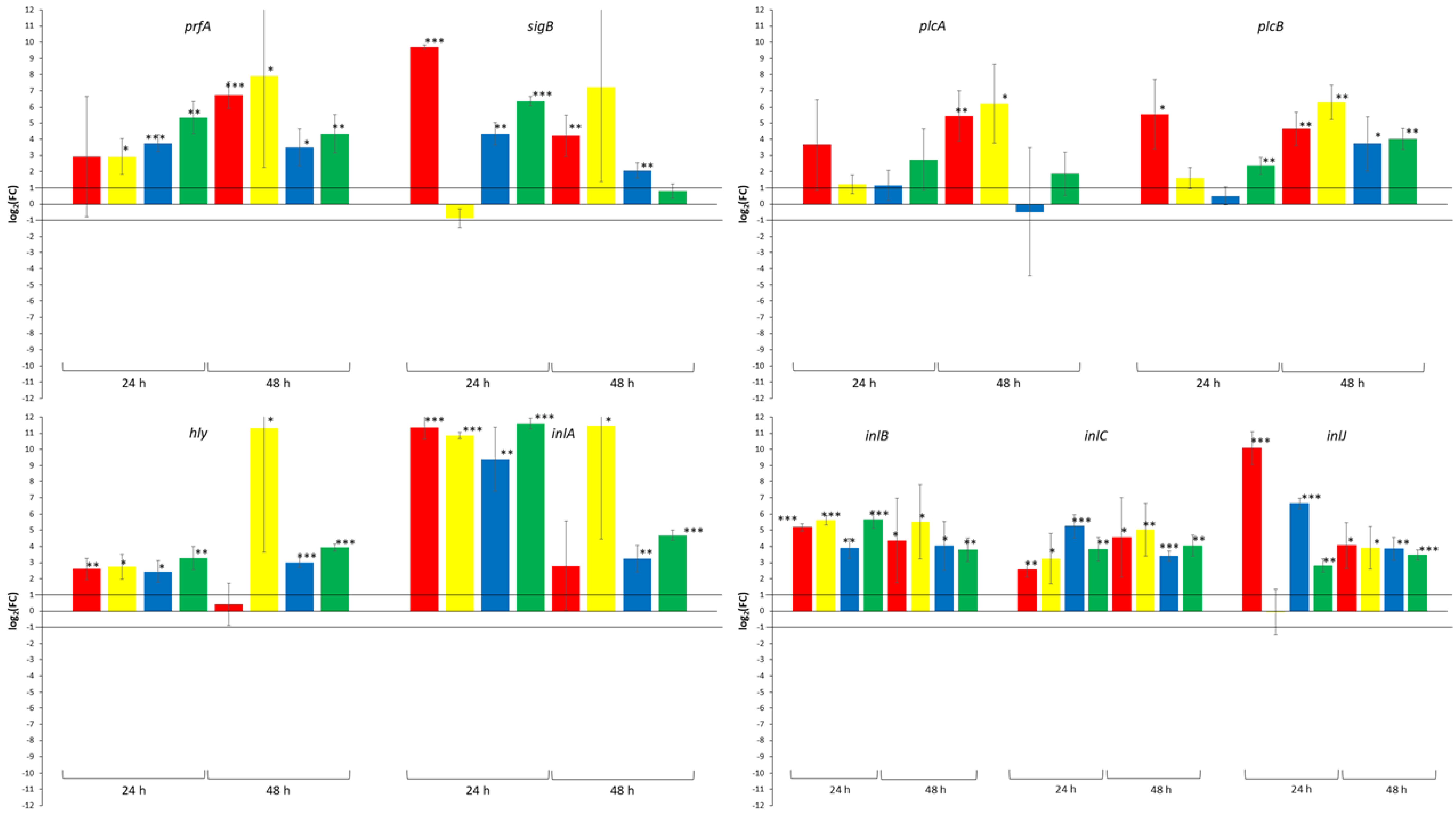
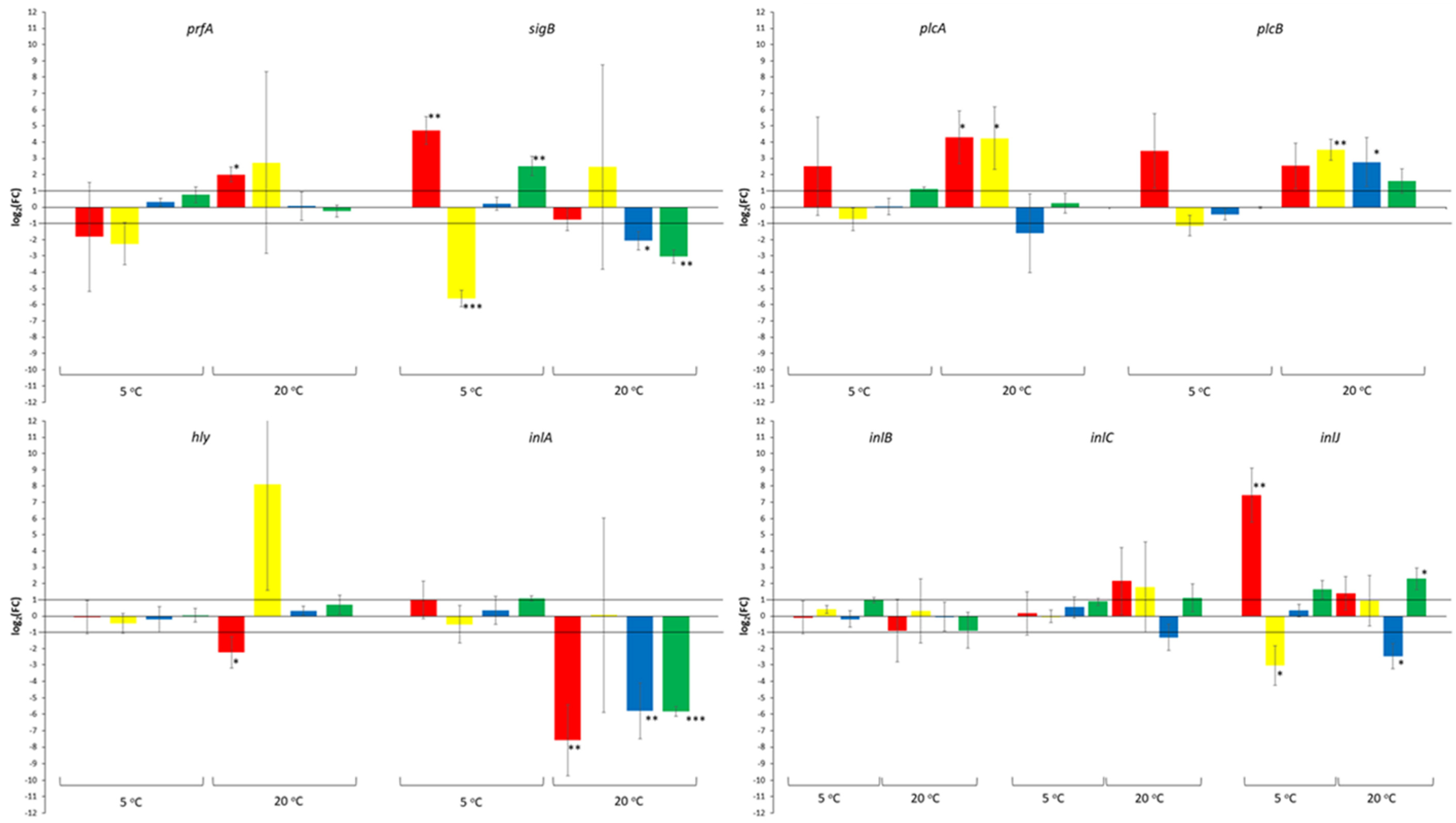
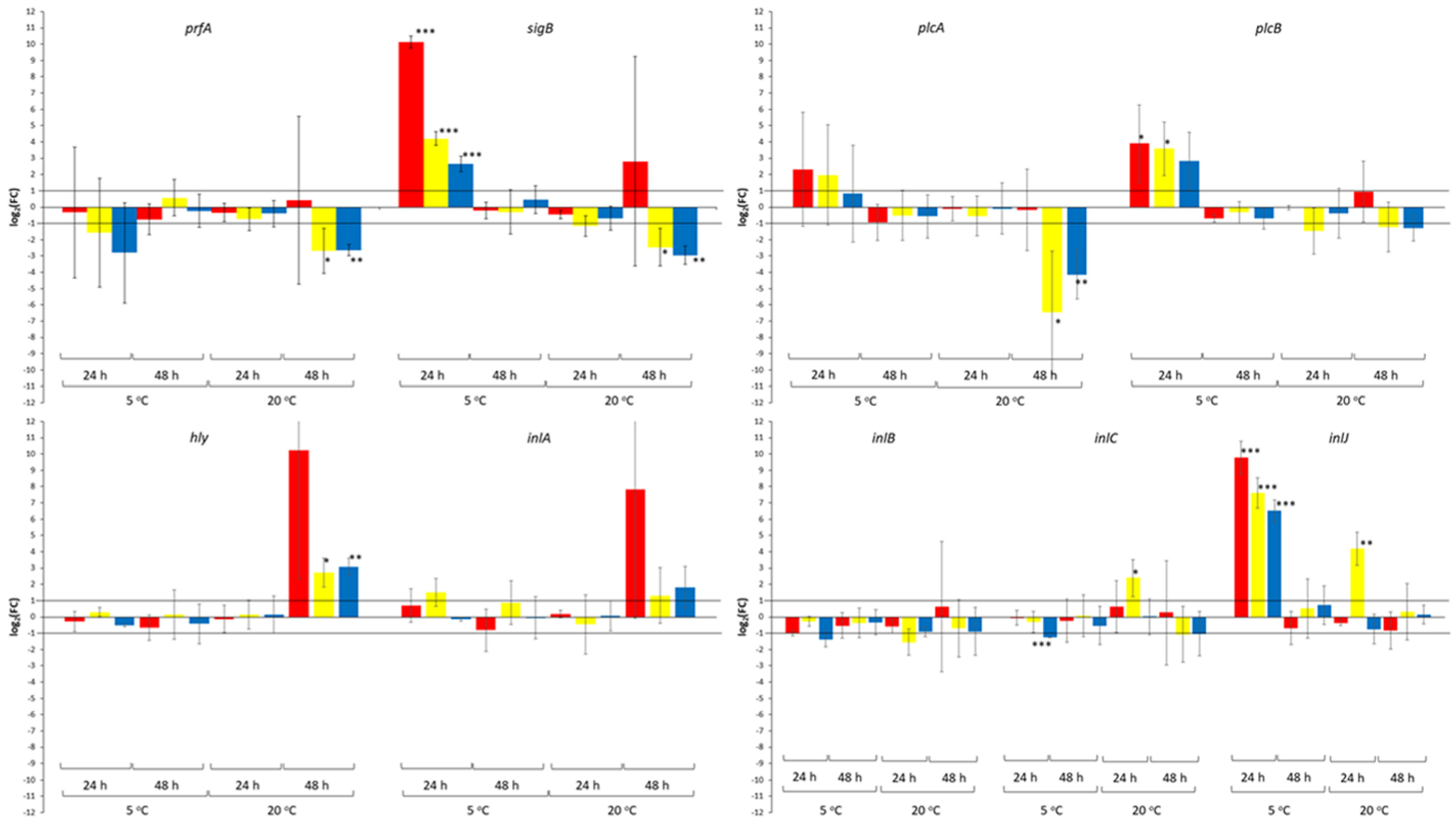
Figure 1. Effect of co-culture with S. cerevisiae strains Y32 (red bars), Y34 (yellow bars) and Y37 (blue bars) on the relative transcription of prfA, sigB, hly, plcA, plcB, inlA, inlB, inlC and inlJ, during growth of L. monocytogenes strain LQC 15257 in BHI broth at 5 and 20 °C for 24 and 48 h. Growth of the pathogen in monoculture was used as control. Error bars represent the standard deviation of the mean value. Presence of asterisks indicates that the relative transcription was above 1 or below −1 (the values that were used as threshold) at p value indicated by the number of asterisks (* p < 0.05; ** p < 0.01; *** p < 0.001).
Beverages 07 00055 g001
Figure 2. Effect of incubation time on the relative transcription of prfA, sigB, hly, plcA, plcB, inlA, inlB, inlC and inlJ, during growth of L. monocytogenes strain LQC 15257 in monoculture (red bars) and in coculture with S. cerevisiae strains Y32 (yellow bars), Y34 (blue bars) and Y37 (green bars). Growth of the pathogen in mono-or co-culture for 24 h was used as control. Error bars represent the standard deviation of the mean value. Presence of asterisks indicates that the relative transcription was above 1 or below −1 (the values that were used as threshold) at p value indicated by the number of asterisks (* p < 0.05; ** p < 0.01; *** p < 0.001).
Beverages 07 00055 g002
Figure 3. Effect of incubation temperature on the relative transcription of prfA, sigB, hly, plcA, plcB, inlA, inlB, inlC and inlJ, during growth of L. monocytogenes strain LQC 15257 in monoculture (red bars) and in coculture with S. cerevisiae strains Y32 (yellow bars), Y34 (blue bars) and Y37 (green bars). Growth of the pathogen in mono-or co-culture at 5 °C was used as control. Error bars represent the standard deviation of the mean value. Presence of asterisks indicates that the relative transcription was above 1 or below −1 (the values that were used as threshold) at p value indicated by the number of asterisks (* p < 0.05; ** p < 0.01; *** p < 0.001).
Beverages 07 00055 g003
Table 1. L. monocytogenes strain LQC 15257 and S. cerevisiae strains Y32, Y34 and Y37 population dynamics (log CFU/mL) during mono-and co-culture in BHI broth at 5 and 20 °C for 24 and 48 h.
L. monocytogenes LQC 15257S. cerevisiae Y32S. cerevisiae Y34S. cerevisiae Y37
Initial inoculum7.12 (0.20)4.10 (0.15)3.95 (0.24)4.20 (0.22)
Mono-culture
5 °C 24 h7.61 (0.27) a4.12 (0.20) a4.27 (0.23) a4.20 (0.20) a
5 °C 48 h7.72 (0.18) a4.35 (0.15) a4.52 (0.20) a4.40 (0.34) a
20 °C 24 h9.29 (0.21) a5.68 (0.21) a5.49 (0.18) a5.60 (0.25) a
20 °C 48 h9.00 (0.17) a6.12 (0.24) b5.95 (0.10) b6.10 (0.11) b
Co-culture
5 °C 24 h7.75 (0.19) a4.20 (0.10) a--
7.70 (0.31) a-4.36 (0.21) a-
7.71 (0.12) a--4.25 (0.15) a
5 °C 48 h7.77 (0.07) a4.31 (0.12) a--
7.59 (0.14) a-4.40 (0.08) a-
7.57 (0.18) a--4.35 (0.14) a
20 °C 24 h9.24 (0.12) a5.57 (0.20) a--
9.17 (0.14) a-5.57 (0.15) a-
9.10 (0.15) a--5.69 (0.19) a
20 °C 48 h9.07 (0.30) a6.10 (0.15) b--
9.14 (0.21) a-5.97 (0.12) b-
9.11 (0.15) a--6.12 (0.17) b
Statistically significant differences between the population of a strain incubated at the same temperature are indicated by different superscript letters.
Table 2. Number of samples in which the relative transcription of the genes under study was below, above or within the threshold set. Percentage is given in parenthesis.
log2(FC)Effect of Yeast Strains 1Effect of Incubation Time 2Effect of Incubation Temperature 3
prfA
<−12 (16.7)0 (0.0)0 (0.0)
−1 to 110 (83.3)7 (87.5)1 (12.5)
>10 (0.0)1 (12.5)7 (87.5)
sigB
<−12 (16.7)3 (37.5)0 (0.0)
−1 to 17 (33.3)3 (37.5)3 (37.5)
>13 (25.0)2 (25.0)5 (62.5)
plcA
<−12 (16.7)0 (0.0)0 (0.0)
−1 to 110 (83.3)6 (75.0)6 (75.0)
>10 (0.0)2 (25.0)2 (25.0)
plcB
<−10 (0.0)0 (0.0)0 (0.0)
−1 to 110 (83.3)6 (75.0)2 (25.0)
>12 (16.7)2 (25.0)6 (75.0)
hly
<−10 (0.0)1 (12.5)0 (0.0)
−1 to 110 (83.3)7 (87.5)1 (12.5)
>12 (16.7)0 (0.0)7 (87.5)
inlA
<−10 (0.0)3 (37.5)0 (0.0)
−1 to 112 (100.0)5 (62.5)1 (12.5)
>10 (0.0)0 (0.0)7 (87.5)
inlB
<−10 (0.0)0 (0.0)0 (0.0)
−1 to 112 (100.0)8 (100.0)0 (0.0)
>10 (0.0)0 (0.0)8 (100.0)
inlC
<−11 (8.3)0 (0.0)0 (0.0)
−1 to 110 (83.3)8 (100.0)0 (0.0)
>11 (8.3)0 (0.0)8 (100.0)
inlJ
<−10 (0.0)2 (25.0)0 (0.0)
−1 to 18 (66.6)4 (50.0)1 (12.5)
>14 (33.3)2 (25.0)7 (87.5)
Total
<−17 (6.5)9 (12.5)0 (0.0)
−1 to 189 (82.5)54 (75.0)15 (20.8)
>112 (11.0)9 (12.5)57 (79.2)
1 the effect of yeast strains was assessed by comparing the transcription of the genes under study during growth of the pathogen in co-culture with each of the yeast strains using as control the growth of the pathogen in monoculture. 2 the effect of incubation time was assessed by comparing the transcription of the genes under study during growth of the pathogen in mono-or co-culture for 48 h using as control the growth of the pathogen in mono-or co-culture for 24 h. 3 the effect of incubation temperature was assessed by comparing the transcription of the genes under study during growth of the pathogen in mono-or co-culture at 20 °C using as control the growth of the pathogen in mono-or co-culture at 5 °C.
Table 3. Pearson product moment (r) correlations between the relative transcription of each pair of genes.
prfAsigBplcAplcBhlyinlAinlBinlC
sigBr0.4792
p0.0099
plcAr0.69610.6497
p0.00000.0002
plcBr0.60650.70280.7614
p0.00060.00000.0000
hlyr0.48620.29250.22650.3533
p0.00870.13090.24650.0652
inlAr0.56980.56400.28970.31610.6024
p0.00150.00180.13490.10130.0007
inlBr0.84610.46650.48980.54490.46460.8140
p0.00000.01230.00810.00270.01280.0000
inlCr0.90040.39300.62350.47460.38790.59140.8211
p0.00000.03850.00040.01070.04140.00090.0000
inlJr0.18370.79200.47190.6052−0.02840.31450.24030.3003
p0.34950.00000.01120.00060.88600.10310.21800.1205
Statistically significant correlations (p < 0.05) are presented in bold.
Publisher’s Note: MDPI stays neutral with regard to jurisdictional claims in published maps and institutional affiliations.

