Extracellular Vesicles and Their Role in Osteogenesis



Abstract
1. Introduction
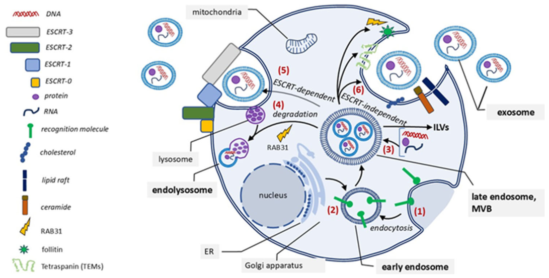
2. Biology of Extracellular Vesicles
3. Role of EVs in Bone Regeneration
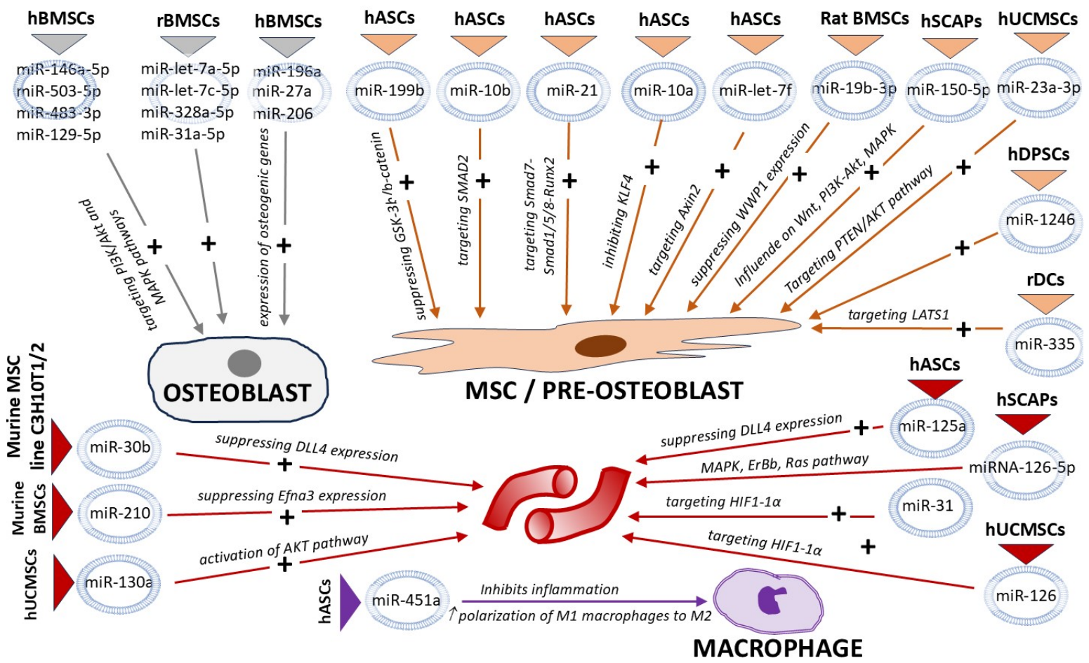
3.1. Promotion of Osteogenesis
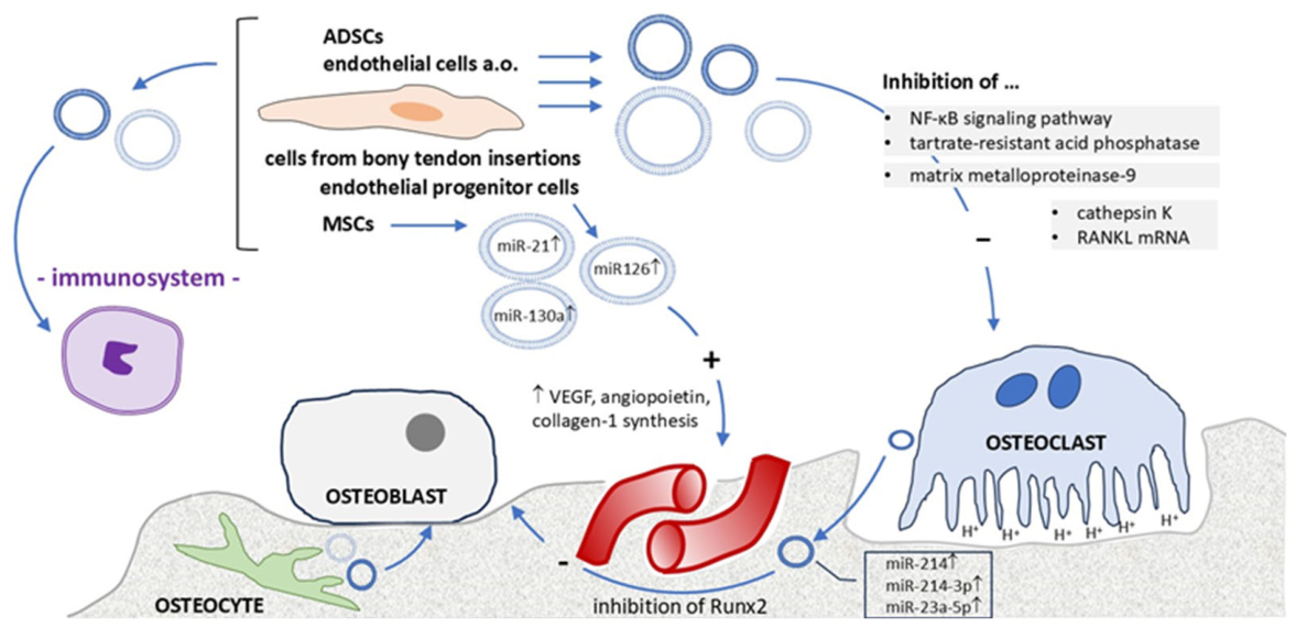
3.2. Regulation of Osteoclast Activity
- Downregulation of miR-214 expression and inhibition of the NF-κB signaling pathway (EVs from prostate cells) [50].
- Inhibition of the expression of tartrate-resistant acid phosphatase, cathepsin K, and matrix metalloproteinase-9 [51].
- Inhibition of RANKL mRNA and protein expression (EVs from ADSCs) [52].
- Reduction in the bone resorption rate (EVs from endothelial cells) [53].
3.3. Promotion of Angiogenesis
3.4. Immunomodulatory Effects of EVs
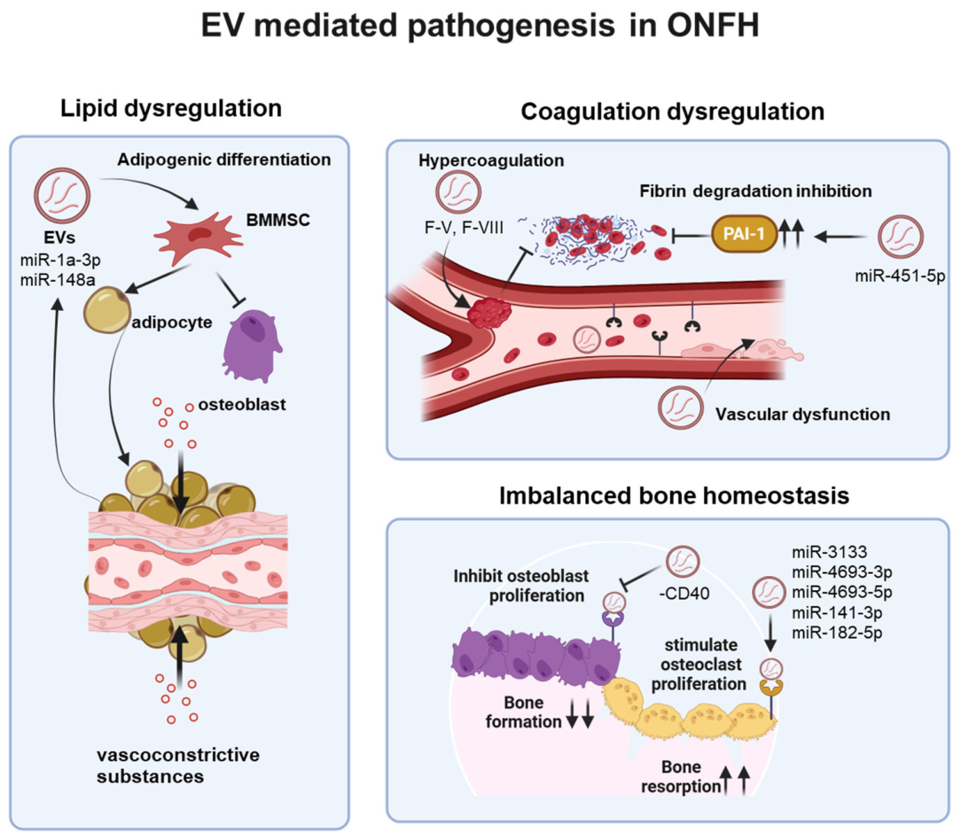
4. Pathophysiological Effects of EVs in Osteonecrosis of the Femoral Head
5. Engineering and Modification of EVs for Bone Regeneration

Biomaterial-Assisted EV Delivery

6. Isolation of EVs
7. Preclinical Application and Clinical Trials of EVs for Bone Regeneration
8. Conclusions and Future Perspectives
Author Contributions
Funding
Institutional Review Board Statement
Informed Consent Statement
Data Availability Statement
Acknowledgments
Conflicts of Interest
Abbreviations
| ABC | ATP-binding cassette | MMPs | matrix metalloproteinases |
| ADSCs | adipose-derived stromal cells | MVB | multivesicular body |
| Akt | protein kinase B | MYD88 | myeloid differentiation primary response 88 |
| Ale | alendronate | NF-κB | nuclear factor kappa B |
| ALP | alkaline phosphatase | OCN | osteocalcin |
| ARDS | acute respiratory distress syndrome | ONFH | osteonecrosis of the femoral head |
| AVN | avascular osteonecrosis | PDCD6IP | programmed cell death 6-interacting protein |
| BM-MSCs | bone marrow-derived mesenchymal stromal cells | PEG | polyethylene glycol |
| BMP | bone morphogenetic protein | PEGDA | polyethylene glycol diacrylate |
| CD | cluster of differentiation | PI3K | phosphoinositide 3-kinase |
| DBCO | dibenzocyclooctyne | PLGA | poly(lactic-co-glycolic acid) |
| DNA | deoxyribonucleic acid | PRP | platelet-rich plasma |
| EGFR | epidermal growth factor receptor | RANKL | receptor activator of nuclear factor kappa-B ligand |
| ESCRT | endosomal sorting complex required for transport | RGD | arginine-glycine-aspartic acid |
| EV | extracellular vesicle | Ror2 | receptor tyrosine kinase-like orphan receptor 2 |
| FasL | Fas ligand | RT | room temperature |
| HA | hyaluronic acid | RUNX2 | runt-related transcription factor 2 |
| HIF | hypoxia-inducible factor | siRNA | small interfering RNA |
| HIPPO | Hippo signaling pathway | SLC | solute carrier |
| HSP | heat shock protein | Smad | TGF-β signaling mediator |
| HUVECs | human umbilical vein endothelial cells | SNARE | soluble N-ethylmaleimide-sensitive factor attachment receptor |
| ICAM | intercellular adhesion molecule | SSRT | surgical site-released tissue |
| IL-1β | interleukin-1 beta | TEM | transmission electron microscopy |
| IL-6 | interleukin-6 | TEMs | tetraspanin-enriched microdomains |
| ILV | intraluminal vesicle | TGF-β | transforming growth factor beta |
| iPS-MSCs | induced pluripotent stem cell-derived mesenchymal stromal cells | TNF-α | tumor necrosis factor alpha |
| ISEV | International Society for Extracellular Vesicles | TSG101 | tumor susceptibility gene 101 |
| lncRNA | long non-coding RNA | TSPAN4 | tetraspanin 4 |
| MAPK | mitogen-activated protein kinase | VEGF | vascular endothelial growth factor |
| MC3T3 | mouse osteoblast precursor cell line | Wnt | Wnt signaling pathway |
| MFGE8 | milk fat globule EGF factor 8 | β-TCP | beta-tricalcium phosphate |
| MHC | major histocompatibility complex | ||
References
- Tertel, T.; Rebmann, V.; Bielefeld, C.; Haversath, M.; Jager, M.; Wegner, A.; Busch, A.; Giebel, B. Cd9+ and cd82+ extracellular vesicles in synovial fluid differentiate aseptic from septic endoprosthesis loosening. Extracell. Vesicles Circ. Nucl. Acids 2025, 6, 336–349. [Google Scholar] [CrossRef] [Scilit] [PubMed]
- Zhang, H.; Wang, J.; Yang, R.; Song, X.; Liu, R.; Wang, J.; Meng, L.; Xu, Z.; Vinnikov, I.A.; Yang, G.; et al. A native bioactive interface functionalized with osteoprogenitor stem cell-derived migrasomes for enhanced bone regeneration. Research 2026, 9, 1220. [Google Scholar] [CrossRef] [Scilit] [PubMed]
- Sheta, M.; Taha, E.A.; Lu, Y.; Eguchi, T. Extracellular vesicles: New classification and tumor immunosuppression. Biology 2023, 12, 110. [Google Scholar] [CrossRef] [Scilit] [PubMed]
- Doyle, L.M.; Wang, M.Z. Overview of extracellular vesicles, their origin, composition, purpose, and methods for exosome isolation and analysis. Cells 2019, 8, 727. [Google Scholar] [CrossRef] [Scilit] [PubMed]
- Mateescu, B.; Jones, J.C.; Alexander, R.P.; Alsop, E.; An, J.Y.; Asghari, M.; Boomgarden, A.; Bouchareychas, L.; Cayota, A.; Chang, H.C.; et al. Phase 2 of extracellular rna communication consortium charts next-generation approaches for extracellular rna research. iScience 2022, 25, 104653. [Google Scholar] [CrossRef] [Scilit] [PubMed]
- Das, S.; Extracellular, R.N.A.C.C.; Ansel, K.M.; Bitzer, M.; Breakefield, X.O.; Charest, A.; Galas, D.J.; Gerstein, M.B.; Gupta, M.; Milosavljevic, A.; et al. The extracellular rna communication consortium: Establishing foundational knowledge and technologies for extracellular rna research. Cell 2019, 177, 231–242. [Google Scholar] [CrossRef] [Scilit] [PubMed]
- Thery, C.; Witwer, K.W.; Aikawa, E.; Alcaraz, M.J.; Anderson, J.D.; Andriantsitohaina, R.; Antoniou, A.; Arab, T.; Archer, F.; Atkin-Smith, G.K.; et al. Minimal information for studies of extracellular vesicles 2018 (misev2018): A position statement of the international society for extracellular vesicles and update of the misev2014 guidelines. J. Extracell. Vesicles 2018, 7, 1535750. [Google Scholar] [CrossRef] [Scilit] [PubMed]
- Quesenberry, P.J.; Aliotta, J.; Camussi, G.; Abdel-Mageed, A.B.; Wen, S.; Goldberg, L.; Zhang, H.G.; Tetta, C.; Franklin, J.; Coffey, R.J.; et al. Potential functional applications of extracellular vesicles: A report by the nih common fund extracellular rna communication consortium. J. Extracell. Vesicles 2015, 4, 27575. [Google Scholar] [CrossRef] [Scilit] [PubMed]
- Lotvall, J.; Hill, A.F.; Hochberg, F.; Buzas, E.I.; Di Vizio, D.; Gardiner, C.; Gho, Y.S.; Kurochkin, I.V.; Mathivanan, S.; Quesenberry, P.; et al. Minimal experimental requirements for definition of extracellular vesicles and their functions: A position statement from the international society for extracellular vesicles. J. Extracell. Vesicles 2014, 3, 26913. [Google Scholar] [CrossRef] [Scilit] [PubMed]
- Welsh, J.A.; Goberdhan, D.C.I.; O’Driscoll, L.; Buzas, E.I.; Blenkiron, C.; Bussolati, B.; Cai, H.; Di Vizio, D.; Driedonks, T.A.P.; Erdbrugger, U.; et al. Minimal information for studies of extracellular vesicles (misev2023): From basic to advanced approaches. J. Extracell. Vesicles 2024, 13, e12404. [Google Scholar] [CrossRef] [Scilit] [PubMed]
- Nagelkerke, A.; Ojansivu, M.; van der Koog, L.; Whittaker, T.E.; Cunnane, E.M.; Silva, A.M.; Dekker, N.; Stevens, M.M. Extracellular vesicles for tissue repair and regeneration: Evidence, challenges and opportunities. Adv. Drug Deliv. Rev. 2021, 175, 113775. [Google Scholar] [CrossRef] [Scilit] [PubMed]
- Liao, W.; Du, Y.; Zhang, C.; Pan, F.; Yao, Y.; Zhang, T.; Peng, Q. Exosomes: The next generation of endogenous nanomaterials for advanced drug delivery and therapy. Acta Biomater. 2019, 86, 1–14. [Google Scholar] [CrossRef] [Scilit] [PubMed]
- Arenaccio, C.; Chiozzini, C.; Ferrantelli, F.; Leone, P.; Olivetta, E.; Federico, M. Exosomes in therapy: Engineering, pharmacokinetics and future applications. Curr. Drug Targets 2019, 20, 87–95. [Google Scholar] [CrossRef] [Scilit] [PubMed]
- Escudier, B.; Dorval, T.; Chaput, N.; Andre, F.; Caby, M.P.; Novault, S.; Flament, C.; Leboulaire, C.; Borg, C.; Amigorena, S.; et al. Vaccination of metastatic melanoma patients with autologous dendritic cell (dc) derived-exosomes: Results of thefirst phase i clinical trial. J. Transl. Med. 2005, 3, 10. [Google Scholar] [CrossRef] [Scilit] [PubMed]
- Zhang, L.; Lin, Y.; Zhang, X.; Shan, C. Research progress of exosomes in orthopedics. Front. Genet. 2022, 13, 915141. [Google Scholar] [CrossRef] [Scilit] [PubMed]
- Liu, X.; Wang, C.; Meng, H.; Liao, S.; Zhang, J.; Guan, Y.; Tian, H.; Peng, J. Research progress on exosomes in osteonecrosis of the femoral head. Orthop. Surg. 2022, 14, 1951–1957. [Google Scholar] [CrossRef] [Scilit] [PubMed]
- Liu, F.; Sun, T.; An, Y.; Ming, L.; Li, Y.; Zhou, Z.; Shang, F. The potential therapeutic role of extracellular vesicles in critical-size bone defects: Spring of cell-free regenerative medicine is coming. Front. Bioeng. Biotechnol. 2023, 11, 1050916. [Google Scholar] [CrossRef] [Scilit] [PubMed]
- Li, H.; Liu, H.; Zhou, Y.; Cheng, L.; Wang, B.; Ma, J. The multifaceted roles of extracellular vesicles in osteonecrosis of the femoral head. J. Orthop. Transl. 2025, 52, 70–84. [Google Scholar] [CrossRef] [Scilit] [PubMed]
- Chen, S.; Tang, Y.; Liu, Y.; Zhang, P.; Lv, L.; Zhang, X.; Jia, L.; Zhou, Y. Exosomes derived from mir-375-overexpressing human adipose mesenchymal stem cells promote bone regeneration. Cell Prolif. 2019, 52, e12669. [Google Scholar] [CrossRef] [Scilit] [PubMed]
- Narayanan, R.; Huang, C.C.; Ravindran, S. Hijacking the cellular mail: Exosome mediated differentiation of mesenchymal stem cells. Stem Cells Int. 2016, 2016, 3808674. [Google Scholar] [CrossRef] [Scilit] [PubMed]
- Henze, K.; Herten, M.; Haversath, M.; Busch, A.; Brandau, S.; Hackel, A.; Flohe, S.B.; Jager, M. Surgical vacuum filter-derived stromal cells are superior in proliferation to human bone marrow aspirate. Stem Cell Res. Ther. 2019, 10, 338. [Google Scholar] [CrossRef] [Scilit] [PubMed]
- Rehage, E.; Sowislok, A.; Busch, A.; Papaeleftheriou, E.; Jansen, M.; Jager, M. Surgical site-released tissue is potent to generate bone onto tcp and pcl-tcp scaffolds in vitro. Int. J. Mol. Sci. 2023, 24, 15877. [Google Scholar] [CrossRef] [Scilit] [PubMed]
- Groven, R.V.M.; Blokhuis, J.T.; Poeze, M.; van Griensven, M.; Blokhuis, T.J. Surgical suction filter-derived bone graft displays osteogenic mirna and mrna patterns. Eur. J. Trauma Emerg. Surg. 2024, 50, 315–326. [Google Scholar] [CrossRef] [Scilit] [PubMed]
- Sowislok, A.; Gruber, G.; Kaschani, F.; Kaiser, M.; Papaeleftheriou, E.; Jager, M. Intraoperative biologization of beta-tcp and pcl-tcp by autologous proteins. J. Funct. Biomater. 2025, 16, 340. [Google Scholar] [CrossRef] [Scilit] [PubMed]
- Jager, M.; Busch, A.; Sowislok, A. Bioactivation of scaffolds in osteonecrosis. Orthopadie 2022, 51, 808–814. [Google Scholar] [CrossRef] [Scilit] [PubMed]
- Papaeleftheriou, E.; Sowislok, A.; Rehage, E.; Wegner, A.; Haversath, M.; Jansen, M.; Jäger, M. Intraoperative bioactivation of bone substitutes using a surgical suction handle: A prospective clinical pilot study. J. Funct. Biomater. 2026, 17, 245. [Google Scholar] [CrossRef] [Scilit] [PubMed]
- Joly, L.M.; Tertel, T.; Sowislok, A.; Giebel, B.; Jager, M. A new subpopulation of extracellular vesicles harvested from osteogenically induced mesenchymal stromal cells of surgical site-released tissue. Biomolecules 2026, 16, 289. [Google Scholar] [CrossRef] [Scilit] [PubMed]
- Kang, M.; Lee, C.S.; Lee, M. Bioactive scaffolds integrated with liposomal or extracellular vesicles for bone regeneration. Bioengineering 2021, 8, 137. [Google Scholar] [CrossRef] [Scilit] [PubMed]
- Yan, H.C.; Yu, T.T.; Li, J.; Qiao, Y.Q.; Wang, L.C.; Zhang, T.; Li, Q.; Zhou, Y.H.; Liu, D.W. The delivery of extracellular vesicles loaded in biomaterial scaffolds for bone regeneration. Front. Bioeng. Biotechnol. 2020, 8, 1015. [Google Scholar] [CrossRef] [Scilit] [PubMed]
- Irfan, D.; Ahmad, I.; Patra, I.; Margiana, R.; Rasulova, M.T.; Sivaraman, R.; Kandeel, M.; Mohammad, H.J.; Al-Qaim, Z.H.; Jawad, M.A.; et al. Stem cell-derived exosomes in bone healing: Focusing on their role in angiogenesis. Cytotherapy 2023, 25, 353–361. [Google Scholar] [CrossRef] [Scilit] [PubMed]
- Schank, M.B.; Zhao, J.; Wang, L.; Moorman, J.P.; Yao, Z.Q. Extracellular vesicles: A comprehensive review of their origins, functions, and therapeutic potential. Biomedicines 2026, 14, 495. [Google Scholar] [CrossRef] [Scilit] [PubMed]
- Zhang, X.; Yao, L.; Meng, Y.; Li, B.; Yang, Y.; Gao, F. Migrasome: A new functional extracellular vesicle. Cell Death Discov. 2023, 9, 381. [Google Scholar] [CrossRef] [Scilit] [PubMed]
- Zhang, Y.; Wu, J.; Liu, L.; Zhu, Y.; Zhang, J.; Xu, J.; Shi, X.; Yu, Y.; Wang, C.; Xiao, J.; et al. M2 microglia-derived migrasome-enriched extracellular vesicles restore mitochondrial homeostasis to orchestrate neurovascular unit recovery after ischemic stroke. J. Nanobiotechnol. 2026, 24, 737. [Google Scholar] [CrossRef] [Scilit] [PubMed]
- Wang, M.; Zhang, S.; Kutibiding, A.; Zhou, X.; Tian, X.; Yuan, S.; Lv, M.; Yang, Y.; Bai, S. M2 macrophage-derived migrasomes enhance bone regeneration by directing osteogenic differentiation of bone marrow mesenchymal stem cells (bmscs). Tissue Cell 2026, 100, 103394. [Google Scholar] [CrossRef] [Scilit] [PubMed]
- Wickman, G.; Julian, L.; Olson, M.F. How apoptotic cells aid in the removal of their own cold dead bodies. Cell Death Differ. 2012, 19, 735–742. [Google Scholar] [CrossRef] [Scilit] [PubMed]
- Guo, Y.; Du, W.; Cheng, S.; Li, Y.; Xiao, S.; Yang, L.; Wang, L.; Pathak, J.L. Apoptotic extracellular vesicles act as master regulators of the bone healing niche. J. Nanobiotechnol. 2026, 24, 453. [Google Scholar] [CrossRef] [Scilit] [PubMed]
- Phan, J.; Kumar, P.; Hao, D.; Gao, K.; Farmer, D.; Wang, A. Engineering mesenchymal stem cells to improve their exosome efficacy and yield for cell-free therapy. J. Extracell. Vesicles 2018, 7, 1522236. [Google Scholar] [CrossRef] [Scilit] [PubMed]
- Qin, Y.; Wang, L.; Gao, Z.; Chen, G.; Zhang, C. Bone marrow stromal/stem cell-derived extracellular vesicles regulate osteoblast activity and differentiation in vitro and promote bone regeneration in vivo. Sci. Rep. 2016, 6, 21961. [Google Scholar] [CrossRef] [Scilit] [PubMed]
- Zhai, M.; Zhu, Y.; Yang, M.; Mao, C. Human mesenchymal stem cell derived exosomes enhance cell-free bone regeneration by altering their mirnas profiles. Adv. Sci. 2020, 7, 2001334. [Google Scholar] [CrossRef] [Scilit] [PubMed]
- Yang, X.; Yang, J.; Lei, P.; Wen, T. Lncrna malat1 shuttled by bone marrow-derived mesenchymal stem cells-secreted exosomes alleviates osteoporosis through mediating microrna-34c/satb2 axis. Aging 2019, 11, 8777–8791. [Google Scholar] [CrossRef] [Scilit] [PubMed]
- Fujioka, T.; Nishimura, T.; Kawana, H.; Hirosawa, K.M.; Yamakawa, R.; Sapili, H.; Oono-Yakura, K.; Nakagawa, R.; Inoue, T.; Nureki, O.; et al. Efficient cellular transformation via protein delivery through the protrusion-derived extracellular vesicles. Nat. Commun. 2025, 16, 10900. [Google Scholar] [CrossRef] [Scilit] [PubMed]
- Cao, Z.; Wu, Y.; Yu, L.; Zou, L.; Yang, L.; Lin, S.; Wang, J.; Yuan, Z.; Dai, J. Exosomal mir-335 derived from mature dendritic cells enhanced mesenchymal stem cell-mediated bone regeneration of bone defects in athymic rats. Mol. Med. 2021, 27, 20. [Google Scholar] [CrossRef] [Scilit] [PubMed]
- Qi, X.; Zhang, J.; Yuan, H.; Xu, Z.; Li, Q.; Niu, X.; Hu, B.; Wang, Y.; Li, X. Exosomes secreted by human-induced pluripotent stem cell-derived mesenchymal stem cells repair critical-sized bone defects through enhanced angiogenesis and osteogenesis in osteoporotic rats. Int. J. Biol. Sci. 2016, 12, 836–849. [Google Scholar] [CrossRef] [Scilit] [PubMed]
- Zhang, J.; Liu, X.; Li, H.; Chen, C.; Hu, B.; Niu, X.; Li, Q.; Zhao, B.; Xie, Z.; Wang, Y. Exosomes/tricalcium phosphate combination scaffolds can enhance bone regeneration by activating the pi3k/akt signaling pathway. Stem Cell Res. Ther. 2016, 7, 136. [Google Scholar] [CrossRef] [Scilit] [PubMed]
- Li, W.; Liu, Y.; Zhang, P.; Tang, Y.; Zhou, M.; Jiang, W.; Zhang, X.; Wu, G.; Zhou, Y. Tissue-engineered bone immobilized with human adipose stem cells-derived exosomes promotes bone regeneration. ACS Appl. Mater. Interfaces 2018, 10, 5240–5254. [Google Scholar] [CrossRef] [Scilit] [PubMed]
- Ge, M.; Ke, R.; Cai, T.; Yang, J.; Mu, X. Identification and proteomic analysis of osteoblast-derived exosomes. Biochem. Biophys. Res. Commun. 2015, 467, 27–32. [Google Scholar] [CrossRef] [Scilit] [PubMed]
- Ge, M.; Wu, Y.; Ke, R.; Cai, T.; Yang, J.; Mu, X. Value of osteoblast-derived exosomes in bone diseases. J. Craniofac. Surg. 2017, 28, 866–870. [Google Scholar] [CrossRef] [Scilit] [PubMed]
- Song, X.; Xu, L.; Zhang, W. Biomimetic synthesis and optimization of extracellular vesicles for bone regeneration. J. Control. Release 2023, 355, 18–41. [Google Scholar] [CrossRef] [Scilit] [PubMed]
- Infante, A.; Alcorta-Sevillano, N.; Macias, I.; Rodriguez, C.I. Educating evs to improve bone regeneration: Getting closer to the clinic. Int. J. Mol. Sci. 2022, 23, 1865. [Google Scholar] [CrossRef] [Scilit] [PubMed]
- Duan, Y.; Tan, Z.; Yang, M.; Li, J.; Liu, C.; Wang, C.; Zhang, F.; Jin, Y.; Wang, Y.; Zhu, L. Pc-3-derived exosomes inhibit osteoclast differentiation by downregulating mir-214 and blocking nf-kappab signaling pathway. BioMed Res. Int. 2019, 2019, 8650846. [Google Scholar] [CrossRef] [Scilit] [PubMed]
- Karlsson, T.; Lundholm, M.; Widmark, A.; Persson, E. Tumor cell-derived exosomes from the prostate cancer cell line tramp-c1 impair osteoclast formation and differentiation. PLoS ONE 2016, 11, e0166284. [Google Scholar] [CrossRef] [Scilit] [PubMed]
- Ren, L.; Song, Z.J.; Cai, Q.W.; Chen, R.X.; Zou, Y.; Fu, Q.; Ma, Y.Y. Adipose mesenchymal stem cell-derived exosomes ameliorate hypoxia/serum deprivation-induced osteocyte apoptosis and osteocyte-mediated osteoclastogenesis in vitro. Biochem. Biophys. Res. Commun. 2019, 508, 138–144. [Google Scholar] [CrossRef] [Scilit] [PubMed]
- Song, H.; Li, X.; Zhao, Z.; Qian, J.; Wang, Y.; Cui, J.; Weng, W.; Cao, L.; Chen, X.; Hu, Y.; et al. Reversal of osteoporotic activity by endothelial cell-secreted bone targeting and biocompatible exosomes. Nano Lett. 2019, 19, 3040–3048. [Google Scholar] [CrossRef] [Scilit] [PubMed]
- Yang, J.X.; Xie, P.; Li, Y.S.; Wen, T.; Yang, X.C. Osteoclast-derived mir-23a-5p-containing exosomes inhibit osteogenic differentiation by regulating runx2. Cell. Signal. 2020, 70, 109504. [Google Scholar] [CrossRef] [Scilit] [PubMed]
- Li, D.; Liu, J.; Guo, B.; Liang, C.; Dang, L.; Lu, C.; He, X.; Cheung, H.Y.; Xu, L.; Lu, C.; et al. Osteoclast-derived exosomal mir-214-3p inhibits osteoblastic bone formation. Nat. Commun. 2016, 7, 10872. [Google Scholar] [CrossRef] [Scilit] [PubMed]
- Wang, L.; Jia, P.; Shan, Y.; Hao, Y.; Wang, X.; Jiang, Y.; Yuan, Y.; Du, Q.; Zhang, H.; Yang, F.; et al. Synergistic protection of bone vasculature and bone mass by desferrioxamine in osteoporotic mice. Mol. Med. Rep. 2017, 16, 6642–6649. [Google Scholar] [CrossRef] [Scilit] [PubMed]
- Jia, P.; Chen, H.; Kang, H.; Qi, J.; Zhao, P.; Jiang, M.; Guo, L.; Zhou, Q.; Qian, N.D.; Zhou, H.B.; et al. Deferoxamine released from poly(lactic-co-glycolic acid) promotes healing of osteoporotic bone defect via enhanced angiogenesis and osteogenesis. J. Biomed. Mater. Res. A 2016, 104, 2515–2527. [Google Scholar] [CrossRef] [Scilit] [PubMed]
- Jia, Y.; Zhu, Y.; Qiu, S.; Xu, J.; Chai, Y. Exosomes secreted by endothelial progenitor cells accelerate bone regeneration during distraction osteogenesis by stimulating angiogenesis. Stem Cell Res. Ther. 2019, 10, 12. [Google Scholar] [CrossRef] [Scilit] [PubMed]
- Vizoso, F.J.; Eiro, N.; Cid, S.; Schneider, J.; Perez-Fernandez, R. Mesenchymal stem cell secretome: Toward cell-free therapeutic strategies in regenerative medicine. Int. J. Mol. Sci. 2017, 18, 1852. [Google Scholar] [CrossRef] [Scilit] [PubMed]
- Zhang, B.; Huang, J.; Liu, J.; Lin, F.; Ding, Z.; Xu, J. Injectable composite hydrogel promotes osteogenesis and angiogenesis in spinal fusion by optimizing the bone marrow mesenchymal stem cell microenvironment and exosomes secretion. Mater. Sci. Eng. C Mater. Biol. Appl. 2021, 123, 111782. [Google Scholar] [CrossRef] [Scilit] [PubMed]
- Wu, D.; Qin, H.; Wang, Z.; Yu, M.; Liu, Z.; Peng, H.; Liang, L.; Zhang, C.; Wei, X. Bone mesenchymal stem cell-derived sev-encapsulated thermosensitive hydrogels accelerate osteogenesis and angiogenesis by release of exosomal mir-21. Front. Bioeng. Biotechnol. 2021, 9, 829136. [Google Scholar] [CrossRef] [Scilit] [PubMed]
- Wang, Z.; Yuan, Y.; Ji, X.; Xiao, X.; Li, Z.; Yi, X.; Zhu, Y.; Guo, T.; Wang, Y.; Chen, L.; et al. The hippo-taz axis mediates vascular endothelial growth factor c in glioblastoma-derived exosomes to promote angiogenesis. Cancer Lett. 2021, 513, 1–13. [Google Scholar] [CrossRef] [Scilit] [PubMed]
- Takeuchi, R.; Katagiri, W.; Endo, S.; Kobayashi, T. Exosomes from conditioned media of bone marrow-derived mesenchymal stem cells promote bone regeneration by enhancing angiogenesis. PLoS ONE 2019, 14, e0225472. [Google Scholar] [CrossRef] [Scilit] [PubMed]
- Zhang, L.; Jiao, G.; Ren, S.; Zhang, X.; Li, C.; Wu, W.; Wang, H.; Liu, H.; Zhou, H.; Chen, Y. Exosomes from bone marrow mesenchymal stem cells enhance fracture healing through the promotion of osteogenesis and angiogenesis in a rat model of nonunion. Stem Cell Res. Ther. 2020, 11, 38. [Google Scholar] [CrossRef] [Scilit] [PubMed]
- Jing, X.; Wang, S.; Tang, H.; Li, D.; Zhou, F.; Xin, L.; He, Q.; Hu, S.; Zhang, T.; Chen, T.; et al. Dynamically bioresponsive DNA hydrogel incorporated with dual-functional stem cells from apical papilla-derived exosomes promotes diabetic bone regeneration. ACS Appl. Mater. Interfaces 2022, 14, 16082–16099. [Google Scholar] [CrossRef] [Scilit] [PubMed]
- Liu, L.; Liu, Y.; Feng, C.; Chang, J.; Fu, R.; Wu, T.; Yu, F.; Wang, X.; Xia, L.; Wu, C.; et al. Lithium-containing biomaterials stimulate bone marrow stromal cell-derived exosomal mir-130a secretion to promote angiogenesis. Biomaterials 2019, 192, 523–536. [Google Scholar] [CrossRef] [Scilit] [PubMed]
- Liu, W.; Li, L.; Rong, Y.; Qian, D.; Chen, J.; Zhou, Z.; Luo, Y.; Jiang, D.; Cheng, L.; Zhao, S.; et al. Hypoxic mesenchymal stem cell-derived exosomes promote bone fracture healing by the transfer of mir-126. Acta Biomater. 2020, 103, 196–212. [Google Scholar] [CrossRef] [Scilit] [PubMed]
- Cosenza, S.; Toupet, K.; Maumus, M.; Luz-Crawford, P.; Blanc-Brude, O.; Jorgensen, C.; Noel, D. Mesenchymal stem cells-derived exosomes are more immunosuppressive than microparticles in inflammatory arthritis. Theranostics 2018, 8, 1399–1410. [Google Scholar] [CrossRef] [Scilit] [PubMed]
- Casado, J.G.; Blazquez, R.; Vela, F.J.; Alvarez, V.; Tarazona, R.; Sanchez-Margallo, F.M. Mesenchymal stem cell-derived exosomes: Immunomodulatory evaluation in an antigen-induced synovitis porcine model. Front. Vet. Sci. 2017, 4, 39. [Google Scholar] [CrossRef] [Scilit] [PubMed]
- Lindenbergh, M.F.S.; Stoorvogel, W. Antigen presentation by extracellular vesicles from professional antigen-presenting cells. Annu. Rev. Immunol. 2018, 36, 435–459. [Google Scholar] [CrossRef] [Scilit] [PubMed]
- Qian, X.; An, N.; Ren, Y.; Yang, C.; Zhang, X.; Li, L. Immunosuppressive effects of mesenchymal stem cells-derived exosomes. Stem Cell Rev. Rep. 2021, 17, 411–427. [Google Scholar] [CrossRef] [Scilit] [PubMed]
- Chen, W.; Huang, Y.; Han, J.; Yu, L.; Li, Y.; Lu, Z.; Li, H.; Liu, Z.; Shi, C.; Duan, F.; et al. Immunomodulatory effects of mesenchymal stromal cells-derived exosome. Immunol. Res. 2016, 64, 831–840. [Google Scholar] [CrossRef] [Scilit] [PubMed]
- Bozec, A.; Soulat, D. Latest perspectives on macrophages in bone homeostasis. Pflug. Arch. 2017, 469, 517–525. [Google Scholar] [CrossRef] [Scilit] [PubMed]
- Wasnik, S.; Rundle, C.H.; Baylink, D.J.; Yazdi, M.S.; Carreon, E.E.; Xu, Y.; Qin, X.; Lau, K.W.; Tang, X. 1,25-dihydroxyvitamin d suppresses m1 macrophages and promotes m2 differentiation at bone injury sites. JCI Insight 2018, 3, e98773. [Google Scholar] [CrossRef] [Scilit] [PubMed]
- Guan, P.; Liu, C.; Xie, D.; Mao, S.; Ji, Y.; Lin, Y.; Chen, Z.; Wang, Q.; Fan, L.; Sun, Y. Exosome-loaded extracellular matrix-mimic hydrogel with anti-inflammatory property facilitates/promotes growth plate injury repair. Bioact. Mater. 2022, 10, 145–158. [Google Scholar] [CrossRef] [Scilit] [PubMed]
- Kim, D.S.; Lee, J.K.; Kim, J.H.; Lee, J.; Kim, D.S.; An, S.; Park, S.B.; Kim, T.H.; Rim, J.S.; Lee, S.; et al. Advanced plga hybrid scaffold with a bioactive pdrn/bmp2 nanocomplex for angiogenesis and bone regeneration using human fetal mscs. Sci. Adv. 2021, 7, eabj1083. [Google Scholar] [CrossRef] [Scilit] [PubMed]
- Fan, C.; Ma, X.; Wang, Y.; Lv, L.; Zhu, Y.; Liu, H.; Liu, Y. A notch1/lsd1/bmp2 co-regulatory network mediated by mir-137 negatively regulates osteogenesis of human adipose-derived stem cells. Stem Cell Res. Ther. 2021, 12, 417. [Google Scholar] [CrossRef] [Scilit] [PubMed]
- Li, R.; Li, D.; Wang, H.; Chen, K.; Wang, S.; Xu, J.; Ji, P. Exosomes from adipose-derived stem cells regulate m1/m2 macrophage phenotypic polarization to promote bone healing via mir-451a/mif. Stem Cell Res. Ther. 2022, 13, 149. [Google Scholar] [CrossRef] [Scilit] [PubMed]
- Masaoutis, C.; Theocharis, S. The role of exosomes in bone remodeling: Implications for bone physiology and disease. Dis. Markers 2019, 2019, 9417914. [Google Scholar] [CrossRef] [Scilit] [PubMed]
- Duan, P.; Yu, Y.L.; Cheng, Y.N.; Nie, M.H.; Yang, Q.; Xia, L.H.; Ji, Y.X.; Pan, Z.Y. Exosomal miR-1a-3p derived from glucocorticoid-stimulated M1 macrophages promotes the adipogenic differentiation of BMSCs in glucocorticoid-associated osteonecrosis of the femoral head by targeting Cebpz. J. Nanobiotechnol. 2024, 22, 648. [Google Scholar] [CrossRef] [Scilit] [PubMed]
- Duan, D.Y.; Tang, J.; Tian, H.T.; Shi, Y.Y.; Jia, J. Adipocyte-secreted microvesicle-derived miR-148a regulates adipogenic and osteogenic differentiation by targeting Wnt5a/Ror2 pathway. Life Sci. 2021, 278, 119548. [Google Scholar] [CrossRef] [Scilit] [PubMed]
- Li, S.; Kong, Z.; Ma, B.; Wang, H.; Han, Y.; Zhao, H.; Shi, X.; Lv, P.; Yue, H.; Grässel, S.; et al. Low miR-182-5p expressing extracellular vesicles derived from human bone marrow stromal cells of subjects with steroid-induced osteonecrosis of the femoral head aggravate disease progression. J. Bone Miner. Res. 2023, 38, 976–993. [Google Scholar] [CrossRef] [Scilit] [PubMed]
- Wu, Z.; Ji, C.; Li, H.; Qiu, G.; Gao, C.; Weng, X. Elevated level of membrane microparticles in the disease of steroid-induced vascular osteonecrosis. J. Craniofac. Surg. 2013, 24, 1252–1256. [Google Scholar] [CrossRef] [Scilit] [PubMed]
- Kang, P.; Shen, B.; Yang, J.; Pei, F. Circulating platelet-derived microparticles and endothelium-derived microparticles may be a potential cause of microthrombosis in patients with osteonecrosis of the femoral head. Thromb. Res. 2008, 123, 367–373. [Google Scholar] [CrossRef] [Scilit] [PubMed]
- Huang, Q.; Li, B.; Lin, C.; Chen, X.; Wang, T.; Liu, J.; Liu, Y.; Lu, R.; Liao, S.; Ding, X. MicroRNA sequence analysis of plasma exosomes in early Legg-Calvé-Perthes disease. Cell. Signal. 2022, 91, 110184. [Google Scholar] [CrossRef] [Scilit] [PubMed]
- Zha, Y.; Li, Y.; Lin, T.; Chen, J.; Zhang, S.; Wang, J. Progenitor cell-derived exosomes endowed with vegf plasmids enhance osteogenic induction and vascular remodeling in large segmental bone defects. Theranostics 2021, 11, 397–409. [Google Scholar] [CrossRef] [Scilit] [PubMed]
- Liang, Z.; Luo, Y.; Lv, Y. Mesenchymal stem cell-derived microvesicles mediate bmp2 gene delivery and enhance bone regeneration. J. Mater. Chem. B 2020, 8, 6378–6389. [Google Scholar] [CrossRef] [Scilit] [PubMed]
- Grosso, A.; Burger, M.G.; Lunger, A.; Schaefer, D.J.; Banfi, A.; Di Maggio, N. It takes two to tango: Coupling of angiogenesis and osteogenesis for bone regeneration. Front. Bioeng. Biotechnol. 2017, 5, 68. [Google Scholar] [CrossRef] [Scilit] [PubMed]
- Hu, K.; Olsen, B.R. Osteoblast-derived vegf regulates osteoblast differentiation and bone formation during bone repair. J. Clin. Investig. 2016, 126, 509–526. [Google Scholar] [CrossRef] [Scilit] [PubMed]
- Stickney, Z.; Losacco, J.; McDevitt, S.; Zhang, Z.; Lu, B. Development of exosome surface display technology in living human cells. Biochem. Biophys. Res. Commun. 2016, 472, 53–59. [Google Scholar] [CrossRef] [Scilit] [PubMed]
- Hu, Y.; Li, X.; Zhang, Q.; Gu, Z.; Luo, Y.; Guo, J.; Wang, X.; Jing, Y.; Chen, X.; Su, J. Exosome-guided bone targeted delivery of antagomir-188 as an anabolic therapy for bone loss. Bioact. Mater. 2021, 6, 2905–2913. [Google Scholar] [CrossRef] [Scilit] [PubMed]
- Martins, M.; Ribeiro, D.; Martins, A.; Reis, R.L.; Neves, N.M. Extracellular vesicles derived from osteogenically induced human bone marrow mesenchymal stem cells can modulate lineage commitment. Stem Cell Rep. 2016, 6, 284–291. [Google Scholar] [CrossRef] [Scilit] [PubMed]
- Li, H.; Liu, D.; Li, C.; Zhou, S.; Tian, D.; Xiao, D.; Zhang, H.; Gao, F.; Huang, J. Exosomes secreted from mutant-hif-1alpha-modified bone-marrow-derived mesenchymal stem cells attenuate early steroid-induced avascular necrosis of femoral head in rabbit. Cell Biol. Int. 2017, 41, 1379–1390. [Google Scholar] [CrossRef] [Scilit] [PubMed]
- Ying, C.; Wang, R.; Wang, Z.; Tao, J.; Yin, W.; Zhang, J.; Yi, C.; Qi, X.; Han, D. Bmsc-exosomes carry mutant hif-1alpha for improving angiogenesis and osteogenesis in critical-sized calvarial defects. Front. Bioeng. Biotechnol. 2020, 8, 565561. [Google Scholar] [CrossRef] [Scilit] [PubMed]
- Johnsen, K.B.; Gudbergsson, J.M.; Skov, M.N.; Christiansen, G.; Gurevich, L.; Moos, T.; Duroux, M. Evaluation of electroporation-induced adverse effects on adipose-derived stem cell exosomes. Cytotechnology 2016, 68, 2125–2138. [Google Scholar] [CrossRef] [Scilit] [PubMed]
- Pascucci, L.; Cocce, V.; Bonomi, A.; Ami, D.; Ceccarelli, P.; Ciusani, E.; Vigano, L.; Locatelli, A.; Sisto, F.; Doglia, S.M.; et al. Paclitaxel is incorporated by mesenchymal stromal cells and released in exosomes that inhibit in vitro tumor growth: A new approach for drug delivery. J. Control. Release 2014, 192, 262–270. [Google Scholar] [CrossRef] [Scilit] [PubMed]
- Narayanan, K.; Kumar, S.; Padmanabhan, P.; Gulyas, B.; Wan, A.C.A.; Rajendran, V.M. Lineage-specific exosomes could override extracellular matrix mediated human mesenchymal stem cell differentiation. Biomaterials 2018, 182, 312–322. [Google Scholar] [CrossRef] [Scilit] [PubMed]
- Liu, A.; Lin, D.; Zhao, H.; Chen, L.; Cai, B.; Lin, K.; Shen, S.G. Optimized bmsc-derived osteoinductive exosomes immobilized in hierarchical scaffold via lyophilization for bone repair through bmpr2/acvr2b competitive receptor-activated smad pathway. Biomaterials 2021, 272, 120718. [Google Scholar] [CrossRef] [Scilit] [PubMed]
- Liang, B.; Liang, J.M.; Ding, J.N.; Xu, J.; Xu, J.G.; Chai, Y.M. Dimethyloxaloylglycine-stimulated human bone marrow mesenchymal stem cell-derived exosomes enhance bone regeneration through angiogenesis by targeting the akt/mtor pathway. Stem Cell Res. Ther. 2019, 10, 335. [Google Scholar] [CrossRef] [Scilit] [PubMed]
- Lu, T.; Liu, Y.; Huang, X.; Sun, S.; Xu, H.; Jin, A.; Wang, X.; Gao, X.; Liu, J.; Zhu, Y.; et al. Early-responsive immunoregulation therapy improved microenvironment for bone regeneration via engineered extracellular vesicles. Adv. Healthc. Mater. 2023, 13, e2303681. [Google Scholar] [CrossRef] [Scilit] [PubMed]
- Huang, C.C.; Kang, M.; Lu, Y.; Shirazi, S.; Diaz, J.I.; Cooper, L.F.; Gajendrareddy, P.; Ravindran, S. Functionally engineered extracellular vesicles improve bone regeneration. Acta Biomater. 2020, 109, 182–194. [Google Scholar] [CrossRef] [Scilit] [PubMed]
- Wei, F.; Li, M.; Crawford, R.; Zhou, Y.; Xiao, Y. Exosome-integrated titanium oxide nanotubes for targeted bone regeneration. Acta Biomater. 2019, 86, 480–492. [Google Scholar] [CrossRef] [Scilit] [PubMed]
- Sakaguchi, K.; Katagiri, W.; Osugi, M.; Kawai, T.; Sugimura-Wakayama, Y.; Hibi, H. Periodontal tissue regeneration using the cytokine cocktail mimicking secretomes in the conditioned media from human mesenchymal stem cells. Biochem. Biophys. Res. Commun. 2017, 484, 100–106. [Google Scholar] [CrossRef] [Scilit] [PubMed]
- Kim, M.S.; Haney, M.J.; Zhao, Y.; Mahajan, V.; Deygen, I.; Klyachko, N.L.; Inskoe, E.; Piroyan, A.; Sokolsky, M.; Okolie, O.; et al. Development of exosome-encapsulated paclitaxel to overcome mdr in cancer cells. Nanomedicine 2016, 12, 655–664. [Google Scholar] [CrossRef] [Scilit] [PubMed]
- Fuhrmann, G.; Serio, A.; Mazo, M.; Nair, R.; Stevens, M.M. Active loading into extracellular vesicles significantly improves the cellular uptake and photodynamic effect of porphyrins. J. Control. Release 2015, 205, 35–44. [Google Scholar] [CrossRef] [Scilit] [PubMed]
- Yan, Y.; Du, C.; Duan, X.; Yao, X.; Wan, J.; Jiang, Z.; Qin, Z.; Li, W.; Pan, L.; Gu, Z.; et al. Inhibiting collagen i production and tumor cell colonization in the lung via mir-29a-3p loading of exosome-/liposome-based nanovesicles. Acta Pharm. Sin. B 2022, 12, 939–951. [Google Scholar] [CrossRef] [Scilit] [PubMed]
- Wan, Y.; Wang, L.; Zhu, C.; Zheng, Q.; Wang, G.; Tong, J.; Fang, Y.; Xia, Y.; Cheng, G.; He, X.; et al. Aptamer-conjugated extracellular nanovesicles for targeted drug delivery. Cancer Res. 2018, 78, 798–808. [Google Scholar] [CrossRef] [Scilit] [PubMed]
- Cui, Y.; Li, Z.; Guo, Y.; Qi, X.; Yang, Y.; Jia, X.; Li, R.; Shi, J.; Gao, W.; Ren, Z.; et al. Bioinspired nanovesicles convert the skeletal endothelium-associated secretory phenotype to treat osteoporosis. ACS Nano 2022, 16, 11076–11091. [Google Scholar] [CrossRef] [Scilit] [PubMed]
- Sato, Y.T.; Umezaki, K.; Sawada, S.; Mukai, S.A.; Sasaki, Y.; Harada, N.; Shiku, H.; Akiyoshi, K. Engineering hybrid exosomes by membrane fusion with liposomes. Sci. Rep. 2016, 6, 21933. [Google Scholar] [CrossRef] [Scilit] [PubMed]
- Tian, T.; Zhang, H.X.; He, C.P.; Fan, S.; Zhu, Y.L.; Qi, C.; Huang, N.P.; Xiao, Z.D.; Lu, Z.H.; Tannous, B.A.; et al. Surface functionalized exosomes as targeted drug delivery vehicles for cerebral ischemia therapy. Biomaterials 2018, 150, 137–149. [Google Scholar] [CrossRef] [Scilit] [PubMed]
- Zhao, Z.; McGill, J.; Gamero-Kubota, P.; He, M. Microfluidic on-demand engineering of exosomes towards cancer immunotherapy. Lab Chip 2019, 19, 1877–1886. [Google Scholar] [CrossRef] [Scilit] [PubMed]
- Yang, Z.; Li, X.; Gan, X.; Wei, M.; Wang, C.; Yang, G.; Zhao, Y.; Zhu, Z.; Wang, Z. Hydrogel armed with Bmp2 mRNA-enriched exosomes enhances bone regeneration. J. Nanobiotechnol. 2023, 21, 119. [Google Scholar] [CrossRef] [Scilit] [PubMed]
- Ma, Y.; Sun, L.; Zhang, J.; Chiang, C.-L.; Pan, J.; Wang, X.; Kwak, K.J.; Li, H.; Zhao, R.; Rima, X.Y.; et al. Exosomal mRNAs for Angiogenic-Osteogenic Coupled Bone Repair. Adv. Sci. 2023, 10, e2302622. [Google Scholar] [CrossRef] [Scilit] [PubMed]
- Yerneni, S.S.; Adamik, J.; Weiss, L.E.; Campbell, P.G. Cell trafficking and regulation of osteoblastogenesis by extracellular vesicle associated bone morphogenetic protein 2. J. Extracell. Vesicles 2021, 10, e12155. [Google Scholar] [CrossRef] [Scilit] [PubMed]
- Du, Z.; Rizzo, S.A.; Sarrafian, T.L.; Bagwell, M.S.; Mahlberg, R.C.; Amontree, A.; Schiebel, P.; Tauferner, D.M.; LeBrasseur, Z.S.; Witt, T.A.; et al. Engineered BMP2/BMP7 extracellular vesicles induce autocrine BMP release driving SMAD phosphorylation to promote bone formation. npj Regen. Med. 2025, 10, 26. [Google Scholar] [CrossRef] [Scilit] [PubMed]
- Jafari, D.; Shajari, S.; Jafari, R.; Mardi, N.; Gomari, H.; Ganji, F.; Forouzandeh Moghadam, M.; Samadikuchaksaraei, A. Designer exosomes: A new platform for biotechnology therapeutics. BioDrugs 2020, 34, 567–586. [Google Scholar] [CrossRef] [Scilit] [PubMed]
- Guo, R.; Wu, C.; Liu, F.; Dong, T.; Zhang, T. Biomimetic composite hydrogel promotes new bone formation in rat bone defects through regulation of mir-19b-3p/wwp1 axis by loaded extracellular vesicles. J. Nanobiotechnol. 2023, 21, 459. [Google Scholar] [CrossRef] [Scilit] [PubMed]
- Chen, L.; Mou, S.; Li, F.; Zeng, Y.; Sun, Y.; Horch, R.E.; Wei, W.; Wang, Z.; Sun, J. Self-assembled human adipose-derived stem cell-derived extracellular vesicle-functionalized biotin-doped polypyrrole titanium with long-term stability and potential osteoinductive ability. ACS Appl. Mater. Interfaces 2019, 11, 46183–46196. [Google Scholar] [CrossRef] [Scilit] [PubMed]
- Hu, H.; Zhang, H.; Bu, Z.; Liu, Z.; Lv, F.; Pan, M.; Huang, X.; Cheng, L. Small extracellular vesicles released from bioglass/hydrogel scaffold promote vascularized bone regeneration by transferring mir-23a-3p. Int. J. Nanomed. 2022, 17, 6201–6220. [Google Scholar] [CrossRef] [Scilit] [PubMed]
- Wang, L.; Wei, X.; He, X.; Xiao, S.; Shi, Q.; Chen, P.; Lee, J.; Guo, X.; Liu, H.; Fan, Y. Osteoinductive dental pulp stem cell-derived extracellular vesicle-loaded multifunctional hydrogel for bone regeneration. ACS Nano 2024, 18, 8777–8797. [Google Scholar] [CrossRef] [Scilit] [PubMed]
- Gong, M.; Yu, B.; Wang, J.; Wang, Y.; Liu, M.; Paul, C.; Millard, R.W.; Xiao, D.S.; Ashraf, M.; Xu, M. Mesenchymal stem cells release exosomes that transfer mirnas to endothelial cells and promote angiogenesis. Oncotarget 2017, 8, 45200–45212. [Google Scholar] [CrossRef] [Scilit] [PubMed]
- Wang, N.; Chen, C.; Yang, D.; Liao, Q.; Luo, H.; Wang, X.; Zhou, F.; Yang, X.; Yang, J.; Zeng, C.; et al. Mesenchymal stem cells-derived extracellular vesicles, via mir-210, improve infarcted cardiac function by promotion of angiogenesis. Biochim. Biophys. Acta Mol. Basis Dis. 2017, 1863, 2085–2092. [Google Scholar] [CrossRef] [Scilit] [PubMed]
- Todorova, D.; Simoncini, S.; Lacroix, R.; Sabatier, F.; Dignat-George, F. Extracellular vesicles in angiogenesis. Circ. Res. 2017, 120, 1658–1673. [Google Scholar] [CrossRef] [Scilit] [PubMed]
- Fan, J.; Lee, C.S.; Kim, S.; Chen, C.; Aghaloo, T.; Lee, M. Generation of small rna-modulated exosome mimetics for bone regeneration. ACS Nano 2020, 14, 11973–11984. [Google Scholar] [CrossRef] [Scilit] [PubMed]
- Aghayan, A.H.; Mohammadi, D.; Samarkhazan, H.S.; Atashi, A.; Jamalpoor, Z. Therapeutic efficacy of mesenchymal stem cell-derived extracellular vesicles combined with injectable hydrogels in bone defect repair: A systematic review and meta-analysis of preclinical studies. J. Biol. Eng. 2026, 20, 104. [Google Scholar] [CrossRef] [Scilit] [PubMed]
- Wang, Z.; Dong, L.; Han, L.; Wang, K.; Lu, X.; Fang, L.; Qu, S.; Chan, C.W. Self-assembled biodegradable nanoparticles and polysaccharides as biomimetic ecm nanostructures for the synergistic effect of rgd and bmp-2 on bone formation. Sci. Rep. 2016, 6, 25090. [Google Scholar] [CrossRef] [Scilit] [PubMed]
- Huang, Y.; Xie, H. Extracellular vesicle-integrated biomaterials in bone tissue engineering applications: Current progress and future perspectives. Int. J. Nanomed. 2025, 20, 7653–7683. [Google Scholar] [CrossRef] [Scilit] [PubMed]
- Zou, J.; Shi, M.; Liu, X.; Jin, C.; Xing, X.; Qiu, L.; Tan, W. Aptamer-functionalized exosomes: Elucidating the cellular uptake mechanism and the potential for cancer-targeted chemotherapy. Anal. Chem. 2019, 91, 2425–2430. [Google Scholar] [CrossRef] [Scilit] [PubMed]
- Cui, Y.; Guo, Y.; Kong, L.; Shi, J.; Liu, P.; Li, R.; Geng, Y.; Gao, W.; Zhang, Z.; Fu, D. A bone-targeted engineered exosome platform delivering sirna to treat osteoporosis. Bioact. Mater. 2022, 10, 207–221. [Google Scholar] [CrossRef] [Scilit] [PubMed]
- Wu, Q.; Fu, X.; Li, X.; Li, J.; Han, W.; Wang, Y. Modification of adipose mesenchymal stem cells-derived small extracellular vesicles with fibrin-targeting peptide creka for enhanced bone repair. Bioact. Mater. 2023, 20, 208–220. [Google Scholar] [CrossRef] [Scilit] [PubMed]
- Zha, Y.; Lin, T.; Li, Y.; Zhang, X.; Wang, Z.; Li, Z.; Ye, Y.; Wang, B.; Zhang, S.; Wang, J. Exosome-mimetics as an engineered gene-activated matrix induces in-situ vascularized osteogenesis. Biomaterials 2020, 247, 119985. [Google Scholar] [CrossRef] [Scilit] [PubMed]
- Franz, S.; Allenstein, F.; Kajahn, J.; Forstreuter, I.; Hintze, V.; Moller, S.; Simon, J.C. Artificial extracellular matrices composed of collagen i and high-sulfated hyaluronan promote phenotypic and functional modulation of human pro-inflammatory m1 macrophages. Acta Biomater. 2013, 9, 5621–5629. [Google Scholar] [CrossRef] [Scilit] [PubMed]
- Pol, F.; Longoni, A.; Levato, R.; Gawlitta, D.; Man, K. Extracellular vesicles in osteoimmunomodulation: Orchestrating immune-driven bone regeneration. Int. J. Biol. Macromol. 2026, 338, 149614. [Google Scholar] [CrossRef] [Scilit] [PubMed]
- Chen, F.; Liu, J.; Liu, K.; Tian, L.; Li, X.; Zhu, X.; Chen, X.; Zhang, X. Osteo-immunomodulatory effects of macrophage-derived extracellular vesicles treated with biphasic calcium phosphate ceramics on bone regeneration. Biomed. Mater. 2024, 19, 045025. [Google Scholar] [CrossRef] [Scilit] [PubMed]
- Pantazi, P.; Clements, T.; Parsons, H.T.; Kaforou, M.; Heesom, K.J.; Bennett, P.R.; Guller, S.; Abrahams, V.M.; Holder, B. Quantitative proteomics reveals differential extracellular vesicle cargo from m1 and m2 monocyte-derived human macrophages. bioRxiv 2024. [Google Scholar] [CrossRef] [Scilit] [PubMed]
- Jo, H.Y.; Kim, M.K.; Kim, K.T.; Choi, C.; Rhee, W.J. Extracellular vesicles for macrophage reprogramming: An emerging paradigm in immunomodulatory therapeutics. J. Biol. Eng. 2025, 20, 13. [Google Scholar] [CrossRef] [Scilit] [PubMed]
- Cheng, Y.; Dong, X.; Shi, J.; Wu, G.; Tao, P.; Ren, N.; Zhao, Y.; Li, F.; Wang, Z. Immunomodulation with m2 macrophage-derived extracellular vesicles for enhanced titanium implant osseointegration under diabetic conditions. Mater. Today Bio 2025, 30, 101385. [Google Scholar] [CrossRef] [Scilit] [PubMed]
- Tam, N.W.; Becker, A.; Mangiarotti, A.; Cipitria, A.; Dimova, R. Extracellular vesicle mobility in collagen i hydrogels is influenced by matrix-binding integrins. ACS Nano 2024, 18, 29585–29601. [Google Scholar] [CrossRef] [Scilit] [PubMed]
- Wang, Y.; Yao, J.; Cai, L.; Liu, T.; Wang, X.; Zhang, Y.; Zhou, Z.; Li, T.; Liu, M.; Lai, R.; et al. Bone-targeted extracellular vesicles from mesenchymal stem cells for osteoporosis therapy. Int. J. Nanomed. 2020, 15, 7967–7977. [Google Scholar] [CrossRef] [Scilit] [PubMed]
- Haraszti, R.A.; Didiot, M.C.; Sapp, E.; Leszyk, J.; Shaffer, S.A.; Rockwell, H.E.; Gao, F.; Narain, N.R.; DiFiglia, M.; Kiebish, M.A.; et al. High-resolution proteomic and lipidomic analysis of exosomes and microvesicles from different cell sources. J. Extracell. Vesicles 2016, 5, 32570. [Google Scholar] [CrossRef] [Scilit] [PubMed]
- Zarovni, N.; Corrado, A.; Guazzi, P.; Zocco, D.; Lari, E.; Radano, G.; Muhhina, J.; Fondelli, C.; Gavrilova, J.; Chiesi, A. Integrated isolation and quantitative analysis of exosome shuttled proteins and nucleic acids using immunocapture approaches. Methods 2015, 87, 46–58. [Google Scholar] [CrossRef] [Scilit] [PubMed]
- Muller, L.; Hong, C.S.; Stolz, D.B.; Watkins, S.C.; Whiteside, T.L. Isolation of biologically-active exosomes from human plasma. J. Immunol. Methods 2014, 411, 55–65. [Google Scholar] [CrossRef] [Scilit] [PubMed]
- Livshits, M.A.; Khomyakova, E.; Evtushenko, E.G.; Lazarev, V.N.; Kulemin, N.A.; Semina, S.E.; Generozov, E.V.; Govorun, V.M. Isolation of exosomes by differential centrifugation: Theoretical analysis of a commonly used protocol. Sci. Rep. 2015, 5, 17319. [Google Scholar] [CrossRef] [Scilit] [PubMed]
- Gao, F.; Jiao, F.; Xia, C.; Zhao, Y.; Ying, W.; Xie, Y.; Guan, X.; Tao, M.; Zhang, Y.; Qin, W.; et al. A novel strategy for facile serum exosome isolation based on specific interactions between phospholipid bilayers and tio(2). Chem. Sci. 2019, 10, 1579–1588. [Google Scholar] [CrossRef] [Scilit] [PubMed]
- Zhang, M.; Jin, K.; Gao, L.; Zhang, Z.; Li, F.; Zhou, F.; Zhang, L. Methods and technologies for exosome isolation and characterization. Small Methods 2018, 2, 1800021. [Google Scholar] [CrossRef] [Scilit]
- Tauro, B.J.; Greening, D.W.; Mathias, R.A.; Ji, H.; Mathivanan, S.; Scott, A.M.; Simpson, R.J. Comparison of ultracentrifugation, density gradient separation, and immunoaffinity capture methods for isolating human colon cancer cell line lim1863-derived exosomes. Methods 2012, 56, 293–304. [Google Scholar] [CrossRef] [Scilit] [PubMed]
- Simons, M.; Raposo, G. Exosomes--vesicular carriers for intercellular communication. Curr. Opin. Cell Biol. 2009, 21, 575–581. [Google Scholar] [CrossRef] [Scilit] [PubMed]
- Gatti, S.; Bruno, S.; Deregibus, M.C.; Sordi, A.; Cantaluppi, V.; Tetta, C.; Camussi, G. Microvesicles derived from human adult mesenchymal stem cells protect against ischaemia-reperfusion-induced acute and chronic kidney injury. Nephrol. Dial. Transplant. 2011, 26, 1474–1483. [Google Scholar] [CrossRef] [Scilit] [PubMed]
- Akbar, A.; Malekian, F.; Baghban, N.; Kodam, S.P.; Ullah, M. Methodologies to isolate and purify clinical grade extracellular vesicles for medical applications. Cells 2022, 11, 186. [Google Scholar] [CrossRef] [Scilit] [PubMed]
- Richter, M.; Vader, P.; Fuhrmann, G. Approaches to surface engineering of extracellular vesicles. Adv. Drug Deliv. Rev. 2021, 173, 416–426. [Google Scholar] [CrossRef] [Scilit] [PubMed]
- Zuo, R.; Liu, M.; Wang, Y.; Li, J.; Wang, W.; Wu, J.; Sun, C.; Li, B.; Wang, Z.; Lan, W.; et al. Bm-msc-derived exosomes alleviate radiation-induced bone loss by restoring the function of recipient bm-mscs and activating wnt/beta-catenin signaling. Stem Cell Res. Ther. 2019, 10, 30. [Google Scholar] [CrossRef] [Scilit] [PubMed]
- Liu, G.; Xie, Z.; Fu, X.; Wu, M. Functional extracellular vesicles enable injectable hydrogels with stable osteogenic properties for minimally invasive bone defect repair. Sci. Rep. 2025, 16, 2350. [Google Scholar] [CrossRef] [Scilit] [PubMed]
- Ye, P.; Bai, R.; Ding, X.; Chen, H.; Bai, Z.; Wu, L. Mesenchymal stem cell-derived exosome nr4a2 inhibits fracture healing in aged rat by blocking nell2-mediated osteogenic differentiation. Mol. Immunol. 2026, 197, 115–129. [Google Scholar] [CrossRef] [Scilit] [PubMed]
- Mandal, S.; Kim, L.; Kang, M.; Elamin, I.; Rao, M.; Elsayed, I.S.; Xu, C.; Aghaloo, T.L.; Lee, M.; Fan, J. Mesenchymal stem/stromal cells-derived exosomal micro-rna delivery enhances bone repair in osteoporotic conditions. Tissue Eng. Part A 2026. [Google Scholar] [CrossRef] [PubMed]
- Ma, T.; Yang, M.; Zhou, X.; Hu, X.; Wei, D.; Guo, W. Mir-329-3p in bone marrow mesenchymal stem cells-derived exosomes promotes bone regeneration in diabetic fractures by alleviating the sirt3-mediated oxidative stress via inhibiting lsd1. Stem Cells Int. 2026, 2026, 6280416. [Google Scholar] [CrossRef] [Scilit] [PubMed]
- Giebel, B.; Kordelas, L.; Borger, V. Clinical potential of mesenchymal stem/stromal cell-derived extracellular vesicles. Stem Cell Investig. 2017, 4, 84. [Google Scholar] [CrossRef] [Scilit] [PubMed]
- Mizenko, R.R.; Feaver, M.; Bozkurt, B.T.; Lowe, N.; Nguyen, B.; Huang, K.W.; Wang, A.; Carney, R.P. A critical systematic review of extracellular vesicle clinical trials. J. Extracell. Vesicles 2024, 13, e12510. [Google Scholar] [CrossRef] [Scilit] [PubMed]
- Carney, R.P.; Mizenko, R.R.; Bozkurt, B.T.; Lowe, N.; Henson, T.; Arizzi, A.; Wang, A.; Tan, C.; George, S.C. Harnessing extracellular vesicle heterogeneity for diagnostic and therapeutic applications. Nat. Nanotechnol. 2025, 20, 14–25. [Google Scholar] [CrossRef] [Scilit] [PubMed]
- Ma, Y.; Dong, S.; Wu, A.; Jeong, S.D.; Lee, A.S.; Jiang, W.; Kim, B.Y.S. Engineering challenges and translational opportunities in emerging gene delivery platforms. Nat. Biomed. Eng. 2026, 10, 1060–1083. [Google Scholar] [CrossRef] [Scilit] [PubMed]



| EV Subtype | Typical Reported Size (Shape) | Commonly Reported Markers | Biogenesis/Origin | |
|---|---|---|---|---|
| Exosomes | ~30–150 nm (mostly round) | Tetraspanins, Alix, TSG101, PDCD6IP, flotillin, MFGE8, CD63, CD82, CD9 | Endolysosomal pathway; release of ILVs from MVBs |  |
| Microvesicles | ~50–1000 nm (irregular) | Integrins, MMPs, selectins, CD40 | Outward budding of the plasma membrane |  |
| Migrasomes | ~500–3000 nm | Tetraspanin-enriched macrodomains, TSPAN4, integrin, phosphatidylserine | Form at tips and intersections of retracting fibers |  |
| Apoptotic bodies | ~50–5000 nm (heterogeneous) | Phosphatidylserine, genomic DNA, caspase 3, histones | Released from cells undergoing apoptosis |  |
| Modification Technique | Principle and Limitations |
|---|---|
| Genetic modification [90,91] | Transfection via viral gene transfer or plasmids (e.g., RUNX2 gene [92] or HIF-1α [93,94]) → high technical complexity, long preparation time |
| Electroporation [95] | Transfer of exogenous proteins, siRNA, or miRNA by electroporation of the EV membrane → risk of destruction of membrane integrity; potential alterations in the biological properties of producer cells or EVs |
| Preconditioning through co-cultivation [96] | + osteogenic induction medium [97,98] induces expression of miRNAs such as let-7a-5p-, let-7c-5p, miR-328a-5p and miR31a-5p + Dimethyloxalylglycine stabilizes HIF-1α [99] + BMP-2 leads to improved bone healing (rats) [100,101,102] + Hypoxia in progenitor cells promotes osteogenesis and angiogenesis [93,103] → Low loading capacity; in some cases, toxic effects of the substances on the source cell or EVs |
| Sonication [104] | Use of ultrasound → risk of compromising membrane integrity, unstable biological properties of producer cells or EVs |
| Mechanical extrusion [105,106,107,108] | Mechanical pressing of substances (e.g., using magnetic nanoparticles) → risk of compromising membrane integrity; unstable biological properties of producer cells or EVs |
| Freezing/thawing [109] | Freeze–thaw cycles → low loading capacity; possible changes in the biological activity of EVs or cargo |
| Chemical conjugation [110,111] | Covalent bonding of substances → technically complex; loading is limited to specific functional groups |
| Therapeutic Cargo | EV Source | Delivery | Bone Model | Main Outcome |
|---|---|---|---|---|
| Osteogenic/RUNX2 [92] | modified BM-MSCs | In vitro | MSC osteogenesis | ↑ Osteogenesis |
| HIF-1α [94] | HIF-1α-modified BM-MSCs | Local | Rat calvarial defect | ↑ Osteogenesis, ↑ angiogenesis |
| CXCR4/antagomir-188 [91] | NIH-3T3 EV/liposome hybrids | i.v. | Mouse osteoporosis | ↑ Bone targeting, ↑ bone mass |
| BMP-related [101] | BMP-2-overexpressing MSCs | Local | Rat calvarial defect | ↑ Osteogenesis, ↑ bone regeneration |
| TIM3 [100] | TIM3-overexpressing BM-MSCs | Local | Mouse calvarial defect | ↓ Inflammation; ↑ bone regeneration |
| Proangiogenic [99] | DMOG-preconditioned BM-MSCs | Local | Rat calvarial defect | ↑ Angiogenesis, ↑ bone formation |
| Endogenous [102] | BMP-2-stimulated macrophages | Local | MSC osteogenesis | ↑ Osteogenic differentiation |
| miR-1246 [108] | Hypoxia-conditioned iPSC-ECs | i.v. | Mouse osteoporosis | ↑ Bone targeting, vascularization and bone formation |
| Lineage-specific [97] | Osteoblast-/adipocyte-derived EVs | In vitro | MSC differentiation | ↑ Lineage-specific differentiation |
| BMP-2 mRNA [112] | Modified donor cells | Local | Rat calvarial defect | ↑ Bone regeneration |
| VEGF-A/BMP-2 mRNAs [113] | Modified ADSCs | Local | Rat femoral defect | ↑ Angiogenesis, ↑ bone regeneration |
| BMP-2 [114] | Modified donor cells | Local | In vitro/in vivo | ↑ Osteogenesis, ↑ BMP-2 stability |
| BMP-2/BMP-7 [115] | Modified MSCs | Local | Rat calvarial defect | ↑ Bone formation, comparable to rhBMP-2 |
| Isolation Technique | Main Principle | Main Limitation |
|---|---|---|
| Differential ultracentrifugation | Separation by sedimentation rate | Limited specificity; co-isolation of contaminants |
| Density gradient centrifugation | Separation by particle density | Lower yield; longer processing time |
| Rate-zonal centrifugation | Separation primarily by sedimentation rate | Gradient-based; technically demanding |
| Isopycnic centrifugation | Separation by particle density at equilibrium | Relatively time-consuming |
| Ultrafiltration | Separation according to membrane pore size | Potential EV loss or membrane fouling |
| Size-exclusion chromatography | Separation according to particle size | Dilution; limited sample capacity |
| Precipitation | Polymer- or reagent-based EV precipitation | Lower purity; possible reagent contamination |
| Affinity-based isolation | Capture via specific surface markers | Marker-dependent; potentially limited EV recovery |
Disclaimer/Publisher’s Note: The statements, opinions and data contained in all publications are solely those of the individual author(s) and contributor(s) and not of MDPI and/or the editor(s). MDPI and/or the editor(s) disclaim responsibility for any injury to people or property resulting from any ideas, methods, instructions or products referred to in the content. |
© 2026 by the authors. Licensee MDPI, Basel, Switzerland. This article is an open access article distributed under the terms and conditions of the Creative Commons Attribution (CC BY) license.
Share and Cite
Jäger, M.; Sowislok, A. Extracellular Vesicles and Their Role in Osteogenesis. Bioengineering 2026, 13, 1086. https://doi.org/10.3390/bioengineering13091086
Jäger M, Sowislok A. Extracellular Vesicles and Their Role in Osteogenesis. Bioengineering. 2026; 13(9):1086. https://doi.org/10.3390/bioengineering13091086
Chicago/Turabian StyleJäger, Marcus, and Andrea Sowislok. 2026. "Extracellular Vesicles and Their Role in Osteogenesis" Bioengineering 13, no. 9: 1086. https://doi.org/10.3390/bioengineering13091086
APA StyleJäger, M., & Sowislok, A. (2026). Extracellular Vesicles and Their Role in Osteogenesis. Bioengineering, 13(9), 1086. https://doi.org/10.3390/bioengineering13091086