Preservation and Reuse of KLEx Lenticules: From Biobanking to Therapeutic and Refractive Innovations

 , and
, and 
Abstract
1. Introduction

2. Lenticule Banking
2.1. Short-Term Storage and Transportation
2.2. Long-Term Preservation Techniques
Quality Assurance, Screening, and Regulatory Considerations

| Preservation Technique | Storage Condition | Approach | Structural Integrity | Keratocytes Viability | Advantages | Limitations |
|---|---|---|---|---|---|---|
| Cryopreservation | −80 to −196 °C | Cryopreservation with cryoprotectants (e.g., DMSO) | Alterations in collagen organization have been reported | Preserved | Well-established approach for long-term storage | Technically complex and costly |
| Glycerol-based preservation | Maintained collagen integrity | Not preserved | Dehydrating and antimicrobial properties | Technically complex and costly | ||
| Dehydration | 4 °C to 25 °C | Air-drying under a laminar-flow hood followed by silica gel storage | Stromal architecture and collagen organization maintained | Not preserved | Simple and cost effective | Limited evidence regarding long-term storage |
| Controlled dehydration under low pressure and mild heat | Stromal architecture and collagen organization maintained | Not preserved | Simple and cost effective | Limited evidence regarding long-term storage | ||
| Hydrogel nutrient capsules | −80 °C | Encapsulation in a hydrogel composed of natural polysaccharides | Preserved stromal integrity and collagen organization | Preserved | Maintains transparency and collagen organization; fewer internal cavitation bubbles reported | Novel approach; limited evidence and clinical experience |
3. Refractive Uses of KLEx Lenticules
3.1. KLEx Lenticule Implantation for Hyperopia Correction
| Study | Eyes (n) | Lenticule Type | Technique/Depth | Sphere (D) | Cylinder (D) | SE (D) | |||
|---|---|---|---|---|---|---|---|---|---|
| Pre-op | Post-op | Pre-op | Post-op | Pre-op | Post-op | ||||
| ° Pradhan et al. (2013) [25] | 1 | Allogeneic | Pocket (110 µm) | 12.00 | −1.50 | 7.50 | −3.00 | 15.75 | −3.00 |
| # Ganesh et al. (2014) [36] | 9 | Allogeneic | Pocket (160 µm) | 5.11 ± 2.37 | 0.61 ± 1.10 | 0.28 ± 0.36 | 0.78 ± 0.38 | 4.5 ± 1.11 | 1.14 ± 1.14 |
| * Brar and Ganesh et al. (2022) [62] | 42 | Allogeneic | Pocket (160 µm) | 5.24 ± 1.96 | 0.56 ± 0.94 | 0.51 ± 0.48 | 0.19 ± 0.67 | 5.54 ± 1.96 | 0.66 ± 1.18 |
| # Sun et al. (2015) [65] | 5 | Autologous | Flap (110 µm) preceded by excimer ablation | 4.75 ± 2.27 | −0.85 ± 1.43 | −0.8 ± 0.71 | −0.65 ± 0.45 | 4.34 ± 1.93 | −1.62 ±0.78 |
| Δ Li et al. (2017) [66] | 10 | Autologous | Flap-based | 5.20 ± 1.77 | −0.18 ± 1.39 | −1.48 ± 1.68 | −0.70 ± 0.42 | 4.46 ± 1.97 | −0.53 ± 1.45 |
| Δ Lin et al. (2023) [67] | 22 | Allogeneic (n = 14) | Flap-based (110 µm) | 6.55 ± 2.45 | NA | −1.05 ± 0.8 | NA | 6.03 ± 2.54 | −0.24 ± 0.96 |
| Autologus (n = 8) | 4.59 ± 1.36 | NA | −1.22 ± 1.78 | NA | 3.98 ± 1.87 | −0.61 ± 1.60 | |||
| * Hou et al. (2022) [68] | 31 | Allogeneic | Pocket (120 µm) | NA | NA | NA | NA | 5.95 ± 2.1 | 0.52 ± 0.82 |
| Δ Lin et al. (2023) [70] | 10 | Allogeneic | Pocket (100 µm) | 6.10 ± 1.05 | NA | −0.98 ± 0.34 | NA | 5.61 ± 1.08 | −0.60 ± 1.20 |
| Δ Zhang et al. (2022) [71] | 42 | Allogeneic | Pocket (120 µm; n = 20) | 4.62 ± 0.97 | 0.96 ± 0.37 | 0.68 ± 0.53 | −0.66 ± 0.48 | 4.96 ± 0.93 | 0.63 ± 0.28 |
| Flap (110 µm; n = 22) | 4.66 ± 0.76 | 1.22 ± 0.61 | 0.95 ± 0.58 | −0.41 ± 0.46 | 5.13 ± 0.70 | 1.01 ± 0.57 | |||
| ° Moshirfar et al. (2020) [72] | 1 | Allogeneic | Flap-based | 6.50 | 0.00 | −1.00 | −1.25 | 6.00 | −0.62 |
| ° Lazaridis et al. (2016) [73] | 1 | Allogeneic | Flap-based (post-LASIK) | 6.50 | −6.25 | −9.00 | −4.00 | 2.00 | −8.25 |
| # Liu et al. (2021) [74] | 14 | Allogeneic | Pocket (100 µm) | 5.99 ± 1.46 | 0.48 ± 1.13 | −0.89 ± 0.33 | −1.59 ± 0.95 | 5.54 ± 1.45 | −0.60 ± 1.20 |
| * Dong et al. (2024) [77] | 17 | Allogeneic | SMI-LIKE (small-incision) | NA | NA | −1.26 ± 0.95 | NA | 5.37 ± 1.80 | NA |
| # Wu et al. (2025) [69] | 18 | Allogenic | Flap (110–120 µm) | 6.64 ± 0.99 | NA | −0.97 ± 0.54 | NA | 6.15 ± 0.97 | −0.01 ± 0.96 |
| # Tanriverdi et al. (2021) [78] | 28 | Allogeneic (excimer-shaped ACI) | Flap (110 µm) | NA | NA | NA | NA | 3.60 ± 1.51 | 0.21 ± 0.56 |
3.2. KLEx Lenticule Implantation for Presbyopia Treatment
4. Therapeutic Uses of KLEx Lenticules
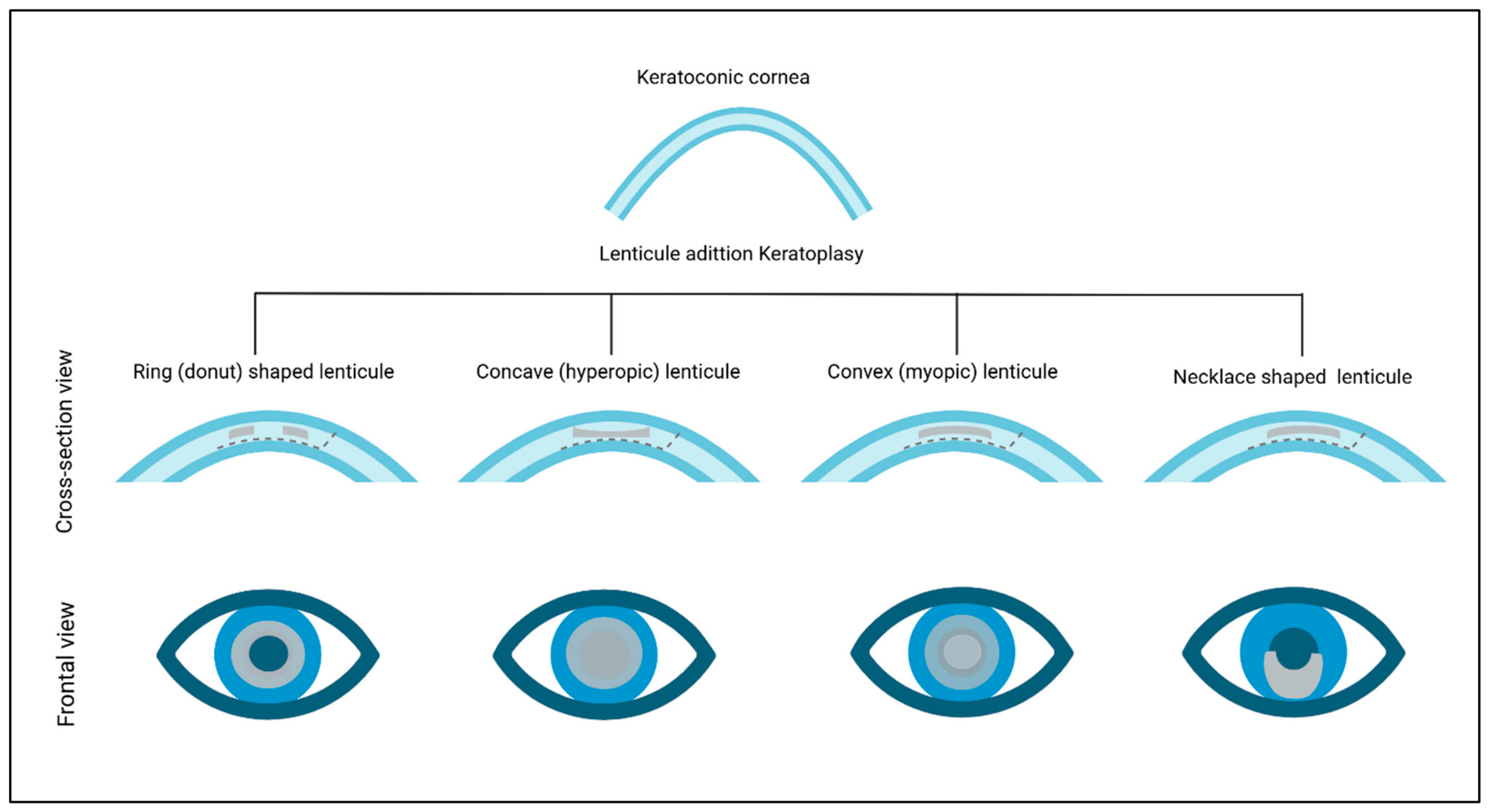
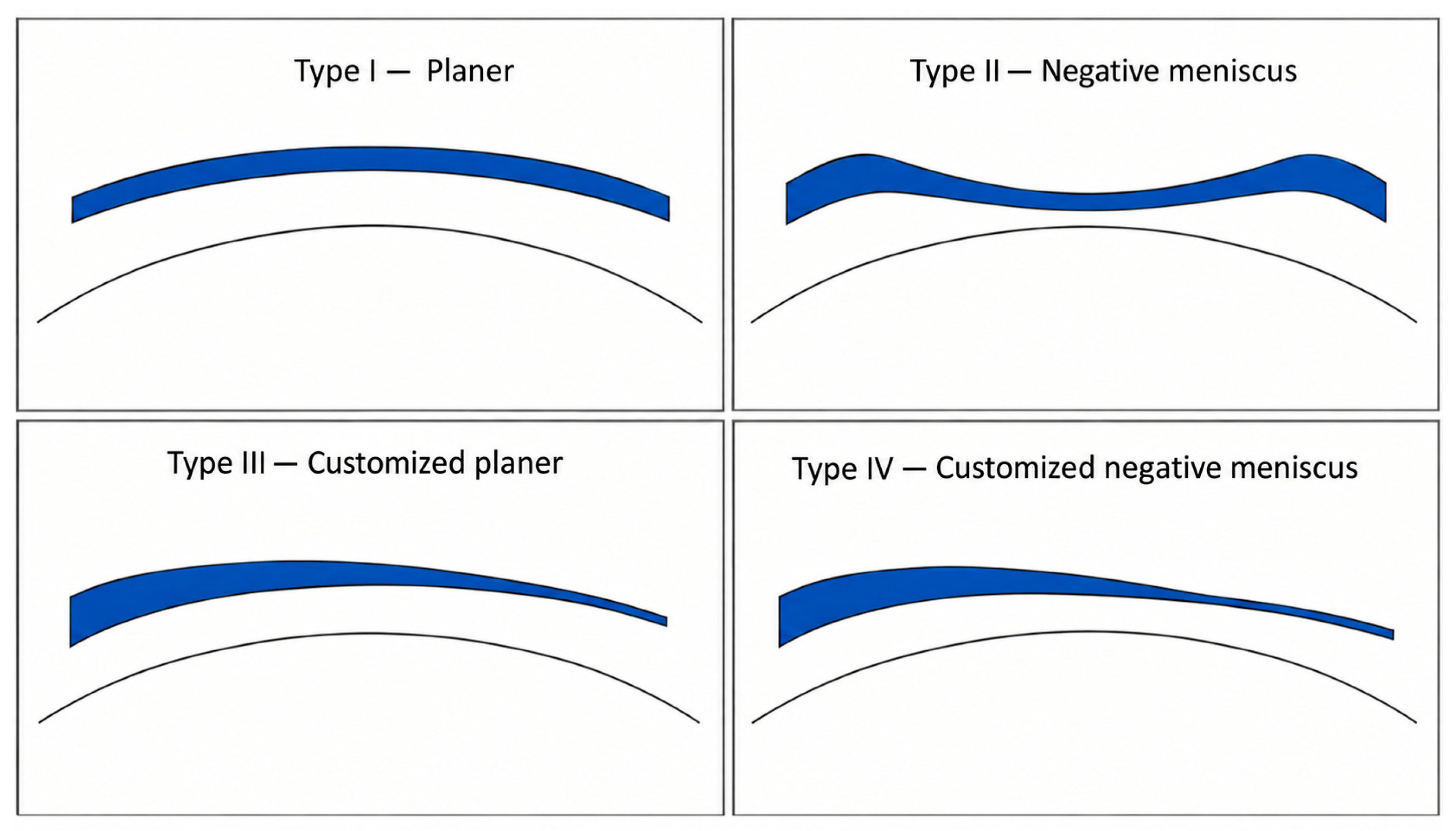
4.1. The Use of KLEx Lenticule for the Treatment of Keratoconus and Post-LASIK Ectasia
4.2. The Use of KLEx-Derived Lenticules as a Scaffold for Cells
| Approach | Category | Agent | Mechanism of Action | Advantages | Limitations | References |
|---|---|---|---|---|---|---|
| Chemical | Alcohols | Ethanol, Acetone | Cell dehydration and lysis through lipid membrane disruption | Simple, rapid, and highly efficient | ECM ultrastructural damage and tissue transparency alteration due to dehydration | Ponce Marquez et al., 2009, Lumpkins et al., 2008 [107,108] |
| Ionic detergents | Sodium dodecyl sulfate (SDS) | Solubilization of the cell membrane and denaturation of proteins | Highly effective decellularization | Induced protein denaturation that might affect the ECM organization and protein composition; high cytotoxicity if not fully removed | Yam et al. 2016, Du et al. 2011, Yoeruek et al. 2012, Alio et al. 2018, and Wilson et al. 2016, Marin Tapia et al. 2021 [109,110,111,112,117,133] | |
| Non-ionic detergents | Triton X-100 | Disruption of lipid–lipid and lipid–protein interactions | Minimal disruption of ECM architecture and collagen organization compared to other surfactants | Incomplete removal of cellular and nuclear debris; requires combinations with other agents | Du and Wu, 2011, Wilson et al. 2016, Yam 2016 -decellularization, Marin Tampa et al. 2021 [109,110,117,133] | |
| Zwitterionic detergents | 3-[(3-cholamidopropyl)dimethylamminio]-1 propanesulfonate (CHAPS) | Solubilization of the cell membrane | Better ECM preservation compared to ionic detergents | Poor cellular removal and ECM protein-induced damage; requires combination with other agents | Du et al. 2011, Marin Tapia et al. 2021 [110,133] | |
| Chelating agents | Ethylene-diamine-tetraacetic acid (EDTA) | Ca2+/Mg2+ chelating; disrupting cell adhesion and causing cell dissociation | Optical properties retention | Ineffective alone; requires combination protocols | Bayyoud et al., 2012, Oh et al., 2009, Yoeruek et al., 2012, Poornejad et al., 2016, Huh et al., 2018 [111,115,118,134,135] | |
| Hypotonic and hypertonic solutions | Sodium chloride solution | Disrupt the cell membrane through osmotic shock | Minimal damage to the ECM architecture and proteoglycans (GAG); good optical property retention | Low removal efficacy of cellular and nuclear debris | Gilbert et al., 2006, Yam et al., Wilson et al. 2016, Zang 2015, Gonzales-Andreas et al. 2011 [109,117,136,137,138] | |
| Acid and basic solutions | Formic acid (HCOOH), Ammonium hydroxide (NH4OH) | Hydrolytic degradation of biomolecules and nucleic acids | Highly effective decellularization | ECM protein damage; altered biomechanics | Choi et al., 2010, Zang 2015, Lin et al. 2019 [137,139,140] | |
| Biological | Esterase | Phospholipase A2 | Hydrolysis of ester bonds in phospholipids, contributing to cell membrane degradation | Potentially milder than proteases; supports lipid removal | ECM component disruption | Wu et al., 2009 [113] |
| Protease | Trypsin | Proteolytic cleavage of peptide bonds, disrupting cell–matrix adhesions | Efficient cell removal | ECM degradation and weakening if overexposed | He et al. 2016, Huh et al. 2018, Marquez et al., 2009, Procházková, 2024 [108,114,115,116] | |
| Nucleases | DNase and RNase | Cleavage of phosphodiester bonds in nucleic acids | Minimal damage to the ECM architecture and components | Requires combination with other agents for membrane disruption and access to nucleic acids; difficult to be cleared from the ECM | Wilson et al. 2016, Oh et al., 2009, He et al., 2016 [116,117,118] | |
| Physical | Freeze and thaw | Intracellular ice crystal formation causes mechanical rupture of cell membranes | Simple; no chemical residues; good optical properties compared to chemical detergents | Must be combined with other decellularizing agents to remove DNA remnants from the ECM scaffold | Oh et al., 2009, Crapo et al. 2011 [106,118] | |
| Supercritical carbon dioxide | Membrane lipid solubilization via supercritical fluid extraction | No toxic residue; simultaneous tissue sterilization; reduced decellularization time | Requires complex ScCO2 reactor system | Sawada et al., 2008, Huang et al. 2017, Guler et al. 2017, Liang et al. 2022 [119,120,121,122] | ||
| Pressurization | High hydrostatic pressure (HHP) (>600 MPa) induces irreversible cellular membrane phase transition and protein denaturation. | Efficient decellularization with the maintenance of the collagen fibril matrix | Expensive specialized equipment that generates high HHP (~1GPa) is required | Sasaki et al., 2009, Hashimoto et al. 2010 [123,124] | ||
| Ultrasonication | Acoustic cavitation induces mechanical rupture of the cell membrane | Uniform treatment and short treatment time | May require a combination with detergent (SDS) to complete decellularization | Azhim et al., 2014 [128] | ||
| Gamma radiation | Ionizing radiation induces DNA fragmentation and cellular damage | Simultaneous tissue sterilization; reduced decellularization time | May cause loss of tissue transparency | Henner et al., 1983, Stevenson et al., 2012, Utine et al., 2011 [125,126,127] | ||
| Electroporation | High voltage electrical field induces irreversible nanopore formation and cell lysis | ECM structural preservation | Incomplete removal of cellular and nuclear debris; requires combinations with other agents | Chang et al., 2017 [129] |
| Study | Cell Type | Experimental Model | Application | Key Finding/Outcomes |
|---|---|---|---|---|
| Alió et al. (2015) [130] | h-ADASCs | Rabbit | Stromal tissue engineering | Human ADASCs differentiated into keratocytes in vivo; clinical study showed improved vision with no adverse effects, but the recellularization benefit was unclear |
| Alió et al. (2018) [112] | Autologous h-ADASCs | Clinical | ||
| Aghamollaei et al. (2021) [131] | WJ-MSCs | Rabbit | Stromal tissue engineering | Good graft integration with no signs of rejection; recellularized lenticules showed higher expression of keratocyte markers (lumican, keratocan) compared to acellular control |
| Yam et al. (2016) [109] | Stromal fibroblast (In vitro) | In vitro + Rabbit (SDS-decellularized lenticule) | Stromal tissue engineering | In vitro: cell adhesion, low cytotoxicity; In vivo: good integration with no signs of necrosis, neovascularization, inflammation or extrusion |
| Ghiasi et al. (2023) [132] | h-ADASCs + KCM | Rabbit | Stromal tissue engineering | KCM increased keratocyte-specific gene and protein expression; in vivo results showed good lenticule integration, with no signs of inflammation or neovascularization |
| Hong et al. (2018) [141] | LESCs | Rabbit LSCD model | LSCD (epithelial reconstruction) | The developed collagen-lenticule biocomposite successfully restored stratified epithelium in rabbit LSCD models without neovascularization. |
| Qin et al. (2019) [142] | iPSCs | In vitro | LSCD (epithelial reconstruction) | Formation of stratified epithelial-like sheets on decellularized lenticules |
| Hazra et al. (2023) [143] | hCECs | Ex vivo (donor human cornea) | Endothelial Keratoplasty | Proof-of-concept showing a confluent endothelial monolayer; successfully reduced corneal thickness in an ex vivo edema model. |
| Gu et al. (2019) [144] | RPE cells in iPSC-CM | Rabbit | RPE cell therapy | PSC-conditioned medium enhanced RPE density and barrier function; lenticules supported RPE polarization and maturation; good biocompatibility following subretinal implantation |
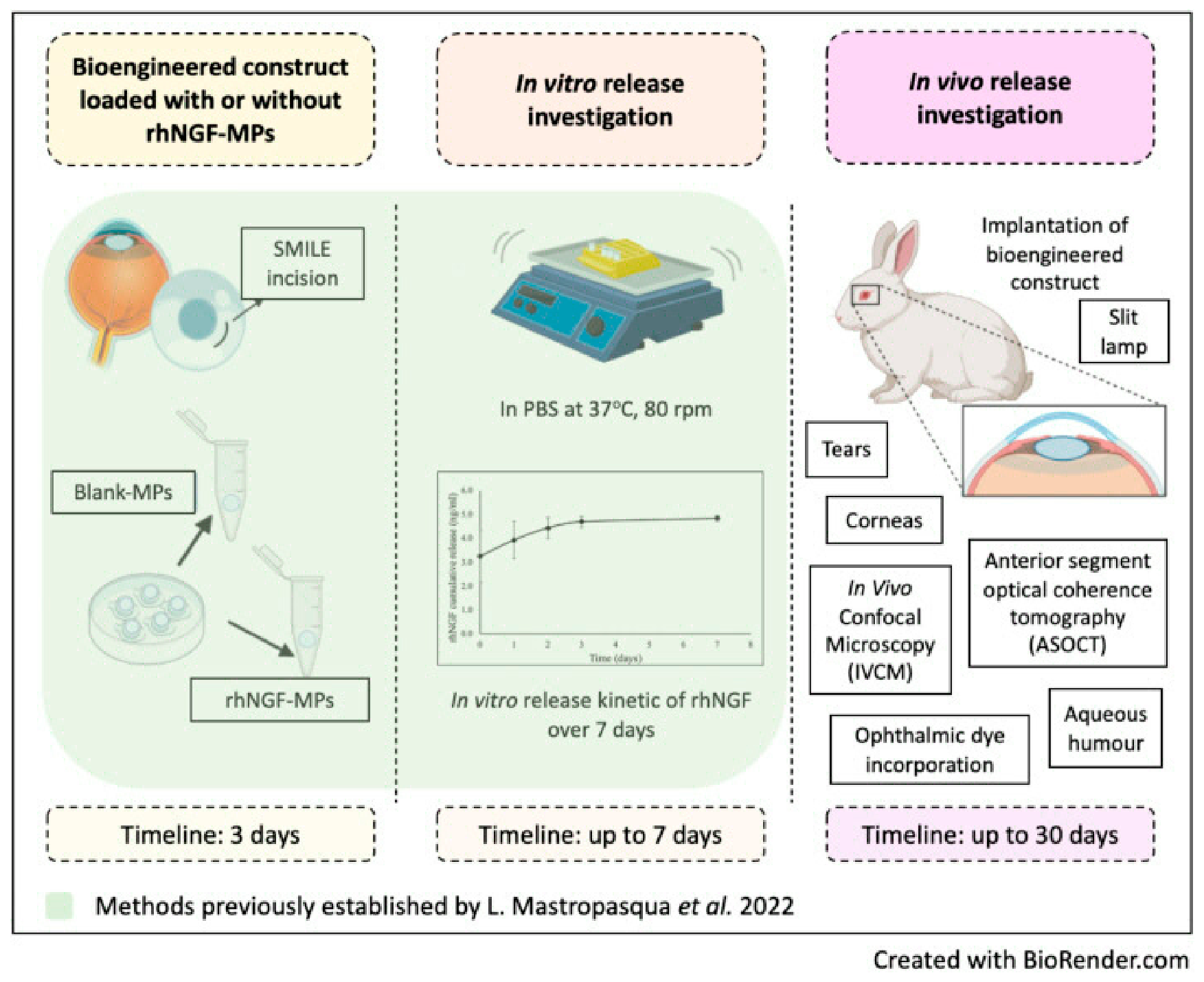
4.3. The Use of KLEx Lenticules as Drug Delivery System
4.4. The Use of KLEx Lenticule for Tectonic Keratoplasty
4.4.1. Biological Patches for Corneal Perforation and Macular Hole
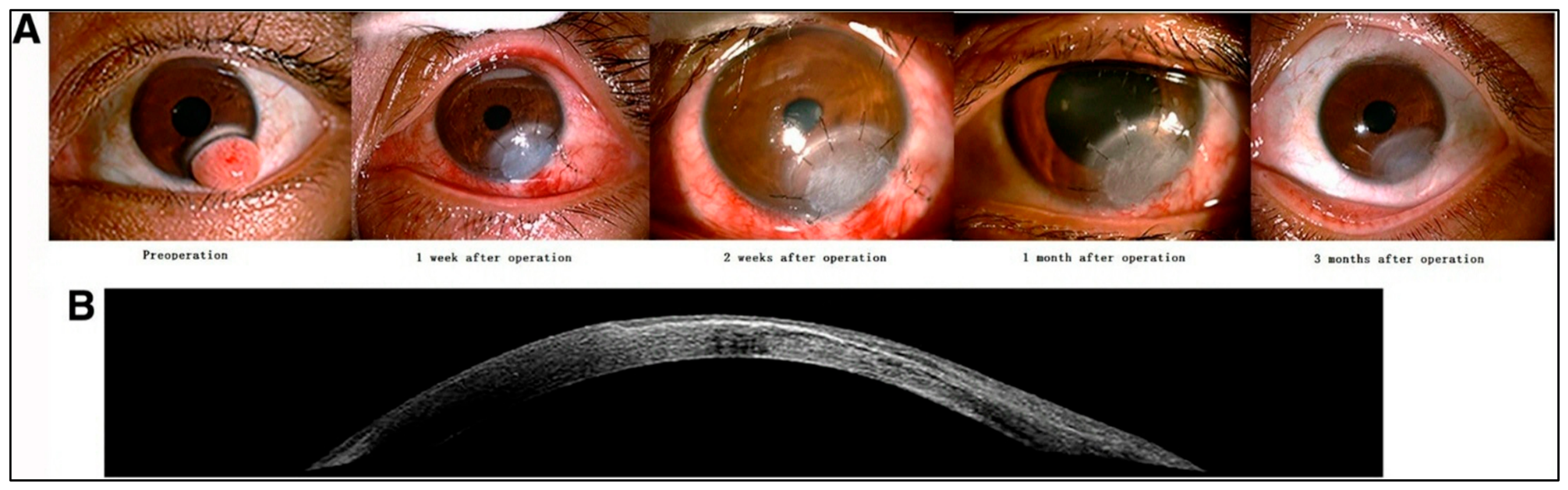
4.4.2. KLEx-Derived Lenticules for Pterygium and Dermoid Treatment
4.4.3. The Use of KLEx Lenticules as a Patch Graft in Glaucoma Drainage Implant Surgery
5. Conclusions and Future Outlook
Author Contributions
Funding
Data Availability Statement
Conflicts of Interest
References
- Nishida, T.; Saika, S.; Morishige, N. Cornea and Sclera: Anatomy and Physiology. In Cornea: Fundamentals, Diagnosis and Management, 4th ed.; Mannis, M.J., Holland, E.J., Eds.; Elsevier: New York, NY, USA, 2017; pp. 1–22. [Google Scholar]
- Olsen, T. On the calculation of power from curvature of the cornea. Br. J. Ophthalmol. 1986, 70, 152–154. [Google Scholar] [CrossRef] [Scilit] [PubMed]
- Wang, J.; Bao, J.; Song, W.; Li, S.; Hao, Y.; Tian, L.; Jie, Y. Assessment of Eyelid Pressure Using a Novel Pressure Measurement Device in Patients With Moderate-to-Severe Dry Eye Disease. Front. Med. 2022, 9, 833576. [Google Scholar] [CrossRef] [Scilit] [PubMed]
- Bates, W.H. A suggestion of an operation to correct astigmatism. J. Refract. Surg. 1989, 5, 58–59. [Google Scholar] [CrossRef] [Scilit]
- Lans, L.J. Experimentelle Untersuchungen über Entstehung von Astigmatismus durch nicht-perforirende Corneawunden. Albrecht Von Graefes Arch. Ophthalmol. 1898, 45, 117–152. [Google Scholar] [CrossRef] [Scilit]
- Barker, B.A.; Swinger, C.A. Keratophakia and keratomileusis. Int. Ophthalmol. Clin. 1988, 28, 126–133. [Google Scholar] [CrossRef] [Scilit] [PubMed]
- Barraquer, J.I. Method for Cutting Lamellar Grafts in Frozen Cornea, New Orientation for Refractive Surgery. Arch. Soc. Am. Ophthalmic Optom. 1958, 1, 271–286. [Google Scholar]
- Friedlander, M.H.; Rich, L.F.; Werblin, T.P.; Kaufman, H.E.; Granet, N. Keratophakia Using Preserved Lenticules. Ophthalmology 1980, 87, 687–692. [Google Scholar] [CrossRef] [Scilit] [PubMed]
- Friedlander, M.H.; Werblin, T.P.; Kaufman, H.E.; Granet, N.S. Clinical Results of Keratophakia and Keratomileusis. Ophthalmology 1981, 88, 716–720. [Google Scholar] [CrossRef] [Scilit] [PubMed]
- Friedlander, M.H.; Safir, A.; McDonald, M.B.; Kaufman, H.E.; Granet, N. Update on Keratophakia. Ophthalmology 1983, 90, 365–368. [Google Scholar] [CrossRef] [Scilit] [PubMed]
- Riau, A.K.; Liu, Y.C.; Yam, G.H.F.; Mehta, J.S. Stromal keratophakia: Corneal inlay implantation. Prog. Retin Eye Res. 2020, 75, 100780. [Google Scholar] [CrossRef] [Scilit] [PubMed]
- Swinger, C.A.; Barraquer, J.I. Keratophakia and keratomileusis—Clinical results. Ophthalmology 1981, 88, 709–715. [Google Scholar] [CrossRef] [Scilit] [PubMed]
- Salchow, D.J.; Zirm, M.E.; Stieldorf, C.; Parisi, A. Laser in situ keratomileusis for myopia and myopic astigmatism. J. Cataract Refract. Surg. 1998, 24, 175–182. [Google Scholar] [CrossRef] [Scilit] [PubMed]
- Werblin, T.P.; Kaufman, H.E.; Friedlander, M.H.; McDonald, M.B.; Sehon, K.L. Epikeratophakia—The surgical correction of aphakia. Update 1981. Ophthalmology 1982, 89, 916–920. [Google Scholar] [CrossRef] [Scilit] [PubMed]
- Cheng, K.P.; Hiles, D.A.; Biglan, A.W.; Pettapiece, M.C.; Behler, S.C.; Moore, M.B. Risk factors for complications following pediatric epikeratoplasty. J. Cataract Refract. Surg. 1992, 18, 270–279. [Google Scholar] [CrossRef] [Scilit] [PubMed]
- Busin, M.; Cusumano, A.; Spitznas, M. Epithelial interface cysts after epikeratophakia. Ophthalmology 1993, 100, 1225–1229. [Google Scholar] [CrossRef] [Scilit] [PubMed]
- Miller, S.M.; Sitto, M.M.; Moin, K.A.; Hoopes, P.C.; Moshirfar, M. Comparing The Existing Myopic Keratorefractive Lenticule Extraction (KLEx) Platforms: A Narrative Review. Clin. Ophthalmol. 2025, 19, 2189–2202. [Google Scholar] [CrossRef] [Scilit] [PubMed]
- Blum, M.; Khoramnia, R.; Sekundo, W. Keratorefractive Lenticule Extraction—KLEx. Klin. Monbl. Augenheilkd. 2025, 242, 795–802. [Google Scholar] [CrossRef] [Scilit] [PubMed]
- Sandoval, H.P.; de Castro, L.E.; Vroman, D.T.; Solomon, K.D. Refractive Surgery Survey 2004. J. Cataract Refract. Surg. 2005, 31, 221–233. [Google Scholar] [CrossRef] [Scilit] [PubMed]
- Gain, P.; Jullienne, R.; He, Z.; Aldossary, M.; Acquart, S.; Cognasse, F.; Thuret, G. Global Survey of Corneal Transplantation and Eye Banking. JAMA Ophthalmol. 2016, 134, 167–173. [Google Scholar] [CrossRef] [Scilit] [PubMed]
- Zhang, Y.; Li, J.; Wu, Z.; Li, Y.; Wu, G.; Wei, S. The Preservation and Reuse of Lenticules Extracted via Small Incision Lenticule Extraction (SMILE): A Narrative Review. Bioengineering 2025, 12, 380. [Google Scholar] [CrossRef] [Scilit] [PubMed]
- Santra, M.; Liu, Y.C.; Jhanji, V.; Yam, G.H. Human SMILE-Derived Stromal Lenticule Scaffold for Regenerative Therapy: Review and Perspectives. Int. J. Mol. Sci. 2022, 23, 7967. [Google Scholar] [CrossRef] [Scilit] [PubMed]
- Riau, A.K.; Angunawela, R.I.; Chaurasia, S.S.; Lee, W.S.; Tan, D.T.; Mehta, J.S. Reversible femtosecond laser-assisted myopia correction: A non-human primate study of lenticule re-implantation after refractive lenticule extraction. PLoS ONE 2013, 8, e67058. [Google Scholar] [CrossRef] [Scilit] [PubMed]
- Angunawela, R.I.; Riau, A.K.; Chaurasia, S.S.; Tan, D.T.; Mehta, J.S. Refractive lenticule re-implantation after myopic ReLEx: A feasibility study of stromal restoration after refractive surgery in a rabbit model. Investig. Ophthalmol. Vis. Sci. 2012, 53, 4975–4985. [Google Scholar] [CrossRef] [Scilit] [PubMed]
- Pradhan, K.R.; Reinstein, D.Z.; Carp, G.I.; Archer, T.J.; Gobbe, M.E.; Gurung, R. Femtosecond laser-assisted keyhole endokeratophakia: Correction of hyperopia by implantation of an allogeneic lenticule obtained by SMILE from a myopic donor. J. Refract. Surg. 2013, 29, 777–782. [Google Scholar] [CrossRef] [Scilit] [PubMed]
- Ganesh, S.; Brar, S. Femtosecond Intrastromal Lenticular Implantation Combined with Accelerated Collagen Cross-Linking for the Treatment of Keratoconus-Initial Clinical Result in 6 Eyes. Cornea 2015, 34, 1331–1339. [Google Scholar] [CrossRef] [Scilit] [PubMed]
- Pelusi, L.; Hurst, J.; Detta, N.; Pipino, C.; Lamolinara, A.; Conte, G.; Mastropasqua, R.; Allegretti, M.; Di Pietrantonio, N.; Romeo, T.; et al. Effects of mesenchymal stromal cells and human recombinant Nerve Growth Factor delivered by bioengineered human corneal lenticule on an innovative model of diabetic retinopathy. Front. Endocrinol. 2024, 15, 1462043. [Google Scholar] [CrossRef] [Scilit] [PubMed]
- Kilic, A.; Cummings, A.; Stodulka, P.; Daya, S.; Hamada, S.; Gobbe, M.; Muller, D.; Mrochen, M. Corneal Allograft for Near Vision Improvement in Emmetropic Presbyopia: 4-Year Safety and Efficacy Results from a Prospective European Multicenter Clinical Trial. J. Refract. Surg. 2026, 42, e100–e107. [Google Scholar] [CrossRef] [Scilit] [PubMed]
- Abd Elaziz, M.S.; Zaky, A.G.; El SaebaySarhan, A.R. Stromal lenticule transplantation for management of corneal perforations; one year results. Graefes Arch. Clin. Exp. Ophthalmol. 2017, 255, 1179–1184. [Google Scholar] [CrossRef] [Scilit] [PubMed]
- Bievel-Radulescu, R.; Ferrari, S.; Piaia, M.; Mandatori, D.; Pandolfi, A.; Nubile, M.; Mastropasqua, L.; Stanca, H.T.; Ponzin, D. Banking of post-SMILE stromal lenticules for additive keratoplasty: A new challenge for eye banks? Int. Ophthalmol. 2024, 44, 355. [Google Scholar] [CrossRef] [Scilit] [PubMed]
- Nemcokova, M.; Dite, J.; Klimesova, Y.M.; Netukova, M.; Studeny, P. Preservation of corneal stromal lenticule: Review. Cell Tissue Bank. 2022, 23, 627–639. [Google Scholar] [CrossRef] [Scilit] [PubMed]
- Liu, Y.C.; Williams, G.P.; George, B.L.; Soh, Y.Q.; Seah, X.Y.; Peh, G.S.L.; Yam, G.H.F.; Mehta, J.S. Corneal lenticule storage before reimplantation. Mol. Vis. 2017, 23, 753–764. [Google Scholar] [PubMed]
- Christodoulou, R.; Mandatori, D.; Boldini, A.; Russo, A.; Mastropasqua, L.; Zarif, M.E.; Ruzza, A.; Pelusi, L.; Pandolfi, A.; Pietrangelo, L.; et al. Biological assessment and preservation strategies for SMILE- derived lenticules: Towards a corneal regenerative biobank. Sci. Rep. 2026, 16, 19882. [Google Scholar] [CrossRef] [Scilit] [PubMed]
- Soo, J.Y.W.; Tan, G.; Han, E.; Boey, K.P.Y.; Liu, Y.C.; Mehta, J.S.; Riau, A.K. Impact of Transportation on the Suitability of Cryopreserved Corneal Lenticule for Implantation. ACS Appl. Bio Mater. 2025, 8, 2065–2077. [Google Scholar] [CrossRef] [Scilit] [PubMed]
- Mohamed-Noriega, K.; Toh, K.P.; Poh, R.; Balehosur, D.; Riau, A.; Htoon, H.M.; Peh, G.S.; Chaurasia, S.S.; Tan, D.T.; Mehta, J.S. Cornea lenticule viability and structural integrity after refractive lenticule extraction (ReLEx) and cryopreservation. Mol. Vis. 2011, 17, 3437–3449. [Google Scholar] [PubMed]
- Ganesh, S.; Brar, S.; Rao, P.A. Cryopreservation of extracted corneal lenticules after small incision lenticule extraction for potential use in human subjects. Cornea 2014, 33, 1355–1362. [Google Scholar] [CrossRef] [Scilit] [PubMed]
- Li, J.; Shi, S.; Zhang, X.; Ni, S.; Wang, Y.; Curcio, C.A.; Chen, W. Comparison of different methods of glycerol preservation for deep anterior lamellar keratoplasty eligible corneas. Investig. Ophthalmol. Vis. Sci. 2012, 53, 5675–5685. [Google Scholar] [CrossRef] [Scilit] [PubMed]
- Tripathi, H.; Mehdi, M.U.; Gupta, D.; Sen, S.; Kashyap, S.; Nag, T.C.; Purwar, M.; Jassal, M.; Agrawal, A.K.; Mohanty, S.; et al. Long-term preservation of donor corneas in glycerol for keratoplasty: Exploring new protocols. Br. J. Ophthalmol. 2016, 100, 284–290. [Google Scholar] [CrossRef] [Scilit] [PubMed]
- Chen, W.; Lin, Y.; Zhang, X.; Wang, L.; Liu, M.; Liu, J.; Ye, Y.; Sun, L.; Ma, H.; Qu, J. Comparison of fresh corneal tissue versus glycerin-cryopreserved corneal tissue in deep anterior lamellar keratoplasty. Investig. Ophthalmol. Vis. Sci. 2010, 51, 775–781. [Google Scholar] [CrossRef] [Scilit] [PubMed]
- Riau, A.K.; Boey, K.P.Y.; Binte M Yusoff, N.Z.; Goh, T.W.; Yam, G.H.F.; Tang, K.F.; Phua, C.S.H.; Chen, H.J.; Chiew, Y.F.; Liu, Y.C.; et al. Experiment-Based Validation of Corneal Lenticule Banking in a Health Authority-Licensed Facility. Tissue Eng. Part A 2022, 28, 69–83. [Google Scholar] [CrossRef] [Scilit] [PubMed]
- Parekh, M.; Ferrari, S.; Ruzza, A.; Leon, P.; Franch, A.; Camposampiero, D.; Gadhvi, K.A.; Ponzin, D.; Ahmad, S.; Romano, V. Biobanking corneal tissues for emergency procedures during COVID-19 era. Indian J. Ophthalmol. 2021, 69, 167–168. [Google Scholar] [CrossRef] [Scilit] [PubMed]
- Romano, V.; Levis, H.J.; Gallon, P.; Lace, R.; Borroni, D.; Ponzin, D.; Ruzza, A.; Kaye, S.B.; Ferrari, S.; Parekh, M. Biobanking of Dehydrated Human Donor Corneal Stroma to Increase the Supply of Anterior Lamellar Grafts. Cornea 2019, 38, 480–484. [Google Scholar] [CrossRef] [Scilit] [PubMed]
- Vautier, A.; Bourges, J.L.; Gabison, E.; Chaventre, F.; Lefevre, S.; Toubeau, D.; Arnoult, C.; Albou-Ganem, C.; Boyer, O.; Muraine, M. An Efficient Technique for the Long-term Preservation of SMILE Lenticules Using Desiccation. J. Refract. Surg. 2023, 39, 491–498. [Google Scholar] [CrossRef] [Scilit] [PubMed]
- Bovone, C.; De Rosa, L.; Pellegrini, M.; Ruzza, A.; Ferrari, S.; Camposampiero, D.; Ponzin, D.; Zauli, G.; Yu, A.C.; Busin, M. Deep Anterior Lamellar Keratoplasty Using Dehydrated versus Standard Organ Culture-Stored Donor Corneas: Prospective Randomized Trial. Ophthalmology 2024, 131, 674–681. [Google Scholar] [CrossRef] [Scilit] [PubMed]
- Zhao, J.; Zhang, Z.; Xia, F.; Li, G.; Saiding, Q.; Xiang, L.; Xu, H.; Yao, P.; Wang, X.; Cai, Z.; et al. Nutrient capsules maintain tear film homeostasis for human corneal lenticule transplantation. Chem. Eng. J. 2022, 450, 138078. [Google Scholar] [CrossRef] [Scilit]
- Zhang, Z.; Sun, B.; Xia, F.; Yu, Y.; Shen, Y.; Yao, P.; Wang, X.; Zhou, X.; Zhao, J. Study on the biological properties of SMILE-derived corneal stromal lenticules after long-term cryopreservation in nutrient capsules. Exp. Eye Res. 2024, 239, 109756. [Google Scholar] [CrossRef] [Scilit] [PubMed]
- Zhang, Z.; Yu, Y.; Sun, B.; Li, M.; Yao, P.; Wang, X.; Zhou, X.; Zhao, J. Long-term follow-up of SMILE-derived corneal stromal lenticules preserved in nutrient capsules for treating corneal diseases. BMC Ophthalmol. 2025, 25, 64. [Google Scholar] [CrossRef] [Scilit] [PubMed]
- Xia, F.; Zhao, J.; Fu, D.; Xu, Y.; Yao, P.; Li, M.; Aruma, A.; Zhou, X. Optical transmittance and ultrastructure of SMILE-derived lenticules subjected to three different preservative methods. Exp. Eye Res. 2020, 201, 108357. [Google Scholar] [CrossRef] [Scilit] [PubMed]
- Hourd, P.; Medcalf, N.; Segal, J.; Williams, D.J. A 3D bioprinting exemplar of the consequences of the regulatory requirements on customized processes. Regen. Med. 2015, 10, 863–883. [Google Scholar] [CrossRef] [Scilit] [PubMed]
- Lambert, N.G.; Chamberlain, W.D. The structure and evolution of eye banking: A review on eye banks’ historical, present, and future contribution to corneal transplantation. J. Biorepository Sci. Appl. Med. 2017, 5, 23–40. [Google Scholar] [CrossRef] [Scilit]
- Parekh, M.; Wongvisavavit, R.; Cubero Cortes, Z.M.; Wojcik, G.; Romano, V.; Tabernero, S.S.; Ferrari, S.; Ahmad, S. Alternatives to endokeratoplasty: An attempt towards reducing global demand of human donor corneas. Regen. Med. 2022, 17, 461–475. [Google Scholar] [CrossRef] [Scilit] [PubMed]
- Hyman, L. Myopic and hyperopic refractive error in adults: An overview. Ophthalmic Epidemiol. 2007, 14, 192–197. [Google Scholar] [CrossRef] [Scilit] [PubMed]
- Hashemi, H.; Fotouhi, A.; Yekta, A.; Pakzad, R.; Ostadimoghaddam, H.; Khabazkhoob, M. Global and regional estimates of prevalence of refractive errors: Systematic review and meta-analysis. J. Curr. Ophthalmol. 2017, 30, 3–22. [Google Scholar] [CrossRef] [Scilit] [PubMed]
- Williams, G.P.; Wu, B.; Liu, Y.C.; Teo, E.; Nyein, C.L.; Peh, G.; Tan, D.T.; Mehta, J.S. Hyperopic refractive correction by LASIK, SMILE or lenticule reimplantation in a non-human primate model. PLoS ONE 2018, 13, e0194209. [Google Scholar] [CrossRef] [Scilit] [PubMed]
- Lee, B.S. Accuracy and stability of hyperopic treatments. Curr. Opin. Ophthalmol. 2014, 25, 281–285. [Google Scholar] [CrossRef] [Scilit] [PubMed]
- Alió, J.L.; El Aswad, A.; Vega-Estrada, A.; Javaloy, J. Laser in situ keratomileusis for high hyperopia (>5.0 diopters) using optimized aspheric profiles: Efficacy and safety. J. Cataract Refract. Surg. 2013, 39, 519–527. [Google Scholar] [CrossRef] [Scilit] [PubMed]
- Aslanides, I.M.; Mukherjee, A.N. Adjuvant corneal crosslinking to prevent hyperopic LASIK regression. Clin. Ophthalmol. 2013, 7, 637–641. [Google Scholar] [CrossRef] [Scilit] [PubMed]
- Settas, G.; Settas, C.; Minos, E.; Yeung, I.Y. Photorefractive keratectomy (PRK) versus laser assisted in situ keratomileusis (LASIK) for hyperopia correction. Cochrane Database Syst. Rev. 2012, 2012, CD007112. [Google Scholar] [CrossRef] [Scilit] [PubMed]
- Mimouni, M.; Vainer, I.; Shapira, Y.; Levartovsky, S.; Sela, T.; Munzer, G.; Kaiserman, I. Factors Predicting the Need for Retreatment After Laser Refractive Surgery. Cornea 2016, 35, 607–612. [Google Scholar] [CrossRef] [Scilit] [PubMed]
- Pesudovs, K. Wavefront aberration outcomes of LASIK for high myopia and high hyperopia. J. Refract. Surg. 2005, 21, S508–S512. [Google Scholar] [CrossRef] [Scilit] [PubMed]
- Barraquer, J.I. Queratoplastia refractiva. Estud. Inf. Oftalmol. 1948, 2, 10–14. [Google Scholar]
- Brar, S.; Ganesh, S.; Sriganesh, S.S.; Bhavsar, H. Femtosecond Intrastromal Lenticule Implantation (FILI) for Management of Moderate to High Hyperopia: 5-Year Outcomes. J. Refract. Surg. 2022, 38, 348–354. [Google Scholar] [CrossRef] [Scilit] [PubMed]
- Dave, R.; O’Brart, D.P.; Wagh, V.K.; Lim, W.S.; Patel, P.; Lee, J.; Marshall, J. Sixteen-year follow-up of hyperopic laser in situ keratomileusis. J. Cataract Refract. Surg. 2016, 42, 717–724. [Google Scholar] [CrossRef] [Scilit] [PubMed]
- Thirunavukarasu, A.J.; Han, E.; Nedumaran, A.M.; Kurz, A.C.; Shuman, J.; Yusoff, N.Z.B.M.; Liu, Y.C.; Foo, V.; Czarny, B.; Riau, A.K.; et al. Electron beam-irradiated donor cornea for on-demand lenticule implantation to treat corneal diseases and refractive error. Acta Biomater. 2023, 169, 334–347. [Google Scholar] [CrossRef] [Scilit] [PubMed]
- Sun, L.; Yao, P.; Li, M.; Shen, Y.; Zhao, J.; Zhou, X. The Safety and Predictability of Implanting Autologous Lenticule Obtained by SMILE for Hyperopia. J. Refract. Surg. 2015, 31, 374–379. [Google Scholar] [CrossRef] [Scilit] [PubMed]
- Li, M.; Li, M.; Sun, L.; Ni, K.; Zhou, X. Predictive Formula for Refraction of Autologous Lenticule Implantation for Hyperopia Correction. J. Refract. Surg. 2017, 33, 827–833. [Google Scholar] [CrossRef] [Scilit] [PubMed]
- Lin, F.; Cheng, C.; Li, M.; Liu, S.; Zhou, X. Visual outcomes and corneal densitometry after allogenic and autologous lenticule intrastromal keratoplasty for the correction of moderate-to-high hyperopia. Graefes Arch. Clin. Exp. Ophthalmol. 2023, 261, 3015–3022. [Google Scholar] [CrossRef] [Scilit] [PubMed]
- Hou, J.; Wang, Y.; Zhang, J.; Lei, Y.; Ma, Z.; Zhang, Y.; Zheng, X. Corneal densitometry after allogeneic small-incision intrastromal lenticule implantation for hyperopia correction. BMC Ophthalmol. 2022, 22, 286. [Google Scholar] [CrossRef] [Scilit] [PubMed]
- Wu, J.; Xiong, L.; Wang, Z.; Thornton, I. Change in Corneal Tomography Following Femtosecond Laser-Assisted Lenticule Intrastromal Keratoplasty (LIKE) in Correcting Moderate to High Hyperopia. J. Refract. Surg. 2025, 41, e998–e1007. [Google Scholar] [CrossRef] [Scilit] [PubMed]
- Lin, F.; Liu, S.; Fu, D.; Zhang, L.; Wei, R.; Li, M.; Zhou, X. Comparison of Visual Outcomes and Higher-order Aberrations Between FS-LASIK and SMI-LIKE for Moderate to High Hyperopia: A 2-Year Result. Cornea 2023, 42, 1506–1512. [Google Scholar] [CrossRef] [Scilit] [PubMed]
- Zhang, L.; Zhou, Y.H.; Zhai, C.B.; Zhang, J.; Zheng, Y. Comparison of clinical outcome of small-incision lenticule intrastromal keratoplasty and FS-LASIK for correction of moderate and high hyperopia. Int. J. Ophthalmol. 2022, 15, 780–785. [Google Scholar] [CrossRef] [Scilit] [PubMed]
- Moshirfar, M.; Hopping, G.C.; Somani, A.N.; Vaidyanathan, U.; Liu, H.; Barnes, J.R.; Linn, S.; Ronquillo, Y.C.; Hoopes, P.C. Human allograft refractive lenticular implantation for high hyperopiccorrection. J. Cataract Refract. Surg. 2020, 46, 305–311. [Google Scholar] [CrossRef] [Scilit] [PubMed]
- Lazaridis, A.; Reinstein, D.Z.; Archer, T.J.; Schulze, S.; Sekundo, W. Refractive Lenticule Transplantation for Correction of Iatrogenic Hyperopia and High Astigmatism After LASIK. J. Refract. Surg. 2016, 32, 780–786. [Google Scholar] [CrossRef] [Scilit] [PubMed]
- Liu, S.; Wei, R.; Choi, J.; Li, M.; Zhou, X. Visual Outcomes After Implantation of Allogenic Lenticule in a 100-µm Pocket for Moderate to High Hyperopia: 2-Year Results. J. Refract. Surg. 2021, 37, 734–740. [Google Scholar] [CrossRef] [Scilit] [PubMed]
- Liu, S.; Yu, L.; Zhao, Y.; Zhou, X. Changes in the posterior corneal surface after femtosecond laser-assisted lenticule intrastromal keratoplasty (LIKE) performed into a pocket (SMI-LIKE) or under a flap (FS-LIKE). Eye Vis. 2023, 10, 23. [Google Scholar] [CrossRef] [Scilit] [PubMed]
- Damgaard, I.B.; Ivarsen, A.; Hjortdal, J. Biological Lenticule Implantation for Correction of Hyperopia: An Ex Vivo Study in Human Corneas. J. Refract. Surg. 2018, 34, 245–252. [Google Scholar] [CrossRef] [Scilit] [PubMed]
- Dong, Y.; Hou, J.; Zhang, J.; Lei, Y.; Yang, X.; Sun, F. Epithelial thickness remodeling after small incision lenticule intrastromal keratoplasty in correcting hyperopia measured by RTVue OCT. BMC Ophthalmol. 2024, 24, 13. [Google Scholar] [CrossRef] [Scilit] [PubMed]
- Tanriverdi, C.; Ozpinar, A.; Haciagaoglu, S.; Kilic, A. Sterile Excimer Laser Shaped Allograft Corneal Inlay for Hyperopia: One-year Clinical Results in 28 Eyes. Curr. Eye Res. 2021, 46, 630–637. [Google Scholar] [CrossRef] [Scilit] [PubMed]
- Markoulli, M.; Fricke, T.R.; Arvind, A.; Frick, K.D.; Hart, K.M.; Joshi, M.R.; Kandel, H.; Filipe Macedo, A.; Makrynioti, D.; Retallic, N.; et al. BCLA CLEAR Presbyopia: Epidemiology and impact. Contact Lens Anterior Eye 2024, 47, 102157. [Google Scholar] [CrossRef] [Scilit] [PubMed]
- Charman, W.N. Developments in the correction of presbyopia II: Surgical approaches. Ophthalmic Physiol. Opt. 2014, 34, 397–426. [Google Scholar] [CrossRef] [Scilit] [PubMed]
- Konstantopoulos, A.; Mehta, J.S. Surgical compensation of presbyopia with corneal inlays. Expert Rev. Med. Devices 2015, 12, 341–352. [Google Scholar] [CrossRef] [Scilit] [PubMed]
- Papadopoulos, P.A.; Papadopoulos, A.P. Current management of presbyopia. Middle East Afr. J. Ophthalmol. 2014, 21, 10–17. [Google Scholar] [CrossRef] [Scilit] [PubMed]
- Mulet, M.E.; Alio, J.L.; Knorz, M.C. Hydrogel intracorneal inlays for the correction of hyperopia: Outcomes and complications after 5 years of follow-up. Ophthalmology 2009, 116, 1455–1460.e1. [Google Scholar] [CrossRef] [Scilit] [PubMed]
- Moarefi, M.A.; Bafna, S.; Wiley, W. A Review of Presbyopia Treatment with Corneal Inlays. Ophthalmol. Ther. 2017, 6, 55–65. [Google Scholar] [CrossRef] [Scilit] [PubMed]
- Ong, H.S.; Chan, A.S.; Yau, C.W.; Mehta, J.S. Corneal Inlays for Presbyopia Explanted Due to Corneal Haze. J. Refract. Surg. 2018, 34, 357–360. [Google Scholar] [CrossRef] [Scilit] [PubMed]
- Jacob, S.; Kumar, D.A.; Agarwal, A.; Agarwal, A.; Aravind, R.; Saijimol, A.I. Preliminary Evidence of Successful Near Vision Enhancement With a New Technique: PrEsbyopic Allogenic Refractive Lenticule (PEARL) Corneal Inlay Using a SMILE Lenticule. J. Refract. Surg. 2017, 33, 224–229. [Google Scholar] [CrossRef] [Scilit] [PubMed]
- Pinsky, P.M. Three-dimensional modeling of metabolic species transport in the cornea with a hydrogel intrastromal inlay. Investig. Ophthalmol. Vis. Sci. 2014, 55, 3093–3106. [Google Scholar] [CrossRef] [Scilit] [PubMed]
- Fenner, B.J.; Liu, Y.C.; Koh, S.K.; Gao, Y.; Deng, L.; Beuerman, R.W.; Zhou, L.; Theng, J.T.S.; Mehta, J.S. Mediators of Corneal Haze Following Implantation of Presbyopic Corneal Inlays. Investig. Ophthalmol. Vis. Sci. 2019, 60, 868–876. [Google Scholar] [CrossRef] [Scilit] [PubMed]
- Liu, Y.C.; Teo, E.P.W.; Ang, H.P.; Seah, X.Y.; Lwin, N.C.; Yam, G.H.F.; Mehta, J.S. Biological corneal inlay for presbyopia derived from small incision lenticule extraction (SMILE). Sci. Rep. 2018, 8, 1831. [Google Scholar] [CrossRef] [Scilit] [PubMed]
- Kanellopoulos, A.J.; Pe, L.H.; Perry, H.D.; Donnenfeld, E.D. Modified intracorneal ring segment implantations (INTACS) for the management of moderate to advanced keratoconus: Efficacy and complications. Cornea 2006, 25, 29–33. [Google Scholar] [CrossRef] [Scilit] [PubMed]
- Coskunseven, E.; Kymionis, G.D.; Tsiklis, N.S.; Atun, S.; Arslan, E.; Siganos, C.S.; Jankov, M.; Pallikaris, I.G. Complications of intrastromal corneal ring segment implantation using a femtosecond laser for channel creation: A survey of 850 eyes with keratoconus. Acta Ophthalmol. 2011, 89, 54–57. [Google Scholar] [CrossRef] [Scilit] [PubMed]
- Riau, A.K.; Htoon, H.M.; Alió Del Barrio, J.L.; Nubile, M.; El Zarif, M.; Mastropasqua, L.; Alió, J.L.; Mehta, J.S. Femtosecond laser-assisted stromal keratophakia for keratoconus: A systemic review and meta-analysis. Int. Ophthalmol. 2021, 41, 1965–1979. [Google Scholar] [CrossRef] [Scilit] [PubMed]
- Sachdev, M.S.; Gupta, D.; Sachdev, G.; Sachdev, R. Tailored stromal expansion with a refractive lenticule for crosslinking the ultrathin cornea. J. Cataract Refract. Surg. 2015, 41, 918–923. [Google Scholar] [CrossRef] [Scilit] [PubMed]
- Dong, Y.; Zhang, J.; Xu, Y.; Xu, L.; Sun, F.; Hou, J.; Ma, Z.; Lei, Y. Combined femtosecond intrastromal lenticular implantation and corneal crosslinking to treat advanced keratoconus: A 6-month observation including epithelial remodeling. BMC Ophthalmol. 2025, 25, 338. [Google Scholar] [CrossRef] [Scilit] [PubMed]
- Mastropasqua, L.; Nubile, M.; Salgari, N.; Mastropasqua, R. Femtosecond Laser-Assisted Stromal Lenticule Addition Keratoplasty for the Treatment of Advanced Keratoconus: A Preliminary Study. J. Refract. Surg. 2018, 34, 36–44. [Google Scholar] [CrossRef] [Scilit] [PubMed]
- Sinha, R.; Anjum, S.; Gupta, K.; Agarwal, T.; Sharma, N.; Nair, S.; Kapoor, S.; Titiyal, J.S. Femtosecond laser-assisted corneal intrastromal negative meniscus lenticule implantation with accelerated collagen crosslinking (FILAC) in advanced keratoconus in Indian eyes: A prospective interventional study. Indian J. Ophthalmol. 2025, 73, 974–979. [Google Scholar] [CrossRef] [Scilit] [PubMed]
- Reinstein, D.Z.; Gobbe, M.; Archer, T.J.; Silverman, R.H.; Coleman, D.J. Epithelial, stromal, and total corneal thickness in keratoconus: Three-dimensional display with artemis very-high frequency digital ultrasound. J. Refract. Surg. 2010, 26, 259–271. [Google Scholar] [CrossRef] [Scilit] [PubMed]
- Nubile, M.; Salgari, N.; Mehta, J.S.; Calienno, R.; Erroi, E.; Bondì, J.; Lanzini, M.; Liu, Y.C.; Mastropasqua, L. Epithelial and stromal remodelling following femtosecond laser-assisted stromal lenticule addition keratoplasty (SLAK) for keratoconus. Sci. Rep. 2021, 11, 2293. [Google Scholar] [CrossRef] [Scilit] [PubMed]
- Pedrotti, E.; Bonacci, E.; Fasolo, A.; Fraccaroli, S.; Anastasi, M.; Vinciguerra, R.; Vinciguerra, P.; Giorgio, M. Meniscus-Shaped Stromal Lenticule Addition Keratoplasty for Corneal Regularization and Thickening in Advanced Keratoconus. Cornea 2023, 42, 1221–1228. [Google Scholar] [CrossRef] [Scilit] [PubMed]
- Pedrotti, E.; Bonacci, E.; Fasolo, A.; Longo, R.; Pastore, G.; Vinciguerra, R.; Vinciguerra, P.; Marchini, G. Corneal Biomechanical Evaluation After Meniscus-Shaped Stromal Lenticule Addition Keratoplasty (MS-SLAK) for Keratoconus. J. Refract. Surg. 2023, 39, 499–504. [Google Scholar] [CrossRef] [Scilit] [PubMed]
- Niazi, S.; Del Barrio, J.A.; Doroodgar, F.; Sanginabadi, A.; Alinia, C.; Hashemian, S.J.; Hashemi, H.; Alio, J.L. Biomechanical changes in keratoconus after customized stromal augmentation. Taiwan J. Ophthalmol. 2024, 14, 59–69. [Google Scholar] [CrossRef] [Scilit] [PubMed]
- Nubile, M.; Alio Del Barrio, J.L.; Cerino, L.; Salgari, N.; El Zarif, M.; Totta, M.; Lanzini, M.; Mastropasqua, L. Ex Vivo Lenticule Customization for Stromal Lenticule Addition Keratoplasty. Investig. Ophthalmol. Vis. Sci. 2024, 65, 9. [Google Scholar] [CrossRef] [Scilit] [PubMed]
- Wei, Q.; Ding, H.; Nie, K.; Jin, H.; Zhong, T.; Yu, H.; Yang, Z.; Hu, S.; He, L.; Zhong, X. Long-Term Clinical Outcomes of Small-Incision Femtosecond Laser-Assisted Intracorneal Concave Lenticule Implantation in Patients with Keratoconus. J. Ophthalmol. 2022, 2022, 9774448. [Google Scholar] [CrossRef] [Scilit] [PubMed]
- Li, M.; Wei, R.; Qin, B.; Chang, J.S.M.; Zhou, X. Long-term results of allogenic corneal lenticule of hyperopic SMILE for post-LASIK ectasia. iScience 2024, 27, 110689. [Google Scholar] [CrossRef] [Scilit] [PubMed]
- Wang, L.X.; Deng, Y.P.; Xie, M.Z.; Ma, K.; Yin, H.B.; Wang, Q.; Gong, R.; Tang, J. Stromal lenticule addition keratoplasty with corneal crosslinking for corneal ectasia secondary to FS-LASIK: A case series. Int. J. Ophthalmol. 2024, 17, 596–602. [Google Scholar] [CrossRef] [Scilit] [PubMed]
- Crapo, P.M.; Gilbert, T.W.; Badylak, S.F. An overview of tissue and whole organ decellularization processes. Biomaterials 2011, 32, 3233–3243. [Google Scholar] [CrossRef] [Scilit] [PubMed]
- Lumpkins, S.B.; Pierre, N.; McFetridge, P.S. A mechanical evaluation of three decellularization methods in the design of a xenogeneic scaffold for tissue engineering the temporomandibular joint disc. Acta Biomater. 2008, 4, 808–816. [Google Scholar] [CrossRef] [Scilit] [PubMed]
- Ponce Márquez, S.; Martínez, V.S.; McIntosh Ambrose, W.; Wang, J.; Gantxegui, N.G.; Schein, O.; Elisseeff, J. Decellularization of bovine corneas for tissue engineering applications. Acta Biomater. 2009, 5, 1839–1847. [Google Scholar] [CrossRef] [Scilit] [PubMed]
- Yam, G.H.; Yusoff, N.Z.; Goh, T.W.; Setiawan, M.; Lee, X.W.; Liu, Y.C.; Mehta, J.S. Decellularization of human stromal refractive lenticules for corneal tissue engineering. Sci. Rep. 2016, 6, 26339. [Google Scholar] [CrossRef] [Scilit] [PubMed]
- Du, L.; Wu, X.; Pang, K.; Yang, Y. Histological evaluation and biomechanical characterisation of an acellular porcine cornea scaffold. Br. J. Ophthalmol. 2011, 95, 410–414. [Google Scholar] [CrossRef] [Scilit] [PubMed]
- Yoeruek, E.; Bayyoud, T.; Maurus, C.; Hofmann, J.; Spitzer, M.S.; Bartz-Schmidt, K.U.; Szurman, P. Decellularization of porcine corneas and repopulation with human corneal cells for tissue-engineered xenografts. Acta Ophthalmol. 2012, 90, e125–e131. [Google Scholar] [CrossRef] [Scilit] [PubMed]
- Alió Del Barrio, J.L.; El Zarif, M.; Azaar, A.; Makdissy, N.; Khalil, C.; Harb, W.; El Achkar, I.; Jawad, Z.A.; de Miguel, M.P.; Alió, J.L. Corneal Stroma Enhancement With Decellularized Stromal Laminas With or Without Stem Cell Recellularization for Advanced Keratoconus. Am. J. Ophthalmol. 2018, 186, 47–58. [Google Scholar] [CrossRef] [Scilit] [PubMed]
- Wu, Z.; Zhou, Y.; Li, N.; Huang, M.; Duan, H.; Ge, J.; Xiang, P.; Wang, Z. The use of phospholipase A(2) to prepare acellular porcine corneal stroma as a tissue engineering scaffold. Biomaterials 2009, 30, 3513–3522. [Google Scholar] [CrossRef] [Scilit] [PubMed]
- Procházková, A.; Poláchová, M.; Dítě, J.; Netuková, M.; Studený, P. Chemical, Physical, and Biological Corneal Decellularization Methods: A Review of Literature. J. Ophthalmol. 2024, 2024, 1191462. [Google Scholar] [CrossRef] [Scilit] [PubMed]
- Huh, M.I.; Lee, K.P.; Kim, J.; Yi, S.; Park, B.U.; Kim, H.K. Generation of Femtosecond Laser-Cut Decellularized Corneal Lenticule Using Hypotonic Trypsin-EDTA Solution for Corneal Tissue Engineering. J. Ophthalmol. 2018, 2018, 2590536. [Google Scholar] [CrossRef] [Scilit] [PubMed]
- He, Z.; Forest, F.; Bernard, A.; Gauthier, A.S.; Montard, R.; Peoc’h, M.; Jumelle, C.; Courrier, E.; Perrache, C.; Gain, P.; et al. Cutting and Decellularization of Multiple Corneal Stromal Lamellae for the Bioengineering of Endothelial Grafts. Investig. Ophthalmol. Vis. Sci. 2016, 57, 6639–6651. [Google Scholar] [CrossRef] [Scilit] [PubMed]
- Wilson, S.L.; Sidney, L.E.; Dunphy, S.E.; Dua, H.S.; Hopkinson, A. Corneal Decellularization: A Method of Recycling Unsuitable Donor Tissue for Clinical Translation? Curr. Eye Res. 2016, 41, 769–782. [Google Scholar] [CrossRef] [Scilit] [PubMed]
- Oh, J.Y.; Kim, M.K.; Lee, H.J.; Ko, J.H.; Wee, W.R.; Lee, J.H. Processing porcine cornea for biomedical applications. Tissue Eng. Part C Methods 2009, 15, 635–645. [Google Scholar] [CrossRef] [Scilit] [PubMed]
- Sawada, K.; Terada, D.; Yamaoka, T.; Kitamura, S.; Fujisato, T. Cell removal with supercritical carbon dioxide for acellular artificial tissue. J. Chem. Technol. Biotechnol. 2008, 83, 943–949. [Google Scholar] [CrossRef] [Scilit]
- Huang, Y.H.; Tseng, F.W.; Chang, W.H.; Peng, I.C.; Hsieh, D.J.; Wu, S.W.; Yeh, M.L. Preparation of acellular scaffold for corneal tissue engineering by supercritical carbon dioxide extraction technology. Acta Biomater. 2017, 58, 238–243. [Google Scholar] [CrossRef] [Scilit] [PubMed]
- Guler, S.; Aslan, B.; Hosseinian, P.; Aydin, H.M. Supercritical Carbon Dioxide-Assisted Decellularization of Aorta and Cornea. Tissue Eng. Part C Methods 2017, 23, 540–547. [Google Scholar] [CrossRef] [Scilit] [PubMed]
- Liang, C.M.; Hsieh, D.J.; Tseng, F.W.; Srinivasan, P.; Yeh, M.L.; Tai, M.C. Acellular Porcine Cornea Produced by Supercritical Carbon Dioxide Extraction: A Potential Substitute for Human Corneal Regeneration. Cornea 2022, 41, 328–338. [Google Scholar] [CrossRef] [Scilit] [PubMed]
- Hashimoto, Y.; Funamoto, S.; Sasaki, S.; Honda, T.; Hattori, S.; Nam, K.; Kimura, T.; Mochizuki, M.; Fujisato, T.; Kobayashi, H.; et al. Preparation and characterization of decellularized cornea using high-hydrostatic pressurization for corneal tissue engineering. Biomaterials 2010, 31, 3941–3948. [Google Scholar] [CrossRef] [Scilit] [PubMed]
- Sasaki, S.; Funamoto, S.; Hashimoto, Y.; Kimura, T.; Honda, T.; Hattori, S.; Kobayashi, H.; Kishida, A.; Mochizuki, M. In vivo evaluation of a novel scaffold for artificial corneas prepared by using ultrahigh hydrostatic pressure to decellularize porcine corneas. Mol. Vis. 2009, 15, 2022–2028. [Google Scholar] [PubMed]
- Henner, W.D.; Grunberg, S.M.; Haseltine, W.A. Enzyme action at 3′ termini of ionizing radiation-induced DNA strand breaks. J. Biol. Chem. 1983, 258, 15198–15205. [Google Scholar] [CrossRef] [Scilit]
- Stevenson, W.; Cheng, S.F.; Emami-Naeini, P.; Hua, J.; Paschalis, E.I.; Dana, R.; Saban, D.R. Gamma-irradiation reduces the allogenicity of donor corneas. Investig. Ophthalmol. Vis. Sci. 2012, 53, 7151–7158. [Google Scholar] [CrossRef] [Scilit] [PubMed]
- Utine, C.A.; Tzu, J.H.; Akpek, E.K. Lamellar keratoplasty using gamma-irradiated corneal lenticules. Am. J. Ophthalmol. 2011, 151, 170–174.e1. [Google Scholar] [CrossRef] [Scilit] [PubMed]
- Azhim, A.; Syazwani, N.; Morimoto, Y.; Furukawa, K.S.; Ushida, T. The use of sonication treatment to decellularize aortic tissues for preparation of bioscaffolds. J. Biomater. Appl. 2014, 29, 130–141. [Google Scholar] [CrossRef] [Scilit] [PubMed]
- Chang, T.T.; Zhou, V.X.; Rubinsky, B. Using non-thermal irreversible electroporation to create an in vivo niche for exogenous cell engraftment. Biotechniques 2017, 62, 229–231. [Google Scholar] [CrossRef] [Scilit] [PubMed]
- Alio del Barrio, J.L.; Chiesa, M.; Garagorri, N.; Garcia-Urquia, N.; Fernandez-Delgado, J.; Bataille, L.; Rodriguez, A.; Arnalich-Montiel, F.; Zarnowski, T.; Álvarez de Toledo, J.P.; et al. Acellular human corneal matrix sheets seeded with human adipose-derived mesenchymal stem cells integrate functionally in an experimental animal model. Exp. Eye Res. 2015, 132, 91–100. [Google Scholar] [CrossRef] [Scilit] [PubMed]
- Aghamollaei, H.; Hashemian, H.; Safabakhsh, H.; Halabian, R.; Baghersad, M.; Jadidi, K. Safety of grafting acellular human corneal lenticule seeded with Wharton’s Jelly-Derived Mesenchymal Stem Cells in an experimental animal model. Exp. Eye Res. 2021, 205, 108451. [Google Scholar] [CrossRef] [Scilit] [PubMed]
- Ghiasi, M.; Hashemi, M.; Salimi, A.; Jadidi, K.; Tavallaie, M.; Aghamollaei, H. Combination of natural scaffolds and conditional medium to induce the differentiation of adipose-derived mesenchymal stem cells into keratocyte-like cells and its safety evaluation in the animal cornea. Tissue Cell 2023, 82, 102117. [Google Scholar] [CrossRef] [Scilit] [PubMed]
- Marin-Tapia, H.A.; Romero-Salazar, L.; Arteaga-Arcos, J.C.; Rosales-Ibáñez, R.; Mayorga-Rojas, M. Micro-mechanical properties of corneal scaffolds from two different bio-models obtained by an efficient chemical decellularization. J. Mech. Behav. Biomed. Mater. 2021, 119, 104510. [Google Scholar] [CrossRef] [Scilit] [PubMed]
- Bayyoud, T.; Thaler, S.; Hofmann, J.; Maurus, C.; Spitzer, M.S.; Bartz-Schmidt, K.U.; Szurman, P.; Yoeruek, E. Decellularized bovine corneal posterior lamellae as carrier matrix for cultivated human corneal endothelial cells. Curr. Eye Res. 2012, 37, 179–186. [Google Scholar] [CrossRef] [Scilit] [PubMed]
- Poornejad, N.; Momtahan, N.; Salehi, A.S.; Scott, D.R.; Fronk, C.A.; Roeder, B.L.; Reynolds, P.R.; Bundy, B.C.; Cook, A.D. Efficient decellularization of whole porcine kidneys improves reseeded cell behavior. Biomed. Mater. 2016, 11, 025003. [Google Scholar] [CrossRef] [Scilit] [PubMed]
- Gilbert, T.W.; Sellaro, T.L.; Badylak, S.F. Decellularization of tissues and organs. Biomaterials 2006, 27, 3675–3683. [Google Scholar] [CrossRef] [Scilit] [PubMed]
- Zhang, Z.; Niu, G.; Choi, J.S.; Giegengack, M.; Atala, A.; Soker, S. Bioengineered multilayered human corneas from discarded human corneal tissue. Biomed. Mater. 2015, 10, 035012. [Google Scholar] [CrossRef] [Scilit] [PubMed]
- Gonzalez-Andrades, M.; de la Cruz Cardona, J.; Ionescu, A.M.; Campos, A.; Del Mar Perez, M.; Alaminos, M. Generation of bioengineered corneas with decellularized xenografts and human keratocytes. Investig. Ophthalmol. Vis. Sci. 2011, 52, 215–222. [Google Scholar] [CrossRef] [Scilit] [PubMed]
- Choi, J.S.; Williams, J.K.; Greven, M.; Walter, K.A.; Laber, P.W.; Khang, G.; Soker, S. Bioengineering endothelialized neo-corneas using donor-derived corneal endothelial cells and decellularized corneal stroma. Biomaterials 2010, 31, 6738–6745. [Google Scholar] [CrossRef] [Scilit] [PubMed]
- Lin, H.J.; Wang, T.J.; Li, T.W.; Chang, Y.Y.; Sheu, M.T.; Huang, Y.Y.; Liu, D.Z. Development of Decellularized Cornea by Organic Acid Treatment for Corneal Regeneration. Tissue Eng. Part A 2019, 25, 652–662. [Google Scholar] [CrossRef] [Scilit] [PubMed]
- Hong, H.; Huh, M.I.; Park, S.M.; Lee, K.P.; Kim, H.K.; Kim, D.S. Decellularized corneal lenticule embedded compressed collagen: Toward a suturable collagenous construct for limbal reconstruction. Biofabrication 2018, 10, 045001. [Google Scholar] [CrossRef] [Scilit] [PubMed]
- Qin, S.; Zheng, S.; Qi, B.; Guo, R.; Hou, G. Decellularized Human Stromal Lenticules Combine with Corneal Epithelial-Like Cells: A New Resource for Corneal Tissue Engineering. Stem Cells Int. 2019, 4252514. [Google Scholar] [CrossRef] [Scilit] [PubMed]
- Hazra, S.; Akepogu, J.; Krishna, S.; Pulipaka, S.; Bagga, B.; Ramachandran, C. Use of Decellularized SMILE (Small-Incision Lenticule Extraction) Lenticules for Engineering the Corneal Endothelial Layer: A Proof-of-Concept. Curr. Eye Res. 2023, 48, 251–262. [Google Scholar] [CrossRef] [Scilit] [PubMed]
- Gu, J.; Wang, Y.; Cui, Z.; Li, H.; Li, S.; Yang, X.; Yan, X.; Ding, C.; Tang, S.; Chen, J. The Construction of Retinal Pigment Epithelium Sheets with Enhanced Characteristics and Cilium Assembly Using iPS Conditioned Medium and Small Incision Lenticule Extraction Derived Lenticules. Acta Biomater. 2019, 92, 115–131. [Google Scholar] [CrossRef] [Scilit] [PubMed]
- Jumelle, C.; Gholizadeh, S.; Annabi, N.; Dana, R. Advances and limitations of drug delivery systems formulated as eye drops. J. Control. Release 2020, 321, 1–22. [Google Scholar] [CrossRef] [Scilit] [PubMed]
- Ghate, D.; Edelhauser, H.F. Ocular drug delivery. Expert Opin. Drug Deliv. 2006, 3, 275–287. [Google Scholar] [CrossRef] [Scilit] [PubMed]
- Imperiale, J.C.; Acosta, G.B.; Sosnik, A. Polymer-based carriers for ophthalmic drug delivery. J. Control. Release 2018, 285, 106–141. [Google Scholar] [CrossRef] [Scilit] [PubMed]
- Patel, A.; Cholkar, K.; Agrahari, V.; Mitra, A.K. Ocular drug delivery systems: An overview. World J. Pharmacol. 2013, 2, 47–64. [Google Scholar] [CrossRef] [Scilit] [PubMed]
- Xeroudaki, M.; Thangavelu, M.; Lennikov, A.; Ratnayake, A.; Bisevac, J.; Petrovski, G.; Fagerholm, P.; Rafat, M.; Lagali, N. A porous collagen-based hydrogel and implantation method for corneal stromal regeneration and sustained local drug delivery. Sci. Rep. 2020, 10, 16936. [Google Scholar] [CrossRef] [Scilit] [PubMed]
- Lynch, C.R.; Kondiah, P.P.D.; Choonara, Y.E.; du Toit, L.C.; Ally, N.; Pillay, V. Hydrogel Biomaterials for Application in Ocular Drug Delivery. Front. Bioeng. Biotechnol. 2020, 8, 228. [Google Scholar] [CrossRef] [Scilit] [PubMed]
- Sapino, S.; Chirio, D.; Peira, E.; Abellán Rubio, E.; Brunella, V.; Jadhav, S.A.; Chindamo, G.; Gallarate, M. Ocular Drug Delivery: A Special Focus on the Thermosensitive Approach. Nanomaterials 2019, 9, 884. [Google Scholar] [CrossRef] [Scilit] [PubMed]
- Maulvi, F.A.; Soni, T.G.; Shah, D.O. A review on therapeutic contact lenses for ocular drug delivery. Drug Deliv. 2016, 23, 3017–3026. [Google Scholar] [CrossRef] [Scilit] [PubMed]
- Wan, L.Q.; Zhang, Y.Y.; Wang, H.F.; Chen, C.; Li, H.; Zhang, Y.; Xue, J.F.; Zhou, Q.J.; Xie, L.X. Efficacy of rhNGF-loaded amniotic membrane transplantation for rabbit corneal epithelial and nerve regeneration. Int. J. Ophthalmol. 2021, 14, 1653–1659. [Google Scholar] [CrossRef] [Scilit] [PubMed]
- Zhu, J.; Clark, R.A.F. Fibronectin at select sites binds multiple growth factors and enhances their activity: Expansion of the collaborative ECM-GF paradigm. J. Investig. Dermatol. 2014, 134, 895–901. [Google Scholar] [CrossRef] [Scilit] [PubMed]
- Martino, M.M.; Briquez, P.S.; Maruyama, K.; Hubbell, J.A. Extracellular matrix-inspired growth factor delivery systems for bone regeneration. Adv. Drug Deliv. Rev. 2015, 94, 41–52. [Google Scholar] [CrossRef] [Scilit] [PubMed]
- Tayalia, P.; Mooney, D.J. Controlled growth factor delivery for tissue engineering. Adv. Mater. 2009, 21, 3269–3285. [Google Scholar] [CrossRef] [Scilit] [PubMed]
- Mastropasqua, L.; Nubile, M.; Acerra, G.; Detta, N.; Pelusi, L.; Lanzini, M.; Mattioli, S.; Santalucia, M.; Pietrangelo, L.; Allegretti, M.; et al. Bioengineered Human Stromal Lenticule for Recombinant Human Nerve Growth Factor Release: A Potential Biocompatible Ocular Drug Delivery System. Front. Bioeng. Biotechnol. 2022, 10, 887414. [Google Scholar] [CrossRef] [Scilit] [PubMed]
- Lin, M.T.; Liu, Y.C.; Lee, I.X.Y.; Mandatori, D.; Detta, N.; Han, J.Y.E.; Pelusi, L.; Mastropasqua, L.; Nubile, M.; Cattani, F.; et al. Evaluation of in vitro and in vivo release of recombinant human nerve growth factor from bioengineered human stromal lenticule. Front. Bioeng. Biotechnol. 2025, 13, 1588629. [Google Scholar] [CrossRef] [Scilit] [PubMed]
- Rao, J.; Zhou, Q.; Chen, J.; Gu, J.; Wang, Y.; Liu, Y. Carbodiimide crosslinked decellularized lenticules as a drug carrier for sustained antibacterial eye treatments. Biomed. Mater. 2023, 18, 025009. [Google Scholar] [CrossRef] [Scilit] [PubMed]
- Wang, Q.; Rao, J.; Zhang, M.; Zhou, L.; Chen, X.; Ma, Y.; Guo, H.; Gu, J.; Wang, Y.; Zhou, Q. SMILE-Derived Corneal Stromal Lenticule: Experimental Study as a Corneal Repair Material and Drug Carrier. Cornea 2026, 45, 186–195. [Google Scholar] [CrossRef] [Scilit] [PubMed] [PubMed Central]
- Jhanji, V.; Young, A.L.; Mehta, J.S.; Sharma, N.; Agarwal, T.; Vajpayee, R.B. Management of corneal perforation. Surv. Ophthalmol. 2011, 56, 522–538. [Google Scholar] [CrossRef] [Scilit] [PubMed]
- Lekskul, M.; Fracht, H.U.; Cohen, E.J.; Rapuano, C.J.; Laibson, P.R. Nontraumatic corneal perforation. Cornea 2000, 19, 313–319. [Google Scholar] [CrossRef] [Scilit] [PubMed]
- Sandinha, T.; Zaher, S.S.; Roberts, F.; Devlin, H.C.; Dhillon, B.; Ramaesh, K. Superior forniceal conjunctival advancement pedicles (SFCAP) in the management of acute and impending corneal perforations. Eye 2006, 20, 84–89. [Google Scholar] [CrossRef] [Scilit] [PubMed]
- Zhu, M.; Li, W.; Dong, X.; Yuan, X.; Midgley, A.C.; Chang, H.; Wang, Y.; Wang, H.; Wang, K.; Ma, P.X.; et al. In vivo engineered extracellular matrix scaffolds with instructive niches for oriented tissue regeneration. Nat. Commun. 2019, 10, 4620. [Google Scholar] [CrossRef] [Scilit] [PubMed]
- Ahearne, M.; Fernández-Pérez, J.; Masterton, S.; Madden, P.W.; Bhattacharjee, P. Designing Scaffolds for Corneal Regeneration. Adv. Funct. Mater. 2020, 30, 1908996. [Google Scholar] [CrossRef] [Scilit]
- Wu, F.; Jin, X.; Xu, Y.; Yang, Y. Treatment of corneal perforation with lenticules from small incision lenticule extraction surgery: A preliminary study of 6 patients. Cornea 2015, 34, 658–663. [Google Scholar] [CrossRef] [Scilit] [PubMed]
- Jiang, Y.; Li, Y.; Liu, X.W.; Xu, J. A Novel Tectonic Keratoplasty with Femtosecond Laser Intrastromal Lenticule for Corneal Ulcer and Perforation. Chin. Med. J. 2016, 129, 1817–1821. [Google Scholar] [CrossRef] [Scilit] [PubMed]
- Bhandari, V.; Ganesh, S.; Brar, S.; Pandey, R. Application of the SMILE-Derived Glued Lenticule Patch Graft in Microperforations and Partial-Thickness Corneal Defects. Cornea 2016, 35, 408–412. [Google Scholar] [CrossRef] [Scilit] [PubMed]
- Jacob, S.; Dhawan, P.; Tsatsos, M.; Agarwal, A.; Narasimhan, S.; Kumar, A. Fibrin Glue-Assisted Closure of Macroperforation in Predescemetic Deep Anterior Lamellar Keratoplasty With a Donor Obtained From Small Incision Lenticule Extraction. Cornea 2019, 38, 775–779. [Google Scholar] [CrossRef] [Scilit] [PubMed]
- Yang, H.; Zhou, Y.; Zhao, H.; Xue, J.; Jiang, Q. Application of the SMILE-derived lenticule in therapeutic keratoplasty. Int. Ophthalmol. 2020, 40, 689–695. [Google Scholar] [CrossRef] [Scilit] [PubMed]
- Kotb, A.N.; Elsayed, S.H. Double small incision lenticule extraction lenticular graft in impending corneal perforation. Delta J. Ophthalmol. 2022, 23, 157–161. [Google Scholar] [CrossRef] [Scilit]
- Tawfeek, M.M.M.; Ahmed, H.M.A.H.; Bor’i, A.; Rady, A.M.N.A. SMILE lenticule versus amniotic membrane graft (AMG) augmented with platelet-rich plasma (PRP) for the treatment of perforated corneal ulcer. Int. Ophthalmol. 2023, 43, 2341–2348. [Google Scholar] [CrossRef] [Scilit] [PubMed]
- Min Klimesova, Y.; Nemcokova, M.; Netukova, M.; Baxant, A.D.; Hlavackova, M.; Kacerovska, J.; Studeny, P. Corneal stromal lenticule transplantation for the treatment of corneal ulcers. Biomed. Pap. Med. Fac. Univ. Palacky Olomouc Czech Repub. 2024, 168, 55–61. [Google Scholar] [CrossRef] [Scilit] [PubMed]
- Chen, L.; Dong, Y.; Jiang, L.; Xiao, B.; Cao, J.; Yang, X.; Huo, Y.; Wang, Y. A novel sandwich technique of minimally invasive surgery for corneal perforation. Sci. Rep. 2024, 14, 27675. [Google Scholar] [CrossRef] [Scilit] [PubMed]
- Mergen, B.; Ceylan, A.; Aydin, F.O.; Aksöz Bolat, P.; Kepez Yildiz, B.; Yildirim, Y. A pilot study on SMILE-derived lenticule patch implantation: Stromal augmentation for various corneal pathologies. Can. J. Ophthalmol. 2025, 60, 272–278. [Google Scholar] [CrossRef] [Scilit] [PubMed]
- Chen, S.N.; Yang, C.M. Inverted Internal Limiting Membrane Insertion for Macular Hole-Associated Retinal Detachment in High Myopia. Am. J. Ophthalmol. 2016, 162, 99–106.e1. [Google Scholar] [CrossRef] [Scilit] [PubMed]
- Peng, J.; Chen, C.; Zhang, H.; Zhang, L.; Liu, J.; Ren, J.; Zhao, P. Long-term surgical outcomes of lens capsular flap transplantation in the management of refractory macular hole. Retina 2021, 41, 726–734. [Google Scholar] [CrossRef] [Scilit] [PubMed]
- Khaqan, H.A.; Sahyoun, J.Y.; Haider, M.A.; Buksh, H.M. Amniotic membrane graft for the treatment of large refractory macular hole. Retina 2022, 42, 1479–1483. [Google Scholar] [CrossRef] [Scilit] [PubMed]
- Sonmez, K. Autologous neurosensory retinal transplantation for large refractory idiopathic macular hole. Int. Ophthalmol. 2021, 41, 1415–1425. [Google Scholar] [CrossRef] [Scilit] [PubMed]
- Tsai, D.C.; Huang, Y.H.; Chen, S.J. Parafoveal atrophy after human amniotic membrane graft for macular hole in patients with high myopia. Br. J. Ophthalmol. 2021, 105, 1002–1010. [Google Scholar] [CrossRef] [Scilit] [PubMed]
- Spaide, R.F. “Dissociated optic nerve fiber layer appearance” after internal limiting membrane removal is inner retinal dimpling. Retina 2012, 32, 1719–1726. [Google Scholar] [CrossRef] [Scilit] [PubMed]
- Frisina, R.; Gius, I.; Tozzi, L.; Midena, E. Refractory full thickness macular hole: Current surgical management. Eye 2022, 36, 1344–1354. [Google Scholar] [CrossRef] [Scilit] [PubMed]
- Ding, W.T.; Xu, Y.L.; Li, D.; Qian, X.X.; Li, J.; Ge, H.W.; Zhang, X.H.; Hu, R.J.; Hu, Y.P. Corneal stromal lenticule transplantation for covering macular hole during vitrectomy. Int. J. Ophthalmol. 2025, 18, 1163–1168. [Google Scholar] [CrossRef] [Scilit] [PubMed]
- Zhang, X.; Qiao, G.; He, Y.; Jiang, H.; Liu, Y.; Tang, Z. Corneal stromal lenticule covering technique combined with intraocular tamponade for the treatment of recurrent macular holes and macular hole retinal detachments. BMC Ophthalmol. 2025, 26, 18. [Google Scholar] [CrossRef] [Scilit] [PubMed]
- Wan, C.; Shao, Y.; Wang, C.; Jing, J.; Yang, W. A Novel System for Measuring Pterygium’s Progress Using Deep Learning. Front. Med. 2022, 9, 819971. [Google Scholar] [CrossRef] [Scilit] [PubMed]
- Solomon, A.; Pires, R.T.; Tseng, S.C. Amniotic membrane transplantation after extensive removal of primary and recurrent pterygia. Ophthalmology 2001, 108, 449–460. [Google Scholar] [CrossRef] [Scilit] [PubMed]
- Golchin, B.; Butler, T.K.; Robinson, L.P.; Wechsler, A.W.; Sutton, G.; Robinson, D.I.; McClellan, K. Long-term follow-up results of lamellar keratoplasty as a treatment for recurrent pterygium and for scleral necrosis induced by beta-irradiation. Cornea 2003, 22, 612–618. [Google Scholar] [CrossRef] [Scilit] [PubMed]
- Pant, O.P.; Hao, J.L.; Zhou, D.D.; Wang, F.; Lu, C.W. A novel case using femtosecond laser-acquired lenticule for recurrent pterygium: Case report and literature review. J. Int. Med. Res. 2018, 46, 2474–2480. [Google Scholar] [CrossRef] [Scilit] [PubMed]
- Mutlu, S.N.; Evereklioglu, C.; Najafi, J.; Arda, H. Transparent intrastromal corneal lenticule obtained from SMILE surgery as a free graft for the treatment of primary pterygium: A pilot study. Am. J. Ophthalmol. Case Rep. 2023, 32, 101897. [Google Scholar] [CrossRef] [Scilit] [PubMed]
- Pant, O.P.; Hao, J.L.; Zhou, D.D.; Wang, F.; Zhang, B.J.; Lu, C.W. Lamellar keratoplasty using femtosecond laser intrastromal lenticule for limbal dermoid: Case report and literature review. J. Int. Med. Res. 2018, 46, 4753–4759. [Google Scholar] [CrossRef] [Scilit] [PubMed]
- Wan, Q.; Tang, J.; Han, Y.; Ye, H. Surgical treatment of corneal dermoid by using intrastromal lenticule obtained from small-incision lenticule extraction. Int. Ophthalmol. 2020, 40, 43–49. [Google Scholar] [CrossRef] [Scilit] [PubMed]
- Jacob, S.; Narasimhan, S.; Agarwal, A.; Agarwal, A.; Ai, S. Combined interface tattooing and fibrin glue-assisted sutureless corneal resurfacing with donor lenticule obtained from small-incision lenticule extraction for limbal dermoid. J. Cataract Refract. Surg. 2017, 43, 1371–1375. [Google Scholar] [CrossRef] [Scilit] [PubMed]
- Liu, J.L.; Ji, J.Y.; Ye, Q.; Wei, L.Q.; Zhong, X.; Jiang, L.Z.; Zeng, J. Treatment of corneal dermoid with lenticules from small incision lenticule extraction surgery: A surgery assisted by fibrin glue. Int. J. Ophthalmol. 2023, 16, 547–553. [Google Scholar] [CrossRef] [Scilit] [PubMed]
- Mosaed, S. Update on aqueous shunts in glaucoma. Int. Ophthalmol. Clin. 2011, 51, 155–164. [Google Scholar] [CrossRef] [Scilit] [PubMed]
- Zalta, A.H. Long-term experience of patch graft failure after Ahmed Glaucoma Valve® surgery using donor dura and sclera allografts. Ophthalmic Surg. Lasers Imaging 2012, 43, 408–415. [Google Scholar] [CrossRef] [Scilit] [PubMed]
- Gedde, S.J.; Feuer, W.J.; Lim, K.S.; Barton, K.; Goyal, S.; Ahmed, I.I.; Brandt, J.D. Primary Tube Versus Trabeculectomy Study Group. Postoperative Complications in the Primary Tube Versus Trabeculectomy Study During 5 Years of Follow-up. Ophthalmology 2022, 129, 1357–1367. [Google Scholar] [CrossRef] [Scilit] [PubMed]
- Lind, J.T.; Shute, T.S.; Sheybani, A. Patch graft materials for glaucoma tube implants. Curr. Opin. Ophthalmol. 2017, 28, 194–198. [Google Scholar] [CrossRef] [Scilit] [PubMed]
- Nagi, K.S.; Cumba, R.J.; Bell, N.P.; Blieden, L.S.; Chuang, A.Z.; Mankiewicz, K.A.; Nguyen, D.; Feldman, R.M. Short-Term Outcomes of KeraSys Patch Graft for Glaucoma Drainage Devices: A Case Series. J. Ophthalmol. 2013, 2013, 784709. [Google Scholar] [CrossRef] [Scilit] [PubMed] [PubMed Central]
- Zeppa, L.; Romano, M.R.; Capasso, L.; Tortori, A.; Majorana, M.A.; Costagliola, C. Sutureless human sclera donor patch graft for Ahmed glaucoma valve. Eur. J. Ophthalmol. 2010, 20, 546–551. [Google Scholar] [CrossRef] [Scilit] [PubMed]
- Singh, M.; Chew, P.T.; Tan, D. Corneal patch graft repair of exposed glaucoma drainage implants. Cornea 2008, 27, 1171–1173. [Google Scholar] [CrossRef] [Scilit] [PubMed]
- Spierer, O.; Waisbourd, M.; Golan, Y.; Newman, H.; Rachmiel, R. Partial thickness corneal tissue as a patch graft material for prevention of glaucoma drainage device exposure. BMC Ophthalmol. 2016, 16, 20. [Google Scholar] [CrossRef] [Scilit] [PubMed]
- Ekici, F.; Moster, M.R.; Cvintal, V.; Hu, W.D.; Waisbourd, M. Tube shunt coverage with gamma-irradiated cornea allograft (VisionGraft). Clin. Ophthalmol. 2015, 9, 751–755. [Google Scholar] [CrossRef] [Scilit] [PubMed]
- Song, Y.J.; Kim, S.; Yoon, G.J. Case series: Use of stromal lenticule as patch graft. Am. J. Ophthalmol. Case Rep. 2018, 12, 79–82. [Google Scholar] [CrossRef] [Scilit] [PubMed]
- Wang, Y.; Li, X.; Huang, W.; Liu, J.; Xu, Y.; Chen, M.; Wang, Q. Partial thickness cornea tissue from small incision lenticule extraction: A novel patch graft in glaucoma drainage implant surgery. Medicine 2019, 98, e14500. [Google Scholar] [CrossRef] [Scilit] [PubMed]
- Stone, D.U.; Craven, E.R.; Ahmad, S.I.; AlBeshri, A.; Owaidhah, O.A. Glaucoma Patch Graft Surgery Utilizing Corneas Augmented with Collagen Cross-linking. Middle East Afr. J. Ophthalmol. 2019, 26, 148–152. [Google Scholar] [CrossRef] [Scilit] [PubMed]
- Wang, Y.; Liu, J.; Huang, W.; Xu, Y.; Cheng, M.; Shen, Z. The best thickness of cornea graft from SMILE surgery as patch graft in glaucoma drainage implant surgery. Medicine 2021, 100, e25828. [Google Scholar] [CrossRef] [Scilit] [PubMed]
- Abusayf, M.M.; Han, E.; Liu, Y.C.; Riau, A.K.; Mehta, J.S. Ex Vivo Assessment of Excimer Laser-Assisted Lenticule Customization. Transl. Vis. Sci. Technol. 2026, 15, 10. [Google Scholar] [CrossRef] [Scilit] [PubMed]


| Study | Number of Eyes Treated | Surgical Technique | Clinical Outcome |
|---|---|---|---|
| Δ Wu et al. (2015) [166] | 6 | 2 oversized lenticules sutured over the perforation. An extra AM patch was applied in 5 patients. | 100% sealing; 3 patients (50%) had improved postoperative BSCVA; re-epithelialization in 3–4 weeks; no infection or relapse. |
| Δ Elaziz et al. (2017) [29] | 7 | 1 oversized lenticule sutured over the perforation with an overlying layer of AM. | 100% sealing; 3 patients (42.9%) had improved postoperative BSCVA; no infection or reperforation. |
| # Jiang et al. (2016) [167] | 22 | Trimmed lenticule sutured over the perforation. | 100% globe integrity; mean BCVA improved from 0.17 ± 0.20 to 0.27 ± 0.25; no immune rejection or perforation; 3 cases required repeated surgery due to residual corneal thickness < 250 μm. |
| # Bhandari et al. (2016) [168] | 7 | Customized lenticule patch secured with fibrin glue (sutureless technique). | 100% patch graft acceptance with re-epithelialized surface; significant improvement in CDVA in 5 eyes; no incidence of graft displacement, aqueous leakage, additional stromal lysis, immune rejection, or neovascularization. |
| ° Jacob et al. (2019) [169] | 1 (case report) | Hybrid technique: lenticule sutured with fibrin glue-assisted closure. | Successful sealing, resolution of double anterior chamber, and vision recovery; stable outcome with clear graft at 18 months. |
| # Yang et al. (2020) [170] | 17 | Customized lenticule patch sutured over the perforation, followed by an additional lenticule filling the gap between the graft and the recipient bed. | No adverse events were detected, and complete corneal reepithelialization was achieved within 2 weeks postoperatively. Significant improvements in CDVA were observed in 8 of 17 eyes. |
| Δ Kotb and Elsayed et al. (2022) [171] | 20 | Double-stacked lenticular suture. | 95% globe integrity; improvement in visual acuity in 75%; 2 cases required AM graft augmentation; 1 case required subsequent PK. |
| * Tawfeek et al. (2023) [172] | 40 | Lenticule vs. AM with platelet-rich plasma (comparative study). | 100% closure in both groups; significantly faster healing in the lenticule group. |
| * Klimesova et al. (2024) [173] | 12 | Cryopreserved lenticule implantation: sutured fixation (n = 3) or sutureless intrastromal pocket (n = 9). An overlay amniotic membrane (sutured) was applied in 11 cases. | 58% achieved sealing with re-epithelialization with no infection or rejection; 3 eyes underwent subsequent PK, and 2 eyes required scleral patch surgery. |
| Δ Chen et al. (2024) [174] NCT06233409 | 9 | Intrastromal lenticule insertion (“sandwich” technique). | 100% sealing and anterior chamber restoration occurred immediately; BCVA improved from 0.18 ± 0.12 to 0.52 ± 0.31; no epithelial ingrowth, infection, or rejection. |
| Δ Mergen et al. (2025) [175] NCT06233409 | 16 | Lenticule patch graft with sutures | No evidence of rejection, graft rejection, or failure was observed in any patient; Amniotic membrane transplantation was performed in 3 patients, and PK in 4 patients. |
Disclaimer/Publisher’s Note: The statements, opinions and data contained in all publications are solely those of the individual author(s) and contributor(s) and not of MDPI and/or the editor(s). MDPI and/or the editor(s) disclaim responsibility for any injury to people or property resulting from any ideas, methods, instructions or products referred to in the content. |
© 2026 by the authors. Licensee MDPI, Basel, Switzerland. This article is an open access article distributed under the terms and conditions of the Creative Commons Attribution (CC BY) license.
Share and Cite
Christodoulou, R.; Ferrari, S.; Ponzin, D.; Nubile, M.; Riau, A.K.; Mehta, J.S. Preservation and Reuse of KLEx Lenticules: From Biobanking to Therapeutic and Refractive Innovations. Bioengineering 2026, 13, 1075. https://doi.org/10.3390/bioengineering13091075
Christodoulou R, Ferrari S, Ponzin D, Nubile M, Riau AK, Mehta JS. Preservation and Reuse of KLEx Lenticules: From Biobanking to Therapeutic and Refractive Innovations. Bioengineering. 2026; 13(9):1075. https://doi.org/10.3390/bioengineering13091075
Chicago/Turabian StyleChristodoulou, Raimy, Stefano Ferrari, Diego Ponzin, Mario Nubile, Andri K. Riau, and Jodhbir S. Mehta. 2026. "Preservation and Reuse of KLEx Lenticules: From Biobanking to Therapeutic and Refractive Innovations" Bioengineering 13, no. 9: 1075. https://doi.org/10.3390/bioengineering13091075
APA StyleChristodoulou, R., Ferrari, S., Ponzin, D., Nubile, M., Riau, A. K., & Mehta, J. S. (2026). Preservation and Reuse of KLEx Lenticules: From Biobanking to Therapeutic and Refractive Innovations. Bioengineering, 13(9), 1075. https://doi.org/10.3390/bioengineering13091075