A Subject-Specific Cerebrovascular CFD Modeling Approach Based on a Multimodal Data-Driven Boundary Calibration Framework: A Proof-of-Concept Study

Abstract
1. Introduction
2. Materials and Methods
2.1. Clinical Materials and Data Acquisition
2.2. Geometry and Meshing
2.3. Solution Methods and Boundary Conditions
2.4. Multimodal Data-Driven Boundary Calibration Procedure
- 1.
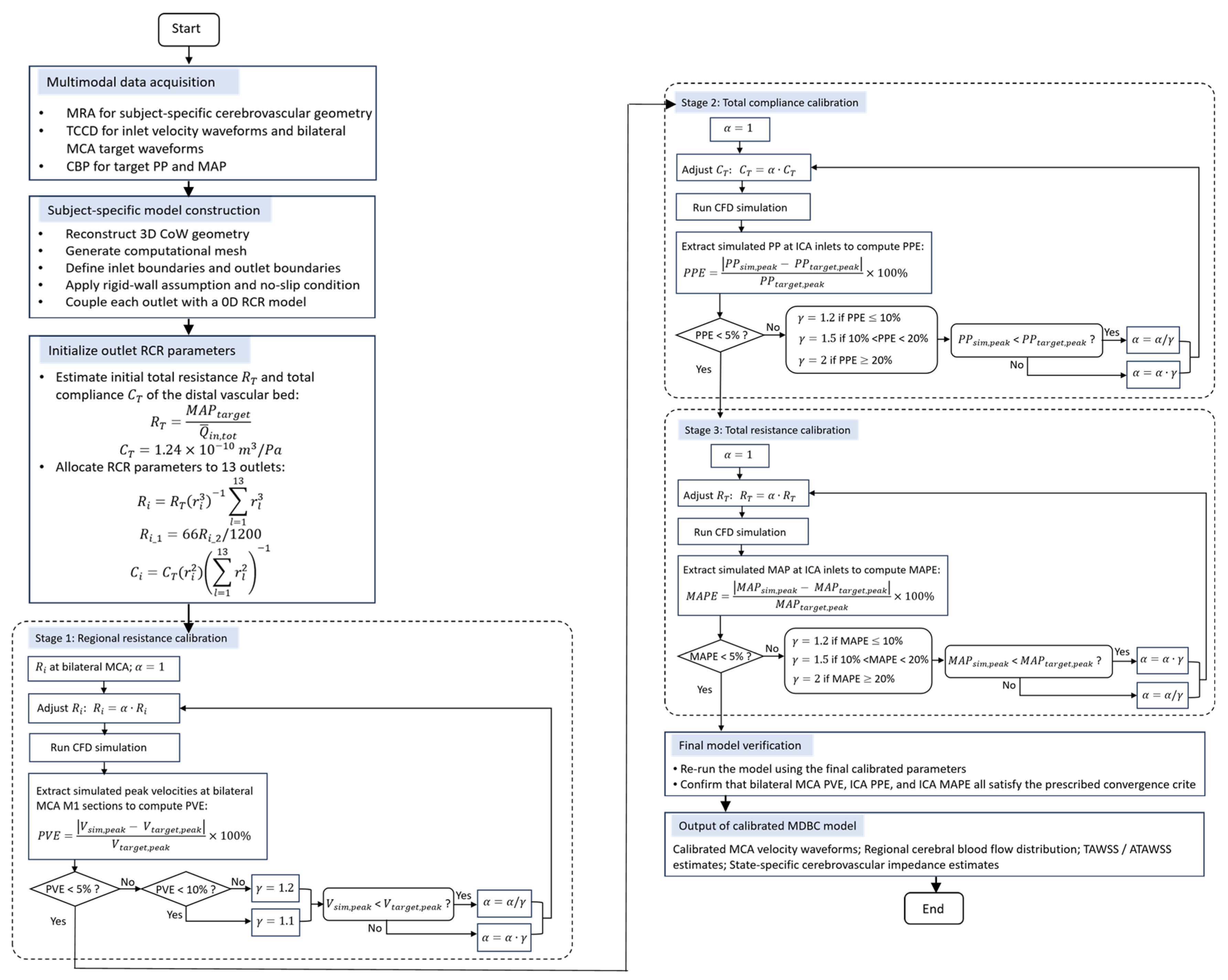
- The initial total resistance and compliance were determined before their allocation among the thirteen outlets. The time-averaged total inlet flow, , was calculated as the sum of the mean flow rates measured at the bilateral ICAs and VAs. The initial total resistance was estimated from the 3 min averaged in vivo MAP () and the total inlet flow as:
- 2.
- While Murray’s law provides a common theoretical framework for distributing boundary impedances based on vessel morphometry, it operates on the idealized assumption of minimum energy expenditure across a population-averaged healthy vascular bed [37]. However, significant physiological deviations exist at the individual level, as Murray’s law fails to account for subject-specific anatomical asymmetry, collateral circulation, and autoregulatory adaptations, indicating that morphology alone cannot fully determine the actual downstream vascular impedance. Nevertheless, because direct measurements of distal cerebrovascular resistance are generally unavailable in vivo, Murray-based allocation remains one of the most widely adopted strategies for initializing outlet BCs in cerebrovascular CFD studies [38,39].
- 3.
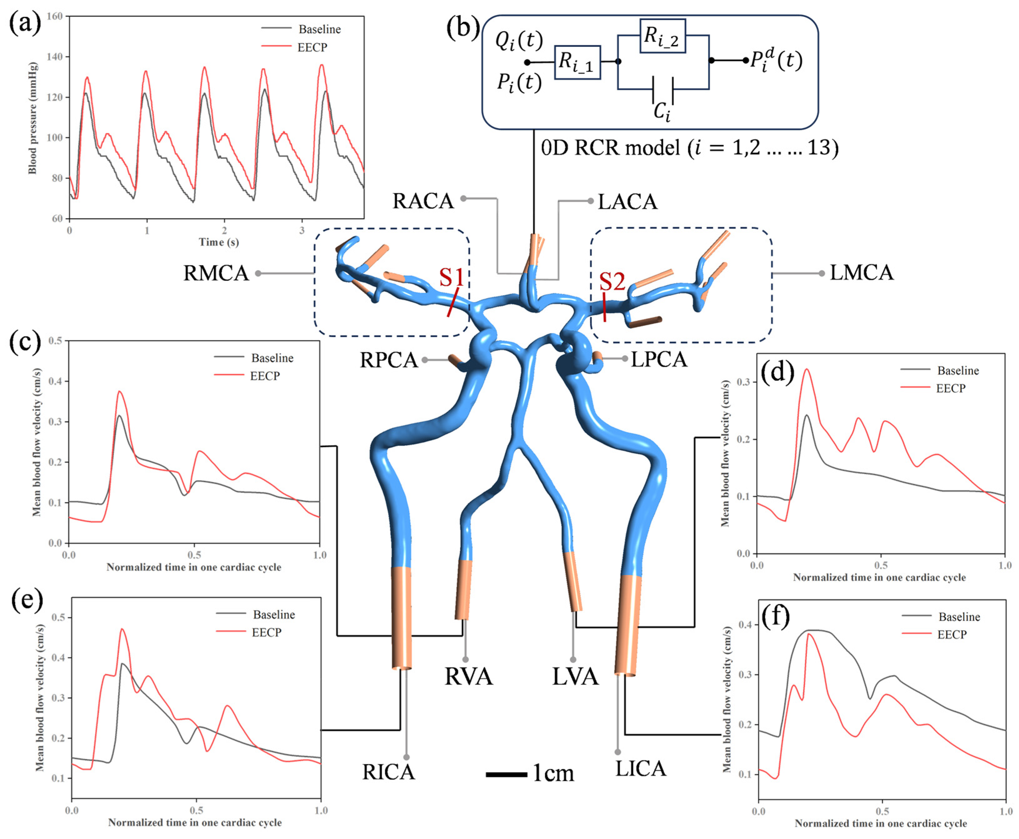
- Following the regional flow matching, the global impedance parameters ( and ) were calibrated to improve agreement between the simulated pressure characteristics and the pressure-derived targets. Firstly, the total compliance was tuned to match the simulated PP () at the ICA inlets with the in vivo target (), which was extracted as the mean PP from a stable 3 min CBP recording (Figure 1a). The objective function was the relative PP Error (PPE):when the was lower than the , was decreased; when the was higher than the , was increased. The adjustment factor was dynamically attenuated based on the residual error: 2.0 (for ), 1.5 (for ), and 1.2 (for ). Convergence was defined strictly as . Subsequently, the total resistance was scaled to align the simulated MAP () with the . The objective function was the relative MAP Error (MAPE):
3. Results and Discussion
3.1. Comparison of Simulated Hemodynamics with In Vivo Measurements

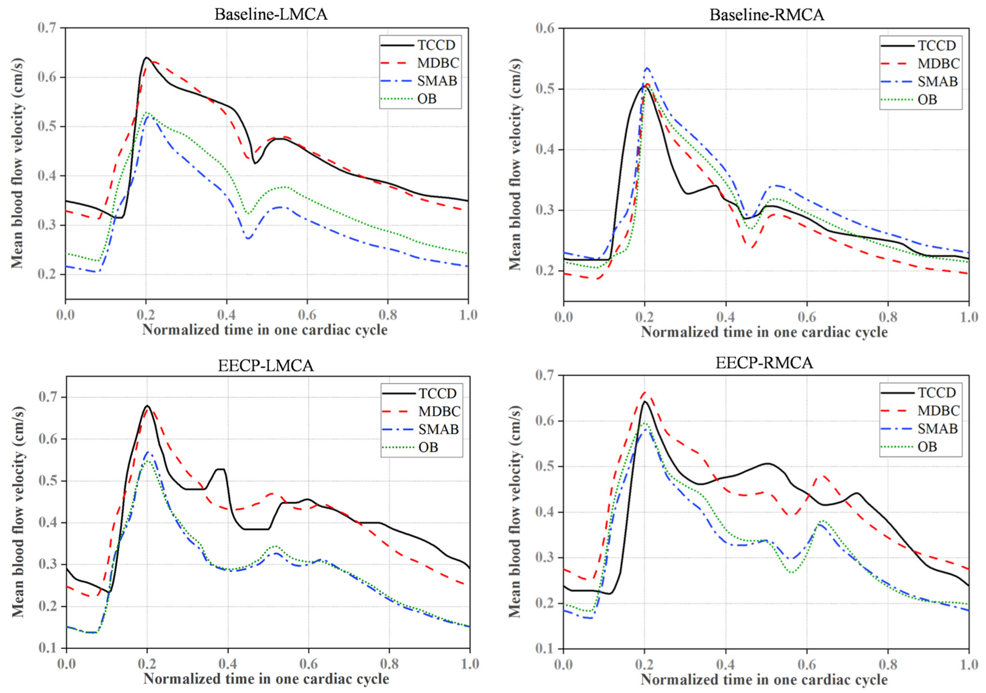
| Physiological State | Artery Branch | Metric | OB | SMAB | MDBC |
|---|---|---|---|---|---|
| Baseline | LMCA | rRMSE | 22.85% | 30.89% | 7.19% |
| PVE | 17.39% | 18.64% | 1.41% | ||
| TAVE | 20.87% | 29.56% | 0.67% | ||
| Baseline | RMCA | rRMSE | 16.76% | 15.67% | 15.44% |
| PVE | 0.05% | 5.98% | 0.82% | ||
| TAVE | 0.73% | 6.92% | 6.88% | ||
| EECP | LMCA | rRMSE | 32.59% | 33.47% | 11.24% |
| PVE | 19.47% | 16.20% | 0.90% | ||
| TAVE | 30.64% | 31.47% | 1.42% | ||
| EECP | RMCA | rRMSE | 27.62% | 27.35% | 17.45% |
| PVE | 7.38% | 9.55% | 3.20% | ||
| TAVE | 17.94% | 20.17% | 4.53% |
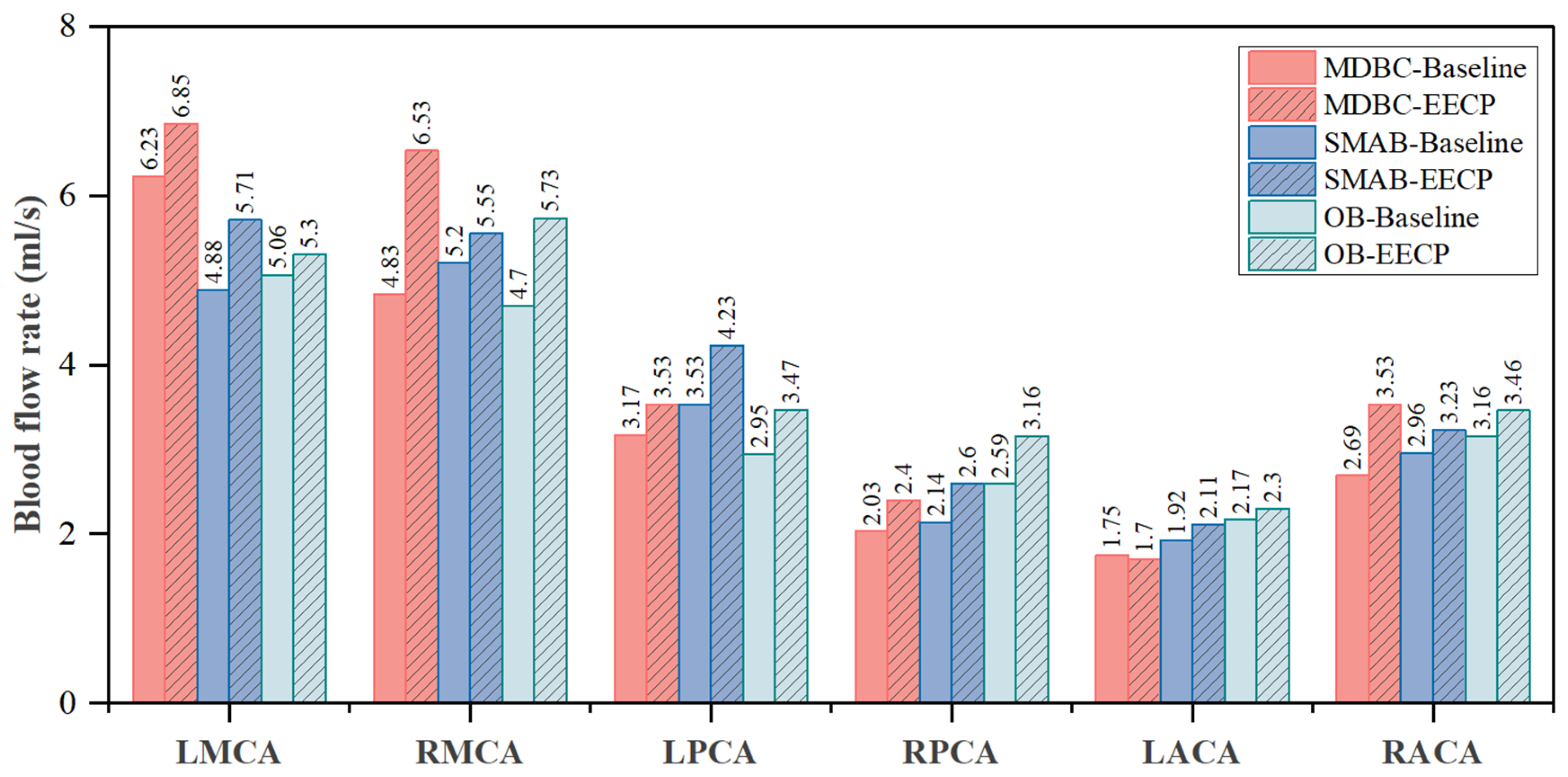
3.2. Boundary Condition Effects on Regional Flow Redistribution
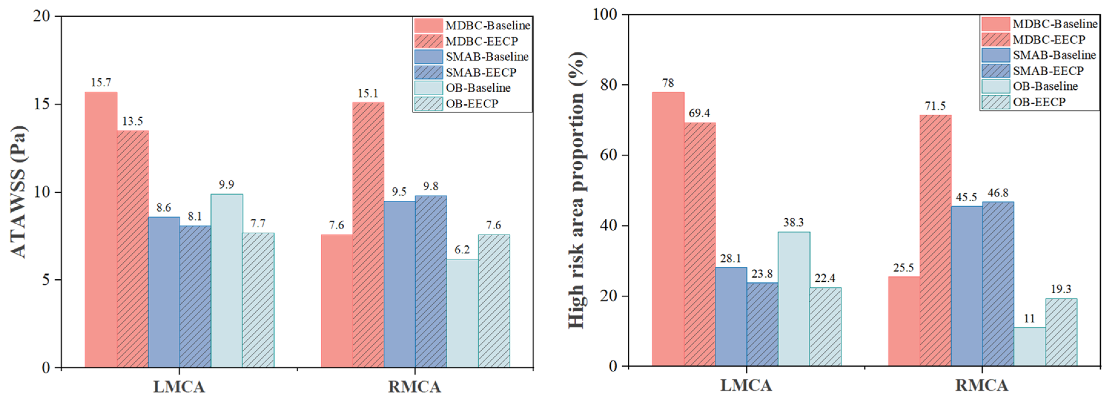
3.3. Boundary Condition Effects on Localized Biomechanical Indices
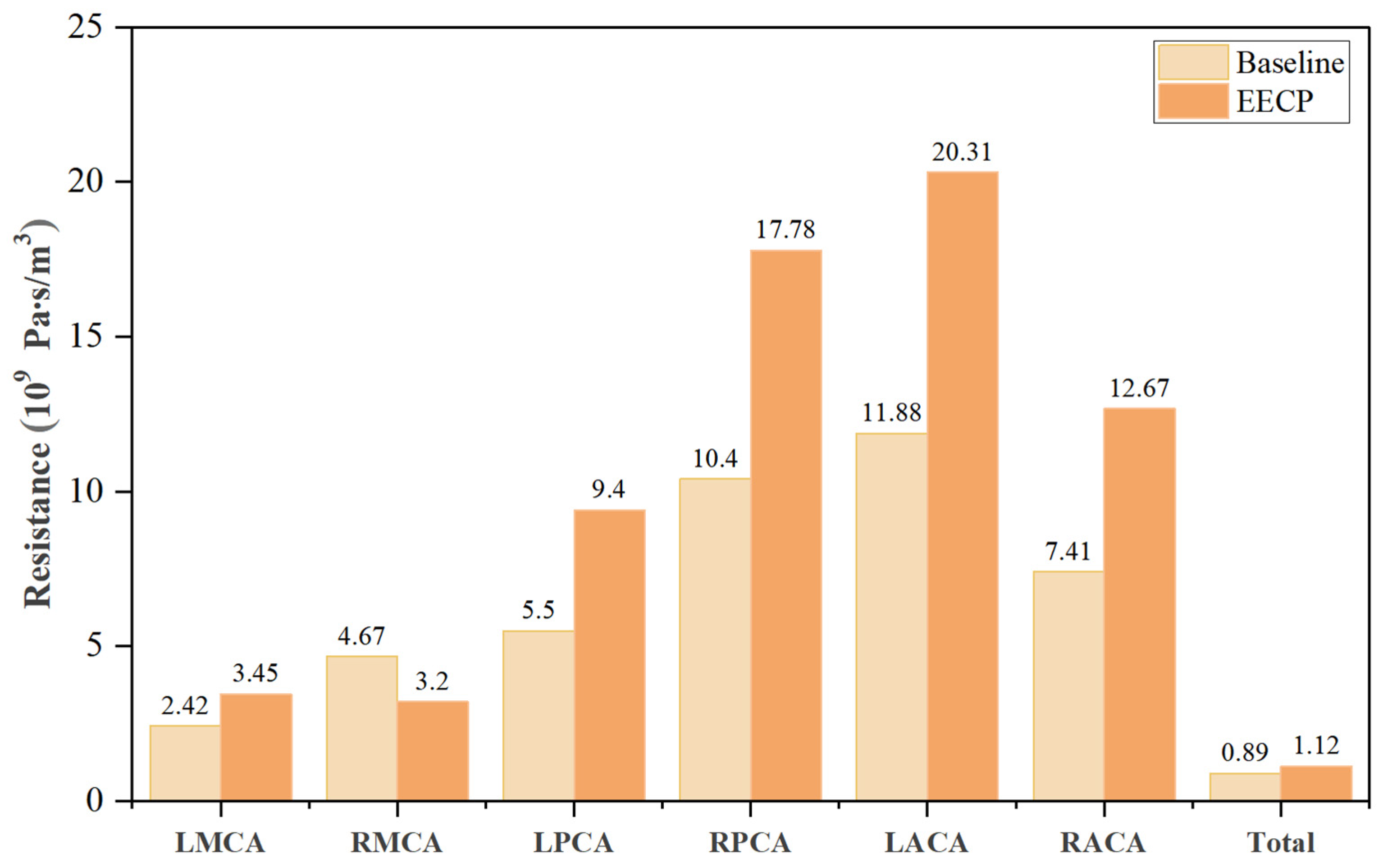
3.4. Estimated State-Specific Cerebrovascular Impedance Changes During EECP
4. Limitations
5. Conclusions
Author Contributions
Funding
Institutional Review Board Statement
Informed Consent Statement
Data Availability Statement
Conflicts of Interest
Abbreviations
| 0D | zero-dimensional |
| 3D | three-dimensional |
| ACA | anterior cerebral artery |
| ATAWSS | area-averaged time-averaged wall shear stress |
| BC | boundary condition |
| CA | cerebral autoregulation |
| CBP | continuous blood pressure |
| CFD | computational fluid dynamics |
| CoW | Circle of Willis |
| EECP | enhanced external counterpulsation |
| FSI | fluid–structure interaction |
| ICA | internal carotid artery |
| LACA | left anterior cerebral artery |
| LICA | left internal carotid artery |
| LMCA | left middle cerebral artery |
| LPCA | left posterior cerebral artery |
| LVA | left vertebral artery |
| MAP | mean arterial pressure |
| MAPE | relative mean arterial pressure error |
| MCA | middle cerebral artery |
| MDBC | multimodal data-driven boundary calibration |
| MRA | magnetic resonance angiography |
| MRI | magnetic resonance imaging |
| OB | open boundary |
| PCA | posterior cerebral artery |
| PCoA | posterior communicating artery |
| PP | pulse pressure |
| PPE | relative pulse pressure error |
| PVE | peak velocity error |
| RACA | right anterior cerebral artery |
| RCR | three-element Windkessel |
| RICA | right internal carotid artery |
| RMCA | right middle cerebral artery |
| RPCA | right posterior cerebral artery |
| rRMSE | relative root mean square error |
| RMSE | root mean square error |
| RVA | right vertebral artery |
| SMAB | static Murray allocation boundary |
| TAVE | time-averaged velocity error |
| TAWSS | time-averaged wall shear stress |
| TCCD | transcranial color-coded Doppler |
| TE | echo time |
| TOF | time-of-flight |
| TR | repetition time |
| UDF | user-defined function |
| VA | vertebral artery |
| WSS | wall shear stress |
References
- Liu, Y.; Li, S.; Liu, H.; Tian, X.; Liu, Y.; Li, Z.; Leung, T.W.; Leng, X. Clinical Implications of Haemodynamics in Symptomatic Intracranial Atherosclerotic Stenosis by Computational Fluid Dynamics Modelling: A Systematic Review. Stroke Vasc. Neurol. 2025, 10, 16–24. [Google Scholar] [CrossRef] [Scilit] [PubMed]
- Ishida, F.; Tsuji, M.; Tanioka, S.; Tanaka, K.; Yoshimura, S.; Suzuki, H. Computational Fluid Dynamics for Cerebral Aneurysms in Clinical Settings. In Trends in Cerebrovascular Surgery and Interventions; Acta Neurochirurgica Supplement; Esposito, G., Regli, L., Cenzato, M., Kaku, Y., Tanaka, M., Tsukahara, T., Eds.; Springer International Publishing: Cham, Switzerland, 2021; Volume 132, pp. 27–32. [Google Scholar]
- Karmonik, C. Toward Improving Fidelity of Computational Fluid Dynamics Simulations: Boundary Conditions Matter. Am. J. Neuroradiol. 2014, 35, 1549–1550. [Google Scholar] [CrossRef] [Scilit] [PubMed]
- Ghaffari, M.; Tangen, K.; Alaraj, A.; Du, X.; Charbel, F.T.; Linninger, A.A. Large-Scale Subject-Specific Cerebral Arterial Tree Modeling Using Automated Parametric Mesh Generation for Blood Flow Simulation. Comput. Biol. Med. 2017, 91, 353–365. [Google Scholar] [CrossRef] [Scilit] [PubMed]
- Berg, P.; Stucht, D.; Janiga, G.; Beuing, O.; Speck, O.; Thévenin, D. Cerebral Blood Flow in a Healthy Circle of Willis and Two Intracranial Aneurysms: Computational Fluid Dynamics Versus Four-Dimensional Phase-Contrast Magnetic Resonance Imaging. J. Biomech. Eng. 2014, 136, 041003. [Google Scholar] [CrossRef] [Scilit] [PubMed]
- Zhu, F.; Qian, Y.; Xu, B.; Gu, Y.; Karunanithi, K.; Zhu, W.; Chen, L.; Mao, Y.; Morgan, M.K. Quantitative Assessment of Changes in Hemodynamics of the Internal Carotid Artery after Bypass Surgery for Moyamoya Disease. J. Neurosurg. 2018, 129, 677–683. [Google Scholar] [CrossRef] [Scilit] [PubMed]
- Sekhane, D.; Mansour, K. Image-Based Computational Fluid Dynamics (CFD) Modeling Cerebral Blood Flow in the Circle of Willis. J. Adv. Res. Phys. 2017, 6, 021601. [Google Scholar]
- Razavi, S.E.; Sahebjam, R. Numerical Simulation of the Blood Flow Behavior in the Circle of Willis. Biolmpacts 2014, 4, 89–94. [Google Scholar] [CrossRef] [Scilit] [PubMed]
- Liu, X.; Gao, Z.; Xiong, H.; Ghista, D.; Ren, L.; Zhang, H.; Wu, W.; Huang, W.; Hau, W.K. Three-Dimensional Hemodynamics Analysis of the Circle of Willis in the Patient-Specific Nonintegral Arterial Structures. Biomech. Model. Mechanobiol. 2016, 15, 1439–1456. [Google Scholar] [CrossRef] [Scilit] [PubMed]
- Neidlin, M.; Büsen, M.; Brockmann, C.; Wiesmann, M.; Sonntag, S.J.; Steinseifer, U.; Kaufmann, T.A.S. A Numerical Framework to Investigate Hemodynamics during Endovascular Mechanical Recanalization in Acute Stroke. Int. J. Numer. Methods Biomed. Eng. 2016, 32, e02748. [Google Scholar] [CrossRef] [Scilit] [PubMed]
- Kashefi, A.; Mahdinia, M.; Firoozabadi, B.; Amirkhosravi, M.; Ahmadi, G.; Saidi, M.S. Multidimensional Modeling of the Stenosed Carotid Artery: A Novel CAD Approach Accompanied by an Extensive Lumped Model. Acta Mech. Sin. 2014, 30, 259–273. [Google Scholar] [CrossRef] [Scilit]
- Lee, K.E.; Ryu, A.-J.; Shin, E.-S.; Shim, E.B. Physiome Approach for the Analysis of Vascular Flow Reserve in the Heart and Brain. Pflüg. Arch. Eur. J. Physiol. 2017, 469, 613–628. [Google Scholar] [CrossRef] [Scilit] [PubMed]
- Zarrinkoob, L.; Ambarki, K.; Wåhlin, A.; Birgander, R.; Eklund, A.; Malm, J. Blood Flow Distribution in Cerebral Arteries. J. Cereb. Blood Flow Metab. 2015, 35, 648–654. [Google Scholar] [CrossRef] [Scilit] [PubMed]
- Vingerhoets, G.; Stroobant, N. Lateralization of Cerebral Blood Flow Velocity Changes During Cognitive Tasks: A Simultaneous Bilateral Transcranial Doppler Study. Stroke 1999, 30, 2152–2158. [Google Scholar] [CrossRef] [Scilit] [PubMed]
- Willie, C.K.; Macleod, D.B.; Shaw, A.D.; Smith, K.J.; Tzeng, Y.C.; Eves, N.D.; Ikeda, K.; Graham, J.; Lewis, N.C.; Day, T.A.; et al. Regional Brain Blood Flow in Man during Acute Changes in Arterial Blood Gases. J. Physiol. 2012, 590, 3261–3275. [Google Scholar] [CrossRef] [Scilit] [PubMed]
- Rayz, V.L.; Cohen-Gadol, A.A. Hemodynamics of Cerebral Aneurysms: Connecting Medical Imaging and Biomechanical Analysis. Annu. Rev. Biomed. Eng. 2020, 22, 231–256. [Google Scholar] [CrossRef] [Scilit] [PubMed]
- Moon, J.Y.; Suh, D.C.; Lee, Y.S.; Kim, Y.W.; Lee, J.S. Considerations of Blood Properties, Outlet Boundary Conditions and Energy Loss Approaches in Computational Fluid Dynamics Modeling. Neurointervention 2014, 9, 1. [Google Scholar] [CrossRef] [Scilit] [PubMed]
- Vignon-Clementel, I.E.; Alberto Figueroa, C.; Jansen, K.E.; Taylor, C.A. Outflow Boundary Conditions for Three-Dimensional Finite Element Modeling of Blood Flow and Pressure in Arteries. Comput. Methods Appl. Mech. Eng. 2006, 195, 3776–3796. [Google Scholar] [CrossRef] [Scilit]
- Taylor, D.J.; Feher, J.; Halliday, I.; Hose, D.R.; Gosling, R.; Aubiniere-Robb, L.; van ‘t Veer, M.; Keulards, D.; Tonino, P.A.L.; Rochette, M.; et al. Refining Our Understanding of the Flow Through Coronary Artery Branches; Revisiting Murray’s Law in Human Epicardial Coronary Arteries. Front. Physiol. 2022, 13, 871912. [Google Scholar] [CrossRef] [Scilit] [PubMed]
- Lin, W.; Xiong, L.; Han, J.; Leung, H.; Leung, T.; Soo, Y.; Chen, X.; Wong, K.S.L. Increasing Pressure of External Counterpulsation Augments Blood Pressure but Not Cerebral Blood Flow Velocity in Ischemic Stroke. J. Clin. Neurosci. 2014, 21, 1148–1152. [Google Scholar] [CrossRef] [Scilit] [PubMed]
- Guluma, K.Z.; Liebeskind, D.S.; Raman, R.; Rapp, K.S.; Ernstrom, K.B.; Alexandrov, A.V.; Shahripour, R.B.; Barlinn, K.; Starkman, S.; Grunberg, I.D.; et al. Feasibility and Safety of Using External Counterpulsation to Augment Cerebral Blood Flow in Acute Ischemic Stroke—The Counterpulsation to Upgrade Forward Flow in Stroke (CUFFS) Trial. J. Stroke Cerebrovasc. Dis. 2015, 24, 2596–2604. [Google Scholar] [CrossRef] [Scilit] [PubMed]
- Xiong, L.; Lin, W.; Han, J.; Chen, X.; Leung, T.; Soo, Y.; Wong, K.S. Enhancing Cerebral Perfusion with External Counterpulsation after Ischaemic Stroke: How Long Does It Last? J. Neurol. Neurosurg. Psychiatry 2016, 87, 531–536. [Google Scholar] [CrossRef] [Scilit] [PubMed]
- Lang, E.W.; Mudaliar, Y.; Lagopoulos, J.; Dorsch, N.; Yam, A.; Griffith, J.; Mulvey, J. A Review of Cerebral Autoregulation: Assessment and Measurements. Australas. Anaesth. 2005, 161–172. [Google Scholar]
- Li, B.; Liu, Y.; Liu, J.; Sun, H.; Feng, Y.; Zhang, Z.; Zhang, L. Cerebral Multi-Autoregulation Model Based Enhanced External Counterpulsation Treatment Planning for Cerebral Ischemic Stroke. J. Cereb. Blood Flow Metab. 2023, 43, 1764–1778. [Google Scholar] [CrossRef] [Scilit] [PubMed]
- Li, B.; Wang, W.; Mao, B.; Zhang, Y.; Chen, S.; Yang, H.; Niu, H.; Du, J.; Li, X.; Liu, Y. Hemodynamic Effects of Enhanced External Counterpulsation on Cerebral Arteries: A Multiscale Study. Biomed. Eng. Online 2019, 18, 91. [Google Scholar] [CrossRef] [Scilit] [PubMed]
- Lin, E.; Kamel, H.; Gupta, A.; RoyChoudhury, A.; Girgis, P.; Glodzik, L. Incomplete Circle of Willis Variants and Stroke Outcome. Eur. J. Radiol. 2022, 153, 110383. [Google Scholar] [CrossRef] [Scilit] [PubMed]
- Siddiqi, H.; Tahir, M.; Lone, K.P. Variations in Cerebral Arterial Circle of Willis in Adult Pakistani Population. J. Coll. Physicians Surg. Pak. 2013, 23, 615–619. [Google Scholar] [PubMed]
- Liu, H.; Lan, L.; Abrigo, J.; Ip, H.L.; Soo, Y.; Zheng, D.; Wong, K.S.; Wang, D.; Shi, L.; Leung, T.W.; et al. Comparison of Newtonian and Non-Newtonian Fluid Models in Blood Flow Simulation in Patients With Intracranial Arterial Stenosis. Front. Physiol. 2021, 12, 718540. [Google Scholar] [CrossRef] [Scilit] [PubMed]
- Zhou, J.; Li, J.; Qin, S.; Liu, J.; Lin, Z.; Xie, J.; Zhang, Z.; Chen, R. High-Resolution Cerebral Blood Flow Simulation with a Domain Decomposition Method and Verified by the TCD Measurement. Comput. Methods Programs Biomed. 2022, 224, 107004. [Google Scholar] [CrossRef] [Scilit] [PubMed]
- Schollenberger, J.; Osborne, N.H.; Hernandez-Garcia, L.; Figueroa, C.A. A Combined Computational Fluid Dynamics and Arterial Spin Labeling MRI Modeling Strategy to Quantify Patient-Specific Cerebral Hemodynamics in Cerebrovascular Occlusive Disease. Front. Bioeng. Biotechnol. 2021, 9, 722445. [Google Scholar] [CrossRef] [Scilit] [PubMed]
- Yan, Z.; Chen, R.; Qi, F.; Cai, X.-C. A Functional Region-Based Approach for the Numerical Simulation of Patient-Specific Cerebral Blood Flows with Clinical Validation 2024. IEEE Trans. Biomed. Eng. 2026, 73, 1346–1357. [Google Scholar] [CrossRef] [Scilit] [PubMed]
- Leng, X.; Scalzo, F.; Ip, H.L.; Johnson, M.; Fong, A.K.; Fan, F.S.Y.; Chen, X.; Soo, Y.O.Y.; Miao, Z.; Liu, L.; et al. Computational Fluid Dynamics Modeling of Symptomatic Intracranial Atherosclerosis May Predict Risk of Stroke Recurrence. PLoS ONE 2014, 9, e97531. [Google Scholar] [CrossRef] [Scilit] [PubMed]
- Leng, X.; Lan, L.; Ip, H.L.; Abrigo, J.; Scalzo, F.; Liu, H.; Feng, X.; Chan, K.L.; Fan, F.S.Y.; Ma, S.H.; et al. Hemodynamics and Stroke Risk in Intracranial Atherosclerotic Disease. Ann. Neurol. 2019, 85, 752–764. [Google Scholar] [CrossRef] [Scilit] [PubMed]
- Lan, L.; Liu, H.; Ip, V.; Soo, Y.; Abrigo, J.; Fan, F.; Ma, S.H.; Ma, K.; Ip, B.; Liu, J.; et al. Regional High Wall Shear Stress Associated With Stenosis Regression in Symptomatic Intracranial Atherosclerotic Disease. Stroke 2020, 51, 3064–3073. [Google Scholar] [CrossRef] [Scilit] [PubMed]
- Lan, H.; Updegrove, A.; Wilson, N.M.; Maher, G.D.; Shadden, S.C.; Marsden, A.L. A Re-Engineered Software Interface and Workflow for the Open-Source SimVascular Cardiovascular Modeling Package. J. Biomech. Eng. 2018, 140, 0245011. [Google Scholar] [CrossRef] [Scilit] [PubMed]
- Chen, R.; Wu, B.; Cheng, Z.; Shiu, W.-S.; Liu, J.; Liu, L.; Wang, Y.; Wang, X.; Cai, X.-C. A Parallel Non-Nested Two-Level Domain Decomposition Method for Simulating Blood Flows in Cerebral Artery of Stroke Patient. Int. J. Numer. Methods Biomed. Eng. 2020, 36, e3392. [Google Scholar] [CrossRef] [Scilit] [PubMed]
- Taylor, D.J.; Saxton, H.; Halliday, I.; Newman, T.; Hose, D.R.; Kassab, G.S.; Gunn, J.P.; Morris, P.D. Systematic Review and Meta-Analysis of Murray’s Law in the Coronary Arterial Circulation. Am. J. Physiol.-Heart Circ. Physiol. 2024, 327, H182–H190. [Google Scholar] [CrossRef] [Scilit] [PubMed]
- Chnafa, C.; Brina, O.; Pereira, V.M.; Steinman, D.A. Better Than Nothing: A Rational Approach for Minimizing the Impact of Outflow Strategy on Cerebrovascular Simulations. Am. J. Neuroradiol. 2018, 39, 337–343. [Google Scholar] [CrossRef] [Scilit] [PubMed]
- Jeken-Rico, P.; Goetz, A.; Meliga, P.; Larcher, A.; Özpeynirci, Y.; Hachem, E. Evaluating the Impact of Domain Boundaries on Hemodynamics in Intracranial Aneurysms within the Circle of Willis. Fluids 2023, 9, 1. [Google Scholar] [CrossRef] [Scilit]
- Avolio, A.P.; Van Bortel, L.M.; Boutouyrie, P.; Cockcroft, J.R.; McEniery, C.M.; Protogerou, A.D.; Roman, M.J.; Safar, M.E.; Segers, P.; Smulyan, H. Role of Pulse Pressure Amplification in Arterial Hypertension: Experts’ Opinion and Review of the Data. Hypertension 2009, 54, 375–383. [Google Scholar] [CrossRef] [Scilit] [PubMed]
- Blanco, P.J.; Watanabe, S.M.; Passos, M.A.R.F.; Lemos, P.A.; Feijoo, R.A. An Anatomically Detailed Arterial Network Model for One-Dimensional Computational Hemodynamics. IEEE Trans. Biomed. Eng. 2015, 62, 736–753. [Google Scholar] [CrossRef] [Scilit] [PubMed]
- Pirola, S.; Cheng, Z.; Jarral, O.A.; O’Regan, D.P.; Pepper, J.R.; Athanasiou, T.; Xu, X.Y. On the Choice of Outlet Boundary Conditions for Patient-Specific Analysis of Aortic Flow Using Computational Fluid Dynamics. J. Biomech. 2017, 60, 15–21. [Google Scholar] [CrossRef] [Scilit] [PubMed]
- Ballout, A.A.; Prochilo, G.; Kaneko, N.; Li, C.; Apfel, R.; Hinman, J.D.; Liebeskind, D.S. Computational Fluid Dynamics in Intracranial Atherosclerotic Disease. Stroke Vasc. Interv. Neurol. 2024, 4, e000792. [Google Scholar] [CrossRef] [Scilit] [PubMed]
- Mechtouff, L.; Rascle, L.; Crespy, V.; Canet-Soulas, E.; Nighoghossian, N.; Millon, A. A Narrative Review of the Pathophysiology of Ischemic Stroke in Carotid Plaques: A Distinction versus a Compromise between Hemodynamic and Embolic Mechanism. Ann. Transl. Med. 2021, 9, 1208. [Google Scholar] [CrossRef] [Scilit] [PubMed]
- Nogueira, R.C.; Beishon, L.; Bor-Seng-Shu, E.; Panerai, R.B.; Robinson, T.G. Cerebral Autoregulation in Ischemic Stroke: From Pathophysiology to Clinical Concepts. Brain Sci. 2021, 11, 511. [Google Scholar] [CrossRef] [Scilit] [PubMed]
- Powers, W.J. Cerebral Hemodynamics in Ischemic Cerebrovascular Disease. Ann. Neurol. 1991, 29, 231–240. [Google Scholar] [CrossRef] [Scilit] [PubMed]
- Liebeskind, D.S. Collateral Circulation. Stroke 2003, 34, 2279–2284. [Google Scholar] [CrossRef] [Scilit] [PubMed]
- Zhu, G.; Yuan, Q.; Yang, J.; Yeo, J.H. Experimental Study of Hemodynamics in the Circle of Willis. Biomed. Eng. Online 2015, 14, S10. [Google Scholar] [CrossRef] [Scilit] [PubMed]
- Guo, Z.-N.; Sun, X.; Liu, J.; Sun, H.; Zhao, Y.; Ma, H.; Xu, B.; Wang, Z.; Li, C.; Yan, X.; et al. The Impact of Variational Primary Collaterals on Cerebral Autoregulation. Front. Physiol. 2018, 9, 759. [Google Scholar] [CrossRef] [Scilit] [PubMed]
- Qureshi, A.I.; Caplan, L.R. Intracranial Atherosclerosis. Lancet 2014, 383, 984–998. [Google Scholar] [CrossRef] [Scilit] [PubMed]
- Malek, A.M. Hemodynamic Shear Stress and Its Role in Atherosclerosis. JAMA 1999, 282, 2035. [Google Scholar] [CrossRef] [Scilit] [PubMed]
- Meng, H.; Tutino, V.M.; Xiang, J.; Siddiqui, A. High WSS or Low WSS? Complex Interactions of Hemodynamics with Intracranial Aneurysm Initiation, Growth, and Rupture: Toward a Unifying Hypothesis. Am. J. Neuroradiol. 2014, 35, 1254–1262. [Google Scholar] [CrossRef] [Scilit] [PubMed]
- Strandgaard, S. Cerebral Autoregulation. Stroke 1984, 15, 3. [Google Scholar] [CrossRef] [Scilit]
- Washio, T.; Watanabe, H.; Ogoh, S. Dynamic Cerebral Autoregulation in Anterior and Posterior Cerebral Circulation during Cold Pressor Test. J. Physiol. Sci. 2020, 70, 1. [Google Scholar] [CrossRef] [Scilit] [PubMed]
- Xiong, L.; Chen, X.; Liu, J.; Wong, L.K.S.; Leung, T.W. Cerebral Augmentation Effect Induced by External Counterpulsation Is Not Related to Impaired Dynamic Cerebral Autoregulation in Ischemic Stroke. Front. Neurol. 2022, 13, 784836. [Google Scholar] [CrossRef] [Scilit] [PubMed]
- Lin, W.; Xiong, L.; Han, J.; Leung, T.W.H.; Soo, Y.O.Y.; Chen, X.; Wong, K.S.L. External Counterpulsation Augments Blood Pressure and Cerebral Flow Velocities in Ischemic Stroke Patients With Cerebral Intracranial Large Artery Occlusive Disease. Stroke 2012, 43, 3007–3011. [Google Scholar] [CrossRef] [Scilit] [PubMed]



| Baseline | EECP | ||||||||
|---|---|---|---|---|---|---|---|---|---|
| Mesh quantity | 1.61 M | 3.70 M | 5.12 M | 6.86 M | 1.61 M | 3.70 M | 5.12 M | 6.86 M | |
| RMSE | ACAs | 10.82% | 7.49% | 5.47% | 3.40% | 11.39% | 7.61% | 5.42% | 3.31% |
| MCAs | 18.60% | 14.62% | 5.59% | 2.32% | 18.42% | 14.60% | 5.55% | 2.24% | |
| PCAs | 8.44% | 7.40% | 5.57% | 2.64% | 8.41% | 7.33% | 5.56% | 2.74% | |
| Physiological State | ICA Inlet | (mmHg) | (mmHg) | (mmHg) | (mmHg) | (%) | (%) |
|---|---|---|---|---|---|---|---|
| Baseline | LICA | 51.98 | 88.71 | 53.83 | 87.11 | 3.43 | 1.84 |
| RICA | 52.33 | 91.10 | 2.79 | 4.58 | |||
| EECP | LICA | 55.26 | 99.42 | 55.40 | 95.52 | 0.25 | 4.08 |
| RICA | 52.84 | 95.90 | 4.62 | 0.40 |
Disclaimer/Publisher’s Note: The statements, opinions and data contained in all publications are solely those of the individual author(s) and contributor(s) and not of MDPI and/or the editor(s). MDPI and/or the editor(s) disclaim responsibility for any injury to people or property resulting from any ideas, methods, instructions or products referred to in the content. |
© 2026 by the authors. Licensee MDPI, Basel, Switzerland. This article is an open access article distributed under the terms and conditions of the Creative Commons Attribution (CC BY) license.
Share and Cite
Hu, J.; Li, H.; Shen, X.; Zhong, Y.; Zheng, H.; Liu, Y.; Luo, B.; Du, J. A Subject-Specific Cerebrovascular CFD Modeling Approach Based on a Multimodal Data-Driven Boundary Calibration Framework: A Proof-of-Concept Study. Bioengineering 2026, 13, 861. https://doi.org/10.3390/bioengineering13080861
Hu J, Li H, Shen X, Zhong Y, Zheng H, Liu Y, Luo B, Du J. A Subject-Specific Cerebrovascular CFD Modeling Approach Based on a Multimodal Data-Driven Boundary Calibration Framework: A Proof-of-Concept Study. Bioengineering. 2026; 13(8):861. https://doi.org/10.3390/bioengineering13080861
Chicago/Turabian StyleHu, Jun, Hongye Li, Xuelian Shen, Yonghao Zhong, Hanxiong Zheng, Yiao Liu, Bin Luo, and Jianhang Du. 2026. "A Subject-Specific Cerebrovascular CFD Modeling Approach Based on a Multimodal Data-Driven Boundary Calibration Framework: A Proof-of-Concept Study" Bioengineering 13, no. 8: 861. https://doi.org/10.3390/bioengineering13080861
APA StyleHu, J., Li, H., Shen, X., Zhong, Y., Zheng, H., Liu, Y., Luo, B., & Du, J. (2026). A Subject-Specific Cerebrovascular CFD Modeling Approach Based on a Multimodal Data-Driven Boundary Calibration Framework: A Proof-of-Concept Study. Bioengineering, 13(8), 861. https://doi.org/10.3390/bioengineering13080861