LLM-Assisted Interpretation of Kinematic Gait Data in Children with Cerebral Palsy: A Pilot Study on Gait Deviation Detection and Surgical Group Recommendations
 , , ,
, , ,
Abstract
1. Introduction
2. Materials and Methods
2.1. Ethics Statement
2.2. Participants
2.3. Experimental Protocol
2.4. GaitSharing Toolkit
2.5. LLM Interpretation
“Deviations 23, 24—Swing plantarflexion with reduced ankle sagittal ROM may reflect dynamic equinus or dorsiflexor weakness, requiring passive ankle examination and selective motor assessment.”
2.6. Expert Review
2.7. Statistical Analysis
2.8. LLM Self-Consistency Analysis
3. Results
4. Discussion
4.1. Gait Deviations
4.2. Surgical Procedures
4.3. LLM Self-Consistency
4.4. Limitations and Future Directions
5. Conclusions
Supplementary Materials
Author Contributions
Funding
Institutional Review Board Statement
Informed Consent Statement
Data Availability Statement
Acknowledgments
Conflicts of Interest
Abbreviations
| CP | Cerebral palsy |
| 3D-IGA | Three-dimensional instrumented gait analysis |
| LLM | Large language model |
| AI | Artificial intelligence |
| EMG | Electromyography |
| ROM | Range of motion |
| EB-GAIT | Evidence-Based Gait Analysis Interpretation Tools |
| ChatGPT | Chat Generative Pre-trained Transformer |
| API | Application Programming Interface |
| NMI | Need more information |
| INC | Inconclusive |
| TAL-TATS | Tendo-Achilles lengthening with tibialis anterior tendon shortening |
| IC | Initial contact |
| GDI | Gait Deviation Index |
References
- Rosenbaum, P.; Paneth, N.; Leviton, A.; Goldstein, M.; Bax, M.; Damiano, D.; Dan, B.; Jacobsson, B. A Report: The Definition and Classification of Cerebral Palsy April 2006. Dev. Med. Child. Neurol. Suppl. 2007, 109, 8–14. [Google Scholar] [CrossRef] [Scilit] [PubMed]
- Graham, H.K.; Thomason, P.; Willoughby, K.; Hastings-Ison, T.; Stralen, R.V.; Dala-Ali, B.; Wong, P.; Rutz, E. Musculoskeletal Pathology in Cerebral Palsy: A Classification System and Reliability Study. Children 2021, 8, 252. [Google Scholar] [CrossRef] [Scilit] [PubMed]
- McIntyre, S.; Goldsmith, S.; Webb, A.; Ehlinger, V.; Hollung, S.J.; McConnell, K.; Arnaud, C.; Smithers-Sheedy, H.; Oskoui, M.; Khandaker, G. Global Prevalence of Cerebral Palsy: A Systematic Analysis. Dev. Med. Child. Neurol. 2022, 64, 1494–1506. [Google Scholar] [CrossRef] [Scilit] [PubMed]
- Schwartz, M.H.; Ries, A.J.; Georgiadis, A.G.; Kainz, H. Demonstrating the Utility of Instrumented Gait Analysis in the Treatment of Children with Cerebral Palsy. PLoS ONE 2024, 19, e0301230. [Google Scholar] [CrossRef] [Scilit] [PubMed]
- Skaggs, D.L.; Rethlefsen, S.A.; Kay, R.M.; Dennis, S.W.; Reynolds, R.A.; Tolo, V.T. Variability in Gait Analysis Interpretation. J. Pediatr. Orthop. 2000, 20, 759–764. [Google Scholar] [CrossRef] [Scilit] [PubMed]
- Wang, K.K.; Stout, J.L.; Ries, A.J.; Novacheck, T.F. Interobserver Reliability in the Interpretation of Three-dimensional Gait Analysis in Children with Gait Disorders. Dev. Med. Child. Neurol. 2019, 61, 710–716. [Google Scholar] [CrossRef] [Scilit] [PubMed]
- Rethlefsen, S.A.; Hanson, A.; Ciccodicola, E.; Hara, R.; Kay, R.M.; Chambers, H.; Wren, T.A. Update on the Reliability of Gait Analysis Interpretation in Cerebral Palsy: Inter-Institution Agreement. Gait Posture 2024, 109, 109–114. [Google Scholar] [CrossRef] [Scilit] [PubMed]
- Schwartz, M.H.; Georgiadis, A.G. Evidence Based Gait Analysis Interpretation Tools (EB-GAIT) Treatment Recommendation and Outcome Prediction Models to Support Decision-Making Based on Clinical Gait Analysis Data. PLoS ONE 2025, 20, e0328036. [Google Scholar] [CrossRef] [Scilit] [PubMed]
- Davoudi, M.; Salami, F.; Reisig, R.; Patikas, D.A.; Wolf, S.I. Rectus Femoris Electromyography Signal Clustering: Data-Driven Management of Crouch Gait in Patients with Cerebral Palsy. PLoS ONE 2024, 19, e0298945. [Google Scholar] [CrossRef] [Scilit] [PubMed]
- Davoudi, M.; Salami, F.; Reisig, R.; Gather, K.S.; Wolf, S.I. Gluteus Medius Muscle Activation Patterns during Gait with Cerebral Palsy (CP): A Hierarchical Clustering Analysis. PLoS ONE 2025, 20, e0309582. [Google Scholar] [CrossRef] [Scilit] [PubMed]
- Reinbolt, J.A.; Fox, M.D.; Schwartz, M.H.; Delp, S.L. Predicting Outcomes of Rectus Femoris Transfer Surgery. Gait Posture 2009, 30, 100–105. [Google Scholar] [CrossRef] [Scilit] [PubMed]
- Pohlmann, P.F.; Glienke, M.; Sandkamp, R.; Gratzke, C.; Schmal, H.; Schoeb, D.S.; Fuchs, A. Assessing the Efficacy of Ortho GPT: A Comparative Study with Medical Students and General LLMs on Orthopedic Examination Questions. Bioengineering 2025, 12, 1290. [Google Scholar] [CrossRef] [Scilit] [PubMed]
- Nori, H.; King, N.; McKinney, S.M.; Carignan, D.; Horvitz, E. Capabilities of Gpt-4 on Medical Challenge Problems. arXiv 2023, arXiv:2303.13375. [Google Scholar]
- Lee, P.; Bubeck, S.; Petro, J. Benefits, Limits, and Risks of GPT-4 as an AI Chatbot for Medicine. N. Engl. J. Med. 2023, 388, 1233–1239. [Google Scholar] [CrossRef] [Scilit] [PubMed]
- Singhal, K.; Azizi, S.; Tu, T.; Mahdavi, S.S.; Wei, J.; Chung, H.W.; Scales, N.; Tanwani, A.; Cole-Lewis, H.; Pfohl, S. Large Language Models Encode Clinical Knowledge. Nature 2023, 620, 172–180. [Google Scholar] [CrossRef] [Scilit] [PubMed]
- Dindorf, C.; Dully, J.; Keilhauer, R.; Lorenz, M.; Fröhlich, M. Evaluating Large Language Models for Gait Classification Using Text-Encoded Kinematic Waveforms. arXiv 2026, arXiv:2603.13317. [Google Scholar]
- Celik, M.; Birsel, S.E.; Demirci, E.; Dogan, O.; Inan, M. Evaluating ChatGPT-4o’s Performance in Gait Pattern Classification Using Sagittal Kinematic Data and Video-Based Inputs in Children with Cerebral Palsy. Gait Posture 2025, 121, 43–44. [Google Scholar] [CrossRef] [Scilit]
- Wang, D.; Bobenrieth, C.; Seo, H. Agir: Assessing 3d Gait Impairment with Reasoning Based on Llms. arXiv 2025, arXiv:2503.18141. [Google Scholar]
- Keilhauer, R.; Lorenz, M.; Dindorf, C.; Ernst, S.; Wang, C.-Y.; Messer, P.; Stricker, D. Exploring Large Language Models for Automated Gait Analysis; IEEE: New York, NY, USA, 2025; pp. 136–142. [Google Scholar]
- Romkes, J.; Freslier, M.; Rutz, E.; Bracht-Schweizer, K. Walking on Uneven Ground: How Do Patients with Unilateral Cerebral Palsy Adapt? Clin. Biomech. 2020, 74, 8–13. [Google Scholar] [CrossRef] [Scilit] [PubMed]
- Sangeux, M.; Viehweger, E.; Romkes, J.; Bracht-Schweizer, K. On the Clinical Interpretation of Overground Gait Stability Indices in Children with Cerebral Palsy. Sci. Rep. 2024, 14, 26363. [Google Scholar] [CrossRef] [Scilit] [PubMed]
- Kadaba, M.P.; Ramakrishnan, H.; Wootten, M. Measurement of Lower Extremity Kinematics during Level Walking. J. Orthop. Res. 1990, 8, 383–392. [Google Scholar] [CrossRef] [Scilit] [PubMed]
- Baker, R.; Leboeuf, F.; Reay, J.; Sangeux, M. The Conventional Gait Model-Success and Limitations. Handb. Hum. Motion 2018, 1, 489–508. [Google Scholar] [CrossRef] [Scilit]
- Dumas, R.; Wojtusch, J. Estimation of the Body Segment Inertial Parameters for the Rigid Body Biomechanical Models Used in Motion Analysis. In Handbook of Human Motion; Müller, B., Wolf, S., Eds.; Springer International Publishing: Cham, Switzerland, 2018; pp. 47–77. [Google Scholar]
- GaitSharing: A Toolkit for Clinical Gait Analysis. Available online: https://github.com/MehrdadDavoudiii/GaitSharingToolkit (accessed on 25 May 2026).
- Documentation for GaitSharingToolkit. Available online: https://mehrdaddavoudiii.github.io/GaitSharingToolkit (accessed on 25 May 2026).
- Michaud, B.; Begon, M. Ezc3d: An Easy C3D File I/O Cross-Platform Solution for C++, Python and MATLAB. J. Open Source Softw. 2021, 6, 2911. [Google Scholar] [CrossRef] [Scilit]
- Wolf, S.; Loose, T.; Schablowski, M.; Döderlein, L.; Rupp, R.; Gerner, H.J.; Bretthauer, G.; Mikut, R. Automated Feature Assessment in Instrumented Gait Analysis. Gait Posture 2006, 23, 331–338. [Google Scholar] [CrossRef] [Scilit] [PubMed]
- Cohen, J. Weighted Kappa: Nominal Scale Agreement Provision for Scaled Disagreement or Partial Credit. Psychol. Bull. 1968, 70, 213. [Google Scholar] [CrossRef] [Scilit] [PubMed]
- Rha, D.; Cahill-Rowley, K.; Young, J.; Torburn, L.; Stephenson, K.; Rose, J. Biomechanical and Clinical Correlates of Stance-Phase Knee Flexion in Persons with Spastic Cerebral Palsy. PM&R 2016, 8, 11–18. [Google Scholar] [CrossRef] [Scilit] [PubMed]
- Schweizer, K.; Romkes, J.; Brunner, R. The Association between Premature Plantarflexor Muscle Activity, Muscle Strength, and Equinus Gait in Patients with Various Pathologies. Res. Dev. Disabil. 2013, 34, 2676–2683. [Google Scholar] [CrossRef] [Scilit] [PubMed]
- Osis, S.T.; Hettinga, B.A.; Macdonald, S.; Ferber, R. Effects of Simulated Marker Placement Deviations on Running Kinematics and Evaluation of a Morphometric-Based Placement Feedback Method. PLoS ONE 2016, 11, e0147111. [Google Scholar] [CrossRef] [Scilit] [PubMed]
- Baker, R.; McGinley, J.L.; Schwartz, M.; Thomason, P.; Rodda, J.; Graham, H.K. The Minimal Clinically Important Difference for the Gait Profile Score. Gait Posture 2012, 35, 612–615. [Google Scholar] [CrossRef] [Scilit] [PubMed]
- Sutherland, D.H.; Davids, J.R. Common Gait Abnormalities of the Knee in Cerebral Palsy. Clin. Orthop. Relat. Res. (1976–2007) 1993, 288, 139–147. [Google Scholar]
- Schwartz, M.H.; Rozumalski, A. The Gait Deviation Index: A New Comprehensive Index of Gait Pathology. Gait Posture 2008, 28, 351–357. [Google Scholar] [CrossRef] [Scilit] [PubMed]
- Widmer, M.; Staganello, M.; Sangeux, M.; Odorizzi, M.; Brunner, R.; Viehweger, E. Single Procedure Tibialis Anterior Tendon Shortening in Combination with Achilles Tendon Lengthening in Unilateral Cerebral Palsy Improves Swing Phase Dorsiflexion in Gait. J. Child. Orthop. 2024, 18, 441–449. [Google Scholar] [CrossRef] [Scilit] [PubMed]
- Rutz, E.; Baker, R.; Tirosh, O.; Romkes, J.; Haase, C.; Brunner, R. Tibialis Anterior Tendon Shortening in Combination with Achilles Tendon Lengthening in Spastic Equinus in Cerebral Palsy. Gait Posture 2011, 33, 152–157. [Google Scholar] [CrossRef] [Scilit] [PubMed]
- Landis, J.R.; Koch, G.G. The Measurement of Observer Agreement for Categorical Data. Biometrics 1977, 33, 159–174. [Google Scholar] [CrossRef] [Scilit] [PubMed]
- Armand, S.; Sawacha, Z.; Goudriaan, M.; Horsak, B.; van der Krogt, M.; Huenaerts, C.; Daly, C.; Kranzl, A.; Boehm, H.; Petrarca, M. Current Practices in Clinical Gait Analysis in Europe: A Comprehensive Survey-Based Study from the European Society for Movement Analysis in Adults and Children (ESMAC) Standard Initiative. Gait Posture 2024, 111, 65–74. [Google Scholar] [CrossRef] [Scilit] [PubMed]



| Group | Parameter | |||||
|---|---|---|---|---|---|---|
| N | Sex | Age (Years) | Height (m) | Weight (kg) | Affected Side | |
| M/F | Mean [Range] | Mean [SD] | Mean [SD] | R/L | ||
| Uni-CP | 8 | 6 M/2 F | 10.3 [7.2–14.8] | 1.39 [0.15] | 36.4 [19.9] | 4 R/4 L |
| TD | 20 | 10 M/10 F | 11.4 [7.6–16.7] | 1.47 [0.18] | 37.3 [12.0] | NA |
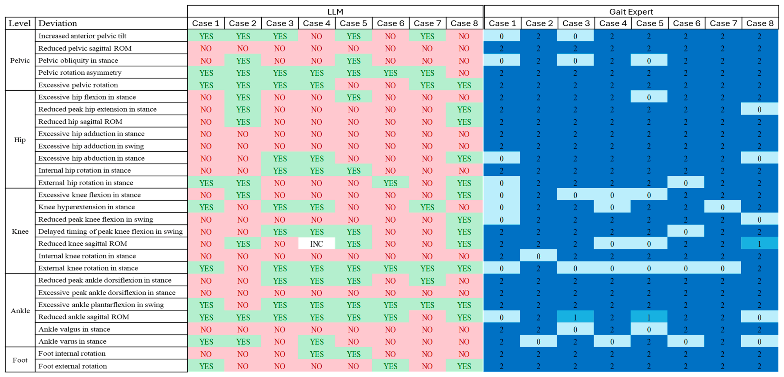
| Joint | Deviation |
|---|---|
| Pelvis | Increased anterior pelvic tilt |
| Reduced pelvic sagittal ROM | |
| Pelvic obliquity in stance | |
| Pelvic rotation asymmetry | |
| Excessive pelvic rotation | |
| Hip | Excessive hip flexion in stance |
| Reduced peak hip extension in stance | |
| Reduced hip sagittal ROM | |
| Excessive hip adduction in stance | |
| Excessive hip adduction in swing | |
| Excessive hip abduction in stance | |
| Internal hip rotation in stance | |
| External hip rotation in stance | |
| Knee | Excessive knee flexion in stance |
| Knee hyperextension in stance | |
| Reduced peak knee flexion in swing | |
| Delayed timing of peak knee flexion in swing | |
| Reduced knee sagittal ROM | |
| Internal knee rotation in stance | |
| External knee rotation in stance | |
| Ankle | Reduced peak ankle dorsiflexion in stance |
| Excessive peak ankle dorsiflexion in stance | |
| Excessive ankle plantarflexion in swing | |
| Reduced ankle sagittal ROM | |
| Ankle valgus in stance | |
| Ankle varus in stance | |
| Foot | Foot internal rotation |
| Foot external rotation |
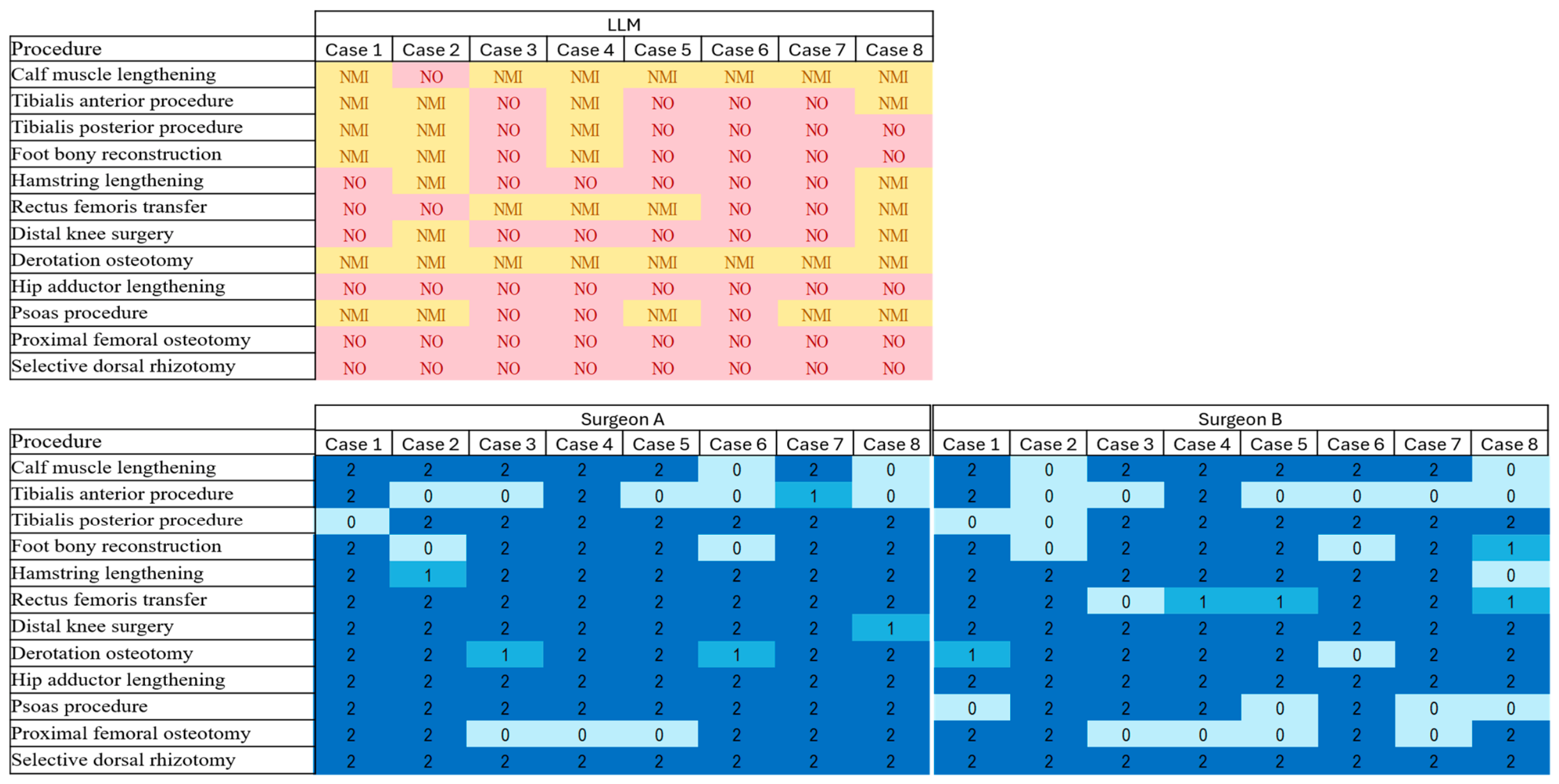
| No. | Procedure Group |
|---|---|
| 1 | Calf muscle lengthening |
| 2 | Tibialis anterior procedure |
| 3 | Tibialis posterior procedure |
| 4 | Foot bony reconstruction |
| 5 | Hamstring lengthening |
| 6 | Rectus femoris transfer |
| 7 | Distal knee surgery |
| 8 | Derotation osteotomy |
| 9 | Hip adductor lengthening |
| 10 | Psoas procedure |
| 11 | Proximal femoral osteotomy |
| 12 | Selective dorsal rhizotomy |
| Sagittal (95% CI) | Frontal (95% CI) | Transverse (95% CI) | Overall (95% CI) | |
|---|---|---|---|---|
| Pelvic | 87.5 (68.8–100) | 62.5 (25–87.5) | 100 (100–100) | 87.5 (75–97.5) |
| Hip | 91.7 (79.2–100) | 91.7 (83.3–100) | 87.5 (68.8–100) | 90.6 (82.8–96.9) |
| Knee | 68.8 (55–82.5) | NA | 56.3 (50–68.8) | 65.2 (54.5–75) |
| Ankle and Foot | 90.6 (82.8–96.9) | 62.5 (50–81.3) | 100 (100–100) | 85.9 (81.3–90.6) |
| Overall | 82.6 (75–90.2) | 77.1 (68.8–85.4) | 85.9 (81.3–90.6) | 82.4 (77–87.5) |
| Concern | Gait Expert Feedback |
|---|---|
| Clinically relevant | The model should be informed of a clinically relevant difference threshold (e.g., 1.5 standard deviation from healthy reference data) to distinguish pathological deviations from normal variability. |
| Phases ROM | Range of motion should be computed over the whole gait cycle only; reporting ROM for stance and swing phases separately is unnecessary and may confuse interpretation. Whole-cycle ROM is most relevant for the knee (stiff knee detection) and pelvic (increased ROM). |
| Pelvic ROM | Pelvic ROM is typically stable across healthy; therefore, an increased ROM should also be considered a deviation, not a reduced ROM. |
| Pelvic obliquity | Pelvic obliquity and rotation asymmetry requires comparison of both left and right gait cycles to distinguish anatomical or functional leg length discrepancy from a true pelvic deviation. Assessing the affected side alone may be insufficient. |
| Knee flexion at IC | Knee flexion angle at IC is an important indicator for downstream decision-making, particularly regarding orthotic prescription, and was not captured. |
| Ankle angle at IC | Ankle angle at IC is clinically important for identifying flat/fore-foot contact patterns (initial flat foot or forefoot contact), which was not included in the deviation list. |
| Ankle valgus/varus | Valgus and varus as measured during gait represent the alignment between the tibial segment and the global laboratory axis, not ankle joint angle. This should be acknowledged when interpreting these items. More accurate information can come from clinical examinations and clinical images. |
| Foot rotation | “Foot internal/external rotation” in the prompt should be renamed to “internal/external foot progression angle,” as the foot rotation signal from marker-based gait analysis is not a reliable measure of true foot rotation. |
| Procedure Group | Agreement of Surgeon A on LLM Across All Cases (95% CI) | Agreement of Surgeon B on LLM Across All Cases (95% CI) |
|---|---|---|
| Calf muscle lengthening | 75 (37.5–100) | 75 (37.5–100) |
| Tibialis anterior procedure | 31.3 (6.3–62.5) | 25 (0–50.3) |
| Tibialis posterior procedure | 87.5 (62.5–100) | 75 (37.5–100) |
| Foot bony reconstruction | 75 (37.5–100) | 68.8 (37.5–100) |
| Hamstring lengthening | 93.8 (81.3–100) | 87.5 (62.5–100) |
| Rectus femoris transfer | 100 (100–100) | 68.8 (43.8–93.8) |
| Distal knee surgery | 93.8 (81.3–100) | 100 (100–100) |
| Derotation osteotomy | 87.5 (68.8–100) | 81.3 (56.3–100) |
| Hip adductor lengthening | 100 (100–100) | 100 (100–100) |
| Psoas procedure | 100 (100–100) | 50 (12.5–87.5) |
| Proximal femoral osteotomy | 62.5 (25–87.5) | 50 (12.5–87.5) |
| Selective dorsal rhizotomy | 100 (100–100) | 100 (100–100) |
| Average | 83.9 (78.1–89.1) | 73.4 (67.7–79.2) |
| Patients | Inter-Surgeon Agreement (95% CI) |
|---|---|
| Case 1 | 83.4 |
| Case 2 | 75 |
| Case 3 | 83.4 |
| Case 4 | 91.7 |
| Case 5 | 83.4 |
| Case 6 | 83.4 |
| Case 7 | 75 |
| Case 8 | 58.3 |
| Average | 79.2 (71.9–85.4) |
| Cohen kappa | 0.59 (0.47–0.7) |
| Case | Surgeon A | Surgeon B |
|---|---|---|
| 1 | This patient is a candidate for the TAL-TATS procedure. An additional assessment for possible bony foot correction is recommended. | Would proceed with calf lengthening if a contracture is confirmed and further consider tibialis anterior shortening based on EMG results. If assessments indicate flatfoot, a bony correction may be needed. Psoas lengthening is not recommended due to the patient’s good function and risk of muscle weakening. |
| 2 | A knee extension osteotomy should be considered. Hip flexor contractures should be assessed to determine whether lengthening is needed. | Need additional information on the patient’s muscle strength, EMG findings, contractures, and foot deformity before planning for surgical treatment. |
| 3 | candidate for TAL-TATS. Torsional alignment should be assessed through rotational range and femoral anteversion. A derotation osteotomy is indicated when internal rotation predominates with limited external rotation and increased anteversion; if external rotation is good, the issue is likely muscle weakness, and no bony correction is needed. | Depending on the clinical data, including clinical assessment, video analysis, and the torsional profile, it is likely to consider tibialis anterior shortening, calf lengthening, and hip derotation. The patient’s age may influence the decision regarding knee valgus correction; in boys between approximately 12–14 years and girls between about 10–12 years, such correction may be considered. |
| 4 | Distal correction is recommended, including calf muscle lengthening and procedures involving the tibialis anterior and posterior. Torsional malalignment should also be evaluated. | Would likely proceed with calf lengthening and tibialis anterior shortening, based on clinical assessment and EMG findings. Hip derotation with foot correction may also be considered depending on torsional values, foot imaging, and kinetic data. |
| 5 | Equinus correction should be performed together with proximal bony realignment. Rectus femoris intervention should also be considered; botulinum toxin injection may serve as an alternative. | Calf lengthening, tibialis anterior shortening, and hip derotation appear to be the most plausible options, but additional clinical data is required before making a final decision. |
| 6 | This patient requires drop foot correction, with a possible tibialis anterior procedure. | A foot deformity seems more likely in this case, and we need additional information at this level to make an informed surgical recommendation. This includes foot imaging and clinical assessment of the calf muscles to better understand muscle function and potential contracture. |
| 7 | Equinus correction is indicated together with a tibialis anterior procedure. Tibial torsional alignment should be verified through clinical examination. | The patient appears to be well-compensated proximally, but at the distal level we may need to consider equinus correction, including tibialis anterior shortening, depending on clinical/EMG. |
| 8 | Clinical evaluation of knee and hip function is recommended prior to any surgical planning. No distal surgical intervention is indicated at this stage. An assessment of the torsional profile is required. | The patient shows good distal function, but more details on foot alignment and ankle range of motion are needed. The significance of observed hip and knee external rotation remains unclear. The unaffected side demonstrates compensatory gait patterns, and all these factors must be clarified before making decision. |
Disclaimer/Publisher’s Note: The statements, opinions and data contained in all publications are solely those of the individual author(s) and contributor(s) and not of MDPI and/or the editor(s). MDPI and/or the editor(s) disclaim responsibility for any injury to people or property resulting from any ideas, methods, instructions or products referred to in the content. |
© 2026 by the authors. Licensee MDPI, Basel, Switzerland. This article is an open access article distributed under the terms and conditions of the Creative Commons Attribution (CC BY) license.
Share and Cite
Davoudi, M.; Romkes, J.; Widmer, M.; Easthope Awai, C.; Viehweger, E. LLM-Assisted Interpretation of Kinematic Gait Data in Children with Cerebral Palsy: A Pilot Study on Gait Deviation Detection and Surgical Group Recommendations. Bioengineering 2026, 13, 862. https://doi.org/10.3390/bioengineering13080862
Davoudi M, Romkes J, Widmer M, Easthope Awai C, Viehweger E. LLM-Assisted Interpretation of Kinematic Gait Data in Children with Cerebral Palsy: A Pilot Study on Gait Deviation Detection and Surgical Group Recommendations. Bioengineering. 2026; 13(8):862. https://doi.org/10.3390/bioengineering13080862
Chicago/Turabian StyleDavoudi, Mehrdad, Jacqueline Romkes, Michèle Widmer, Chris Easthope Awai, and Elke Viehweger. 2026. "LLM-Assisted Interpretation of Kinematic Gait Data in Children with Cerebral Palsy: A Pilot Study on Gait Deviation Detection and Surgical Group Recommendations" Bioengineering 13, no. 8: 862. https://doi.org/10.3390/bioengineering13080862
APA StyleDavoudi, M., Romkes, J., Widmer, M., Easthope Awai, C., & Viehweger, E. (2026). LLM-Assisted Interpretation of Kinematic Gait Data in Children with Cerebral Palsy: A Pilot Study on Gait Deviation Detection and Surgical Group Recommendations. Bioengineering, 13(8), 862. https://doi.org/10.3390/bioengineering13080862