Putative Self-Organizing Human Corneal Organoids Recapitulate Human Corneal Architecture and Cellular Diversity


Abstract
1. Introduction
2. Methods
2.1. Human Embryonic Stem Cells Culture and Maintenance
2.2. 3D SEAM Corneal Cultures
2.3. Immunofluorescence Staining
2.4. Single-Cell RNA Sequencing
3. Results
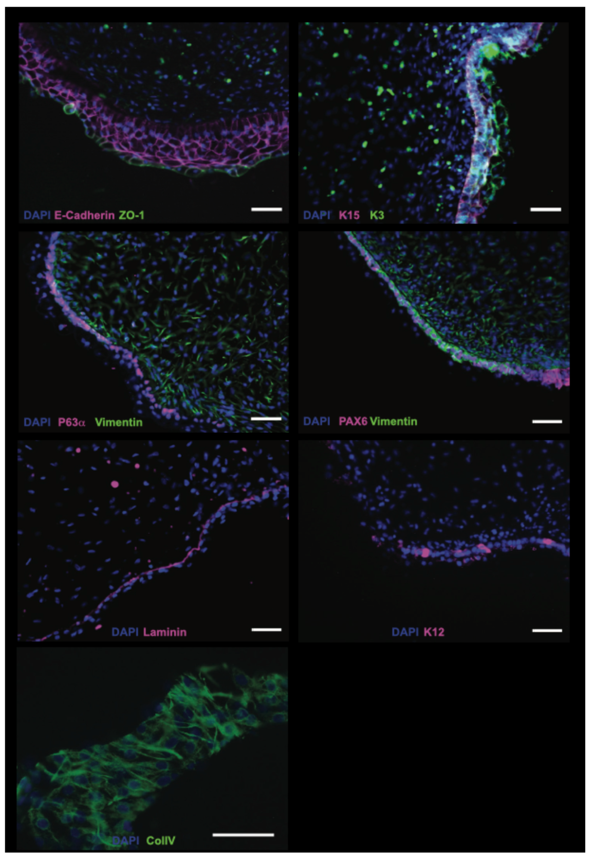
3.1. Immunofluorescent Characterization of D70–80 Organoids Using Corneal Markers
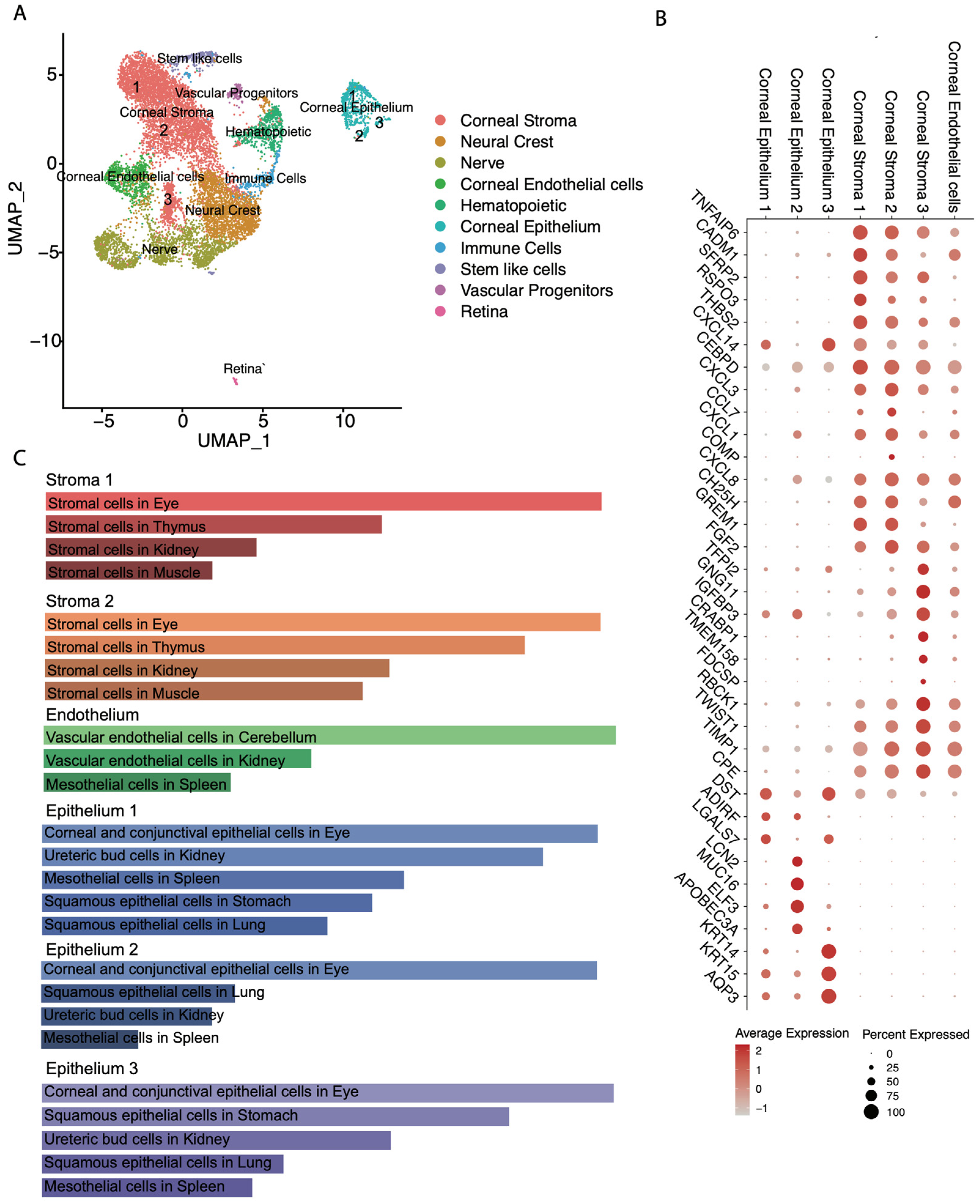
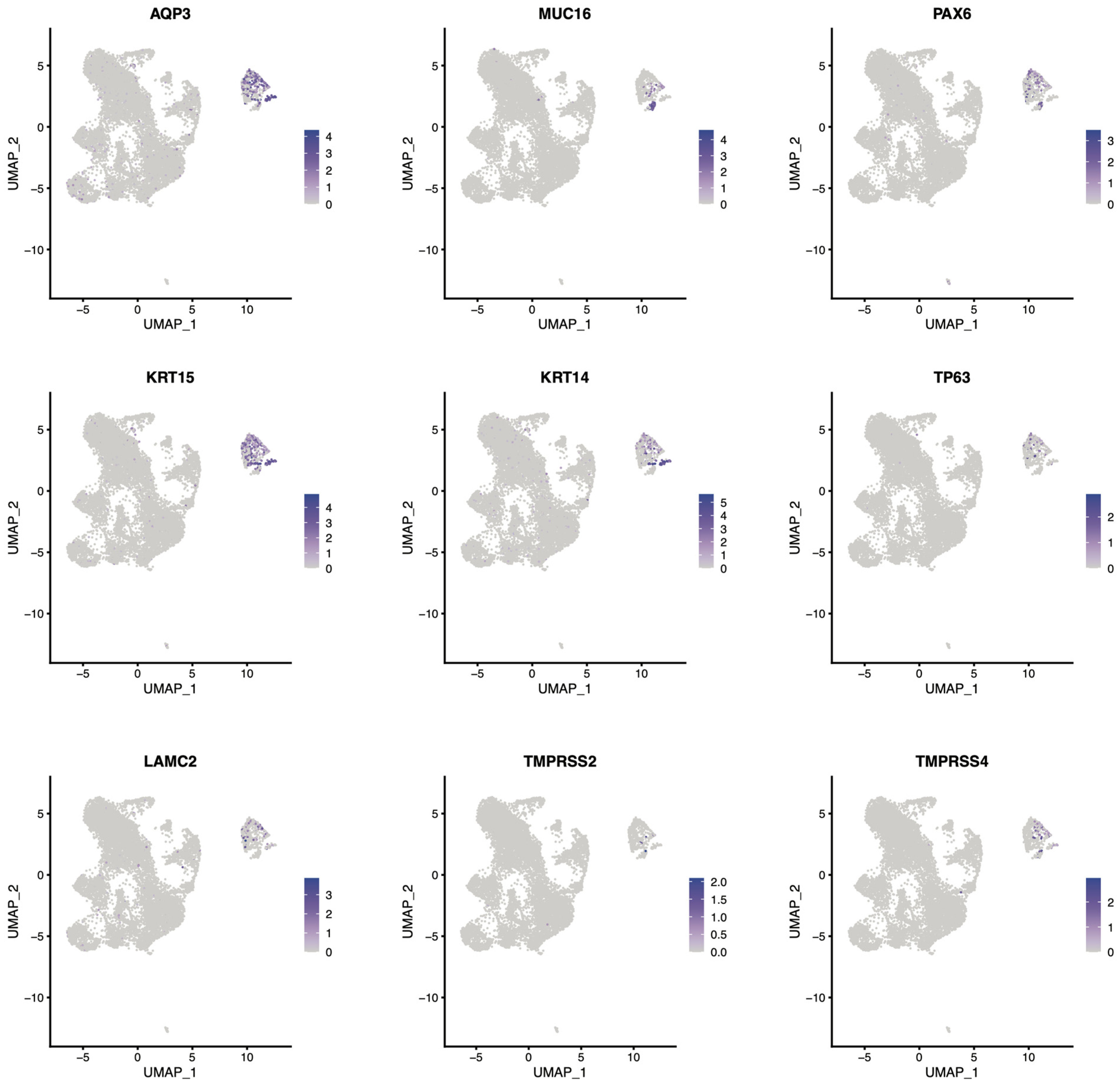
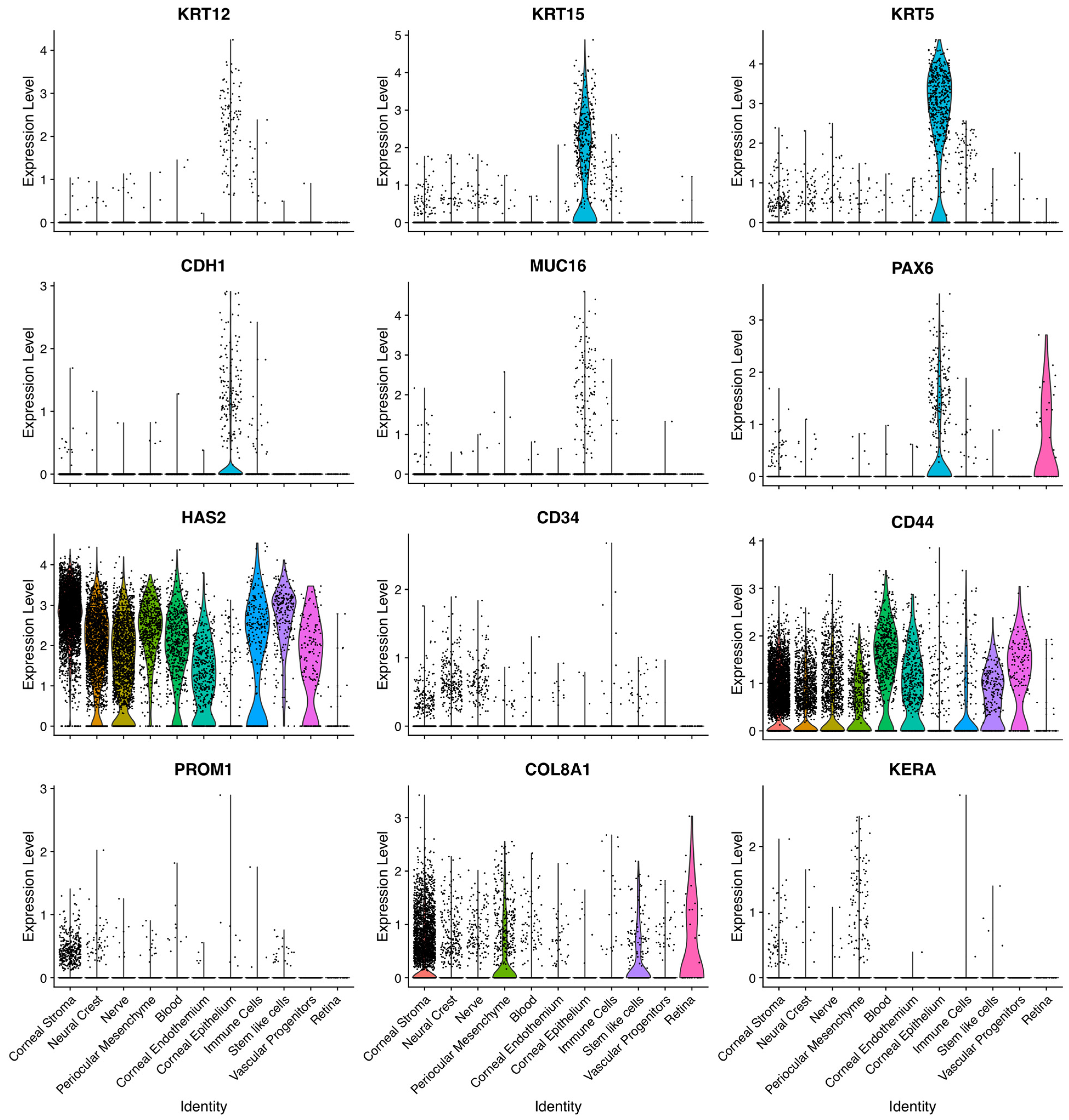
3.2. Single Cell RNA-Seq Characterization of 3D Corneal Organoids
4. Discussion
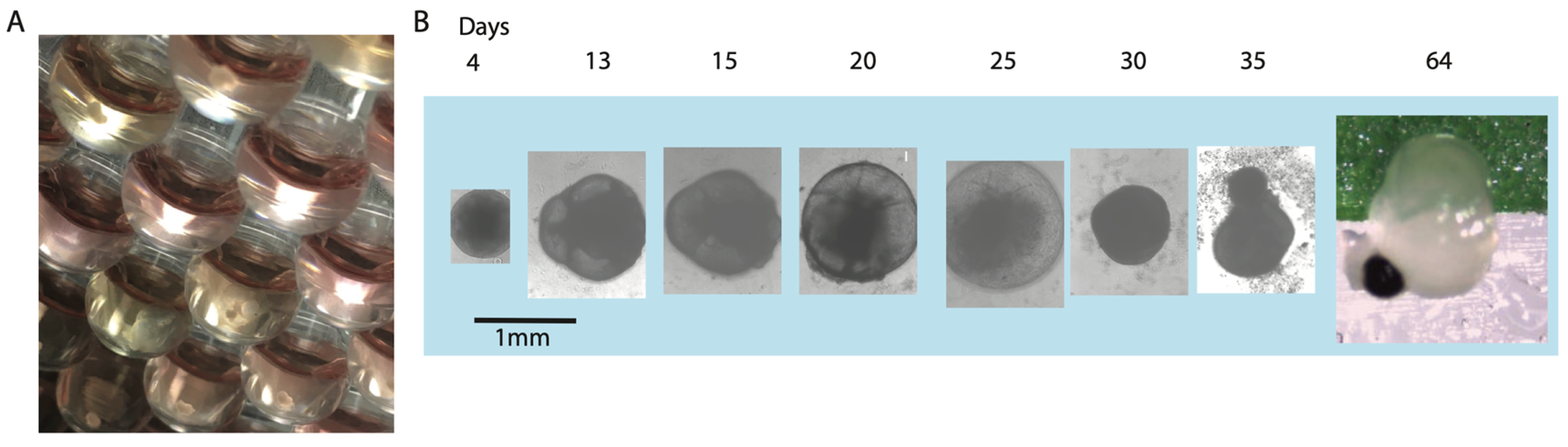
4.1. Development and Maturation Timeline of 3D Corneal Organoids
4.2. Cellular Architecture and Molecular Identity
4.3. Single-Cell Transcriptomic Insights into Cellular Diversity
4.4. Gene Expression Patterns and Comparison to Native Tissue
4.5. Comparison to Existing Corneal Organoid Models
5. Limitations and Future Directions
6. Conclusions
Supplementary Materials
Author Contributions
Funding
Institutional Review Board Statement
Informed Consent Statement
Data Availability Statement
Acknowledgments
Conflicts of Interest
References
- Ma, D.H.; Lai, J.Y.; Cheng, H.Y.; Tsai, C.C.; Yeh, L.K. Carbodiimide cross-linked amniotic membranes for cultivation of limbal epithelial cells. Biomaterials 2010, 31, 6647–6658. [Google Scholar] [CrossRef] [Scilit] [PubMed]
- Tang, X.Y.; Wu, S.; Wang, D.; Chu, C.; Hong, Y.; Tao, M.; Hu, H.; Xu, M.; Guo, X.; Liu, Y. Human organoids in basic research and clinical applications. Signal Transduct. Target. Ther. 2022, 7, 168. [Google Scholar] [CrossRef] [Scilit]
- Zhou, Z.; Cong, L.; Cong, X. Patient-Derived Organoids in Precision Medicine: Drug Screening, Organoid-on-a-Chip and Living Organoid Biobank. Front. Oncol. 2021, 11, 762184. [Google Scholar] [CrossRef] [Scilit]
- Foster, J.W.; Wahlin, K.; Adams, S.M.; Birk, D.E.; Zack, D.J.; Chakravarti, S. Cornea organoids from human induced pluripotent stem cells. Sci. Rep. 2017, 7, 41286. [Google Scholar] [CrossRef] [Scilit]
- Susaimanickam, P.J.; Maddileti, S.; Pulimamidi, V.K.; Boyinpally, S.R.; Naik, R.R.; Naik, M.N.; Reddy, G.B.; Sangwan, V.S.; Mariappan, I. Generating minicorneal organoids from human induced pluripotent stem cells. Development 2017, 144, 2338–2351. [Google Scholar] [CrossRef] [Scilit] [PubMed]
- Sato, T.; Vries, R.G.; Snippert, H.J.; van de Wetering, M.; Barker, N.; Stange, D.E.; van Es, J.H.; Abo, A.; Kujala, P.; Peters, P.J.; et al. Single Lgr5 stem cells build crypt-villus structures in vitro without a mesenchymal niche. Nature 2009, 459, 262–265. [Google Scholar] [CrossRef] [Scilit]
- Swarup, A.; Phansalkar, R.; Morri, M.; Agarwal, A.; Subramaniam, V.; Li, B.; Wu, A.Y. Single-cell transcriptomic analysis of corneal organoids during development. Stem Cell Rep. 2023, 18, 2482–2497. [Google Scholar] [CrossRef] [Scilit]
- Maiti, G.; Hu, N. Single cell RNA-seq of human cornea organoids identifies cell fates of a developing immature cornea. PNAS Nexus 2022, 1, pgac246. [Google Scholar] [CrossRef] [Scilit]
- Yu, Z.; Hao, R.; Du, J.; Wu, X.; Chen, X.; Zhang, Y.; Li, W.; Gu, Z.; Yang, H. A human cornea-on-a-chip for the study of epithelial wound healing by extracellular vesicles. iScience 2022, 25, 104200. [Google Scholar] [CrossRef] [Scilit]
- Wan, X.; Gu, J.; Zhou, X.; Le, Q.; Wang, J.; Xin, C.; Chen, Z.; He, Y.; Hong, J. Establishment of human corneal epithelial organoids for ex vivo modelling dry eye disease. Cell Prolif. 2024, 57, e13704. [Google Scholar] [CrossRef] [Scilit] [PubMed]
- Owen, L.J.; Rainger, J.; Bengani, H.; Kilanowski, F.; FitzPatrick, D.R.; Papanastasiou, A.S. Characterization of an eye field-like state during optic vesicle organoid development. Development 2023, 150, dev201432. [Google Scholar] [CrossRef] [Scilit] [PubMed]
- Soma, T.; Oie, Y.; Takayanagi, H.; Matsubara, S.; Yamada, T.; Nomura, M.; Yoshinaga, Y.; Maruyama, K.; Watanabe, A.; Takashima, K.; et al. Induced pluripotent stem-cell-derived corneal epithelium for transplant surgery: A single-arm, open-label, first-in-human interventional study in Japan. Lancet 2024, 404, 1929–1939. [Google Scholar] [CrossRef] [Scilit]
- Higa, K.; Ishiwata, M.; Kimoto, R.; Hirayama, M.; Yamaguchi, T.; Shimmura, S. Human corneal organoid has a limbal function that supplies epithelium to the cornea with limbal deficiency. Regen. Ther. 2025, 29, 247–253. [Google Scholar] [CrossRef] [Scilit]
- Hayashi, R.; Ishikawa, Y.; Sasamoto, Y.; Katori, R.; Nomura, N.; Ichikawa, T.; Araki, S.; Soma, T.; Kawasaki, S.; Sekiguchi, K.; et al. Co-ordinated ocular development from human iPS cells and recovery of corneal function. Nature 2016, 531, 376–380. [Google Scholar] [CrossRef] [Scilit]
- Hayashi, R.; Ishikawa, Y.; Katori, R.; Sasamoto, Y.; Taniwaki, Y.; Takayanagi, H.; Tsujikawa, M.; Sekiguchi, K.; Quantock, A.J.; Nishida, K. Coordinated generation of multiple ocular-like cell lineages and fabrication of functional corneal epithelial cell sheets from human iPS cells. Nat. Protoc. 2017, 12, 683–696. [Google Scholar] [CrossRef] [Scilit]
- Eriksen, A.Z.; Møller, R.; Makovoz, B.; Uhl, S.A.; tenOever, B.R.; Blenkinsop, T.A. SARS-CoV-2 infects human adult donor eyes and hESC-derived ocular epithelium. Cell Stem Cell 2021, 28, 1205–1220.e7. [Google Scholar] [CrossRef] [Scilit]
- Eriksen, A.Z.; Møller, R.; Makovoz, B.; tenOever, B.R.; Blenkinsop, T.A. Protocols for SARS-CoV-2 infection in primary ocular cells and eye organoids. STAR Protoc. 2022, 3, 101383. [Google Scholar] [CrossRef] [Scilit]
- Makovoz, B.; Eriksen, A.Z.; Warrington, R.E.; Williams, D.; Blenkinsop, T.A. Neuroectoderm-derived iris muscle characterization at the single-cell resolution in native human iris and a pluripotent stem cell eye model. Nat. Commun. 2025, 16, 10648. [Google Scholar] [CrossRef] [Scilit] [PubMed]
- Williams, A.L.; Bohnsack, B.L. Neural crest derivatives in ocular development: Discerning the eye of the storm. Birth Defects Res. C Embryo Today 2015, 105, 87–95. [Google Scholar] [CrossRef] [Scilit] [PubMed]
- Joe, A.W.; Yeung, S.N. Concise review: Identifying limbal stem cells: Classical concepts and new challenges. Stem Cells Transl. Med. 2014, 3, 318–322. [Google Scholar] [CrossRef] [Scilit]
- Das, S.K.; Gupta, I.; Cho, Y.K.; Zhang, X.; Uehara, H.; Muddana, S.K.; Bernhisel, A.A.; Archer, B.; Ambati, B.K. Vimentin knockdown decreases corneal opacity. Investig. Ophthalmol. Vis. Sci. 2014, 55, 4030–4040. [Google Scholar] [CrossRef] [Scilit]
- Wilson, S.E.; Hong, J.W. Bowman’s layer structure and function: Critical or dispensable to corneal function? A hypothesis. Cornea 2000, 19, 417–420. [Google Scholar] [CrossRef] [Scilit]
- Kivelä, T.; Uusitalo, M. Structure, development and function of cytoskeletal elements in non-neuronal cells of the human eye. Prog. Retin. Eye Res. 1998, 17, 385–428. [Google Scholar] [CrossRef] [Scilit]
- Pauklin, M.; Thomasen, H.; Pester, A.; Steuhl, K.P.; Meller, D. Expression of pluripotency and multipotency factors in human ocular surface tissues. Curr. Eye Res. 2011, 36, 1086–1097. [Google Scholar] [CrossRef] [Scilit] [PubMed]
- Gipson, I.K. Distribution of mucins at the ocular surface. Exp. Eye Res. 2004, 78, 379–388. [Google Scholar] [CrossRef] [Scilit]
- Raeder, S.; Utheim, T.P.; Utheim, O.A.; Cai, Y.; Roald, B.; Lyberg, T.; Kjeldsen-Kragh, J.; Ramstad, H.; Messelt, E.; Nicolaissen, B., Jr. Effect of limbal explant orientation on the histology, phenotype, ultrastructure and barrier function of cultured limbal epithelial cells. Acta Ophthalmol. Scand. 2007, 85, 377–386. [Google Scholar] [CrossRef] [Scilit]
- West-Mays, A.; Dwivedi, D.J. The keratocyte: Corneal stromal cell with variable repair phenotypes. Int. J. Biochem. Cell Biol. 2006, 38, 1625–1631. [Google Scholar] [CrossRef] [Scilit]
- Arrigoni, F.; Matarin, M.; Thompson, P.; Michaelides, M.; McClements, M.E.; Redmond, E.; Clarke, L.; Ellins, E.; Mohamed, S.; Pavord, I.; et al. Extended extraocular phenotype of PROM1 mutation in kindreds with known autosomal dominant macular dystrophy. Eur. J. Hum. Genet. 2011, 19, 131–137. [Google Scholar] [CrossRef] [Scilit] [PubMed]
- Guo, N.; Kanter, D.; Funderburgh, M.L.; Mann, M.M.; Du, Y.; Funderburgh, J.L. A rapid transient increase in hyaluronan synthase-2 mRNA initiates secretion of hyaluronan by corneal keratocytes in response to transforming growth factor beta. J. Biol. Chem. 2007, 282, 12475–12483. [Google Scholar] [CrossRef] [Scilit]
- Carlson, E.C.; Liu, C.Y.; Chikama, T.; Hayashi, Y.; Kao, C.W.; Birk, D.E.; Funderburgh, J.L.; Jester, J.V.; Kao, W.W. Keratocan, a cornea-specific keratan sulfate proteoglycan, is regulated by lumican. J. Biol. Chem. 2005, 280, 25541–25547. [Google Scholar] [CrossRef] [Scilit] [PubMed]
- Hopfer, U.; Fukai, N.; Hopfer, H.; Wolf, G.; Joyce, N.; Li, E.; Olsen, B.R. Targeted disruption of Col8a1 and Col8a2 genes in mice leads to anterior segment abnormalities in the eye. FASEB J. 2005, 19, 1232–1244. [Google Scholar] [CrossRef] [Scilit] [PubMed]



Disclaimer/Publisher’s Note: The statements, opinions and data contained in all publications are solely those of the individual author(s) and contributor(s) and not of MDPI and/or the editor(s). MDPI and/or the editor(s) disclaim responsibility for any injury to people or property resulting from any ideas, methods, instructions or products referred to in the content. |
© 2026 by the authors. Licensee MDPI, Basel, Switzerland. This article is an open access article distributed under the terms and conditions of the Creative Commons Attribution (CC BY) license.
Share and Cite
Blenkinsop, T.A.; Eriksen, A.Z. Putative Self-Organizing Human Corneal Organoids Recapitulate Human Corneal Architecture and Cellular Diversity. Bioengineering 2026, 13, 518. https://doi.org/10.3390/bioengineering13050518
Blenkinsop TA, Eriksen AZ. Putative Self-Organizing Human Corneal Organoids Recapitulate Human Corneal Architecture and Cellular Diversity. Bioengineering. 2026; 13(5):518. https://doi.org/10.3390/bioengineering13050518
Chicago/Turabian StyleBlenkinsop, Timothy A., and Anne Z. Eriksen. 2026. "Putative Self-Organizing Human Corneal Organoids Recapitulate Human Corneal Architecture and Cellular Diversity" Bioengineering 13, no. 5: 518. https://doi.org/10.3390/bioengineering13050518
APA StyleBlenkinsop, T. A., & Eriksen, A. Z. (2026). Putative Self-Organizing Human Corneal Organoids Recapitulate Human Corneal Architecture and Cellular Diversity. Bioengineering, 13(5), 518. https://doi.org/10.3390/bioengineering13050518