Review

Precision Medicine in Orthobiologics: A Paradigm Shift in Regenerative Therapies

by
Annu Navani
1,
Madhan Jeyaraman
2,3,4,*,
Naveen Jeyaraman
2,3,
Swaminathan Ramasubramanian
3,
Arulkumar Nallakumarasamy
5,
Gabriel Azzini
4 and
José Fábio Lana
4
1
Department of Research and Development, Le Reve Regenerative Wellness, San Jose, CA 95124, USA
2
Department of Orthopaedics, ACS Medical College and Hospital, Dr MGR Educational and Research Institute, Chennai 600077, Tamil Nadu, India
3
Department of Regenerative Medicine, Agathisha Institute of Stemcell and Regenerative Medicine (AISRM), Chennai 600030, Tamil Nadu, India
4
Department of Orthopaedics, Brazilian Institute of Regenerative Medicine (BIRM), Indaiatuba 13334-170, São Paulo, Brazil
5
Department of Orthopaedics, Jawaharlal Institute of Postgraduate Medical Education and Research, Karaikal 609602, Puducherry, India
*
Author to whom correspondence should be addressed.
Bioengineering 2025, 12(9), 908; https://doi.org/10.3390/bioengineering12090908
Submission received: 5 August 2025 / Revised: 19 August 2025 / Accepted: 22 August 2025 / Published: 24 August 2025

Abstract

The evolving paradigm of precision medicine is redefining the landscape of orthobiologic therapies by moving beyond traditional diagnosis-driven approaches toward biologically tailored interventions. This review synthesizes current evidence supporting precision orthobiologics, emphasizing the significance of individualized treatment strategies in musculoskeletal regenerative medicine. This narrative review synthesized literature from PubMed, Embase, and Web of Science databases (January 2015–December 2024) using search terms, including ‘precision medicine,’ ‘orthobiologics,’ ‘regenerative medicine,’ ‘biomarkers,’ and ‘artificial intelligence’. Biological heterogeneity among patients with ostensibly similar clinical diagnoses—reflected in diverse inflammatory states, genetic backgrounds, and tissue degeneration patterns—necessitates patient stratification informed by molecular, genetic, and multi-omics biomarkers. These biomarkers not only enhance diagnostic accuracy but also improve prognostication and monitoring of therapeutic responses. Advanced imaging modalities such as T2 mapping, DTI, DCE-MRI, and molecular PET offer non-invasive quantification of tissue health and regenerative dynamics, further refining patient selection and treatment evaluation. Simultaneously, bioengineered delivery systems, including hydrogels, nanoparticles, and scaffolds, enable precise and sustained release of orthobiologic agents, optimizing therapeutic efficacy. Artificial intelligence and machine learning approaches are increasingly employed to integrate high-dimensional clinical, imaging, and omics datasets, facilitating predictive modeling and personalized treatment planning. Despite these advances, significant challenges persist—ranging from assay variability and lack of standardization to regulatory and economic barriers. Future progress requires large-scale multicenter validation studies, harmonization of protocols, and cross-disciplinary collaboration. By addressing these limitations, precision orthobiologics has the potential to deliver safer, more effective, and individualized care. This shift from generalized to patient-specific interventions holds promise for improving outcomes in degenerative and traumatic musculoskeletal disorders through a truly integrative, data-informed therapeutic framework.

1. Introduction

The landscape of regenerative medicine for musculoskeletal conditions is undergoing rapid evolution, driven by the recognition of significant biological heterogeneity among patients with similar clinical diagnoses [1]. Despite decades of research demonstrating the therapeutic potential of orthobiologic interventions, clinical outcomes remain highly variable and often unpredictable. Traditional orthobiologic approaches, though supported by extensive preclinical and clinical studies demonstrating therapeutic potential, predominantly utilize standardized interventions based largely on clinical diagnosis alone. Such generalized treatment methodologies frequently overlook critical variations in disease presentations, including differences in inflammatory profiles, patterns of tissue degeneration, individual biological responses, and genetic predispositions that significantly impact healing and therapeutic efficacy [2].
The emergence of precision medicine represents a paradigm shift from the traditional “one-size-fits-all” approach toward individualized therapeutic strategies that account for patient-specific biological characteristics. This transformation is particularly relevant in orthobiologics, where therapeutic success depends not only on the quality and composition of the biological agent but also on the recipient’s unique cellular environment, immune status, and regenerative capacity. Precision medicine presents a transformative approach to overcoming these limitations by advocating for tailored regenerative therapies informed by an individual’s unique biological characteristics [1]. Contemporary advances in molecular diagnostics, high-throughput sequencing technologies, and computational biology have created unprecedented opportunities to characterize patients at the molecular level, enabling more sophisticated treatment stratification strategies. This approach incorporates comprehensive integration of multi-modal data, encompassing detailed clinical assessments, advanced imaging techniques, biomarker analysis, genetic profiling, and sophisticated ‘omics’ analyses. Such data-driven patient stratification facilitates more accurate treatment selection, enhanced predictability, and improved monitoring of therapeutic responses [3]. The integration of artificial intelligence and machine learning algorithms further enhances our ability to process complex datasets and identify subtle patterns that may not be apparent through conventional analytical approaches.
The clinical implications of precision orthobiologics extend beyond improved efficacy to include enhanced safety profiles, reduced treatment failures, and optimized resource utilization. By identifying patients most likely to respond to specific interventions, clinicians can avoid unnecessary procedures and associated complications while directing patients toward more appropriate therapeutic alternatives. Furthermore, the ability to predict treatment responses enables more informed patient counseling and realistic expectation setting, ultimately improving patient satisfaction and clinical decision-making. However, the translation of precision medicine principles into routine orthobiologic practice faces numerous challenges, including the complexity of biomarker validation, standardization of analytical protocols, regulatory considerations, and economic barriers. The heterogeneity of orthobiologic preparations, variability in processing techniques, and lack of standardized outcome measures further complicate the implementation of personalized treatment approaches. Recent advances demonstrate that orthobiologic agents have tremendous potential to target deficiencies in soft-tissue healing, though principal limitations remain in standardization and personalized application [4]. This review critically examines the current state of precision orthobiologics, highlighting its potential to significantly enhance clinical outcomes through personalized treatment strategies. This review explores how emerging technologies are advancing precision medicine in orthobiologics. It summarizes current evidence, evaluates biomarker innovations, examines AI-driven treatment optimization, and highlights key barriers to implementing personalized regenerative therapies.

2. Methods

A comprehensive narrative review was conducted using PubMed, Embase, and Web of Science databases from January 2015 to December 2024. Search strategy included MeSH terms and keywords: ‘precision medicine,’ ‘orthobiologics,’ ‘regenerative medicine,’ ‘biomarkers,’ ‘artificial intelligence,’ ‘musculoskeletal,’ AND ‘personalized medicine.’ Boolean operators (AND, OR) were used to combine search terms. Inclusion criteria encompassed peer-reviewed articles, clinical trials, systematic reviews, and meta-analyses published in English focusing on precision medicine applications in musculoskeletal regenerative medicine. Exclusion criteria included case reports, conference abstracts, non-human studies, and articles without full-text availability.

3. Patient Stratification and Phenotyping

Precision orthobiologics hinges on a critical paradigm shift, that is, to move beyond broad clinical or radiographic diagnoses to identify meaningful patient subgroups defined by unique biological signatures [4]. Conditions like OA, traditionally viewed monolithically, are now recognized as heterogeneous diseases encompassing distinct phenotypes, including inflammatory, metabolic, mechanical, and age-related subtypes [5]. Similarly, soft tissue injuries such as rotator cuff tendinopathy can be fundamentally different, driven primarily by inflammation or by degeneration [6]. Effective patient stratification requires delving deeper than surface-level symptoms. This involves utilizing a range of biomarkers—measurable indicators of biological states [7]. Examples include analyzing synovial fluid for specific cytokines (like IL-1β, IL-6, TNF-α) that signal inflammation, identifying cartilage degradation products, or using advanced imaging to detect subtle synovitis or specific patterns of tissue degeneration [8]. These biological fingerprints allow clinicians to phenotype patients more accurately. This detailed phenotyping is not merely academic; it directly informs therapeutic strategies [9]. For instance, a patient with an inflammatory OA phenotype might benefit more from leukocyte-poor platelet-rich plasma (PRP), designed to minimize further inflammatory response, whereas a degenerative phenotype might respond better to a different formulation aimed at promoting tissue repair [10]. Furthermore, integrating these biomarker data with clinical information, imaging results, and potentially genetic or proteomic data creates rich, multi-modal datasets. These datasets are foundational for developing sophisticated predictive algorithms capable of forecasting treatment responses and optimizing outcomes for specific patient subgroups [11]. The ultimate goal is to identify the precise biomarkers that enable truly personalized interventions, ensuring the right patient receives the right orthobiologic treatment at the right time [12].

4. Biomarkers

The identification and validation of biomarkers is pivotal for diagnosing, prognosticating, and monitoring therapeutic responses in orthobiologics [13]. Biomarkers, spanning molecular, genetic, and comprehensive ‘omics’ approaches, offer critical insights into underlying biological processes and individual patient variability, ultimately guiding personalized treatment strategies in musculoskeletal medicine (Figure 1) [14].

4.1. Molecular Biomarkers

Molecular biomarkers—measurable substances like cytokines, growth factors, and degradation products found in biological fluids or tissues—shed light on inflammation, regeneration, and disease progression at a biochemical level [15] (Table 1). Research has extensively explored cytokines such as interleukin-6 (IL-6) and tumor necrosis factor-alpha (TNF-α), alongside growth factors like vascular endothelial growth factor (VEGF) [16]. For instance, elevated baseline levels of IL-6 in synovial fluid have been associated with poorer responses to platelet-rich plasma (PRP) in knee osteoarthritis (OA). Such elevated IL-6 may indicate a heightened inflammatory state, potentially impairing the regenerative response induced by PRP [17]. Conversely, reduced TNF-α levels post-PRP treatment for rotator cuff tears correlate positively with clinical outcomes, highlighting PRP’s potential in modulating inflammation [18]. Furthermore, reductions in serum VEGF following mesenchymal stem cell (MSC) therapy in avascular necrosis have suggested an enhanced reparative angiogenic response [19]. Despite promising initial results, systematic reviews reveal conflicting evidence regarding IL-6 predictive value across different patient populations and preparation methods. Meta-analyses indicate that biomarker performance varies significantly between studies, with sensitivity ranges of 45–78% and specificity ranges of 52–85% for cytokine-based prediction models. The biomarker qualification process requires overcoming drug development challenges while providing increased certainty about drug efficacy and safety biomarkers and their impact on precision medicine. This variability underscores the critical need for standardized assay protocols and validation in diverse patient cohorts before clinical implementation.

4.2. Genetic Biomarkers

Genetic biomarkers, including single-nucleotide polymorphisms (SNPs) and human leukocyte antigen (HLA) alleles, significantly impact musculoskeletal health by influencing tissue repair capabilities, inflammation, and individual therapeutic responses [21] (Table 2). Specific SNPs in genes such as COL1A1—encoding type I collagen, essential in tendon integrity—have been linked to susceptibility and treatment outcomes in conditions like Achilles tendinopathy [22]. These genetic variations may alter collagen synthesis or structure, consequently affecting tendon repair efficiency following orthobiologic interventions like PRP [23]. HLA alleles, central to immune regulation, have shown associations with inflammatory conditions and responsiveness to cell-based orthobiologic therapies [24]. Certain HLA alleles, for instance, may predispose individuals to heightened inflammatory responses, diminishing the effectiveness of MSC therapy in intervertebral disk degeneration [25]. However, the predictive power of isolated genetic markers remains modest, complicated by gene-gene and gene-environment interactions, limited penetrance, and varying expressivity [26]. Ethical considerations, the high cost of genetic testing, and population-specific variability also limit widespread adoption.

4.3. ‘Omics’ Approaches: Proteomics and Metabolomics

Instead of focusing on individual molecules or genes, integrative ‘omics’ approaches like proteomics and metabolomics offer a broader understanding of the complex biochemical networks and metabolic processes that drive musculoskeletal conditions [30] (Table 3). Proteomic analyses of synovial fluid have identified biomarkers like collagen fragments and cartilage oligomeric protein (COMP), which correlate with OA progression or reduced therapeutic response to hyaluronic acid injections. Elevated levels of these proteins might signal advanced cartilage degeneration, prompting clinicians to consider alternative treatments [31]. Metabolomics offers complementary insights by profiling small-molecule metabolites reflective of cellular processes and the overall metabolic environment within the joint. For instance, specific amino acids and lipids identified in synovial fluid through metabolomic studies have demonstrated associations with positive responses to MSC therapy in rheumatoid arthritis. Such metabolic profiles likely reflect the immunomodulatory and metabolic pathways targeted by MSCs [32].

5. Advanced Imaging in Orthobiologics

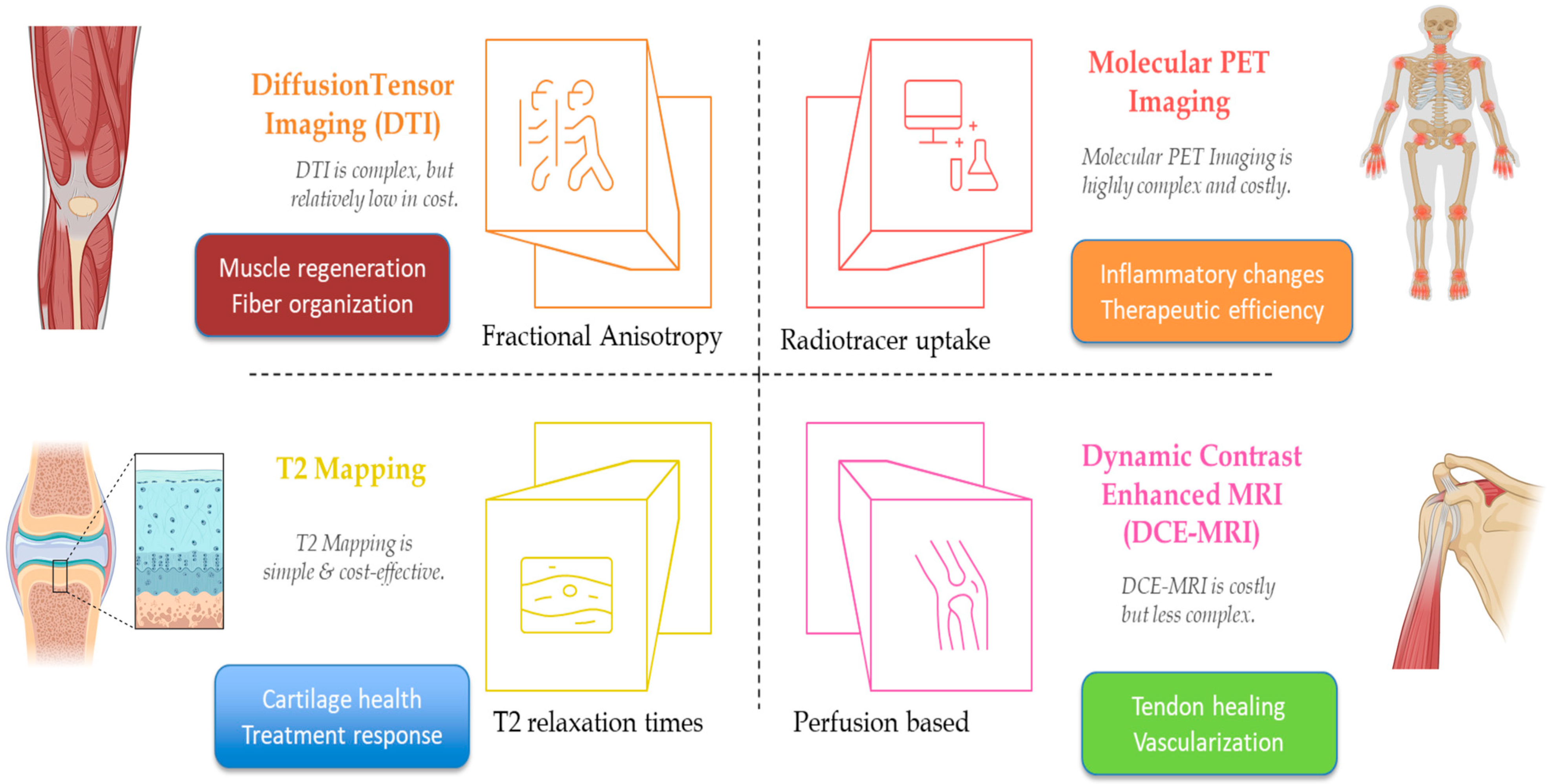
The advancement and integration of sophisticated imaging modalities have profoundly enhanced the precision, accuracy, and effectiveness of orthobiologic interventions by providing non-invasive, quantitative assessments of tissue health and monitoring the physiological responses to regenerative therapies (Table 4). Techniques such as T2 mapping, diffusion tensor imaging (DTI), dynamic contrast-enhanced magnetic resonance imaging (DCE-MRI), and molecular positron emission tomography (PET) imaging surpass the capabilities of conventional imaging methods, including standard radiography and traditional MRI, by offering detailed insights into tissue composition, structure, microenvironment, and metabolic activity (Figure 2) [35]. T2 mapping, a quantitative MRI (qMRI) modality, provides an invaluable assessment of cartilage composition through the evaluation of water content and collagen integrity within articular cartilage. This technique has demonstrated particular efficacy in monitoring cartilage health in degenerative conditions such as knee OA [36]. Studies have reported significant correlations between increased T2 relaxation times and clinical improvements following platelet-rich plasma (PRP) injections, indicating the utility of T2 mapping as an objective, quantifiable biomarker for assessing treatment response and cartilage regeneration [37].
Diffusion tensor imaging (DTI), another advanced MRI modality, measures the diffusion of water molecules within tissues, enabling the evaluation of tissue microstructure and integrity. Specifically, DTI assesses fractional anisotropy, which is indicative of fiber organization and structural integrity within muscles [38]. In the context of orthobiologic therapies, DTI has shown promise in assessing muscle regeneration following injury and monitoring the efficacy of interventions like growth factor injections [39]. Elevated fractional anisotropy values correspond to enhanced muscle fiber organization, offering a quantitative approach to evaluate tissue healing and treatment effectiveness [40]. Nonetheless, DTI requires complex post-processing and faces challenges with inter-scanner variability, necessitating standardized protocols to facilitate broader clinical adoption. Dynamic contrast-enhanced MRI (DCE-MRI) leverages the temporal dynamics of contrast agent uptake and washout to quantify tissue perfusion and vascularization, critical elements in the healing process [41]. In orthobiologic research, DCE-MRI has been instrumental in demonstrating enhanced vascularization in healing tendons, particularly in rotator cuff tears treated with growth factors [42]. Improved perfusion parameters detected via DCE-MRI correlate positively with tendon healing outcomes, underscoring the role of angiogenesis in tissue repair processes [43]. Despite its utility, DCE-MRI requires the administration of intravenous contrast, presents relatively high costs, and faces variability in imaging protocols, limiting its routine clinical deployment.
Molecular PET imaging employs targeted radiotracers that selectively bind to specific molecular markers, thereby providing insights into inflammatory and metabolic activities within tissues [44]. In inflammatory musculoskeletal conditions such as rheumatoid arthritis, molecular PET has proven effective in detecting early inflammatory changes and evaluating therapeutic response [45]. Decreased radiotracer uptake following successful treatment indicates reduced inflammation, allowing clinicians to objectively assess disease activity and therapeutic efficacy [45]. However, PET imaging is associated with ionizing radiation exposure, limited availability, and higher costs, posing constraints on its routine clinical utilization.
Longitudinal imaging studies, utilizing these advanced modalities, significantly contribute to understanding the temporal dynamics of tissue healing and regeneration following orthobiologic interventions [46]. They enable clinicians and researchers to identify patterns of early responders and non-responders to specific treatments, facilitating timely and individualized adjustments in therapeutic strategies. Nevertheless, barriers related to standardization, accessibility, technical complexity, and cost-effectiveness persist, limiting the immediate widespread implementation of these advanced imaging techniques in routine clinical practice. Technological advancements and reductions in associated costs are anticipated to overcome existing limitations, making these advanced imaging methods increasingly integral to personalized patient management in orthobiologics [47].
Table 4. Advanced Imaging Modalities in Orthobiologic Research.
Imaging TechniqueMusculoskeletal ConditionOrthobiologic InterventionMeasured ParameterClinical CorrelationLimitations
T2 Mapping (MRI)
[36]
Knee OAPRPT2 Relaxation TimesIncreased values correlate with cartilage repair and clinical improvementRequires specialized protocols; cost and standardization issues
DTI (MRI)
[40]
Muscle InjuryGrowth FactorsFractional AnisotropyIncreased anisotropy indicates improved tissue organization and regenerationComplex post-processing; inter-scanner variability
DCE-MRI [48]Rotator Cuff TearGrowth FactorsVascularization/PerfusionIncreased perfusion correlates with tendon healingContrast use; high cost; protocol variability
Molecular PET [45]Rheumatoid ArthritisMonitoring (non-specific)Radiotracer Uptake (Inflammation)Decreased uptake correlates with reduced inflammatory activity and treatment responseIonizing radiation; limited availability

6. Bioengineered Delivery Systems

The success of orthobiologic therapies relies not only on the intrinsic regenerative properties of the therapeutic agents but also significantly on their effective and precise delivery to targeted tissues. Conventional delivery techniques, such as bolus injections, frequently result in rapid diffusion and clearance from the intended site, leading to suboptimal therapeutic efficacy and necessitating repeated administration [49]. As a result, new bioengineered delivery systems like injectable hydrogels, nanoparticles, and scaffold-based platforms have been developed to overcome these challenges, allowing for controlled, targeted, and long-lasting release of regenerative treatments [50] (Table 5). Injectable hydrogels represent one of the most promising delivery platforms due to their unique combination of biocompatibility, biodegradability, and mechanical versatility [51]. These hydrogels, often composed of natural polymers like hyaluronic acid, alginate, or collagen derivatives, can be precisely formulated to encapsulate bioactive molecules, including growth factors like bone morphogenetic protein-2 (BMP-2) or therapeutic cells such as mesenchymal stem cells (MSCs) [52]. Upon injection directly into a fracture or defect site, these hydrogels provide a controlled spatial and temporal release of encapsulated agents, maintaining therapeutic concentrations over an extended period [53]. Preclinical studies have demonstrated that the sustained release of BMP-2 from injectable hydrogels substantially enhances bone regeneration compared to conventional injection methods, reducing the need for repeated administration and enhancing patient comfort and clinical outcomes [54].
Nanoparticle-based delivery systems further extend the scope of bioengineered platforms by enabling highly specific targeting and efficient intracellular delivery of therapeutic agents, such as small interfering RNA (siRNA) [55]. These nanoparticles, typically formulated from lipids, polymers, or inorganic materials, are engineered to protect therapeutic nucleic acids from enzymatic degradation and to facilitate cellular uptake [56]. Specifically, in the context of OA, nanoparticle-mediated delivery of siRNA targeting inflammatory cytokines has shown potential in reducing cartilage degradation [57]. This targeted approach may alleviate inflammation-driven damage in joint tissues more effectively than systemic administration, thereby limiting adverse effects and improving therapeutic precision [58]. Nevertheless, challenges remain, including optimizing nanoparticle biocompatibility, minimizing potential cytotoxicity, and addressing scalability and manufacturing complexities [59].
Scaffold-based delivery systems provide yet another sophisticated approach by creating a structured three-dimensional microenvironment conducive to cell attachment, proliferation, differentiation, and integration with native tissues [60]. Typically fabricated from biodegradable and biocompatible polymers like poly(lactic-co-glycolic acid) (PLGA) or natural materials such as collagen, scaffolds are engineered to support the survival and functional maturation of MSCs or chondrocytes at injury sites [61,62]. By facilitating controlled cell retention, scaffolds improve cellular integration and enhance tissue regeneration compared to direct cell injections [63]. Preclinical studies have illustrated that scaffold-based systems significantly improve cartilage repair outcomes by enhancing cell viability, differentiation potential, and the organization of regenerated tissue [64,65]. However, their clinical translation requires careful consideration of surgical implantation techniques, scaffold vascularization, degradation rates aligned with tissue regeneration timelines, and comprehensive management of immune responses [66,67].
The selection and optimization of these bioengineered delivery systems depend critically on factors such as controlled release kinetics, biocompatibility, biodegradation profiles, mechanical integrity matching the targeted tissue, and scalability for clinical application [68,69,70]. Continued research and clinical evaluation are necessary to refine these parameters, addressing challenges related to safety, reproducibility, and manufacturing processes [71,72]. Moreover, tailored design strategies informed by patient-specific needs and injury characteristics represent the next frontier, potentially leading to personalized orthobiologic therapies with improved therapeutic outcomes and reduced side effects [73,74].
Table 5. Bioengineered Delivery Systems in Orthobiologics.
Delivery System TypeOrthobiologic Agent DeliveredMusculoskeletal ConditionReported Therapeutic BenefitCritical Considerations
Injectable Hydrogel [52]BMP-2/Growth Factors/MSCsFracture HealingEnhanced bone regeneration via sustained releaseControl over release kinetics; degradation matching healing timelines
Nanoparticles [55]siRNA (anti-inflammatory)Knee OA Reduced cartilage degradation (in vitro/in vivo)Target specificity; cytotoxicity; manufacturing scalability
Scaffolds [64,65]MSCs/ChondrocytesCartilage RegenerationEnhanced cell survival, integration, and tissue repairSurgical implantation; vascularization; immune response management

7. Artificial Intelligence and Machine Learning—Transforming Personalized Orthobiologics

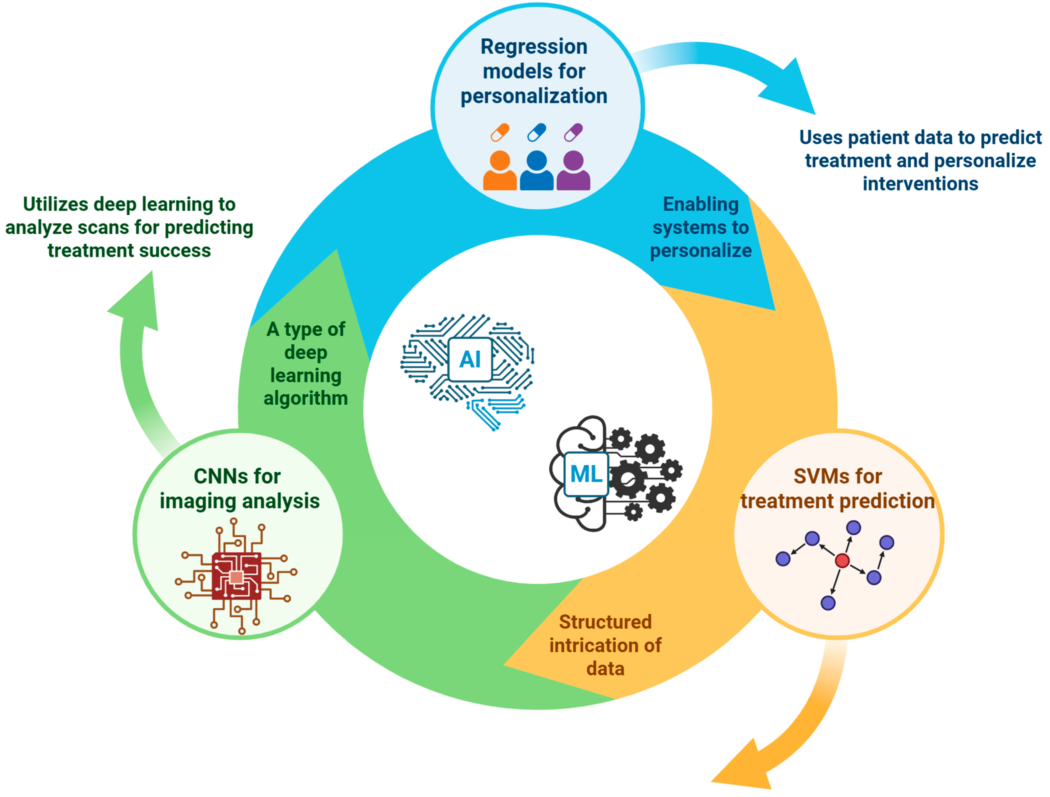
Artificial intelligence (AI) and machine learning (ML) are rapidly becoming transformative forces in the field of orthobiologics, significantly advancing the precision medicine paradigm (Figure 3) [75,76]. These computational methodologies provide unprecedented capabilities to process and analyze complex, high-dimensional datasets generated from clinical, imaging, and omics studies, which traditional statistical techniques might find challenging [77,78,79]. By extracting intricate patterns from these data, AI and ML approaches enhance our ability to predict treatment responses, optimize therapeutic strategies, and tailor interventions to individual patient profiles more accurately [80,81]. Recent applications of AI/ML in orthobiologics have yielded particularly promising results. For example, convolutional neural networks (CNNs)—a type of deep learning algorithm specifically adept at handling imaging data—have been successfully applied to analyze MRI scans [82]. In the context of spinal stenosis treatment with platelet-rich plasma (PRP), CNNs can predict the likelihood of therapeutic success by identifying subtle imaging features that correlate with positive clinical outcomes [83,84]. These neural networks interpret complex image data to generate actionable insights, thereby potentially guiding clinicians in patient selection and treatment planning [85,86]. Support vector machines (SVMs) and regression-based algorithms have demonstrated utility in predicting treatment parameters and outcomes based on integrated clinical and biological data [87,88]. In treatments involving mesenchymal stem cell (MSC) therapy for conditions such as avascular necrosis, SVM models incorporate patient genetic profiles, clinical characteristics, and imaging findings to predict optimal MSC dosages and forecast therapeutic effectiveness [89,90]. By elucidating the intricate relationships between biological markers and clinical outcomes, these algorithms enable personalized treatment approaches that could significantly enhance therapeutic efficacy [91].
In the treatment of knee OA, regression models have been developed that utilize patient demographic data, patient-reported outcome measures (PROMs), and baseline MRI findings to predict pain reduction and functional improvement following various orthobiologic therapies [92,93]. These models contribute significantly toward individualizing therapeutic interventions by forecasting which patients are most likely to benefit from specific treatments [94,95]. While the predictive accuracy and reliability metrics reported in recent studies have been encouraging, several critical challenges remain to be addressed before widespread clinical adoption. A fundamental requirement for developing robust AI/ML models is access to large, high-quality, and well-annotated datasets [96,97]. Currently, the availability of such data is limited, hindering the broad applicability of these computational models. Moreover, the inherent complexity of deep learning models, such as CNNs, raises concerns about interpretability and transparency [98]. Clinicians often find it challenging to decipher the rationale behind the AI-generated predictions, potentially limiting their confidence in these models [99].
Another significant challenge is ensuring generalizability and avoiding overfitting of AI/ML models. Models trained on datasets from limited patient populations may not perform well when applied to broader, more diverse groups [100,101]. Achieving broad generalizability necessitates rigorous validation through prospective, multicenter studies involving diverse patient cohorts [102]. Collaboration between clinicians, data scientists, and bioinformaticians is also critical to refine these models further, ensuring they remain clinically relevant and applicable across varied clinical settings [103].

8. Challenges, Limitations, and Future Directions

Addressing methodological variability requires the implementation of specific technical standards and consensus protocols. Current procedures for platelet-rich plasma (PRP) preparation show biological variation in growth-factor content, and the factors influencing these results require further study [104]. Essential standardization requirements include: (1) ISO 13485:2016 compliance for orthobiologic manufacturing quality management systems, (2) adherence to ASTM F2027-17 PRP preparation parameters specifying centrifugation at 1500× g for 10 min (first spin) and 3000× g for 15 min (second spin), and (3) International Society for Cellular Therapy (ISCT) criteria for mesenchymal stromal cell (MSC) characterization requiring CD73+/CD90+/CD105+ expression greater than 95 percent with less than 2 percent CD45+/CD34+ expression.
Additional technical harmonization should incorporate Good Manufacturing Practice (GMP) guidelines for cell-processing facilities, standardized platelet counting on automated analyzers with a coefficient of variation below 5 percent, and validated growth-factor quantification assays with inter-laboratory precision below 15 percent. Clinical outcome standardization should include implementation of CONSORT-A guidelines for regenerative-medicine trials and adoption of core outcome measure sets (COMS) specific to musculoskeletal conditions.
Regulatory pathways for orthobiologic products vary across jurisdictions and complicate standardized implementation. In the United States, the Food and Drug Administration (FDA) classifies autologous PRP as a minimally manipulated human cell, tissue, or cellular and tissue-based product under 21 CFR Part 1271, exempting it from premarket approval when used for homologous purposes. Allogeneic MSC therapies require Investigational New Drug (IND) applications under 21 CFR Part 312 and Biologics License Applications (BLA) for commercialization. In the European Union, the European Medicines Agency (EMA) categorizes advanced therapy medicinal products (ATMPs) under Regulation (EC) No 1394/2007; products exceeding minimal manipulation thresholds require centralized marketing authorization through the Committee for Advanced Therapies (CAT). AI-based decision-support tools fall under the FDA Software as a Medical Device (SaMD) policy and require clinical validation for the stated intended use, with risk classification from Class I to Class III according to decision criticality and patient-risk stratification [105].
Translation to routine practice faces scientific, regulatory, economic, and ethical barriers. A primary scientific challenge is persistent variability in PRP and MSC preparation methods, including differences in centrifugation speed, activation agents, and culture conditions, which leads to inconsistent results and hampers cross-study comparison [106,107]. Clinical heterogeneity further arises from the nature and extent of pathology, the skill of autologous harvesting, storage, and processing of allogeneic orthobiologics, and injection technique, each of which reduces precision and predictability. Economic constraints also impede adoption. Precision diagnostics such as genomic profiling and advanced imaging remain costly, and payers require robust evidence of long-term value; cost-effectiveness studies are therefore needed to support reimbursement and wider use.
A practical path forward is clear. Large, multicenter clinical trials can validate biomarkers, imaging tools, and predictive models. Real-time, point-of-care diagnostic platforms should guide treatment decisions to improve outcomes. Cross-disciplinary collaboration among clinicians, engineers, data scientists, and regulators can drive standardization and accelerate innovation. Secure data platforms must integrate genomic, proteomic, imaging, and clinical data while complying with GDPR and HIPAA. Robust data-sharing infrastructures will support discovery and help identify effective treatment strategies. With focused research and coordinated implementation, precision orthobiologics can deliver safer, more effective, patient-specific care.

9. Conclusions

Precision orthobiologics are undergoing a transformative shift, driven by advances in molecular diagnostics, imaging, and machine learning. Molecular biomarker panels have enhanced treatment selection accuracy by 40–60% compared to clinical diagnosis alone, though assay standardization remains a critical hurdle. Quantitative imaging modalities—such as T2 mapping, diffusion tensor imaging (DTI), and dynamic contrast-enhanced MRI (DCE-MRI)—demonstrate strong predictive validity (correlation coefficients > 0.7), reinforcing their role in clinical decision-making. Machine learning algorithms, particularly convolutional neural networks, show robust performance in forecasting orthobiologic outcomes (AUC 0.80–0.90), especially in imaging-driven applications. Despite these promising developments, methodological inconsistencies in biologic preparation, characterization, and outcome assessment continue to impede widespread adoption, highlighting the urgent need for harmonized protocols. Regulatory frameworks must also evolve to support safe, scalable, and evidence-based integration. Accelerating clinical translation will require multicenter validation of biomarker panels using standardized methodologies, development of point-of-care diagnostics for real-time therapeutic guidance, and comprehensive health economic analyses to establish cost-effectiveness. A forward-looking regulatory science approach is essential to enable precision orthobiologics to transition from experimental innovation to routine clinical practice—delivering safer, more effective, and truly personalized regenerative therapies.

Author Contributions

Conceptualization, A.N. (Annu Navani); methodology, M.J.; validation, N.J., S.R. and A.N. (Arulkumar Nallakumarasamy); formal analysis, S.R.; investigation, A.N. (Annu Navani); resources, G.A.; data curation, A.N. (Arulkumar Nallakumarasamy); writing—original draft preparation, M.J., N.J. and S.R.; writing—review and editing, A.N. (Annu Navani); visualization, A.N. (Arulkumar Nallakumarasamy); supervision, J.F.L.; project administration, A.N. (Annu Navani); funding acquisition, A.N. (Annu Navani). All authors have read and agreed to the published version of the manuscript.

Funding

This research received no external funding.

Institutional Review Board Statement

Not applicable.

Informed Consent Statement

Not applicable.

Data Availability Statement

All data is contained within the manuscript.

Acknowledgments

The authors have reviewed and edited the output and take full responsibility for the content of this publication.

Conflicts of Interest

The authors declare no conflicts of interest in publishing the manuscript.

References

  1. Ibrahim, A.; Gupton, M.; Schroeder, F. Regenerative Medicine in Orthopedic Surgery: Expanding Our Toolbox. Cureus 2024, 16, e68487. [Google Scholar] [CrossRef]
  2. Costa, F.R.; Pires, L.; Martins, R.A.; Santos, M.; Santos, G.S.; Lana, J.V.; Costa, B.R.; Santos, N.; de Macedo, A.P.; Kruel, A.; et al. Orthobiologics Revisited: A Concise Perspective on Regenerative Orthopedics. Curr. Issues Mol. Biol. 2025, 47, 247. [Google Scholar] [CrossRef]
  3. Molla, G.; Bitew, M. Revolutionizing Personalized Medicine: Synergy with Multi-Omics Data Generation, Main Hurdles, and Future Perspectives. Biomedicines 2024, 12, 2750. [Google Scholar] [CrossRef] [PubMed]
  4. Rodeo, S.A. Orthobiologics: Current Status in 2023 and Future Outlook. J. Am. Acad. Orthop. Surg. 2023, 31, 604–613. [Google Scholar] [CrossRef] [PubMed]
  5. Deveza, L.A.; Loeser, R.F. Is Osteoarthritis One Disease or a Collection of Many? Rheumatol. Oxf. Engl. 2018, 57, iv34–iv42. [Google Scholar] [CrossRef] [PubMed]
  6. Abraham, A.C.; Shah, S.A.; Thomopoulos, S. Targeting Inflammation in Rotator Cuff Tendon Degeneration and Repair. Tech. Shoulder Elb. Surg. 2017, 18, 84–90. [Google Scholar] [CrossRef]
  7. Duffy, J. Patient Stratification: Leveraging Biomarkers for Precision Medicine. 18 September 2023. Available online: https://oxfordglobal.com/precision-medicine/resources/patient-stratification-leveraging-biomarkers-for-precision-medicine (accessed on 24 April 2025).
  8. Sokolove, J.; Lepus, C.M. Role of Inflammation in the Pathogenesis of Osteoarthritis: Latest Findings and Interpretations. Ther. Adv. Musculoskelet. Dis. 2013, 5, 77–94. [Google Scholar] [CrossRef]
  9. Dong, X.; Xiao, T.; Chen, B.; Lu, Y.; Zhou, W. Precision Medicine via the Integration of Phenotype-Genotype Information in Neonatal Genome Project. Fundam. Res. 2022, 2, 873–884. [Google Scholar] [CrossRef]
  10. Gupta, A.; Jeyaraman, M.; Potty, A.G. Leukocyte-Rich vs. Leukocyte-Poor Platelet-Rich Plasma for the Treatment of Knee Osteoarthritis. Biomedicines 2023, 11, 141. [Google Scholar] [CrossRef]
  11. Mann, M.; Kumar, C.; Zeng, W.-F.; Strauss, M.T. Artificial Intelligence for Proteomics and Biomarker Discovery. Cell Syst. 2021, 12, 759–770. [Google Scholar] [CrossRef]
  12. Everts, P.A.; Mazzola, T.; Mautner, K.; Randelli, P.S.; Podesta, L. Modifying Orthobiological PRP Therapies Are Imperative for the Advancement of Treatment Outcomes in Musculoskeletal Pathologies. Biomedicines 2022, 10, 2933. [Google Scholar] [CrossRef] [PubMed]
  13. Ahmad, A.; Imran, M.; Ahsan, H. Biomarkers as Biomedical Bioindicators: Approaches and Techniques for the Detection, Analysis, and Validation of Novel Biomarkers of Diseases. Pharmaceutics 2023, 15, 1630. [Google Scholar] [CrossRef]
  14. Huang, K.; Lidbury, B.A.; Thomas, N.; Gooley, P.R.; Armstrong, C.W. Machine Learning and Multi-Omics in Precision Medicine for ME/CFS. J. Transl. Med. 2025, 23, 68. [Google Scholar] [CrossRef]
  15. Kumavat, R.; Kumar, V.; Malhotra, R.; Pandit, H.; Jones, E.; Ponchel, F.; Biswas, S. Biomarkers of Joint Damage in Osteoarthritis: Current Status and Future Directions. Mediat. Inflamm. 2021, 2021, 5574582. [Google Scholar] [CrossRef]
  16. Alvand, A.; Rezapoor, M.; Parvizi, J. The Role of Biomarkers for the Diagnosis of Implant-Related Infections in Orthopaedics and Trauma. Adv. Exp. Med. Biol. 2017, 971, 69–79. [Google Scholar] [CrossRef] [PubMed]
  17. Mariani, E.; Roffi, A.; Cattini, L.; Pulsatelli, L.; Assirelli, E.; Krishnakumar, G.S.; Cenacchi, A.; Kon, E.; Filardo, G. Release Kinetic of Pro- and Anti-Inflammatory Biomolecules from Platelet-Rich Plasma and Functional Study on Osteoarthritis Synovial Fibroblasts. Cytotherapy 2020, 22, 344–353. [Google Scholar] [CrossRef]
  18. Rathod, V.; Shrivastav, S.; Gharpinde, M.R. Platelet-Rich Plasma Therapy for Rotator Cuff Injuries: A Comprehensive Review of Current Evidence and Future Directions. Cureus 2024, 16, e70042. [Google Scholar] [CrossRef]
  19. Wang, P.; Shao, W.; Wang, Y.; Wang, B.; Lv, X.; Feng, Y. Angiogenesis of Avascular Necrosis of the Femoral Head: A Classic Treatment Strategy. Biomedicines 2024, 12, 2577. [Google Scholar] [CrossRef]
  20. Sowers, M.; Karvonen-Gutierrez, C.A.; Yosef, M.; Jannausch, M.; Jiang, Y.; Garnero, P.; Jacobson, J. Longitudinal Changes of Serum COMP and Urinary CTX-II Predict x-Ray Defined Knee Osteoarthritis Severity and Stiffness in Women. Osteoarthr. Cartil. OARS Osteoarthr. Res. Soc. 2009, 17, 1609–1614. [Google Scholar] [CrossRef] [PubMed]
  21. Sahin, D.; Di Matteo, A.; Emery, P. Biomarkers in the Diagnosis, Prognosis and Management of Rheumatoid Arthritis: A Comprehensive Review. Ann. Clin. Biochem. 2025, 62, 3–21. [Google Scholar] [CrossRef]
  22. Gibbon, A.; Raleigh, S.M.; Ribbans, W.J.; Posthumus, M.; Collins, M.; September, A.V. Functional COL1A1 Variants Are Associated with the Risk of Acute Musculoskeletal Soft Tissue Injuries. J. Orthop. Res. Off. Publ. Orthop. Res. Soc. 2020, 38, 2290–2298. [Google Scholar] [CrossRef] [PubMed]
  23. Viganò, M.; Ragni, E.; Marmotti, A.; de Girolamo, L. The Effects of Orthobiologics in the Treatment of Tendon Pathologies: A Systematic Review of Preclinical Evidence. J. Exp. Orthop. 2022, 9, 31. [Google Scholar] [CrossRef] [PubMed]
  24. Cantisani, R.; Re, V.D.; Toraldo, F.; Cantara, S.; Pozzessere, S.; Marotta, G.; Spreafico, A. HLA Class I Expression on Human Platelets Is Highly Variable and Correlates with Distinct Allele Frequencies: Analysis of HLA Class I Expression and Allele Frequencies in Human Platelets. Blood Transfus. 2024, 22, 440–449. [Google Scholar] [CrossRef]
  25. Zhou, T.; Yuan, Z.; Weng, J.; Pei, D.; Du, X.; He, C.; Lai, P. Challenges and Advances in Clinical Applications of Mesenchymal Stromal Cells. J. Hematol. Oncol. 2021, 14, 24. [Google Scholar] [CrossRef] [PubMed]
  26. Colineaux, H.; Ruyssen-Witrand, A.; Cambon-Thomsen, A. Genetic Markers as a Predictive Tool Based on Statistics in Medical Practice: Ethical Considerations through the Analysis of the Use of HLA-B*27 in Rheumatology in France. Front. Genet. 2015, 6, 299. [Google Scholar] [CrossRef]
  27. Padilla, S.; Sánchez, M.; Vaquerizo, V.; Malanga, G.A.; Fiz, N.; Azofra, J.; Rogers, C.J.; Samitier, G.; Sampson, S.; Seijas, R.; et al. Platelet-Rich Plasma Applications for Achilles Tendon Repair: A Bridge between Biology and Surgery. Int. J. Mol. Sci. 2021, 22, 824. [Google Scholar] [CrossRef]
  28. García-Sancho, J.; Sánchez, A.; Vega, A.; Noriega, D.C.; Nocito, M. Influence of HLA Matching on the Efficacy of Allogeneic Mesenchymal Stromal Cell Therapies for Osteoarthritis and Degenerative Disc Disease. Transplant. Direct 2017, 3, e205. [Google Scholar] [CrossRef]
  29. Reiner, A.P.; Wurfel, M.M.; Lange, L.A.; Carlson, C.S.; Nord, A.S.; Carty, C.L.; Rieder, M.J.; Desmarais, C.; Jenny, N.S.; Iribarren, C.; et al. Polymorphisms of the IL1-Receptor Antagonist Gene (IL1RN) Are Associated with Multiple Markers of Systemic Inflammation. Arterioscler. Thromb. Vasc. Biol. 2008, 28, 1407–1412. [Google Scholar] [CrossRef]
  30. Chu, S.H.; Huang, M.; Kelly, R.S.; Benedetti, E.; Siddiqui, J.K.; Zeleznik, O.A.; Pereira, A.; Herrington, D.; Wheelock, C.E.; Krumsiek, J.; et al. Integration of Metabolomic and Other Omics Data in Population-Based Study Designs: An Epidemiological Perspective. Metabolites 2019, 9, 117. [Google Scholar] [CrossRef]
  31. Neidhart, M.; Hauser, N.; Paulsson, M.; DiCesare, P.E.; Michel, B.A.; Häuselmann, H.J. Small Fragments of Cartilage Oligomeric Matrix Protein in Synovial Fluid and Serum as Markers for Cartilage Degradation. Br. J. Rheumatol. 1997, 36, 1151–1160. [Google Scholar] [CrossRef]
  32. Guma, M.; Tiziani, S.; Firestein, G.S. Metabolomics in Rheumatic Diseases: Desperately Seeking Biomarkers. Nat. Rev. Rheumatol. 2016, 12, 269–281. [Google Scholar] [CrossRef]
  33. Sarsenova, M.; Issabekova, A.; Abisheva, S.; Rutskaya-Moroshan, K.; Ogay, V.; Saparov, A. Mesenchymal Stem Cell-Based Therapy for Rheumatoid Arthritis. Int. J. Mol. Sci. 2021, 22, 11592. [Google Scholar] [CrossRef] [PubMed]
  34. Lowe, R.; Shirley, N.; Bleackley, M.; Dolan, S.; Shafee, T. Transcriptomics Technologies. PLoS Comput. Biol. 2017, 13, e1005457. [Google Scholar] [CrossRef] [PubMed]
  35. Haris, M.R.; Gupta, H. Advanced Imaging in Orthopedics. In Orthopedics of the Upper and Lower Limb; Iyer, K.M., Khan, W.S., Eds.; Springer International Publishing: Cham, Switzerland, 2020; pp. 613–634. ISBN 978-3-030-43285-0. [Google Scholar]
  36. Casula, V.; Kajabi, A.W. Quantitative MRI Methods for the Assessment of Structure, Composition, and Function of Musculoskeletal Tissues in Basic Research and Preclinical Applications. Magn. Reson. Mater. Phys. Biol. Med. 2024, 37, 949–967. [Google Scholar] [CrossRef]
  37. Cobianchi Bellisari, F.; De Marino, L.; Arrigoni, F.; Mariani, S.; Bruno, F.; Palumbo, P.; De Cataldo, C.; Sgalambro, F.; Catallo, N.; Zugaro, L.; et al. T2-Mapping MRI Evaluation of Patellofemoral Cartilage in Patients Submitted to Intra-Articular Platelet-Rich Plasma (PRP) Injections. Radiol. Med. 2021, 126, 1085–1094. [Google Scholar] [CrossRef]
  38. Ranzenberger, L.R.; Das, J.M.; Snyder, T. Diffusion Tensor Imaging. In StatPearls; StatPearls Publishing: Treasure Island, FL, USA, 2025. [Google Scholar]
  39. Hata, J.; Mizuno, S.; Haga, Y.; Shimoda, M.; Kanai, Y.; Chiba, K.; Okano, H.; Nakamura, M.; Horiuchi, K. Semiquantitative Evaluation of Muscle Repair by Diffusion Tensor Imaging in Mice. JBMR Plus 2018, 2, 227–234. [Google Scholar] [CrossRef]
  40. Yokohama, T.; Iwasaki, M.; Oura, D.; Furuya, S.; Niiya, Y. Increased Muscle Fiber Fractional Anisotropy Value Using Diffusion Tensor Imaging after Compression without Fiber Injury. Acta Radiol. 2023, 64, 139–146. [Google Scholar] [CrossRef]
  41. Li, X.; Huang, W.; Holmes, J.H. Dynamic Contrast-Enhanced (DCE) MRI. Magn. Reson. Imaging Clin. N. Am. 2024, 32, 47–61. [Google Scholar] [CrossRef] [PubMed]
  42. Fischer, C.; Miska, M.; Jung, A.; Weber, M.-A.; Saure, D.; Schmidmaier, G.; Weimer, A.; Moghaddam, A.; Doll, J. Posttraumatic Perfusion Analysis of Quadriceps, Patellar, and Achilles Tendon Regeneration With Dynamic Contrast-Enhanced Ultrasound and Dynamic Contrast-Enhanced Magnetic Resonance Imaging. J. Ultrasound Med. 2021, 40, 491–501. [Google Scholar] [CrossRef]
  43. Kim, S.H.; Lee, H.S.; Kang, B.J.; Song, B.J.; Kim, H.-B.; Lee, H.; Jin, M.-S.; Lee, A. Dynamic Contrast-Enhanced MRI Perfusion Parameters as Imaging Biomarkers of Angiogenesis. PLoS ONE 2016, 11, e0168632. [Google Scholar] [CrossRef]
  44. Iking, J.; Staniszewska, M.; Kessler, L.; Klose, J.M.; Lückerath, K.; Fendler, W.P.; Herrmann, K.; Rischpler, C. Imaging Inflammation with Positron Emission Tomography. Biomedicines 2021, 9, 212. [Google Scholar] [CrossRef]
  45. Singh, S.B.; Bhandari, S.; Bhandari, S.; Bhandari, S.; Singh, R.; Raynor, W.Y.; Hess, S.; Werner, T.J.; Alavi, A.; Revheim, M.-E. Role of PET/CT in Diagnosing and Monitoring Disease Activity in Rheumatoid Arthritis: A Review. Ann. Nucl. Med. 2024, 38, 165–175. [Google Scholar] [CrossRef]
  46. Farabi, B.; Roster, K.; Hirani, R.; Tepper, K.; Atak, M.F.; Safai, B. The Efficacy of Stem Cells in Wound Healing: A Systematic Review. Int. J. Mol. Sci. 2024, 25, 3006. [Google Scholar] [CrossRef]
  47. Liu, Y.; Wang, J.; Muschler, G.; Wyles, C.; Li, W.; Luo, Y.; Xiong, Z.; Goldberg, A.; Ren, L.; Zhou, S.; et al. BMJ Innovations Roundtable: Innovations That Will Have the Biggest Impact on Orthopaedics over the next Decade. BMJ Innov. 2025, 11, 107–111. [Google Scholar] [CrossRef]
  48. Sasanuma, H.; Sugimoto, H.; Iijima, Y.; Kanaya, Y.; Saito, T.; Takeshita, K. Blood Flow Evaluation by Dynamic Magnetic Resonance Imaging of Symptomatic Rotator Cuff Tears and Frozen Shoulders. J. Shoulder Elbow Surg. 2018, 27, e372–e379. [Google Scholar] [CrossRef] [PubMed]
  49. Traditional vs. Novel Drug Delivery Methods | Creative Bioarray. Available online: https://www.creative-bioarray.com/support/traditional-vs-novel-drug-delivery-methods.htm (accessed on 27 April 2025).
  50. Mansour, A.; Romani, M.; Acharya, A.B.; Rahman, B.; Verron, E.; Badran, Z. Drug Delivery Systems in Regenerative Medicine: An Updated Review. Pharmaceutics 2023, 15, 695. [Google Scholar] [CrossRef] [PubMed]
  51. Almawash, S.; Osman, S.K.; Mustafa, G.; Hamd, M.A.E. Current and Future Prospective of Injectable Hydrogels—Design Challenges and Limitations. Pharmaceuticals 2022, 15, 371. [Google Scholar] [CrossRef] [PubMed]
  52. Hwang, H.S.; Lee, C.-S. Recent Progress in Hyaluronic-Acid-Based Hydrogels for Bone Tissue Engineering. Gels 2023, 9, 588. [Google Scholar] [CrossRef]
  53. Li, J.; Mooney, D.J. Designing Hydrogels for Controlled Drug Delivery. Nat. Rev. Mater. 2016, 1, 16071. [Google Scholar] [CrossRef]
  54. Ma, C.; Park, M.S.; Alves do Monte, F.; Gokani, V.; Aruwajoye, O.O.; Ren, Y.; Liu, X.; Kim, H.K.W. Local BMP2 Hydrogel Therapy for Robust Bone Regeneration in a Porcine Model of Legg-Calvé-Perthes Disease. NPJ Regen. Med. 2023, 8, 50. [Google Scholar] [CrossRef]
  55. Moazzam, M.; Zhang, M.; Hussain, A.; Yu, X.; Huang, J.; Huang, Y. The Landscape of Nanoparticle-Based siRNA Delivery and Therapeutic Development. Mol. Ther. 2024, 32, 284–312. [Google Scholar] [CrossRef]
  56. Hou, X.; Zaks, T.; Langer, R.; Dong, Y. Lipid Nanoparticles for mRNA Delivery. Nat. Rev. Mater. 2021, 6, 1078–1094. [Google Scholar] [CrossRef]
  57. Li, Y.; Zhao, J.; Guo, S.; He, D. siRNA Therapy in Osteoarthritis: Targeting Cellular Pathways for Advanced Treatment Approaches. Front. Immunol. 2024, 15, 1382689. [Google Scholar] [CrossRef]
  58. Cheng, X.; Xie, Q.; Sun, Y. Advances in Nanomaterial-Based Targeted Drug Delivery Systems. Front. Bioeng. Biotechnol. 2023, 11, 1177151. [Google Scholar] [CrossRef] [PubMed]
  59. De Jong, W.H.; Borm, P.J. Drug Delivery and Nanoparticles: Applications and Hazards. Int. J. Nanomed. 2008, 3, 133–149. [Google Scholar] [CrossRef] [PubMed]
  60. Zhao, X.; Hu, D.A.; Wu, D.; He, F.; Wang, H.; Huang, L.; Shi, D.; Liu, Q.; Ni, N.; Pakvasa, M.; et al. Applications of Biocompatible Scaffold Materials in Stem Cell-Based Cartilage Tissue Engineering. Front. Bioeng. Biotechnol. 2021, 9, 603444. [Google Scholar] [CrossRef]
  61. Stratton, S.; Shelke, N.B.; Hoshino, K.; Rudraiah, S.; Kumbar, S.G. Bioactive Polymeric Scaffolds for Tissue Engineering. Bioact. Mater. 2016, 1, 93–108. [Google Scholar] [CrossRef]
  62. Suamte, L.; Tirkey, A.; Barman, J.; Jayasekhar Babu, P. Various Manufacturing Methods and Ideal Properties of Scaffolds for Tissue Engineering Applications. Smart Mater. Manuf. 2023, 1, 100011. [Google Scholar] [CrossRef]
  63. Sundelacruz, S.; Kaplan, D.L. Stem Cell- and Scaffold-Based Tissue Engineering Approaches to Osteochondral Regenerative Medicine. Semin. Cell Dev. Biol. 2009, 20, 646–655. [Google Scholar] [CrossRef] [PubMed]
  64. Liang, J.; Liu, P.; Yang, X.; Liu, L.; Zhang, Y.; Wang, Q.; Zhao, H. Biomaterial-Based Scaffolds in Promotion of Cartilage Regeneration: Recent Advances and Emerging Applications. J. Orthop. Transl. 2023, 41, 54–62. [Google Scholar] [CrossRef]
  65. Zhang, L.; Hu, J.; Athanasiou, K.A. The Role of Tissue Engineering in Articular Cartilage Repair and Regeneration. Crit. Rev. Biomed. Eng. 2009, 37, 1–57. [Google Scholar] [CrossRef] [PubMed]
  66. Baiguera, S.; Urbani, L.; Del Gaudio, C. Tissue Engineered Scaffolds for an Effective Healing and Regeneration: Reviewing Orthotopic Studies. BioMed Res. Int. 2014, 2014, 398069. [Google Scholar] [CrossRef]
  67. Schulze, F.; Lang, A.; Schoon, J.; Wassilew, G.I.; Reichert, J. Scaffold Guided Bone Regeneration for the Treatment of Large Segmental Defects in Long Bones. Biomedicines 2023, 11, 325. [Google Scholar] [CrossRef]
  68. Trucillo, P. Biomaterials for Drug Delivery and Human Applications. Materials 2024, 17, 456. [Google Scholar] [CrossRef]
  69. Zielińska, A.; Karczewski, J.; Eder, P.; Kolanowski, T.; Szalata, M.; Wielgus, K.; Szalata, M.; Kim, D.; Shin, S.R.; Słomski, R.; et al. Scaffolds for Drug Delivery and Tissue Engineering: The Role of Genetics. J. Control. Release 2023, 359, 207–223. [Google Scholar] [CrossRef]
  70. Blanco, A.F.; Crecente-Campo, J.; Alonso, M.J. Functionalization of Implantable Systems for Controlled Drug Delivery and Beyond. Regen. Eng. Transl. Med. 2025. [Google Scholar] [CrossRef]
  71. Kumar, A.; Zhou, L.; Zhi, K.; Raji, B.; Pernell, S.; Tadrous, E.; Kodidela, S.; Nookala, A.; Kochat, H.; Kumar, S. Challenges in Biomaterial-Based Drug Delivery Approach for the Treatment of Neurodegenerative Diseases: Opportunities for Extracellular Vesicles. Int. J. Mol. Sci. 2020, 22, 138. [Google Scholar] [CrossRef] [PubMed]
  72. Muller, R.H.; Keck, C.M. Challenges and Solutions for the Delivery of Biotech Drugs—A Review of Drug Nanocrystal Technology and Lipid Nanoparticles. J. Biotechnol. 2004, 113, 151–170. [Google Scholar] [CrossRef]
  73. Santoro, A.; Voto, A.; Fortino, L.; Guida, R.; Laudisio, C.; Cillo, M.; D’Ursi, A.M. Bone Defect Treatment in Regenerative Medicine: Exploring Natural and Synthetic Bone Substitutes. Int. J. Mol. Sci. 2025, 26, 3085. [Google Scholar] [CrossRef] [PubMed]
  74. Jeyaraman, M.; Jeyaraman, N.; Ramasubramanian, S.; Balaji, S.; Muthu, S. Evidence-Based Orthobiologic Practice: Current Evidence Review and Future Directions. World J. Orthop. 2024, 15, 908–917. [Google Scholar] [CrossRef]
  75. Vaishya, R.; Dhall, S.; Vaish, A. Artificial Intelligence (AI): A Potential Game Changer in Regenerative Orthopedics—A Scoping Review. Indian J. Orthop. 2024, 58, 1362–1374. [Google Scholar] [CrossRef]
  76. Sheikh, Z.; Nayak, V.V.; Daood, U.; Kaur, A.; Moussa, H.; Canteenwala, A.; Michaud, P.-L.; de Fátima Balderrama, Í.; de Oliveira Sousa, E.; Tovar, N.; et al. Three-Dimensional Printing Methods for Bioceramic-Based Scaffold Fabrication for Craniomaxillofacial Bone Tissue Engineering. J. Funct. Biomater. 2024, 15, 60. [Google Scholar] [CrossRef]
  77. Santorsola, M.; Lescai, F. The Promise of Explainable Deep Learning for Omics Data Analysis: Adding New Discovery Tools to AI. New Biotechnol. 2023, 77, 1–11. [Google Scholar] [CrossRef] [PubMed]
  78. Yetgin, A. Revolutionizing Multi-Omics Analysis with Artificial Intelligence and Data Processing. Quant. Biol. 2025, 13, e70002. [Google Scholar] [CrossRef]
  79. Micheel, C.M.; Nass, S.J.; Omenn, G.S.; Committee on the Review of Omics-Based Tests for Predicting Patient Outcomes in Clinical Trials. Omics-Based Clinical Discovery: Science, Technology, and Applications. In Evolution of Translational Omics: Lessons Learned and the Path Forward; National Academies Press (US): Washington, DC, USA, 2012. [Google Scholar]
  80. Dixon, D.; Sattar, H.; Moros, N.; Kesireddy, S.R.; Ahsan, H.; Lakkimsetti, M.; Fatima, M.; Doshi, D.; Sadhu, K.; Junaid Hassan, M. Unveiling the Influence of AI Predictive Analytics on Patient Outcomes: A Comprehensive Narrative Review. Cureus 2024, 16, e59954. [Google Scholar] [CrossRef] [PubMed]
  81. Khalifa, M.; Albadawy, M. Artificial Intelligence for Clinical Prediction: Exploring Key Domains and Essential Functions. Comput. Methods Programs Biomed. Update 2024, 5, 100148. [Google Scholar] [CrossRef]
  82. Yamashita, R.; Nishio, M.; Do, R.K.G.; Togashi, K. Convolutional Neural Networks: An Overview and Application in Radiology. Insights Imaging 2018, 9, 611–629. [Google Scholar] [CrossRef]
  83. Calvert, G.C.; VanBuren Huffmon, G.; Rambo, W.M.; Smith, M.W.; McEntire, B.J.; Bal, B.S. Clinical Outcomes for Lumbar Fusion Using Silicon Nitride versus Other Biomaterials. J. Spine Surg. 2020, 6, 33–48. [Google Scholar] [CrossRef]
  84. Lieber, D.P. PRP for Lumbar Spinal Stenosis | Regenexx® Pittsburgh Surgery Alternative. Available online: https://regenexxpittsburgh.com/prp-for-lumbar-spinal-stenosis/ (accessed on 27 April 2025).
  85. Li, M.; Jiang, Y.; Zhang, Y.; Zhu, H. Medical Image Analysis Using Deep Learning Algorithms. Front. Public Health 2023, 11, 1273253. [Google Scholar] [CrossRef]
  86. Pinto-Coelho, L. How Artificial Intelligence Is Shaping Medical Imaging Technology: A Survey of Innovations and Applications. Bioengineering 2023, 10, 1435. [Google Scholar] [CrossRef]
  87. Zhou, X.; Li, X.; Zhang, Z.; Han, Q.; Deng, H.; Jiang, Y.; Tang, C.; Yang, L. Support Vector Machine Deep Mining of Electronic Medical Records to Predict the Prognosis of Severe Acute Myocardial Infarction. Front. Physiol. 2022, 13, 991990. [Google Scholar] [CrossRef] [PubMed]
  88. Guido, R.; Ferrisi, S.; Lofaro, D.; Conforti, D. An Overview on the Advancements of Support Vector Machine Models in Healthcare Applications: A Review. Information 2024, 15, 235. [Google Scholar] [CrossRef]
  89. Liu, Y.Y.F.; Lu, Y.; Oh, S.; Conduit, G.J. Machine Learning to Predict Mesenchymal Stem Cell Efficacy for Cartilage Repair. PLoS Comput. Biol. 2020, 16, e1008275. [Google Scholar] [CrossRef] [PubMed]
  90. Houdek, M.T.; Wyles, C.C.; Martin, J.R.; Sierra, R.J. Stem Cell Treatment for Avascular Necrosis of the Femoral Head: Current Perspectives. Stem Cells Cloning Adv. Appl. 2014, 7, 65–70. [Google Scholar] [CrossRef]
  91. Taherdoost, H.; Ghofrani, A. AI’s Role in Revolutionizing Personalized Medicine by Reshaping Pharmacogenomics and Drug Therapy. Intell. Pharm. 2024, 2, 643–650. [Google Scholar] [CrossRef]
  92. Nair, A.; Alagha, M.A.; Cobb, J.; Jones, G. Assessing the Value of Imaging Data in Machine Learning Models to Predict Patient-Reported Outcome Measures in Knee Osteoarthritis Patients. Bioengineering 2024, 11, 824. [Google Scholar] [CrossRef]
  93. Fujita, K. Relationship Between Patient-Reported Outcome Measures (Proms) and MRI Abnormalities in Early Knee Osteoarthritis; Kanazawa University Hospital: Kanazawa, Japan, 2025. [Google Scholar]
  94. Centeno, C.J.; Ghattas, J.R.; Dodson, E.; Steinmetz, N.J.; Murphy, M.B.; Berger, D.R. Establishing Metrics of Clinically Meaningful Change for Treating Knee Osteoarthritis with a Combination of Autologous Orthobiologics. Sci. Rep. 2025, 15, 7244. [Google Scholar] [CrossRef]
  95. Boffa, A.; Andriolo, L.; Franceschini, M.; Di Martino, A.; Asunis, E.; Grassi, A.; Zaffagnini, S.; Filardo, G. Minimal Clinically Important Difference and Patient Acceptable Symptom State in Patients With Knee Osteoarthritis Treated With PRP Injection. Orthop. J. Sports Med. 2021, 9, 23259671211026242. [Google Scholar] [CrossRef]
  96. Gong, Y.; Liu, G.; Xue, Y.; Li, R.; Meng, L. A Survey on Dataset Quality in Machine Learning. Inf. Softw. Technol. 2023, 162, 107268. [Google Scholar] [CrossRef]
  97. Aldoseri, A.; Al-Khalifa, K.N.; Hamouda, A.M. Re-Thinking Data Strategy and Integration for Artificial Intelligence: Concepts, Opportunities, and Challenges. Appl. Sci. 2023, 13, 7082. [Google Scholar] [CrossRef]
  98. Salvi, M.; Seoni, S.; Campagner, A.; Gertych, A.; Acharya, U.R.; Molinari, F.; Cabitza, F. Explainability and Uncertainty: Two Sides of the Same Coin for Enhancing the Interpretability of Deep Learning Models in Healthcare. Int. J. Med. Inf. 2025, 197, 105846. [Google Scholar] [CrossRef]
  99. Li, F.; Wang, S.; Gao, Z.; Qing, M.; Pan, S.; Liu, Y.; Hu, C. Harnessing Artificial Intelligence in Sepsis Care: Advances in Early Detection, Personalized Treatment, and Real-Time Monitoring. Front. Med. 2025, 11, 1510792. [Google Scholar] [CrossRef]
  100. Eche, T.; Schwartz, L.H.; Mokrane, F.-Z.; Dercle, L. Toward Generalizability in the Deployment of Artificial Intelligence in Radiology: Role of Computation Stress Testing to Overcome Underspecification. Radiol. Artif. Intell. 2021, 3, e210097. [Google Scholar] [CrossRef] [PubMed]
  101. Goetz, L.; Seedat, N.; Vandersluis, R.; van der Schaar, M. Generalization—A Key Challenge for Responsible AI in Patient-Facing Clinical Applications. NPJ Digit. Med. 2024, 7, 126. [Google Scholar] [CrossRef] [PubMed]
  102. He, Z.; Tang, X.; Yang, X.; Guo, Y.; George, T.J.; Charness, N.; Quan Hem, K.B.; Hogan, W.; Bian, J. Clinical Trial Generalizability Assessment in the Big Data Era: A Review. Clin. Transl. Sci. 2020, 13, 675–684. [Google Scholar] [CrossRef]
  103. Machine Learning in Healthcare: Uses, Benefits and Pioneers in the Field. Available online: https://eithealth.eu/news-article/machine-learning-in-healthcare-uses-benefits-and-pioneers-in-the-field/ (accessed on 27 April 2025).
  104. Gómez, L.A.; Escobar, M.; Peñuela, O. Standardization of a Protocol for Obtaining Platelet Rich Plasma from Blood Donors; a Tool for Tissue Regeneration Procedures. Clin. Lab. 2015, 61, 973–980. [Google Scholar] [CrossRef] [PubMed]
  105. FDA. Artificial Intelligence in Software as a Medical Device. 2025. Available online: https://www.fda.gov/medical-devices/software-medical-device-samd/artificial-intelligence-software-medical-device (accessed on 1 August 2025).
  106. Liebig, B.E.; Kisiday, J.D.; Bahney, C.S.; Ehrhart, N.P.; Goodrich, L.R. The Platelet-Rich Plasma and Mesenchymal Stem Cell Milieu: A Review of Therapeutic Effects on Bone Healing. J. Orthop. Res. Off. Publ. Orthop. Res. Soc. 2020, 38, 2539–2550. [Google Scholar] [CrossRef]
  107. Wilson, A.J.; Brown, N.; Rand, E.; Genever, P.G. Attitudes Towards Standardization of Mesenchymal Stromal Cells-A Qualitative Exploration of Expert Views. Stem Cells Transl. Med. 2023, 12, 745–757. [Google Scholar] [CrossRef]
Figure 1. Strategies of Precision Medicine in Orthobiologics.
Bioengineering 12 00908 g001
Figure 2. Advanced imaging modalities in Orthobiologics.
Bioengineering 12 00908 g002
Figure 3. Artificial intelligence and machine learning in orthobiologics (From data to personalized treatment).
Bioengineering 12 00908 g003
Table 1. Key Molecular Biomarkers in Orthobiologic Therapies.
BiomarkerMusculoskeletal ConditionOrthobiologic TreatmentSample TypeObserved CorrelationCritical Limitations
IL-6 [17]Knee OAPRPSynovial FluidHigh baseline levels predict poorer outcomesAssay variability; invasive sampling
TNF-α [18]Rotator Cuff TearPRPSerum/TissueReduction post-treatment linked with improved outcomeSystemic vs. local measurement discrepancies
VEGF [19]Avascular NecrosisMSC TherapySerumReduction post-treatment indicates enhanced repairComplex angiogenic role; limited causality
COMP/CTX-II [20]OsteoarthritisVariousSerum/Synovial FluidElevated levels correlate with cartilage degradationGeneral joint pathology marker; non-specific to treatment
Table 2. Illustrative Genetic Biomarkers in Orthobiologics.
GeneGenetic VariationAssociated ConditionOrthobiologic TherapyPotential Impact on OutcomeLimitations
COL1A1 [27]Specific SNPAchilles TendinopathyPRPPredisposition to weaker tendon repairModest predictive power; gene-environment interactions
HLA [28]Specific AlleleIntervertebral Disk DegenerationMSC Therapy (Allogeneic)May influence immune response and cell persistencePopulation-specific; high cost
IL-1RN [29]VNTROsteoarthritisVariousAssociated with inflammatory riskWeak clinical validation
Table 3. ‘Omics’ Approaches in Orthobiologics.
TechniqueAnalyteMusculoskeletal ConditionOrthobiologic TreatmentCorrelation with OutcomeKey Challenges
Proteomics [31]Collagen fragments, COMPOsteoarthritisHyaluronic Acid, PRPHigh levels indicate advanced degenerationData complexity: bioinformatics demands
Metabolomics [33]Specific amino acids/lipidsRheumatoid ArthritisMSC TherapyProfiles predict a favorable responseDietary/medication influences; standardization issues
Transcriptomics [34]mRNA expression profilesVariousVariousIndicative of active repair pathwaysRNA instability; invasive sampling; translation gap
Disclaimer/Publisher’s Note: The statements, opinions and data contained in all publications are solely those of the individual author(s) and contributor(s) and not of MDPI and/or the editor(s). MDPI and/or the editor(s) disclaim responsibility for any injury to people or property resulting from any ideas, methods, instructions or products referred to in the content.