
Finite Element Model of the Effect of Optic Nerve Sheath Anisotropy on Ocular Loading During Horizontal Duction


Abstract
1. Introduction
2. Materials and Methods
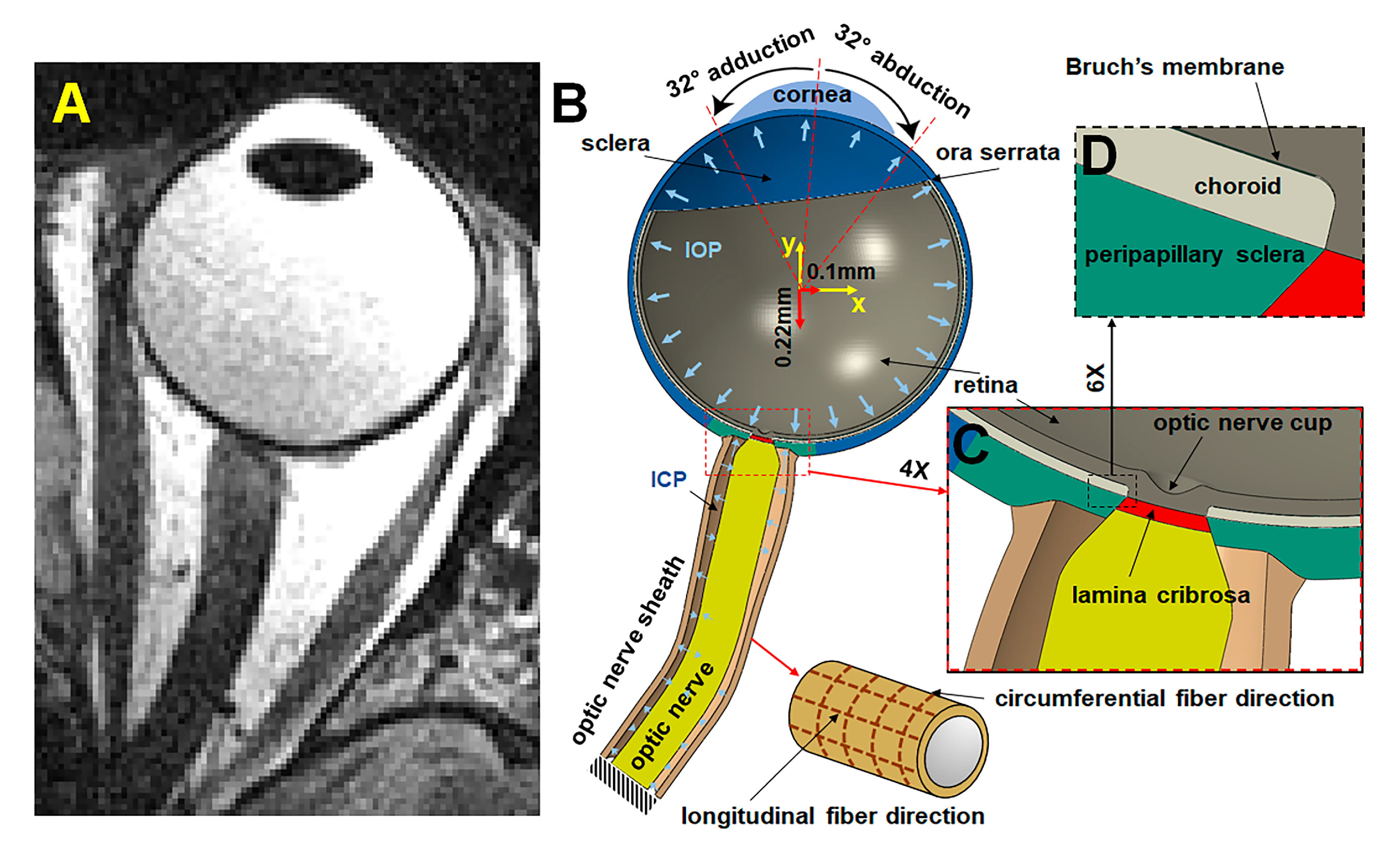
2.1. Anatomy
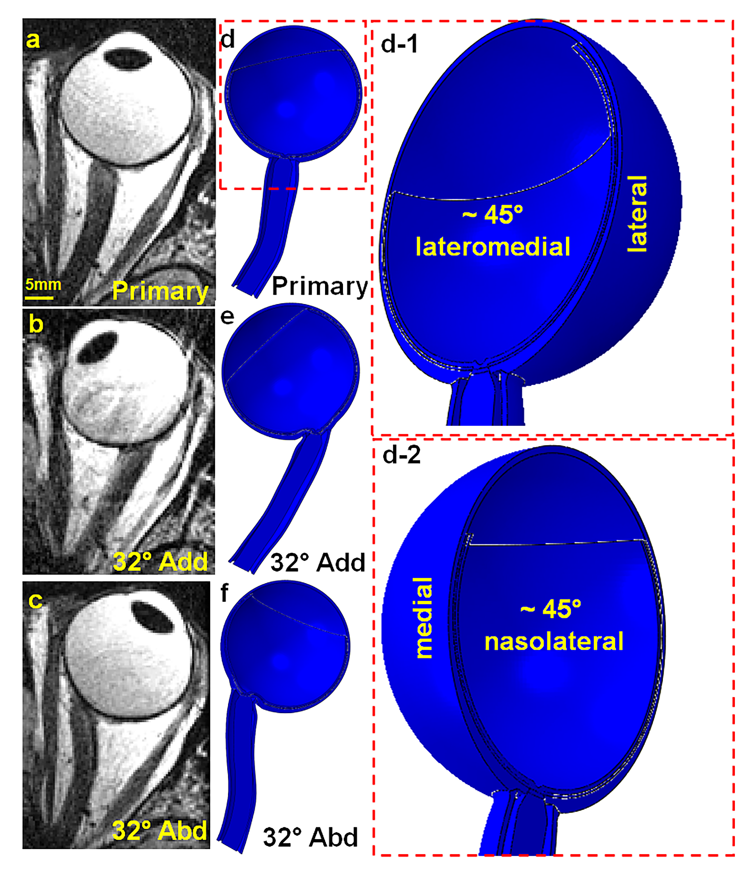
2.2. Geometry
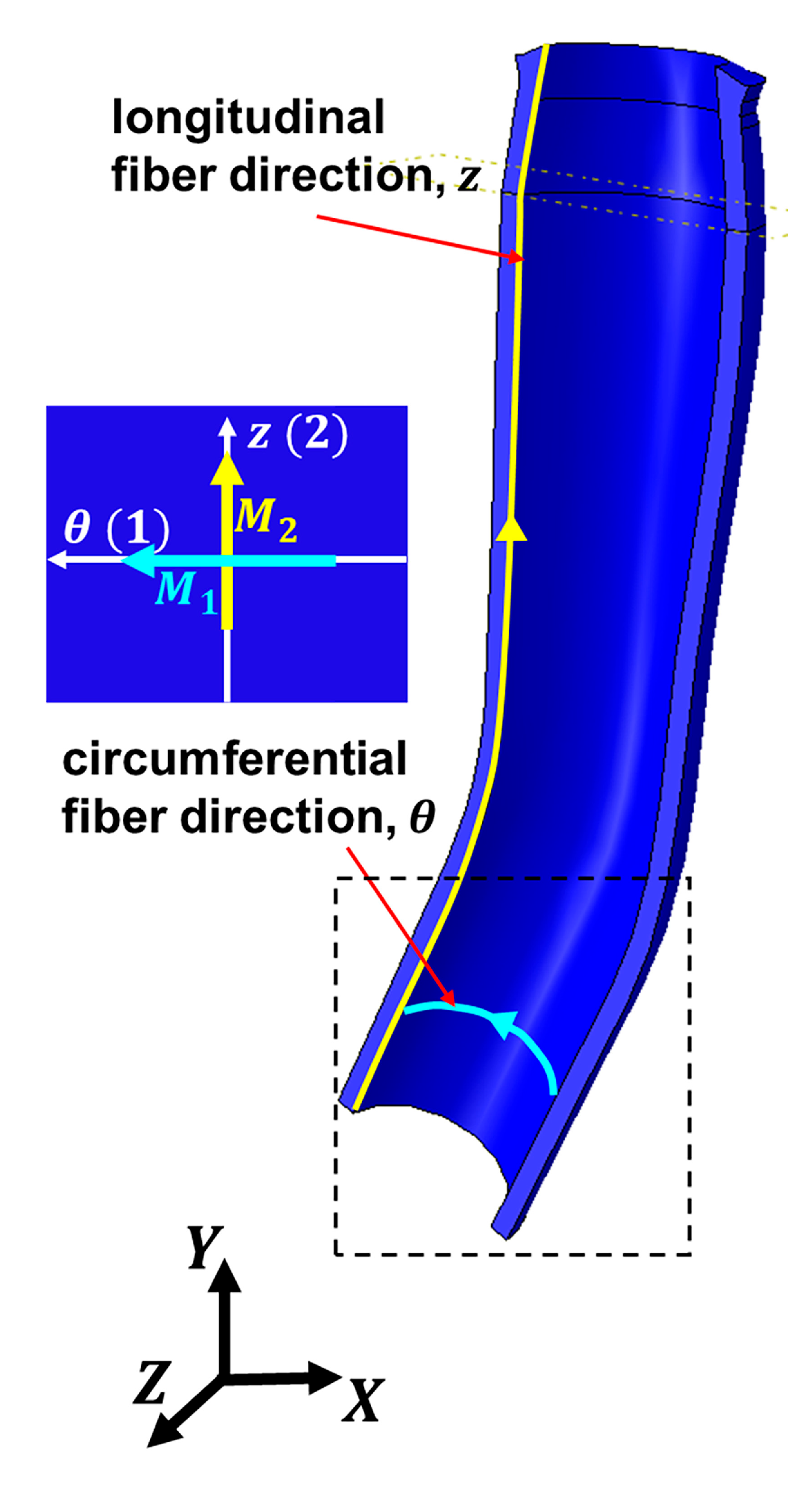
2.3. Material Parameters for Isotropic Eye Components and Anisotropic ONS
2.4. Boundary Conditions
2.5. Numerical Analysis
3. Results
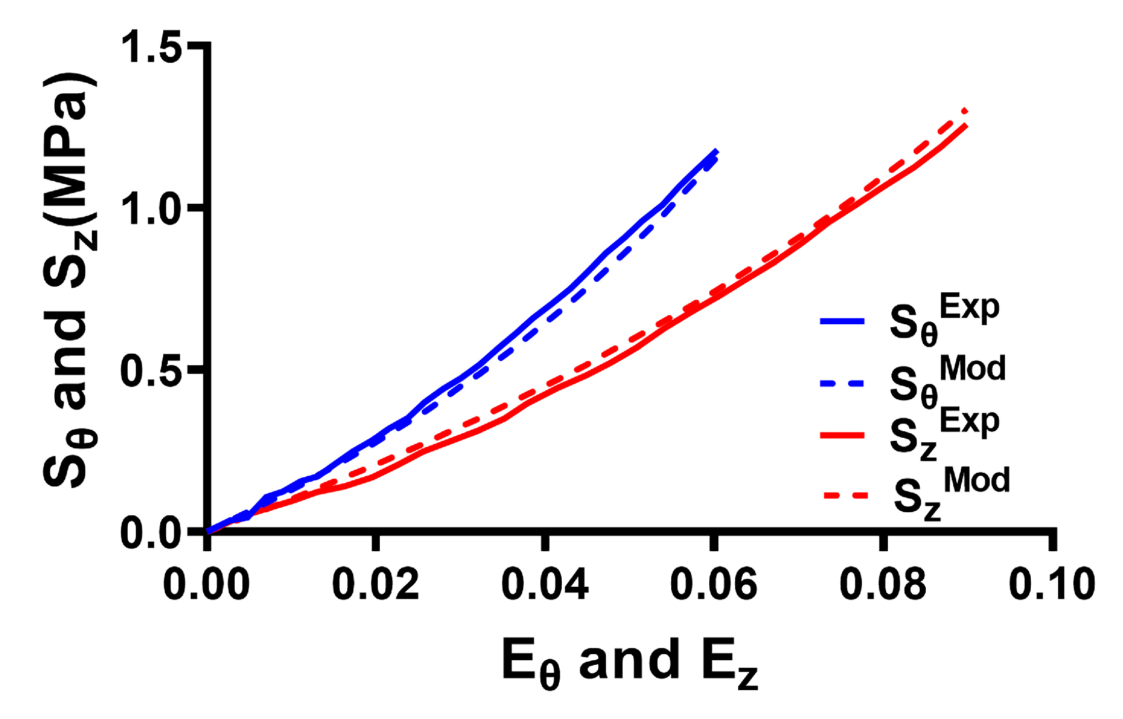
3.1. Fitting Model to Experimental Results for ONS
3.2. On and ONS Traction Force
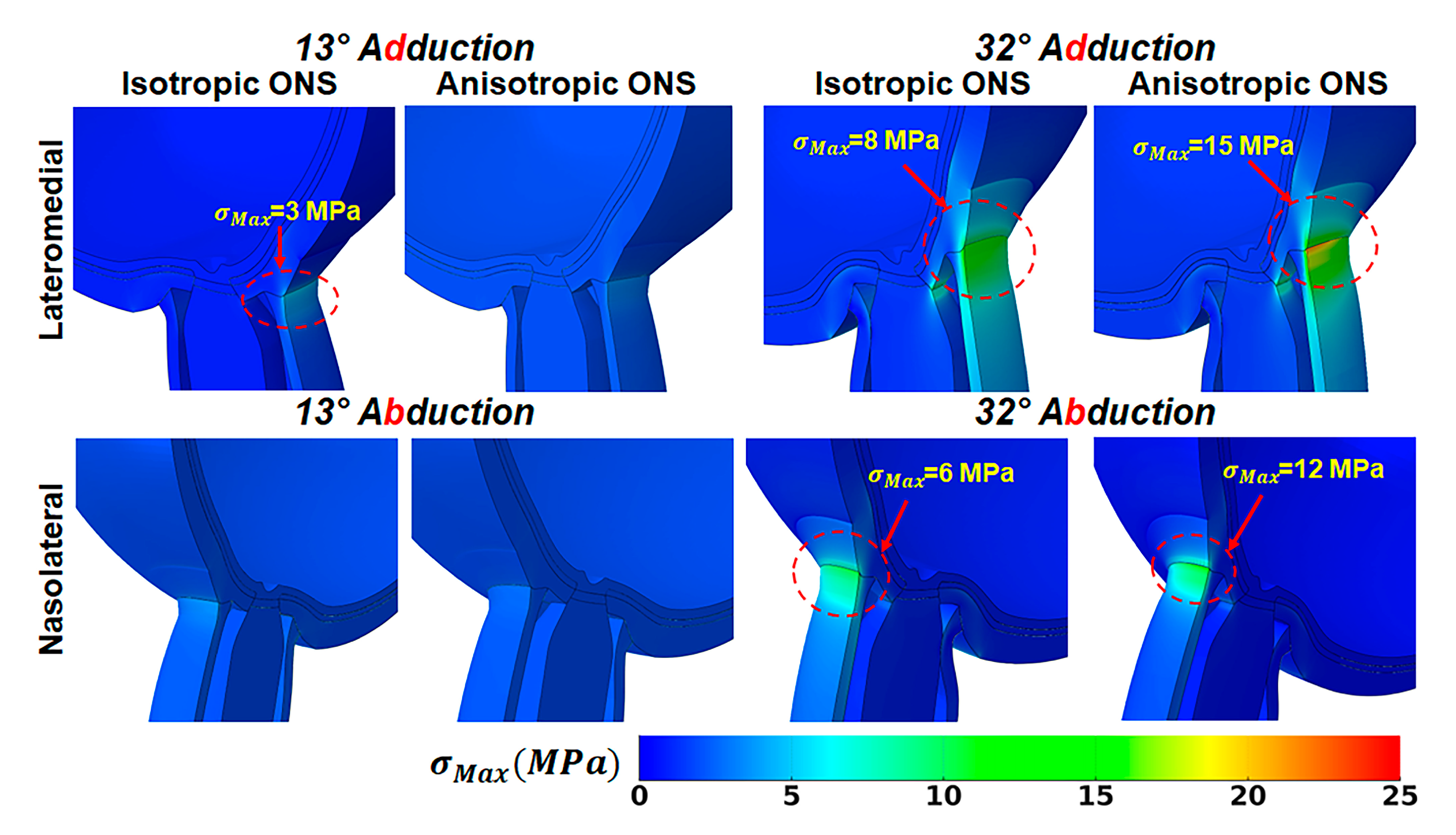
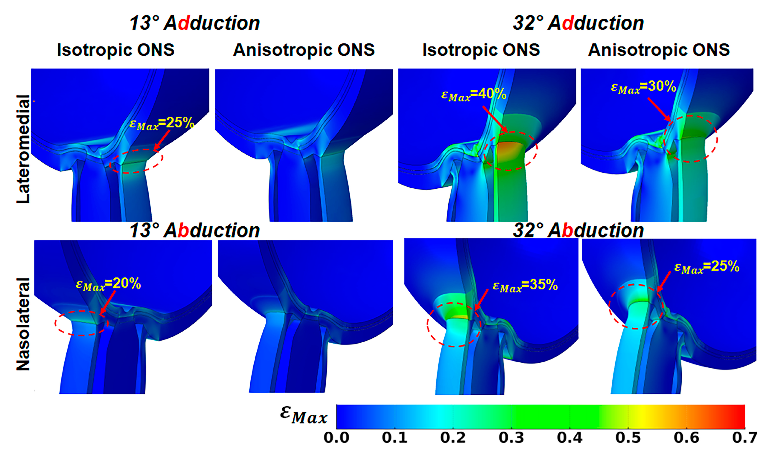
3.3. Effect of ONS Anisotropy on Stress and Strain
3.4. Wrinkling During Large Angle of Duction
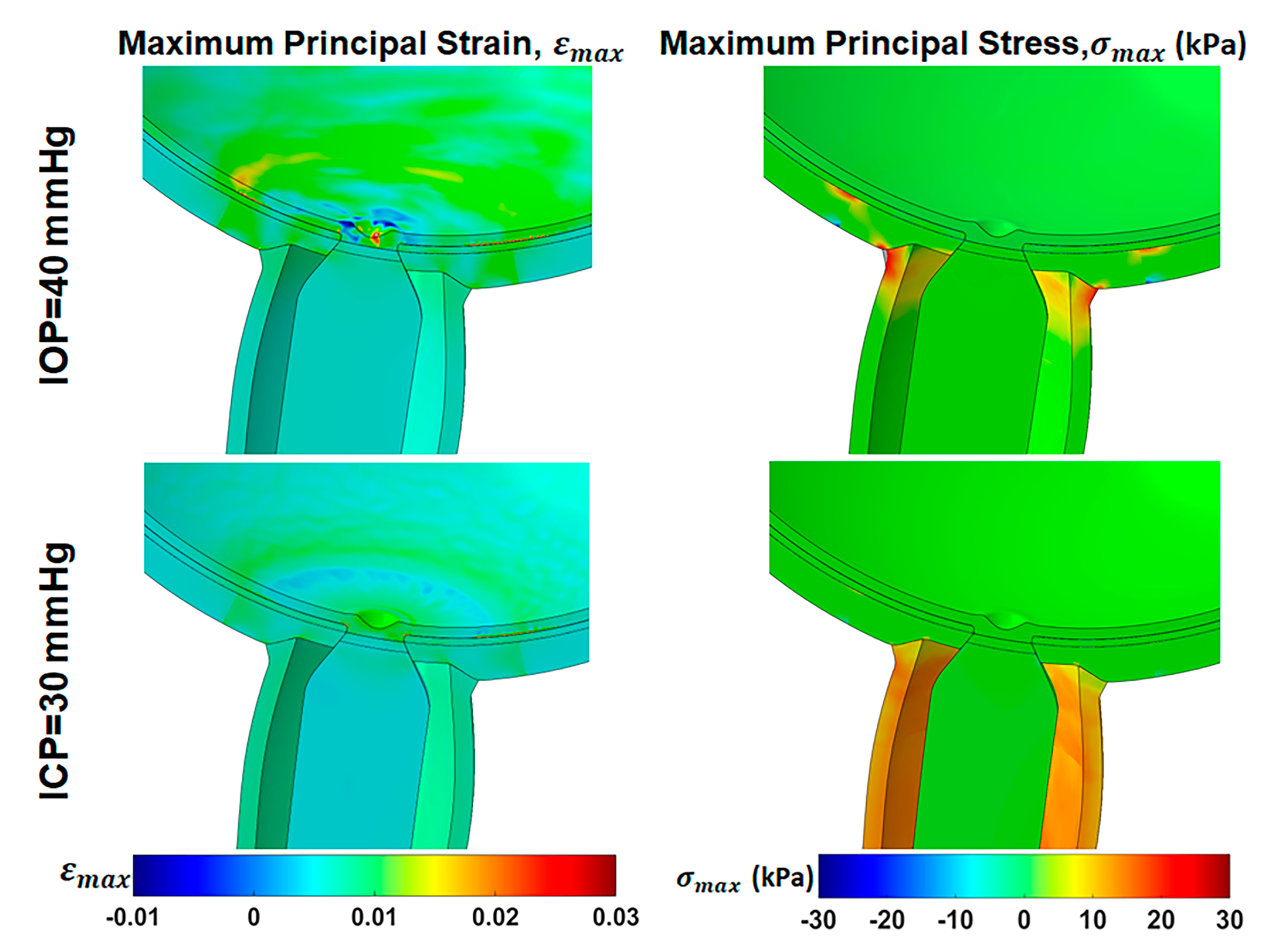
3.5. Effect of Elevated IOP and ICP
4. Discussion
5. Conclusions
Author Contributions
Funding
Institutional Review Board Statement
Informed Consent Statement
Data Availability Statement
Acknowledgments
Conflicts of Interest
References
- Demer, J.L. Optic Nerve Sheath as a Novel Mechanical Load on the Globe in Ocular Duction. Investig. Ophthalmol. Vis. Sci. 2016, 57, 1826–1838. [Google Scholar] [CrossRef] [PubMed]
- Raykin, J.; Forte, T.E.; Wang, R.; Feola, A.; Samuels, B.C.; Myers, J.G.; Mulugeta, L.; Nelson, E.S.; Gleason, R.L.; Ethier, C.R. Characterization of the Mechanical Behavior of the Optic Nerve Sheath and its Role in Spaceflight-Induced Ophthalmic Changes. Biomech. Model. Mechanobiol. 2017, 16, 33–43. [Google Scholar] [CrossRef] [PubMed]
- Shin, A.; Park, J.; Le, A.; Poukens, V.; Demer, J.L. Bilaminar Mechanics of the Human Optic Nerve Sheath. Curr. Eye Res. 2020, 45, 854–863. [Google Scholar] [CrossRef] [PubMed]
- Park, J.; Shin, A.; Jafari, S.; Demer, J.L. Material Properties and Effect of Preconditioning of Human Sclera, Optic Nerve, and Optic Nerve Sheath. Biomech. Model. Mechanbiol. 2021, 20, 1353–1363. [Google Scholar] [CrossRef] [PubMed]
- Demer, J.L.; Clark, R.A.; Suh, S.Y.; Giaconi, J.A.; Nouri-Mahdavi, K.; Law, S.K.; Bonelli, L.; Coleman, A.L.; Caprioli, J. Optic Nerve Traction During Adduction in Open Angle Glaucoma with Normal versus Elevated Intraocular Pressure. Curr. Eye Res. 2020, 45, 199–210. [Google Scholar] [CrossRef]
- Wang, X.; Fisher, L.K.; Milea, D.; Jonas, J.B.; Girard, M.J. Predictions of Optic Nerve Traction Forces and Peripapillary Tissue Stresses Following Horizontal Eye Movements. Investig. Ophthalmol. Vis. Sci. 2017, 58, 2044–2053. [Google Scholar] [CrossRef]
- Wang, X.; Rumpel, H.; Lim, W.E.; Baskaran, M.; Perera, S.A.; Nongpiur, M.E.; Aung, T.; Milea, D.; Girard, M.J. Finite Element Analysis Predicts Large Optic Nerve Head Strains During Horizontal Eye Movements. Investig. Ophthalmol. Vis. Sci. 2016, 57, 2452–2462. [Google Scholar] [CrossRef]
- Ji, F.; Bansal, M.; Wang, B.; Hua, Y.; Islam, M.R.; Matuschke, F.; Axer, M.; Sigal, I.A. A Direct Fiber Approach to Model Sclera Collagen Architecture and Biomechanics. Exp. Eye Res. 2023, 232, 109510. [Google Scholar] [CrossRef]
- Coudrillier, B.; Tian, J.; Alexander, S.; Myers, K.M.; Quigley, H.A.; Nguyen, T.D. Biomechanics of the Human Posterior Sclera: Age- and Glaucoma-Related Changes Measured Using Inflation Testing. Investig. Ophthalmol. Vis. Sci. 2012, 53, 1714–1728. [Google Scholar] [CrossRef]
- Grytz, R.; Fazio, M.A.; Girard, M.J.; Libertiaux, V.; Bruno, L.; Gardiner, S.; Girkin, C.A.; Downs, J.C. Material Properties of the Posterior Human Sclera. J. Mech. Behav. Biomed. Mater. 2014, 29, 602–617. [Google Scholar] [CrossRef]
- Jafari, S.; Lu, Y.; Park, J.; Demer, J.L. Finite Element Model of Ocular Adduction by Active Extraocular Muscle Contraction. Investig. Ophthalmol. Vis. Sci. 2021, 62, 1. [Google Scholar] [CrossRef]
- Karimi, A.; Rahmati, S.M.; Razaghi, R.; Girkin, C.A.; Downs, J.C. Finite Element Modeling of the Complex Anisotropic Mechanical Behavior of the Human Sclera and Pia Mater. Comput. Methods Programs Biomed. 2022, 215, 106618. [Google Scholar] [CrossRef] [PubMed]
- Jafari, S.; Park, J.; Lu, Y.; Demer, J.L. Finite Element Model Of Ocular Adduction with Unconstrained Globe Translation. Biomech. Model. Mechanobiol. 2024, 23, 601–614. [Google Scholar] [CrossRef] [PubMed]
- Holzapfel, G.A.; Gasser, T.C.; Ogden, R.W. A new constitutive framework for arterial wall mechanics and a Comparative Study of Material Models. J. Elast. 2000, 61, 1–48. [Google Scholar] [CrossRef]
- Shetye, S.S.; Deault, M.M.; Puttlitz, C.M. Biaxial Response of Ovine Spinal Cord Dura Mater. J. Mech. Behav. Biomed. Mater. 2014, 34, 146–153. [Google Scholar] [CrossRef] [PubMed]
- Cunningham, E.T., Jr.; Riordan-Eva, P. Vaughan & Asbury’s General Ophthalmology; Lange Medical Books/McGraw-Hill Medical Pub. Division: New York, NY, USA, 2011. [Google Scholar]
- Vurgese, S.; Panda-Jonas, S.; Jonas, J.B. Scleral Thickness in Human Eyes. PLoS ONE 2012, 7, e29692. [Google Scholar] [CrossRef]
- Olsen, T.W.; Aaberg, S.Y.; Geroski, D.H.; Edelhauser, H.F. Human Sclera: Thickness and Surface Area. Am. J. Ophthalmol. 1998, 125, 237–241. [Google Scholar] [CrossRef]
- Voorhees, A.P.; Jan, N.J.; Hua, Y.; Yang, B.; Sigal, I.A. Peripapillary Sclera Architecture Revisited: A Tangential Fiber Model and its Biomechanical Implications. Acta Biomater. 2018, 79, 113–122. [Google Scholar] [CrossRef]
- Quigley, H.A.; Brown, A.E.; Morrison, J.D.; Drance, S.M. The Size and Shape of the Optic Disc in Normal Human Eyes. Arch. Ophthalmol. 1990, 108, 51–57. [Google Scholar] [CrossRef]
- Elledge, J.A.; Davis, M.D.; Hubbard, L.D.; Reimers, J.L.; Fink, C.A.; Hafford, D.G.; Susman, R.A. Diameter/Area of the Standardized Optic Disc and accurate Scaling in Retinal Images. Investig. Ophthalmol. Vis. Sci. 2005, 46, 2583. [Google Scholar]
- Myers, C.E.; Klein, B.E.; Meuer, S.M.; Swift, M.K.; Chandler, C.S.; Huang, Y.; Gangaputra, S.; Pak, J.W.; Danis, R.P.; Klein, R. Retinal Thickness Measured by Spectral-Domain Optical Coherence Tomography in Eyes without Retinal Abnormalities: The Beaver Dam Eye Study. Am. J. Ophthalmol. 2015, 159, 445–456.e441. [Google Scholar] [CrossRef] [PubMed]
- Alamouti, B.; Funk, J. Retinal Thickness Decreases with Age: An OCT Study. Br. J. Ophthalmol. 2003, 87, 899–901. [Google Scholar] [CrossRef] [PubMed]
- Manjunath, V.; Taha, M.; Fujimoto, J.G.; Duker, J.S. Choroidal thickness in Normal Eyes Measured Using Cirrus Hd Optical Coherence Tomography. Am. J. Ophthalmol. 2010, 150, 325–329.e321. [Google Scholar] [CrossRef] [PubMed]
- Tan, K.A.; Gupta, P.; Agarwal, A.; Chhablani, J.; Cheng, C.Y.; Keane, P.A.; Agrawal, R. State of Science: Choroidal Thickness and Systemic Health. Surv. Ophthalmol. 2016, 61, 566–581. [Google Scholar] [CrossRef]
- Sohn, E.H.; Khanna, A.; Tucker, B.A.; Abramoff, M.D.; Stone, E.M.; Mullins, R.F. Structural and Biochemical Analyses of Choroidal Thickness in Human Donor Eyes. Investig. Ophthalmol. Vis. Sci. 2014, 55, 1352–1360. [Google Scholar] [CrossRef]
- Newsome, D.A.; Huh, W.; Green, W.R. Bruch′s Membrane Age-Related Changes Vary by Region. Curr. Eye Res. 1987, 6, 1211–1221. [Google Scholar] [CrossRef]
- Miller, K. Method of Testing Very Soft Biological Tissues in Compression. J. Biomech. 2005, 38, 153–158. [Google Scholar] [CrossRef]
- Sacks, M.S.; Sun, W. Multiaxial Mechanical Behavior of Biological Materials. Annu. Rev. Biomed. Eng. 2003, 5, 251–284. [Google Scholar] [CrossRef]
- Wex, C.; Arndt, S.; Stoll, A.; Bruns, C.; Kupriyanova, Y. Isotropic Incompressible Hyperelastic Models for Modelling the Mechanical Behaviour of Biological Tissues: A Review. Biomed. Tech. 2015, 60, 577–592. [Google Scholar] [CrossRef]
- Le, A.; Shin, A.; Park, J.; Poukens, V.; Demer, J.L. Bilaminar Structure of the Human Optic Nerve Sheath. Curr. Eye Res. 2020, 45, 864–872. [Google Scholar] [CrossRef]
- Stevens, R.R.F.; Gommer, E.D.; Aries, M.J.H.; Ertl, M.; Mess, W.H.; Huberts, W.; Delhaas, T. Optic Nerve Sheath Diameter Assessment by Neurosonology: A Review of Methodologic Discrepancies. J. Neuroimaging 2021, 31, 814–825. [Google Scholar] [CrossRef] [PubMed]
- Roy, D.; Holzapfel, G.A.; Kauffmann, C.; Soulez, G. Finite Element Analysis of Abdominal Aortic Aneurysms: Geometrical and Structural Reconstruction with Application of an Anisotropic Material Model. IMA J. Appl. Math. 2014, 79, 1011–1026. [Google Scholar] [CrossRef]
- Sigal, I.A. Interactions between Geometry and Mechanical Properties on the Optic Nerve Head. Investig. Ophthalmol. Vis. Sci. 2009, 50, 2785–2795. [Google Scholar] [CrossRef] [PubMed]
- Sigal, I.A.; Grimm, J.L.; Schuman, J.S.; Kagemann, L.; Ishikawa, H.; Wollstein, G. A Method to Estimate Biomechanics and Mechanical Properties of Optic Nerve Head Tissues from Parameters Measurable Using Optical Coherence Tomography. IEEE Trans. Med. Imaging 2014, 33, 1381–1389. [Google Scholar] [CrossRef]
- Sigal, I.A.; Flanagan, J.G.; Tertinegg, I.; Ethier, C.R. Predicted Extension, Compression and Shearing of Optic Nerve Head Tissues. Exp. Eye Res. 2007, 85, 312–322. [Google Scholar] [CrossRef]
- Miller, K. How to Test Very Soft Biological Tissues in Extension? J. Biomech. 2001, 34, 651–657. [Google Scholar] [CrossRef]
- Pitre, J.J., Jr.; Kirby, M.A.; Li, D.S.; Shen, T.T.; Wang, R.K.; O′Donnell, M.; Pelivanov, I. Nearly-Incompressible Transverse Isotropy (NITI) of Cornea Elasticity: Model and Experiments with Acoustic Micro-Tapping OCE. Sci. Rep. 2020, 10, 12983. [Google Scholar] [CrossRef]
- Chan, W.; Hussain, A.; Marshall, J. Youngs Modulus of Bruchs Membrane: Implications for AMD. Investig. Ophthalmol. Vis. Sci. 2007, 48, 2187. [Google Scholar]
- Chen, K.; Rowley, A.P.; Weiland, J.D.; Humayun, M.S. Elastic Properties of Human Posterior Eye. J. Biomed. Mater. Res. A 2014, 102, 2001–2007. [Google Scholar] [CrossRef]
- Kaskar, O.; Fleischman, D.; Kuznetsov, A.V.; Grace, L. A Finite Element Model Investigating the Cyclic Strains in the Lamina Cribrosa and Their Potential Role in Glaucoma. Model. Artif. Intell. Ophthalmol. 2022, 4, 1–10. [Google Scholar] [CrossRef]
- Gasser, T.C.; Ogden, R.W.; Holzapfel, G.A. Hyperelastic Modelling of Arterial Layers with Distributed Collagen Fibre Orientations. J. R. Soc. Interface 2006, 3, 15–35. [Google Scholar] [CrossRef] [PubMed]
- Trosset, M.W. What Is Simulated Annealing? Optim. Eng. 2001, 2, 201–213. [Google Scholar] [CrossRef]
- Demer, J.L.; Clark, R.A. Translation and Eccentric Rotation in Ocular Motor Modeling. Prog. Brain Res. 2019, 248, 117–126. [Google Scholar] [CrossRef] [PubMed]
- Munoz Sarmiento, D.M.; Rodriguez Montano, O.L.; Alarcon Castiblancoa, J.D.; Cortes Rodriguez, C.J. The Im-pact of horizontal eye movements versus intraocular pressure on Optic Nerve Head Biomechanics: A Tridimen-sional Finite Element Analysis Study. Heliyon 2023, 9, e13634. [Google Scholar] [CrossRef]
- Burgoyne, C.F.; Downs, J.C.; Bellezza, A.J.; Suh, J.K.F.; Hart, R.T. The Optic Nerve Head as a Biomechanical Structure: A New Paradigm for Understanding the Role of IOP-Related Stress and Strain in the Pathophysiology of Glaucomatous Optic Nerve Head Damage. Prog. Retin. Eye Res. 2005, 24, 39–73. [Google Scholar] [CrossRef]
- Coudrillier, B.; Boote, C.; Quigley, H.A.; Nguyen, T.D. Scleral Anisotropy and its Effects on the Mechanical Response of the Optic Nerve Head. Biomech. Model. Mechanobiol. 2013, 12, 941–963. [Google Scholar] [CrossRef]
- Colin, J.; Holland, M.A. Layer Wrinkling in an Inhomogeneous Matrix. Int. J. Solids Struct. 2019, 156, 119–125. [Google Scholar] [CrossRef]
- Sibony, P.A.; Kupersmith, M.J.; Feldon, S.E.; Wang, J.K.; Garvin, M.; OCT Substudy Group for the NORDIC Idiopathic Intracranial Hypertension Treatment Trial. Retinal and Choroidal Folds in Papilledema. Investig. Ophthalmol. Vis. Sci. 2015, 56, 5670–5680. [Google Scholar] [CrossRef]
- Del Priore, L.V. Stiffness of Retinal and Choroidal Tissue: A Surface Wrinkling Analysis of Epiretinal Membranes and Choroidal Folds. Am. J. Ophthalmol. 2006, 142, 435–440. [Google Scholar] [CrossRef]
- Kita, M.; Marmor, M.F. Retinal adhesive force in living rabbit, cat, and monkey eyes. Normative data and enhancement by mannitol and acetazolamide. Investig. Ophthalmol. Vis. Sci. 1992, 33, 1879–1882. [Google Scholar]
- Sigal, I.A.; Flanagan, J.G.; Tertinegg, I.; Ethier, C.R. Finite Element Modeling of Optic Nerve Head Biomechanics. Investig. Ophthalmol. Vis. Sci. 2004, 45, 4378–4387. [Google Scholar] [CrossRef] [PubMed]
- Muñoz-Sarmiento, D.M.; Rodríguez-Montaño, Ó.L.; Alarcón-Castiblanco, J.D.; Gamboa-Márquez, M.A.; Corredor-Gómez, J.P.; Cortés-Rodríguez, C.J. A Finite Element Study of Posterior Eye Biomechanics: The Influence of Intraocular and Cerebrospinal Pressure on the Optic Nerve Head, Peripapillary Region, Subarachnoid Space and Meninges. Inform. Med. Unlocked 2019, 15, 100185. [Google Scholar] [CrossRef]
- Feola, A.J.; Myers, J.G.; Raykin, J.; Mulugeta, L.; Nelson, E.S.; Samuels, B.C.; Ethier, C.R. Finite Element Modeling of Factors Influencing Optic Nerve Head Deformation Due to Intracranial Pressure. Investig. Ophthalmol. Vis. Sci. 2016, 57, 1901–1911. [Google Scholar] [CrossRef] [PubMed]
- Schoemaker, I.; Hoefnagel, P.P.W.; Mastenbroek, T.J.; Kolff, C.F.; Schutte, S.; van der Helm, F.C.T.; Picken, S.J.; Gerritsen, A.F.C.; Wielopolski, P.A.; Spekreijse, H.; et al. Elasticity, Viscosity, and Deformation of Orbital Fat. Investig. Ophthalmol. Vis. Sci. 2006, 47, 4819–4826. [Google Scholar] [CrossRef]


| first invariant of right Cauchy–Green deformation tensor without volume change | |
| volume ratio | |
| , | coefficients of strain energy |
| reduced polynomial strain energy | |
| , , | principal stretches in radial, circumferential, and longitudinal directions |
| , | volumetric and isochoric terms of strain energy |
| , | original and modified right Cauchy–Green tensor |
| , , | invariants of modified right Cauchy–Green tensor |
| , | unit vectors of elastin fibers in reference state |
| fiber dispersion parameter | |
| parameters related to Holzapfel model | |
| , | circumferential and longitudinal components of 2nd Piola–Kirchhoff stress tensor |
| , | circumferential and longitudinal components of Green–Lagrange strain tensor |
| and | weighting factors |
| Eye Component | Model | Reference | ||
|---|---|---|---|---|
| 2nd Order Reduced Polynomial | C10 (MPa) | C20 (MPa) | ||
| optic nerve | 0.23 | 8.18 | ||
| peripapillary sclera | 0.20 | 11.60 | ||
| anterior sclera | 2.25 | 102.20 | ||
| equatorial sclera | 0.87 | 91.30 | ||
| posterior sclera | 0.80 | 32.8 | ||
| Neo-Hookean | C10 (MPa) | D1 (MPa)−1 | ||
| Bruch’s membrane | 1.82 | 0.022 | [39] | |
| choroid | 0.063 | 0.64 | [40] | |
| retina | 0.003 | 15.5 | ||
| Lamina cribrosa | Ogden | µ1 | α1 | [41] |
| 0.36 | 10.4 |
| Case | Isotropic ONS | Anisotropic ONS | 32° Adduction | 32° Abduction | Normal IOP and ICP | IOP Change | ICP Change |
|---|---|---|---|---|---|---|---|
| 1 | ✓ | ͞ | ✓ | ͞ | ✓ | ͞ | ͞ |
| 2 | ͞ | ✓ | ✓ | ͞ | ✓ | ͞ | ͞ |
| 3 | ✓ | ͞ | ͞ | ✓ | ✓ | ͞ | ͞ |
| 4 | ͞ | ✓ | ͞ | ✓ | ✓ | ͞ | ͞ |
| 5 | ͞ | ✓ | ͞ | ͞ | ͞ | ✓ | ͞ |
| 6 | ͞ | ✓ | ͞ | ͞ | ͞ | ͞ | ✓ |
| Parameter | Magnitude | |
|---|---|---|
| Anisotropic | Isotropic | |
| (MPa) | 0.8 | 2.3 |
| (MPa) | 32.2 | - |
| 79.1 | - | |
| 0.29 | - | |
| Tissue | Optic Nerve Sheath Isotropy | Maximum Stress (MPa) | |||||||
|---|---|---|---|---|---|---|---|---|---|
| 13° Add | 13° Abd | 32° Add | 32° Abd | 13° Add | 13° Abd | 32° Add | 32° Abd | ||
| ONH | Isotropic | 40 | 20 | 500 | 300 | 0.9 | 0.2 | 5.0 | 2.0 |
| Anisotropic | 70 | 15 | 1100 | 350 | 0.3 | 0.1 | 3.0 | 2.0 | |
| ONS-Sclerajunction | Isotropic | 1500 | 360 | 6000 | 2600 | 3.0 | 1.5 | 8.0 | 6.0 |
| Anisotropic | 770 | 130 | 5300 | 2100 | 4.0 | 4.0 | 15 | 12 | |
Disclaimer/Publisher’s Note: The statements, opinions and data contained in all publications are solely those of the individual author(s) and contributor(s) and not of MDPI and/or the editor(s). MDPI and/or the editor(s) disclaim responsibility for any injury to people or property resulting from any ideas, methods, instructions or products referred to in the content. |
© 2025 by the authors. Licensee MDPI, Basel, Switzerland. This article is an open access article distributed under the terms and conditions of the Creative Commons Attribution (CC BY) license (https://creativecommons.org/licenses/by/4.0/).
Share and Cite
Jafari, S.; Cai, S.; Demer, J.L. Finite Element Model of the Effect of Optic Nerve Sheath Anisotropy on Ocular Loading During Horizontal Duction. Bioengineering 2025, 12, 587. https://doi.org/10.3390/bioengineering12060587
Jafari S, Cai S, Demer JL. Finite Element Model of the Effect of Optic Nerve Sheath Anisotropy on Ocular Loading During Horizontal Duction. Bioengineering. 2025; 12(6):587. https://doi.org/10.3390/bioengineering12060587
Chicago/Turabian StyleJafari, Somaye, Shengqiang Cai, and Joseph L. Demer. 2025. "Finite Element Model of the Effect of Optic Nerve Sheath Anisotropy on Ocular Loading During Horizontal Duction" Bioengineering 12, no. 6: 587. https://doi.org/10.3390/bioengineering12060587
APA StyleJafari, S., Cai, S., & Demer, J. L. (2025). Finite Element Model of the Effect of Optic Nerve Sheath Anisotropy on Ocular Loading During Horizontal Duction. Bioengineering, 12(6), 587. https://doi.org/10.3390/bioengineering12060587