Article

Vanillin Quantum–Classical Photodynamics and Photostatic Optical Spectra

by
Vladimir Pomogaev
1,2,* and
Olga Tchaikovskaya
1,3
1
Laboratory of Photophysics and Photochemistry of Molecules, Department of Physics, National Research Tomsk State University, Lenin Ave. 36, 634050 Tomsk, Russia
2
Institute of Chemistry, Saint-Petersburg State University, Universitetskaya nab. 7/9, 199034 Saint Petersburg, Russia
3
Institute of Electrophysics, Ural Branch of the Russian Academy of Sciences, 106 Amundsena St., 620146 Yekaterinburg, Russia
*
Author to whom correspondence should be addressed.
ChemEngineering 2025, 9(4), 76; https://doi.org/10.3390/chemengineering9040076
Submission received: 15 April 2025 / Revised: 11 July 2025 / Accepted: 17 July 2025 / Published: 23 July 2025

Abstract

Vanillin photoinduced deprotonation was evaluated and analyzed. Vibronic states and transitions were computationally investigated. Optimizations and vertical electron transitions in the gas phase and with the continuum solvation model were computed using the time-dependent density functional theory. Static absorption and emission (photostatic optical) spectra were statistically averaged over the excited instantaneous molecular conformers fluctuating on quantum–classical molecular dynamic trajectories. Photostatic optical spectra were generated using the hybrid quantum–classical molecular dynamics for explicit solvent models. Conical intersection searching and nonadiabatic molecular dynamics simulations defined potential energy surface propagations, intersections, dissipations, and dissociations. The procedure included mixed-reference spin–flip excitations for both procedures and trajectory surface hopping for photodynamics. Insignificant structural deformations vs. hydroxyl bond cleavage followed by deprotonation were demonstrated starting from different initial structural conditions, which included optimized, transition state, and several other important fluctuating configurations in various environments. Vanillin electronic structure changes were illustrated and analyzed at the key points on conical intersection and nonadiabatic molecular dynamics trajectories by investigating molecular orbital symmetry and electron density difference. The hydroxyl group decomposed on transition to a σ-molecular orbital localized on the elongated O–H bond.

1. Introduction

Vanillin’s petrochemical properties are well known and widely used for industrial purposes; its photochemical features are intensively studied and applied in pharmaceutical, food, and cosmetic industries. Skin phototoxicity due to the application of cosmetics is increasing day by day because many natural extracts with low toxicity in the dark are used on the skin on sunny days. The phenolic carbonyl, common in biomass combustion emissions, forms triplet excited states upon exposure to sunlight, resulting in the formation of aqueous secondary organic aerosol [1]. In atmospheric photochemical reactions, oligomers and hydroxylated products were detected through photosensitized reactions involving vanillin. There are few research studies on vanillin photochemistry despite the great interest in its molecular electronic properties. Understanding the relationship between structure and physicochemical properties is necessary to develop desirable photoacids for various applications [2]. Moreover, nowadays, scientists are increasingly focusing on the proton transfer polymerization of vanillin through the click reaction, which is important in the process of imprinting lithography [3].
Vanillin (4-hydroxy-3-methoxybenzaldehyde, C8H8O3), abbreviated here as VNL, is an aldehyde, hydroxyl, and ether-substituted parent phenol, which makes vanillin’s photochemical dynamics more complicated than the basic molecule because of richer localization and the types of low-lying excited electronic states. When considering vanillin as an object of study, it is necessary to take into account the presence of substituents in the meta- and para-positions (alkoxyl and hydroxyl groups) in the structure. The OH group in the vanillin molecule determines its acidic properties. The ether and carbonyl groups in vanillin affect the distribution of electron density in the molecule and determine the ability to ionize in solution [4]. Vanillin has a pKa value that is highly dependent on temperature, ionic strength, and solvent, ranging from 7.40 in water to 17.07 in tert-butyl alcohol. Light excitation of vanillin results in a decrease in pKa values. Therefore, an aqueous solution becomes acidic in the presence of vanillin. The functional group (carbonyl) in vanillin is responsible for the decrease in pKa due to its electron-withdrawing ability upon excitation. The unique acid–base properties of vanillin affect its light-absorbing properties and can enhance deprotonation in aqueous aerosols. The presence of sulfate ions in an aqueous solution of vanillin causes a hyperchromic effect and a bathochromic shift, while nitrate ions cause a hypsochromic shift in the absorption spectrum of vanillin under the influence of sunlight [1]. The participation of various ionic forms of vanillin in its formation and photolytic activity is still being discussed [5].
Vanillin is traditionally used as an important food additive for flavor and astringency. Vanillin acts as a basis for the organic synthesis of many vital drugs [6]. In addition, it is common in a wide range of bioactive natural products for medicine, such as steroid hormones, thyroid hormones, and monoamine neurotransmitters, including estrone, estradiol, and others [7]. The study of vanillin photo-transformation is extremely relevant to the desire to know how phenols are converted into various products with a wide range of useful chemical reactions and pharmaceutical activities. The photoinduced structural transformations of phenol derivatives, including vanillin, inherit some electron properties of the parent cycle and remarkably depend on environmental phases and excitation conditions [8,9,10,11], which requires comprehensive and comparative theoretical analysis.
The anionic, cationic, and neutral protolytic forms of vanillin were observed in aqueous solutions with different pH values, which were identified using absorption and fluorescence spectra [12]. The neutral state has a maxima of absorption at 306 nm and 279 nm and fluorescence at 330 nm and 425 nm, whereas the anionic and cationic forms undergo bathochromic shifts in absorption (345 nm vs. 333 nm) and in emission (395 nm, 520 nm vs. 450 nm), respectively. Acidity tuning provides proton richness or poverty in the solution, resulting in an excess or deficiency of hydrogen atoms in the molecule. Solute–solvent hydrogen exchange is a complicated interaction between vanillin and the water cluster network, but simplified theoretical models allow for omitting some details and studying the process in different stages. The initial steps are calculations of spectral-luminescence and photodynamical properties of the neutral form, followed by photochemical simulations of proton addition to or removal from the hydroxyl bond. The simplest approach is the decomposition of the isolated anionic vanillin by cleavage of the hydroxyl bond, followed by deprotonation.
Comprehensive theoretical spectral-luminescence and photochemical analysis is conducted by means of computational investigation of conventional TD-DFT vibronic (electronic and vibrations) states and photoinduced transitions; absorption and emission static (photostatic optical) spectra, averaged over excited chromophores oscillating along the hybrid SQMMD propagations [10,11,13,14,15,16,17,18]; CoIn searching; and NAMD conversions of the chosen molecular compound. The hybrid methodology unifies an explicit classical surrounding with a photoactive chromophore, incorporating quantum electronic effects through gradient-derived driving forces and electrostatic potential fitting (ESPF) to point charges [19,20]. CoIn and NAMD simulations of electronic transitions with lifetimes, excited energy dissipations, and photochemical reactions are reasonably reproduced using the multiconfigurational LR-MRSF approach based on TD-DFT, where electronic states are calculated as superpositions of spin–flip orbital transitions, originating from a triplet reference state [21,22,23,24,25,26,27,28]. MRSF demonstrates satisfactory accuracy while requiring significantly fewer computational resources compared to other well-known quantum chemistry methods such as the complete active space self-consistent field, CASSCF with Møller–Plesset correction, multireference configuration interaction, and coupled-cluster with equation of motion.
The present work aims to develop and apply to vanillin a research protocol for comprehensive spectral-luminescence, quantum–classical dynamic, and photochemical calculations for vibronic transitions, photostatic optical spectra, and excited energy dissipations. Hydroxyl group decomposition was investigated through vibronic analysis, CoIn searching of predissociation states, and NAMD of excited states’ dissipation leading to deprotonation. These results advance understanding, predictions, and practical applications of the molecular electronic properties and facilitate future research.

2. Materials and Methods

The TD-DFT B3LYP [29] with The Pople’s split-valence basis sets 6-311G(d) (6-311G*) [30,31] optimized the vanillin structure in the ground and first two excited states, and VEE calculated wavelengths with corresponding oscillator strengths in the framework using the Gaussian16 [32] package. The BH&HLYP functionals [33] in conjunction with the 6-311G(d) basis set [34] were focused on eSQMMD, CoIn, and NAMD simulations of PES propagations, intersections, and dissipations driven by classical forces in combination with gradient calculations and using the atomic charges incorporating the ESPF model in the case of an explicit classical water solvent [10,11,13,14,18,19,20,21,22,23,24,25,26,27,28,35,36,37,38,39,40,41]. An aqueous solvent model was also involved in the implicit forms of the implicit SMD-PCM [42] for all procedures excluding CoIn searching because this function was unavailable.
The NVT ensemble was prepared using the Packmol code [43] as an aqueous droplet capsule centered on a vanillin chromophore under SBC with a radius of 24.321 Å, density ρ = 0.977 g/mol, and T = 298 K containing 2000 water molecules. The initial water capsule centered on a frozen molecule was relaxed using the OPLSAA force field [44] during t = 10 ps classical MD propagation with a step Δτ = 1.0 fs and final QCMD equilibration for t = 1.0 ps with Δτ = 0.5 fs. Following the ergodic theorem [15], absorption and emission static spectra were generated through statistically averaging over the excited molecular conformers vibrationally fluctuating on eSQMMD trajectories due to partial QM motion [14,15,16,45] incorporating gradient calculations for driving forces and ESPF to point charges. Absorption spectra were calculated as conventional TD-DFT vertical excitations along the eSQMMD trajectories of the ground state molecule, while the fluorescence spectrum was constructed directly from the lowest chromophore excitation along the dynamic trajectory [15,16,17]. Instantaneous 1000 oscillating vanillin conformers of the water droplet were taken along the 5 ps QCMD prolongation after each 10 steps for optical spectra summation over non-equilibrium vibronic states of the fully flexible molecule motion under QM driving.
The propagating, crossing, and dissipating photoinduced PESs were defined on trajectories of CoIn searching and NAMD simulations using MRSF excitations and the trajectory surface hopping (TSH) component for stochastic photodynamic propagation. NAMD was applied for up to 300 fs to pass all crossing photodynamic PESs for the chromophore, both isolated and in the SMD water solvent. The initial points for these procedures were the molecule optimized to the minimum energy, the same structure with an elongated hydroxyl bond, and the transition state.
PESs’ evolutions through CoIn were analyzed with Dyson molecular orbitals (DMOs) of the extended Koopmans’ theorem [46,47,48] while canonical MOs are considered for conventional TD-DFT calculations. CoIn, NAMD, and eSQMMD simulations were implemented using GAMESS-US (release 30SEP 2021 R2patch 1) [13,49,50,51] with the Tinker 8.10.1 code for the hybrid ESPF QCMD. The electron densities of excited states on PESs at the initial and final CoIn, as well as key NAMD points, were reconstructed in the ChemCraft suite [52] to obtain and visualize electron density differences (EDDs).

3. Results

3.1. Vertical Electronic Transitions and IR Spectra

The TD-DFT B3LYP with the 6-311G* basis sets was used for optimizations and VEE calculations. An insignificant difference between the geometries optimized using various levels of QM theories with numerous basis sets, especially in the context of comparing the electronic excitation of the basic molecule of the phenolic family, was demonstrated and considered earlier [10,11].
The EDD for vibronic Franck–Condon’s VEE reflects the energy levels and electronic types of the vanillin excited electronic states exhibiting an 0S|0,0〉0S|d,FC〉 transition from ground state (absorption) to dark Sd = S1(πσ*), λ1 = 314 nm localized mostly on the ether group and 0S|0,0〉0S|b,FC〉 to the higher bright Sb = S2(ππ*), λ2 = 293 nm with a small energy gap between the states (Figure 1) that could lead to a potential inversion of the states on photodynamic trajectories and the overlap of these bands in common photostatic absorption spectra. The ether σ* MO of Sd cannot lead to hydroxyl bond cleavage with following deprotonation. Absorption to Sd is almost impossible due to the symmetrical forbidden ππ* → πσ* transitions for plane molecules that reflect the insignificant strength force f = 0.001, whereas excitation to Sb is very intensive: f = 0.534 and f = 0.777 to S3(ππ*) with λ3 = 230 nm. The excited states on absorption spectra longer than 200 nm (Figure 1) λ4(1.068) = 197 nm, λ5(0.001) = 183 nm, λ6(0.034) = 180 nm, λ7(0.033) = 169 nm, and λ1(0.004) = 161 nm also demonstrate no trend for hydroxyl electronic population, which is needed for potential O–H decomposition.
The hydroxyl group vibrations exclusively show responses for frequencies above 3400 cm−1 [53]. The calculated highest vibration frequency corresponding to the intensive harmonic O–H stretching vibrations in the ground state 0S|0,51〉 with frequency ν51(S0,O–H) = 3762 cm−1 vs. dS|d,51〉 where ν51(Sd,O–H) = 3771 cm−1 and bS|b,51〉 with ν51(Sb,O–H) = 3735 cm−1 for the both lowest excited states, which can potentially undertake predissociation. The VEE 0S|b,FC〉 energy lies above the bS|b,51〉 O–H stretching vibration level of the molecule optimized in the bS|b,0〉 bright state. Preliminarily, O–H elongation up to 1.4 Å led to an optimized TS with the lowest state dominantly localized on the hydroxyl group and a bond length equal 1.486 Å or 1.924 Å depending on the motif orientation (Figure 1). The structure with an extended hydroxyl bond is the TS for potential O–H decomposition. On the other hand, the FC fluorescence bS|b,0〉bS|0,FC〉 closely corresponds bS|b,0〉0S|0,44〉 = 383 nm ν44 = 2956 cm−1, which covers the active vibrational mode of hydroxyl O–H stretching and can lead to a potential predissociation condition.

3.2. Statistical Absorption and Fluorescence Spectra

Three models of oscillating vanillin motion were considered—as an isolated molecule, in aqueous SMD solvation, and surrounded by classical SBC water molecules [13,14,19,20]—to generate static absorption spectra and evaluate the differences and applicability of the approaches. Photochemical transformations of the quantum chromophore in a classical explicit aqueous solvent require an interregional uniform forces and charges network that was achieved using the ESPF model and gradient calculations. The eSQMMD absorption and emission manifolds were generated from 1000 electronically excited chromophore snapshots, sampled every 10 steps along 5 ps QCMD trajectories (Figure 2). Absorption bands in the 250–400 nm range are based on the low-intensity ‘dark’ state Sd with the dominant σ-MO contribution localized on the aldehyde group and two intense|π→π*〉 states. The energies of the Sd | σ a * →π and bright Sb|π→π*〉 states are nearly degenerate enough to allow flipping along MD trajectories. The third S3|π→π*〉 state is the brightest and energetically much higher than the two others. Flipping the tight-spaced Sd and Sb could quench fluorescence when the dark state is lower in energy.
The representative instantaneous water cluster around (contours with dmax < 4.0 Å) the chromophore was taken from both absorptive and emissive QCMD trajectories intuitively to reflect and reproduce the max spectral peaks of all the bands at the same time that are in the 775th frame at t = 3.775 ps and the 904th snapshot at t = 4.520 ps on absorptive and emissive QCMD, respectively. The dominant configurations S = Amax|ik′〉 define the type of state with a few corrections by the others. All three excitations provide increasing electron densities on the aldehyde group. The low intensity of the long-wavelength absorption band is defined by the dominant |2〉 σ-type aldehyde contribution to Sd = 0.66|2→0′〉 + 0.15|1→0′〉 + 0.15|0→0′〉 excitation, while both Sb = 0.73|0→0′〉 + 0.22|2→0′〉 and S3 = 0.60|1→0′〉 + 0.22|0→1′〉 + 0.09|0→0′〉 are the mostly |π→π*〉 types because of all dominant π-MOs. The common trend of density redistributions remains on QCMD trajectories.
The vanillin absorption spectrum in SMD similar to the manifold of the chromophore in SBC water with slight bathochromic shift especially for the long-wave wing (~3 nm between the maxima) with a wide overlapping area under the spectra. Contrarily, the isolated molecule coincides less with the explicit aqueous model providing a larger hypochromic shift (~7 nm between the short-wavelength maxima) and absolutely different shapes. The trends are inherited from VEE ground-state optimized vanillin, where transitions in SMD (312.5, 283.3, and 262.5 nm) almost coincide with the absorption maxima of the SBC ensemble (309.7, 281.2, and 261.7 nm) but differ significantly from the isolated chromophore (328.9, 274.5, and 254.7 nm), respectively. The calculated maxima in the water models perfectly coincide with the experimental 279 nm, 306 nm for absorption, and 330 nm for fluorescence in neutral solvent [12]. The spectral maxima and solvatochromic broadening of the frozen molecular structure were obtained by summing excitations in 100 various water solvent configurations to evaluate the environmental influence on the absorption spectral profile. Surprisingly, the ESPF water model affects the frozen molecule spectrum broadening much more strongly than expected. The half-width of the short-wavelength band almost similar to the same spectral wing of the free fluctuating molecule and only twice shorter than for first wide intensive band. This is suggestive of some model error in correlation between the classical aqueous region and the quantum molecule and it requires testing.
The optimized structure was tested in three models, vanillin in the last water configuration of the QCMD trajectory, the bare molecule in the SMD model, and the chromophore with three water molecules in a vacuum because a significant contribution was expected from hydrogen bonds to compare the H-bound complex using pure QM and the ESPF model against the total hybrid SBC ensemble (Table 1). All of the states formed almost exclusively by electron promotion to |0′〉 = LUMO which means transition types are defined through the double-occupied MOs. The electronic systems of the nearly plane molecule are the same for all of the models. The first Sd excitation with infinitesimal oscillator strengths is formed dominantly by transition from mostly σ-|i〉, whereas the two others are |π→π*〉 types.
The last SBC frame excitation is very similar to VNL@SMD but strongly differs from the ESPF H-bound complex of the same configuration which is close to this QM calculated compound. This is one of reasons to calculate the photo-processes of the chromophore isolated in a vacuum or in SMD solvent. Another problem was to transport quantum protons in classical water particles after deprotonation because classical interactions cannot create chemical bonds.

3.3. Conical Intersection Searching for Photo-Predissociation

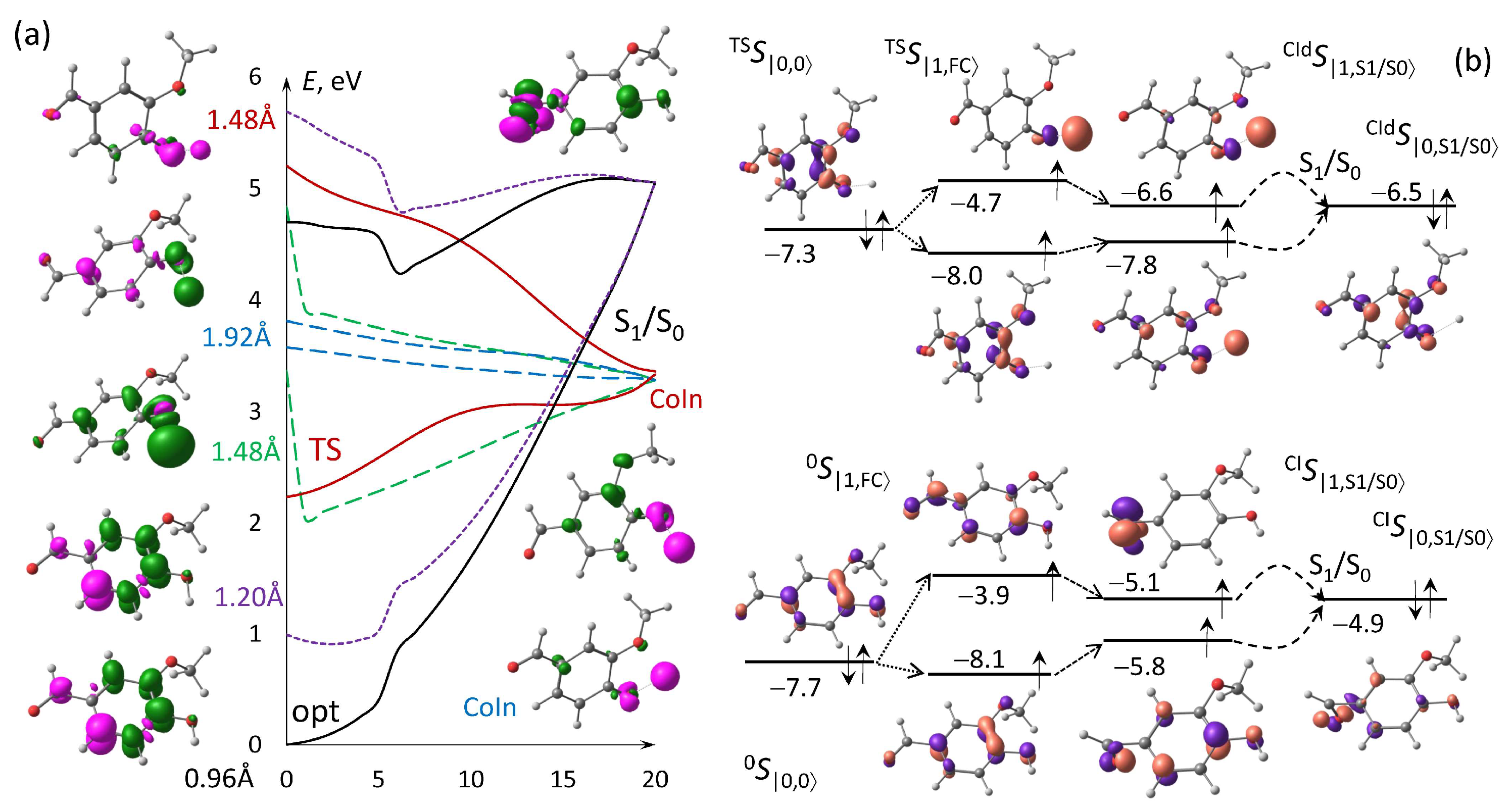
The vanillin deprotonation mechanism requires specifically prepared initial electron states (Figure 1) in contrast to smaller molecules of the phenol family [10,11], but photochemical evolution can also be revealed through CoIn searching and NAMD modeling. Experimental and theoretical investigations demonstrated a strong dependency of the deprotonation probability of the parent phenol on the solvation effect [8,9]. CoIn searching between potential energy surfaces (PESs) calculated using MRSF with BHHLYP&6-311G* for the various initial electronic states considered above (Figure 1) led to deformation or deprotonation depending on the initial O–H stretching. CoIn searching was initiated from Franck–Condon’s theory of the ground-state optimized molecule in 0S|1,FC〉 vs. transition state TSS|1,FC〉. Differences between the bond lengths, bending, and dihedral angles of the initial and final CoIn structures are divided by 20 and the results are added to the current geometric parameters for the next energy calculations that provide PESs’ construction via sequential spatial configurations.
The MRSF electron densities and difference between them ρ(Si)–ρ(Sj) calculated in GAMESS were reconstructed using ChemCraft to elucidate EDD types for the transition at the beginning and the dissipation through the CoIn points (Figure 3a). CoIn searching did not reach the predissociation state starting from the optimization structure and with slight O–H = 1.2 Å elongation when the bond length swiftly restored to the usual covalent distance and arrived to the same structures as those started from the initially optimized structure. There is no hydroxyl contribution to the original electron density delocalization in the both cases, which provides only slight deformation at the CoIn point concerning mostly the aldehyde substitution. The maximal changes in the group from the optimized structure to CoIn are the bond length for |O–H| = 0.11 Å, bending angle ∠OCC = −49.8°, and ∠HCC = 34.0°, with corresponding dihedral torsion angles OC^CC = −88.3° and HC^CC = −65.9°. The phenyl ring remains plain and stable at the S1/S0 CoIn point.
The initial dominant electron density localization on the hydroxyl bond with potential predissociation occurs from the transition state (TS) structure with O–H = 1.486 Å and when the bond is longer. The structural change from TSS|1,FC〉 to S0 concerns mostly the hydroxyl substitution with electron density redistribution to the group (Figure 3a), resulting in an increase in the bond length by 0.32 Å to reach the final S1/S0 CoIn transition with O–H bond cleavage, which can be considered as a predissociation state with potential deprotonation. The averaged remaining bond length is 0.02 Å with a maximum value equal to 0.06 Å. The ∠H,O,C bending change between hydroxyl and phenyl groups is 11.8°, while the changes in the other angles are less than 4.0° in absolute values at S1/S0. The dihedral torsion angles between the hydroxyl group and phenyl ring experience changes of −51.0° accompanied with insignificant methyl hydrogens turning around the CO axis by 177° and with negligible variations of less than 8.0° for the remaining torsion to reach the CoIn point indicating a relatively flat structure.
DMOs in the framework of the extended Koopmans’ theorem [46,47,48] deal with multiconfigurational methods, in the present MRSF, and provide chemically oriented single-configurational electronic transitions from higher doubly occupied (norm ≈ 1) DMOs of the ground state to the highest singly occupied (norm ≈ 0.5) DMO of an excited state with corresponding orbital energies. DMOs define the EDD shapes and types of transitions in CoIn searching evaluation. The tool is applied to consider in detail the O–H cleavage along dissipative trajectories through PES intersections in comparison with the deformed but stable isolated structure at the CoIn points. The lowest Franck–Condon TSS|1,FC〉 = |π→ σ O H * 〉 is excited from TSS|0,0〉 with electron promotion from the highest doubly occupied|π〉 DMO of the unexacted system with an O–H longer than 1.48 Å to the singly occupied | σ O H * 〉 DMO of the first excited electron density (Figure 3b).The almost-complete EDD redistribution from the benzene ring to the hydroxyl group along the dissipative CoIn trajectories indicates a condition conducive to hydroxyl cleavage with potential deprotonation. In the other cases, the first excitation to 0S|1,FC〉 = |π→π*〉 turns to |π→σ*〉 localized on the ether fragment at the CoIn point maintaining the molecules as stable with insignificant structural deformation.

3.4. Photodynamic PES Trajectories Through NAMD

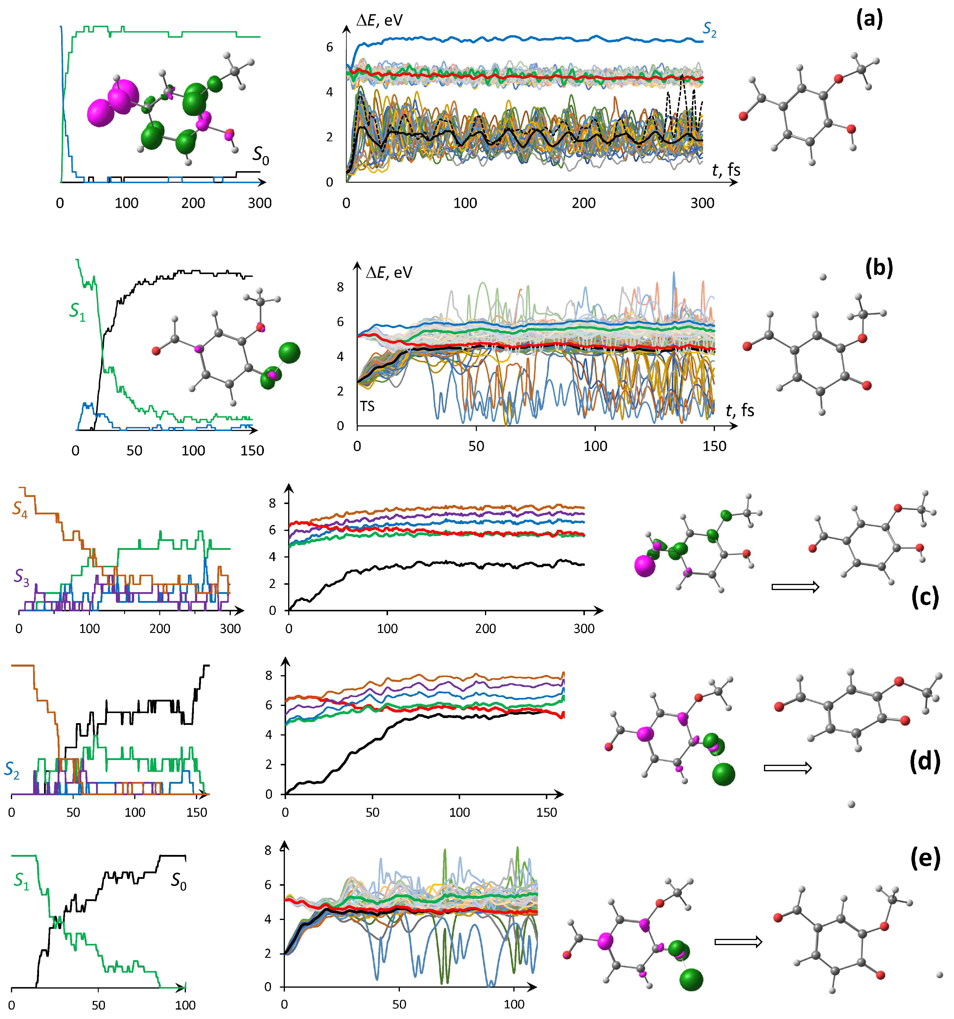
Photodynamic propagations were simulated for VNL solvated with the water SMD model and isolated in a vacuum starting from the ground state of the minimized structure and its several vibrationally fluctuating conformers vs. the transition state of the molecule optimized with the elongated hydroxyl bond. PES propagations together with excited state populations and structural analysis provide statistically averaged stochastic photochemical molecular transformation, predissociation, and further deprotonation. The classical explicit SBC aqueous solvent with the photoexcited chromophore was not considered, since hydroxyl bond cleavage requires subsequent proton travel through a water cluster network and interaction with the electronic system of, at least, the first aqueous shell, which includes no less than a dozen solvent molecules in the large QM region resulting in unacceptably memory-expensive and time-consuming calculations. The 300 fs NAMD trajectories of VML in the water SMD were run five times for each of the six different instantaneous snapshots of fluctuating conformers starting from second excitation of the ground-state optimized molecule through its oscillating structures taken every 1 ps on the static QCMD absorption trajectory (Figure 2), which led once to total dissipation with hydroxyl bond decomposition (Figure 4a).
The averaged O–H distance on the absorption trajectory was 0.964 Å with insignificant ±0.005 Å deviations but, taking into account the stochastic behavior of the PES crossing through CoIn points along NAMD evolutions due to the TSH component with the random function, the weak initial structural changes in the six vibrational frames do not play a role in the photodynamic propagation. Starting from the second excitation, the electronic systems were swiftly in several fs hopping to the lowest excited states and not degrading to the ground state, with one exception, where the mean bond length |O–H| = 0.958 ± 0.015 Å on the observable trajectories. On the other hand, all 62 trials starting from the TS point swiftly degraded from first excitation to the ground state with following deprotonation and possible hydrogen bonding to the other VNL atoms on 150 fs trajectories (Figure 4b) because of the initial lowest excited state localized on the breaking hydroxyl bond. After bond cleavage, the proton migrates along the molecular electron field lines establishing temporary or stable connections with various atoms of the structure (45%), restoring hydroxyl bond vibrations to the normal covalent distance after the initial elongation (5%), or leaving the molecular vicinity with total deprotonation (50%).
In contrast to the SMD model, the isolated chromophore provides almost equal probabilities of keeping the structure stable (14 trials with |O–H| = 0.968 ± 0.035 Å) on 300 fs trajectories vs. O–H decomposition (11 trials) during 150 fs, even starting from the high fourth excitation of the ground state-optimized molecule (Figure 4c,d). The averaged hopping transfer to the lowest excited state appears near 120 fs for stable structure and nearly the same time is needed for total dissipation with following deprotonation. Only the averaged 80 fs was needed for degradation to the ground state starting from the TS but not all led to deprotonation in the ground state (Figure 4e). There are also two main photochemical pathways in this model where the proton escapes (50%) or recombines with other nuclei of the molecules (50%) but in the absence of an external SMD force field that provides a greater variety of VNL isomers with different proton bonding sites.

4. Conclusions

The research protocol for comprehensive spectral-luminescence and photodynamical calculations of vibronic transitions, eSQMMD static optical spectra, CoIn, and NAMD excitation evolution were suggested and applied to study photoinduced vanillin deprotonation in a vacuum and an aqueous solvent. Hydroxyl group decomposition was investigated through vibronic analysis, photostatic optical spectra generation, CoIn searching of predissociation states, and NAMD of excited states dissipation leading to deprotonation. The absorption and emission static spectra of vanillin in a vacuum and different water models were obtained, compared, and demonstrated the advantages of the SMD approach. Insignificant structural deformations against hydroxyl bond cleavage with following deprotonation were demonstrated with CoIn and NAMD simulations initiated from the vanillin structure optimized to either minimum energy or a transition state in the various environment.
CoIn searching starting from the minimum energy molecule or the same structure with an elongated hydroxyl bond shorter than 1.4 Å arrived to a slight molecular deformation, whereas longer O–H or TS bonds led to the initial states of electron redistribution over the group with potential dissociation. NAMD simulation initialized from the TS demonstrated swift deprotonation in a vacuum and SMD water, whereas the nearly totally optimized molecule provided only insignificant structural deformations vs. the same initial condition in a vacuum which led to deprotonation in almost half of the trials.

Author Contributions

Conceptualization, V.P. and O.T.; methodology, V.P.; software, V.P.; validation, V.P. and O.T.; formal analysis, V.P.; investigation, V.P.; resources, O.T.; data curation, O.T.; writing—original draft preparation, V.P.; writing—review and editing, V.P. and O.T.; visualization, V.P.; supervision, O.T.; project administration, O.T.; funding acquisition, O.T. All authors have read and agreed to the published version of the manuscript.

Funding

This work was carried out within the framework of the state program for supporting universities “Priority-2030” (project number NU 2.0.4.25 ML).

Data Availability Statement

All published research data are available from the authors upon request.

Acknowledgments

The authors acknowledge technical support of the TSU-SKIF computational center.

Conflicts of Interest

The authors declare that they have no known competing financial interests or personal relationships that could have appeared to influence the work reported in this paper.

Abbreviations

The following abbreviations are used in this manuscript:
VNLVanillin
CoInConical intersection
QCMDQuantum–classical molecular dynamic
SMD PCMSolvation model of polarizable continuum model
NAMDNonadiabatic molecular dynamics
LR-MRSFLinear response mixed-reference spin–flip
TD-DFTTime-dependent density functional theory
eQCMDExtended statistical quantum-mechanical/molecular dynamic
MOMolecular orbital
TSHTrajectory surface hopping
ESPFEmbedding electrostatic potential fitting
VEEVertical electronic excitation
EDDElectron density differences
PESPotential energy surfaces
DMODyson molecular orbitals
SBCSpherical boundary conditions

References

  1. Go, B.R.; Lyu, Y.; Ji, Y.; Li, Y.J.; Huang, D.D.; Li, X.; Nah, T.; Lam, C.H.; Chan, C.K. Aqueous Secondary Organic Aerosol Formation from the Direct Photosensitized Oxidation of Vanillin in the Absence and Presence of Ammonium Nitrate. Atmos. Chem. Phys. 2022, 22, 273–293. [Google Scholar] [CrossRef]
  2. Al-Hunaiti, A.; Mohaidat, Q.; Bsoul, I.; Mahmood, S.; Taher, D.; Hussein, T. Synthesis and Characterization of Novel Phyto-Mediated Catalyst, and Its Application for a Selective Oxidation of (VAL) into Vanillin under Visible Light. Catalysts 2020, 10, 839. [Google Scholar] [CrossRef]
  3. Liu, Z.; Liu, Q.; Ren, X.; Xie, J.; Chang, N.; Zhang, Q. Novel Photobase Generators Mediated Thiol-Epoxy “Click” Reaction for Self-Healing, Degradable and Recyclable Photoresist Based on Bio-Vanillin and Eugenol. Chem. Eng. J. 2025, 503, 158304. [Google Scholar] [CrossRef]
  4. Loisel, G.; Mekic, M.; Liu, S.; Song, W.; Jiang, B.; Wang, Y.; Deng, H.; Gligorovski, S. Ionic Strength Effect on the Formation of Organonitrate Compounds through Photochemical Degradation of Vanillin in Liquid Water of Aerosols. Atmos. Environ. 2021, 246, 118140. [Google Scholar] [CrossRef]
  5. Carena, L.; Zoppi, B.; Sordello, F.; Fabbri, D.; Minella, M.; Minero, C. Phototransformation of Vanillin in Artificial Snow by Direct Photolysis and Mediated by Nitrite. Environ. Sci. Technol. 2023, 57, 8785–8795. [Google Scholar] [CrossRef] [PubMed]
  6. Arya, S.S.; Rookes, J.E.; Cahill, D.M.; Lenka, S.K. Vanillin: A Review on the Therapeutic Prospects of a Popular Flavouring Molecule. Adv. Tradit. Med. 2021, 21, 1–17. [Google Scholar] [CrossRef]
  7. Liu, B.; Lim, C.-H.; Miyake, G.M. Light-Driven Intermolecular Charge Transfer Induced Reactivity of Ethynylbenziodoxol(on)e and Phenols. J. Am. Chem. Soc. 2018, 140, 12829–12835. [Google Scholar] [CrossRef]
  8. Kusaka, R.; Nihonyanagi, S.; Tahara, T. The Photochemical Reaction of Phenol Becomes Ultrafast at the Air–Water Interface. Nat. Chem. 2021, 13, 306–311. [Google Scholar] [CrossRef]
  9. Ishiyama, T.; Tahara, T.; Morita, A. Why the Photochemical Reaction of Phenol Becomes Ultrafast at the Air–Water Interface: The Effect of Surface Hydration. J. Am. Chem. Soc. 2022, 144, 6321–6325. [Google Scholar] [CrossRef]
  10. Pomogaev, V.; Bocharnikova, E.; Tchaikovskaya, O.; Avramov, P. Phenol Photostatic Spectra and Quantum-Classical Photodynamic Deprotonation. Int. J. Quantum Chem. 2024, 124, e27504. [Google Scholar] [CrossRef]
  11. Pomogaev, V.; Bocharnikova, E.; Avramov, P.; Tchaikovskaya, O. P-Cresol Quantum-Classical Photodynamics, Photostatic UV, IR and Raman Spectra. J. Mol. Struct. 2025, 1344, 142986. [Google Scholar] [CrossRef]
  12. Vusovich, O.V.; Lapin, I.N.; Svetlichnyi, V.A.; Sul’timova, N.B.; Tchaikovskaya, O.N. Comparison of Vanillin and Isovanillin Photolysis in Aqueous Solutions. Russ. Phys. J. 2014, 56, 1287–1291. [Google Scholar] [CrossRef]
  13. Huix-Rotllant, M.; Schwinn, K.; Pomogaev, V.; Farmani, M.; Ferré, N.; Lee, S.; Choi, C.H. Photochemistry of Thymine in Solution and DNA Revealed by an Electrostatic Embedding QM/MM Combined with Mixed-Reference Spin-Flip TDDFT. J. Chem. Theory Comput. 2023, 19, 147–156. [Google Scholar] [CrossRef]
  14. Pomogaev, V.A.; Sadiq, S.; Filatov, M.; Choi, C.H. Modeling of Photochemical Conversions on Nonadiabatic Molecular Dynamics Trajectories; Izvestiya Vuzov. Fizika: Tomsk, Russia, 2023; Volume 66, pp. 25–33. [Google Scholar]
  15. Pomogaev, V.; Pomogaeva, A.; Avramov, P.; Jalkanen, K.J.; Kachin, S. Thermo-Dynamical Contours of Electronic-Vibrational Spectra Simulated Using the Statistical Quantum–Mechanical Methods. Theor. Chem. Acc. 2011, 130, 609–632. [Google Scholar] [CrossRef]
  16. Pomogaev, V.A.; Ramazanov, R.R.; Ruud, K.; Artyukhov, V.Y. Insight into the Fluorescence Quenching of Trp214 at HSA by the Dimetridazole Ligand from Simulation. J. Photochem. Photobiol. A Chem. 2018, 254, 86–100. [Google Scholar] [CrossRef]
  17. Zuehlsdorff, T.J.; Shedge, S.V.; Lu, S.-Y.; Hong, H.; Aguirre, V.P.; Shi, L.; Isborn, C.M. Vibronic and Environmental Effects in Simulations of Optical Spectroscopy. Annu. Rev. Phys. Chem. 2021, 72, 165–188. [Google Scholar] [CrossRef]
  18. Kubenko, V.G.; Pomogaev, V.A.; Buglak, A.A.; Kononov, A.I. Photophysics of 5,6,7,8-Tetrahydrobiopterin on a Femtosecond Time-Scale. J. Photochem. Photobiol. B Biol. 2025, 265, 113134. [Google Scholar] [CrossRef]
  19. Huix-Rotllant, M.; Ferré, N. Analytic Energy, Gradient, and Hessian of Electrostatic Embedding QM/MM Based on Electrostatic Potential-Fitted Atomic Charges Scaling Linearly with the MM Subsystem Size. J. Chem. Theory Comput. 2021, 17, 538–548. [Google Scholar] [CrossRef]
  20. Alías-Rodríguez, M.; Bonfrate, S.; Park, W.; Ferré, N.; Choi, C.H.; Huix-Rotllant, M. Solvent Effects and pH Dependence of the X-Ray Absorption Spectra of Proline from Electrostatic Embedding Quantum Mechanics/Molecular Mechanics and Mixed-Reference Spin-Flip Time-Dependent Density-Functional Theory. J. Phys. Chem. A 2023, 127, 10382–10392. [Google Scholar] [CrossRef]
  21. Park, W.; Shen, J.; Lee, S.; Piecuch, P.; Filatov, M.; Choi, C.H. Internal Conversion between Bright (11 Bu+) and Dark (21 Ag) States in s-Trans-Butadiene and s-Trans-Hexatriene. J. Phys. Chem. Lett. 2021, 12, 9720–9729. [Google Scholar] [CrossRef] [PubMed]
  22. Lee, S.; Kim, E.E.; Nakata, H.; Lee, S.; Choi, C.H. Efficient Implementations of Analytic Energy Gradient for Mixed-Reference Spin-Flip Time-Dependent Density Functional Theory (MRSF-TDDFT). J. Chem. Phys. 2019, 150, 184111. [Google Scholar] [CrossRef] [PubMed]
  23. Sadiq, S.; Park, W.; Mironov, V.; Lee, S.; Filatov (Gulak), M.; Choi, C.H. Prototropically Controlled Dynamics of Cytosine Photodecay. J. Phys. Chem. Lett. 2023, 14, 791–797. [Google Scholar] [CrossRef] [PubMed]
  24. Shostak, S.; Park, W.; Oh, J.; Kim, J.; Lee, S.; Nam, H.; Filatov, M.; Kim, D.; Choi, C.H. Ultrafast Excited State Aromatization in Dihydroazulene. J. Am. Chem. Soc. 2023, 145, 1638–1648. [Google Scholar] [CrossRef] [PubMed]
  25. Lee, I.S.; Ha, J.; Han, D.; Kim, T.I.; Moon, S.W.; Min, S.K. PyUNIxMD: A Python-Based Excited State Molecular Dynamics Package. J. Comput. Chem. 2021, 42, 1755–1766. [Google Scholar] [CrossRef]
  26. Accomasso, D.; Jankowska, J. Quantum-Classical Simulations Reveal the Photoisomerization Mechanism of a Prototypical First-Generation Molecular Motor. Chem. A Eur. J. 2025, 31, e202403768. [Google Scholar] [CrossRef]
  27. Park, W.; Lee, S.; Huix-Rotllant, M.; Filatov, M.; Choi, C.H. Impact of the Dynamic Electron Correlation on the Unusually Long Excited-State Lifetime of Thymine. J. Phys. Chem. Lett. 2021, 12, 4339–4346. [Google Scholar] [CrossRef]
  28. Park, W.; Komarov, K.; Lee, S.; Choi, C.H. Mixed-Reference Spin-Flip Time-Dependent Density Functional Theory: Multireference Advantages with the Practicality of Linear Response Theory. J. Phys. Chem. Lett. 2023, 14, 8896–8908. [Google Scholar] [CrossRef]
  29. Lee, C.; Yang, W.; Parr, R.G. Development of the Colle-Salvetti Correlation-Energy Formula into a Functional of the Electron Density. Phys. Rev. B 1988, 37, 785–789. [Google Scholar] [CrossRef]
  30. Petersson, G.A.; Al-Laham, M.A. A Complete Basis Set Model Chemistry. II. Open-shell Systems and the Total Energies of the First-row Atoms. J. Chem. Phys. 1991, 94, 6081–6090. [Google Scholar] [CrossRef]
  31. Rassolov, V.A.; Ratner, M.A.; Pople, J.A.; Redfern, P.C.; Curtiss, L.A. 6-31G* Basis Set for Third-row Atoms. J. Comput. Chem. 2001, 22, 976–984. [Google Scholar] [CrossRef]
  32. Frisch, M.J.; Trucks, G.W.; Schlegel, H.B.; Scuseria, G.E.; Robb, M.A.; Cheeseman, J.R.; Scalmani, G.; Barone, V.; Mennucci, B.; Petersson, G.A.; et al. Gaussian, 16, Revision C.01; Gaussian Inc.: Wallingford, CT, USA, 2016. [Google Scholar]
  33. Becke, A.D. A New Mixing of Hartree-Fock and Local Density-Functional Theories. J. Chem. Phys. 1993, 98, 1372–1377. [Google Scholar] [CrossRef]
  34. Petersson, G.A.; Bennett, A.; Tensfeldt, T.G.; Al-Laham, M.A.; Shirley, W.A.; Mantzaris, J. A Complete Basis Set Model Chemistry. I. The Total Energies of Closed-Shell Atoms and Hydrides of the First-Row Elements. J. Chem. Phys. 1988, 89, 2193–2218. [Google Scholar] [CrossRef]
  35. Barbatti, M.; Bondanza, M.; Crespo-Otero, R.; Demoulin, B.; Dral, P.O.; Granucci, G.; Kossoski, F.; Lischka, H.; Mennucci, B.; Mukherjee, S.; et al. Newton-X Platform: New Software Developments for Surface Hopping and Nuclear Ensembles. J. Chem. Theory Comput. 2022, 18, 6851–6865. [Google Scholar] [CrossRef] [PubMed]
  36. Toldo, J.M.; Telles do Casal, M.; Ventura, E.; Andrade do Monte, S.; Barbatti, M. Surface Hopping Modeling of Charge and Energy Transfer in Complex Environments. Phys. Chem. Chem. Phys. 2023, 25, 8293–8316. [Google Scholar] [CrossRef] [PubMed]
  37. Shu, Y.; Zhang, L.; Chen, X.; Sun, S.; Huang, Y.; Truhlar, D.G. Nonadiabatic Dynamics Algorithms with Only Potential Energies and Gradients: Curvature-Driven Coherent Switching with Decay of Mixing and Curvature-Driven Trajectory Surface Hopping. J. Chem. Theory Comput. 2022, 18, 1320–1328. [Google Scholar] [CrossRef]
  38. Liu, X.-Y.; Chen, W.-K.; Fang, W.-H.; Cui, G. Nonadiabatic Dynamics Simulations for Photoinduced Processes in Molecules and Semiconductors: Methodologies and Applications. J. Chem. Theory Comput. 2023, 19, 8491–8522. [Google Scholar] [CrossRef]
  39. Crespo-Otero, R.; Barbatti, M. Recent Advances and Perspectives on Nonadiabatic Mixed Quantum–Classical Dynamics. Chem. Rev. 2018, 118, 7026–7068. [Google Scholar] [CrossRef]
  40. Zhang, L.; Ullah, A.; Pinheiro Jr, M.; Dral, P.O.; Barbatti, M. Excited-State Dynamics with Machine Learning. In Quantum Chemistry in the Age of Machine Learning; Elsevier: Amsterdam, The Netherlands, 2023; pp. 329–353. ISBN 978-0-323-90049-2. [Google Scholar]
  41. Ferré, N.; Filatov, M.; Huix-Rotllant, M. (Eds.) Density-Functional Methods for Excited States; Topics in Current Chemistry; Springer International Publishing: Cham, Switzerland, 2016; Volume 368, ISBN 978-3-319-22080-2. [Google Scholar]
  42. Marenich, A.V.; Cramer, C.J.; Truhlar, D.G. Universal Solvation Model Based on Solute Electron Density and on a Continuum Model of the Solvent Defined by the Bulk Dielectric Constant and Atomic Surface Tensions. J. Phys. Chem. B 2009, 113, 6378–6396. [Google Scholar] [CrossRef]
  43. Martínez, L.; Andrade, R.; Birgin, E.G.; Martínez, J.M. P ACKMOL: A Package for Building Initial Configurations for Molecular Dynamics Simulations. J. Comput. Chem. 2009, 30, 2157–2164. [Google Scholar] [CrossRef]
  44. Jorgensen, W.L.; Maxwell, D.S.; Tirado-Rives, J. Development and Testing of the OPLS All-Atom Force Field on Conformational Energetics and Properties of Organic Liquids. J. Am. Chem. Soc. 1996, 118, 11225–11236. [Google Scholar] [CrossRef]
  45. Pomogaev, V.; Pomogaeva, A.; Aoki, Y. Absorption Spectra of Estradiol and Tryptophan Constructed by the Statistical and Elongation Methods. J. Phys. Chem. A 2009, 113, 1429–1433. [Google Scholar] [CrossRef]
  46. Ortiz, J.V. Dyson-Orbital Concepts for Description of Electrons in Molecules. J. Chem. Phys. 2020, 153, 070902. [Google Scholar] [CrossRef]
  47. Filatov, M.; Lee, S.; Nakata, H.; Choi, C.H. Computation of Molecular Electron Affinities Using an Ensemble Density Functional Theory Method. J. Phys. Chem. A 2020, 124, 7795–7804. [Google Scholar] [CrossRef]
  48. Pomogaev, V.; Lee, S.; Shaik, S.; Filatov, M.; Choi, C.H. Exploring Dyson’s Orbitals and Their Electron Binding Energies for Conceptualizing Excited States from Response Methodology. J. Phys. Chem. Lett. 2021, 12, 9963–9972. [Google Scholar] [CrossRef]
  49. Gordon, M.S. GAMESS. Available online: https://www.msg.chem.iastate.edu/gamess/ (accessed on 30 September 2023).
  50. Schmidt, M.W.; Baldridge, K.K.; Boatz, J.A.; Elbert, S.T.; Gordon, M.S.; Jensen, J.H.; Koseki, S.; Matsunaga, N.; Nguyen, K.A.; Su, S.; et al. General Atomic and Molecular Electronic Structure System. J. Comput. Chem. 1993, 14, 1347–1363. [Google Scholar] [CrossRef]
  51. Ponder, J.W. TINKER: Software Tools for Molecular Design, Version 8.10.1. 2021. Available online: http://dasher.wustl.edu/tinker/ (accessed on 25 November 2022).
  52. Chemcraft—Graphical Software for Visualization of Quantum Chemistry Computations Version 1.8, Build 682. Available online: https://www.chemcraftprog.com (accessed on 15 July 2025).
  53. Žagar, E.; Grdadolnik, J. An Infrared Spectroscopic Study of H-Bond Network in Hyperbranched Polyester Polyol. J. Mol. Struct. 2003, 658, 143–152. [Google Scholar] [CrossRef]
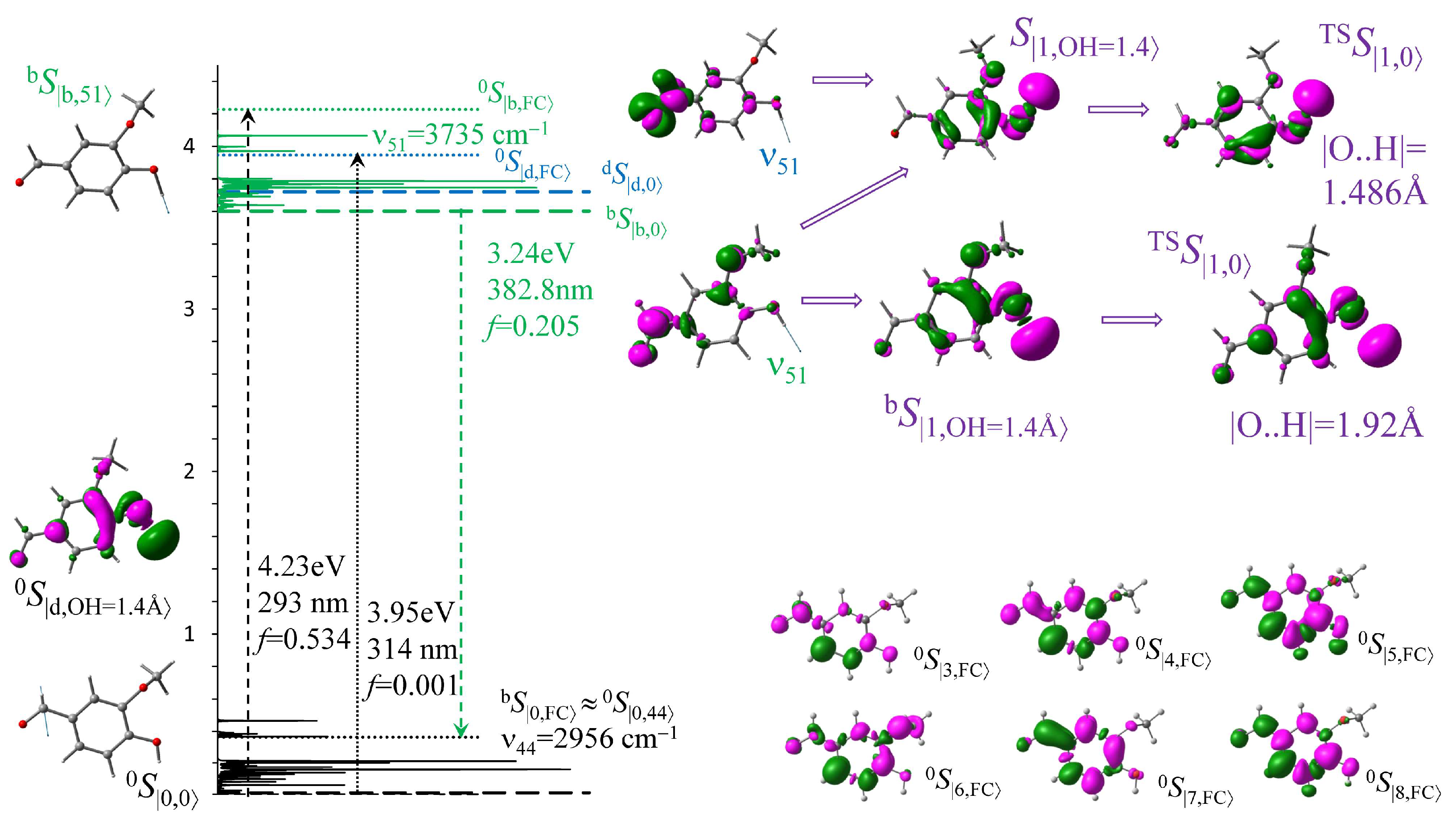
Figure 1. Vibrionic levels of structures optimized in the ground and two excitation states 0S, dS, and bS, respectively, with the TS searching scheme. Electron densities redistribute from pink to green areas for absorption and contrariwise for dissipation.
Chemengineering 09 00076 g001
Figure 2. Normalized absorption spectra isolated (dotted line), in the SMD model (dashed line) vs. the SBC water droplet (last QCMD snapshot) with Sd, Sb, and S3 bands (light brown, green, and blue solid lines, respectively) and two fluorescence bands (red solid and dotted lines) in the water droplet. Transition EDDs redistribute from pink to green areas. VEE-optimized molecule isolated and in SMD vs. the frozen chromophore in 100 different water configurations (straight lines and spectral curves under the axis, respectively) with EDD for the last H-bound frame.
Chemengineering 09 00076 g002
Figure 3. (a) CoIn reaction pathways S1 → S0 initiated from the optimized O–H = 0.96 Å structure (black solid lines), with elongated O–H = 1.20 Å (violet short dashed), O–H = 1.48 Å (green dashed), and O–H = 1.92 Å (blue dashed) vs. the optimized TS (red solid). Electron densities redistribute from pink to green areas for absorption and contrariwise for CoIn. (b) The initial and final DMOs (eV) starting from optimized 0S and TSS to CoIn.
Chemengineering 09 00076 g003
Figure 4. Excited state populations (left graphs) and bunches (multicolor curves) with averaged PES trajectories (right graphs) of the S2 (blue), S1 (green), S0 (black), and Ssys (red) with EDD for the initial structures. PESs in SMD water started from (a) S2 of the ground state and (b) S1 of the TS in contrast with a vacuum: (c) S4 of the optimized molecule remaining stable vs. (d) deprotonated and (e) S1 of the TS.
Chemengineering 09 00076 g004
Table 1. Frozen optimized vanillin in different models.
VNLsbcWM Last FrameVNLespf3WMVNLqm3WMVNL@SMD
Sd308.5 (0.001)0.93|2→0′〉323.2 (0.000)0.95|1→0′〉313.6 (0.000)0.65|3→0′〉312.5 (0.000)0.82|2→0′〉
0.22|2→0′〉 0.13|1→0′〉
Sb285.0 (0.071)0.53|0→0′〉272.3 (0.114)0.65|0→0′〉276.01 (0.151)0.72|1→0′〉283.3 (0.161)0.77|0→0′〉
0.38|1→0′〉 0.25|2→0′〉 0.12|4→0′〉 0.12|1→0′〉
Disclaimer/Publisher’s Note: The statements, opinions and data contained in all publications are solely those of the individual author(s) and contributor(s) and not of MDPI and/or the editor(s). MDPI and/or the editor(s) disclaim responsibility for any injury to people or property resulting from any ideas, methods, instructions or products referred to in the content.