

Dosimetric Implications of Computerised Tomography-Only versus Magnetic Resonance-Fusion Contouring in Stereotactic Body Radiotherapy for Prostate Cancer

Abstract
1. Introduction
2. Materials and Methods
3. Results
4. Discussion
5. Conclusions
Supplementary Materials
Acknowledgments
Author Contributions
Conflicts of Interest
References
- Benedict, S.H.; Yenice, K.M.; Followill, D.; Galvin, J.M.; Hinson, W.; Kavanagh, B.; Keall, P.; Lovelock, M.; Meeks, S.; Papiez, L.; et al. Stereotactic body radiation therapy: The report of aapm task group 101. Med. Phys. 2010, 37, 4078–4101. [Google Scholar] [CrossRef] [Scilit] [PubMed]
- NCCN. National Comprehensive Cancer Network—Clinical Practice Guidelines in Oncology: Prostate Cancer. Version 1.2016. Available online: http://www.nccn.org/professionals/physician_gls/pdf/prostate.pdf (accessed on 27 October 2015).
- ASTRO. Model Policies: Stereotactic Body Radiotherapy. Available online: https://www.astro.org/uploadedFiles/Main_Site/Practice_Management/Reimbursement/2013HPcoding%20guidelines_SBRT_Final.pdf (accessed on 16 September 2014).
- Henderson, D.R.; Tree, A.C.; van As, N.J. Stereotactic body radiotherapy for prostate cancer. Clin. Oncol. 2015, 27, 270–279. [Google Scholar] [CrossRef] [Scilit] [PubMed]
- ClinicalTrials.gov. Prostate Advance in Comparative Evidence (Pace)—nct01584258; The Institute of Cancer Research: Sutton, Surrey, UK, 2017.
- Madsen, B.L.; Hsi, R.A.; Pham, H.T.; Fowler, J.F.; Esagui, L.; Corman, J. Stereotactic hypofractionated accurate radiotherapy of the prostate (sharp), 33.5 gy in five fractions for localized disease: First clinical trial results. Int. J. Radiat. Oncol. Biol. Phys. 2007, 67, 1099–1105. [Google Scholar] [CrossRef] [Scilit] [PubMed]
- Boike, T.P.; Lotan, Y.; Cho, L.C.; Brindle, J.; DeRose, P.; Xie, X.J.; Yan, J.; Foster, R.; Pistenmaa, D.; Perkins, A.; et al. Phase i dose-escalation study of stereotactic body radiation therapy for low- and intermediate-risk prostate cancer. J. Clin. Oncol. 2011, 29, 2020–2026. [Google Scholar] [CrossRef] [Scilit] [PubMed]
- Chen, L.N.; Suy, S.; Uhm, S.; Oermann, E.K.; Ju, A.W.; Chen, V.; Hanscom, H.N.; Laing, S.; Kim, J.S.; Lei, S.; et al. Stereotactic body radiation therapy (sbrt) for clinically localized prostate cancer: The georgetown university experience. Radiat. Oncol. 2013, 8, 58. [Google Scholar] [CrossRef] [Scilit] [PubMed]
- Bolzicco, G.; Favretto, M.S.; Satariano, N.; Scremin, E.; Tambone, C.; Tasca, A. A single-center study of 100 consecutive patients with localized prostate cancer treated with stereotactic body radiotherapy. BMC Urol. 2013, 13, 49. [Google Scholar] [CrossRef] [Scilit] [PubMed]
- Katz, A.J.; Santoro, M.; Diblasio, F.; Ashley, R. Stereotactic body radiotherapy for localized prostate cancer: Disease control and quality of life at 6 years. Radiat. Oncol. 2013, 8, 118. [Google Scholar] [CrossRef] [Scilit] [PubMed]
- Oliai, C.; Lanciano, R.; Sprandio, B.; Yang, J.; Lamond, J.; Arrigo, S.; Good, M.; Mooreville, M.; Garber, B.; Brady, L.W. Stereotactic body radiation therapy for the primary treatment of localized prostate cancer. J. Radiat. Oncol. 2013, 2, 63–70. [Google Scholar] [CrossRef] [Scilit] [PubMed]
- Lee, W.R.; Dignam, J.J.; Amin, M.B.; Bruner, D.W.; Low, D.; Swanson, G.P.; Shah, A.B.; D’Souza, D.P.; Michalski, J.M.; Dayes, I.S.; et al. Randomized phase iii noninferiority study comparing two radiotherapy fractionation schedules in patients with low-risk prostate cancer. J. Clin. Oncol. 2016, 34, 2325–2332. [Google Scholar] [CrossRef] [Scilit] [PubMed]
- Dearnaley, D.; Syndikus, I.; Mossop, H.; Khoo, V.; Birtle, A.; Bloomfield, D.; Graham, J.; Kirkbride, P.; Logue, J.; Malik, Z.; et al. Conventional versus hypofractionated high-dose intensity-modulated radiotherapy for prostate cancer: 5-year outcomes of the randomised, non-inferiority, phase 3 chhip trial. Lancet Oncol. 2016, 17, 1047–1060. [Google Scholar] [CrossRef] [Scilit]
- Hentschel, B.; Oehler, W.; Strauss, D.; Ulrich, A.; Malich, A. Definition of the ctv prostate in ct and mri by using ct-mri image fusion in imrt planning for prostate cancer. Strahlenther. Onkol. 2011, 187, 183–190. [Google Scholar] [CrossRef] [Scilit] [PubMed]
- Debois, M.; Oyen, R.; Maes, F.; Verswijvel, G.; Gatti, G.; Bosmans, H.; Feron, M.; Bellon, E.; Kutcher, G.; Van Poppel, H.; et al. The contribution of magnetic resonance imaging to the three-dimensional treatment planning of localized prostate cancer. Int. J. Radiat. Oncol. Biol. Phys. 1999, 45, 857–865. [Google Scholar] [CrossRef] [Scilit]
- Steenbakkers, R.J.; Deurloo, K.E.; Nowak, P.J.; Lebesque, J.V.; van Herk, M.; Rasch, C.R. Reduction of dose delivered to the rectum and bulb of the penis using mri delineation for radiotherapy of the prostate. Int. J. Radiat. Oncol. Biol. Phys. 2003, 57, 1269–1279. [Google Scholar] [CrossRef] [Scilit]
- Sannazzari, G.L.; Ragona, R.; Ruo Redda, M.G.; Giglioli, F.R.; Isolato, G.; Guarneri, A. Ct-mri image fusion for delineation of volumes in three-dimensional conformal radiation therapy in the treatment of localized prostate cancer. Br. J. Radiol. 2002, 75, 603–607. [Google Scholar] [CrossRef] [Scilit] [PubMed]
- Elias, E.; Helou, J.; Zhang, L.; Cheung, P.; Deabreu, A.; D’Alimonte, L.; Sethukavalan, P.; Mamedov, A.; Cardoso, M.; Loblaw, A. Dosimetric and patient correlates of quality of life after prostate stereotactic ablative radiotherapy. Radiother. Oncol. 2014, 112, 83–88. [Google Scholar] [CrossRef] [Scilit] [PubMed]
- Amdur, R.J.; Gladstone, D.; Leopold, K.A.; Harris, R.D. Prostate seed implant quality assessment using mr and ct image fusion. Int. J. Radiat. Oncol. Biol. Phys. 1999, 43, 67–72. [Google Scholar] [CrossRef] [Scilit]
- Kagawa, K.; Lee, W.R.; Schultheiss, T.E.; Hunt, M.A.; Shaer, A.H.; Hanks, G.E. Initial clinical assessment of ct-mri image fusion software in localization of the prostate for 3D conformal radiation therapy. Int. J. Radiat. Oncol. Biol. Phys. 1997, 38, 319–325. [Google Scholar] [CrossRef] [Scilit]
- Seppala, T.; Visapaa, H.; Collan, J.; Kapanen, M.; Beule, A.; Kouri, M.; Tenhunen, M.; Saarilahti, K. Converting from ct- to mri-only-based target definition in radiotherapy of localized prostate cancer: A comparison between two modalities. Strahlenther. Onkol. 2015, 191, 862–868. [Google Scholar] [CrossRef] [Scilit] [PubMed]
- Parker, C.C.; Damyanovich, A.; Haycocks, T.; Haider, M.; Bayley, A.; Catton, C.N. Magnetic resonance imaging in the radiation treatment planning of localized prostate cancer using intra-prostatic fiducial markers for computed tomography co-registration. Radiother. Oncol. 2003, 66, 217–224. [Google Scholar] [CrossRef] [Scilit]
- Kerkhof, E.M.; van der Put, R.W.; Raaymakers, B.W.; van der Heide, U.A.; van Vulpen, M.; Lagendijk, J.J. Variation in target and rectum dose due to prostate deformation: An assessment by repeated mr imaging and treatment planning. Phys. Med. Biol. 2008, 53, 5623–5634. [Google Scholar] [CrossRef] [Scilit] [PubMed]
- Usmani, N.; Sloboda, R.; Kamal, W.; Ghosh, S.; Pervez, N.; Pedersen, J.; Yee, D.; Danielson, B.; Murtha, A.; Amanie, J.; et al. Can images obtained with high field strength magnetic resonance imaging reduce contouring variability of the prostate? Int. J. Radiat. Oncol. Biol. Phys. 2011, 80, 728–734. [Google Scholar] [CrossRef] [Scilit] [PubMed]
- Tree, A.C.; Ostler, P.; Hoskin, P.; Dankulchai, P.; Nariyangadu, P.; Hughes, R.J.; Wells, E.; Taylor, H.; Khoo, V.S.; van As, N.J. Prostate stereotactic body radiotherapy—First uk experience. Clin. Oncol. 2014, 26, 757–761. [Google Scholar] [CrossRef] [Scilit] [PubMed]
- King, C.R.; Brooks, J.D.; Gill, H.; Presti, J.C., Jr. Long-term outcomes from a prospective trial of stereotactic body radiotherapy for low-risk prostate cancer. Int. J. Radiat. Oncol. Biol. Phys. 2011, 82, 877–882. [Google Scholar] [CrossRef] [Scilit] [PubMed]
- Smith, J.A., Jr.; Chan, R.C.; Chang, S.S.; Herrell, S.D.; Clark, P.E.; Baumgartner, R.; Cookson, M.S. A comparison of the incidence and location of positive surgical margins in robotic assisted laparoscopic radical prostatectomy and open retropubic radical prostatectomy. J. Urol. 2007, 178, 2385–2389. [Google Scholar] [CrossRef] [Scilit] [PubMed]
- Kilby, W.; Dooley, J.R.; Kuduvalli, G.; Sayeh, S.; Maurer, C.R., Jr. The cyberknife robotic radiosurgery system in 2010. Technol. Cancer Res. Treat. 2010, 9, 433–452. [Google Scholar] [CrossRef] [Scilit] [PubMed]
- Loblaw, A.; Cheung, P.; D’Alimonte, L.; Deabreu, A.; Mamedov, A.; Zhang, L.; Tang, C.; Quon, H.; Jain, S.; Pang, G.; et al. Prostate stereotactic ablative body radiotherapy using a standard linear accelerator: Toxicity, biochemical and pathological outcomes. Radiother. Oncol. 2013, 107, 153–158. [Google Scholar] [CrossRef] [Scilit] [PubMed]
- Tree, A.; Jones, C.; Sohaib, A.; Khoo, V.; van As, N. Prostate stereotactic body radiotherapy with simultaneous integrated boost: Which is the best planning method? Radiat. Oncol. 2013, 8, 228. [Google Scholar] [CrossRef] [Scilit] [PubMed]
- Raaymakers, B.W.; Lagendijk, J.J.; Overweg, J.; Kok, J.G.; Raaijmakers, A.J.; Kerkhof, E.M.; van der Put, R.W.; Meijsing, I.; Crijns, S.P.; Benedosso, F.; et al. Integrating a 1.5 t mri scanner with a 6 mv accelerator: Proof of concept. Phys. Med. Biol. 2009, 54, N229–N237. [Google Scholar] [CrossRef] [Scilit] [PubMed]
- Mutic, S.; Dempsey, J.F. The viewray system: Magnetic resonance-guided and controlled radiotherapy. Semin. Radiat. Oncol. 2014, 24, 196–199. [Google Scholar] [CrossRef] [Scilit] [PubMed]

| Parameter | Constrain/Target | Minor Variations |
|---|---|---|
| PTV | V36.25 Gy ≥ 95% | 90–94.9% |
| CTV (prostate + bsv) | V40 Gy ≥ 95% | 90–94.9% |
| CTV-PTV margins | 5 mm, with 3 mm posteriorly | - |
| Rectum | V18.1 Gy < 50% V29 Gy < 20% V36 Gy < 1 cc | - - ≥1 cc but ≤2 cc |
| Bladder | V18.1 Gy < 40% V37 Gy < 10 cc | - ≥10 cc but ≤20 cc |
| Volume | Mean Volume cc (±SD) | p Value vs. MRF |
|---|---|---|
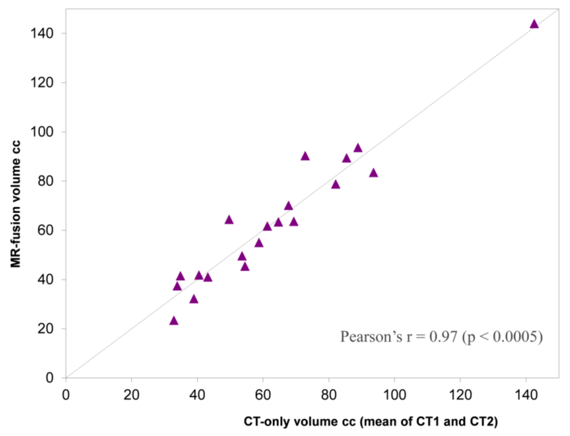
| MRF | 63.5 (±27.9) | - |
| CT1 | 63.2 (±26.5) | 0.84 |
| CT2 | 63.8 (±26.7) | 0.89 |
| Volumes Compared | Mean Dice Coefficient (±SD) |
|---|---|
| MRF vs. CT1 | 0.86 (±0.04) |
| MRF vs. CT2 | 0.85 (±0.05) |
| CT1 vs. CT2 | 0.92 (±0.02) |
| Position | MRF vs. CT1 | MRF vs. CT2 | CT1 vs. CT2 |
|---|---|---|---|
| Mean difference in apex position (mm ±SD; 95% CI) | 1.1 (±3.5; −0.4–2.6) | 1.1 (±3.1; −0.3–2.4) | −0.1 (±2.1; −1.0–0.9) |
| Mean difference in base position (mm; ±SD; 95% CI) | 1.2 (±2.7; 0.0–2.3) | 1.7 (±3.5; 0.1–3.2) | 0.3 (±1.8; −0.5–1.1) |
| MR-Fusion | CT-Only | Comparison | |||
|---|---|---|---|---|---|
| Organ | Constraint * | Mean Volume Receiving ≥ Constraint (±SD) | Mean Difference (95% CI) | p Value | |
| Rectum | V18.1 Gy (<50%) | 33% (±9.2) | 28% (±8.9) | 5.0% (−0.1–10) | 0.05 |
| V29 Gy (<20%) | 11% (±3.2) | 9.4% (±2.5) | 1.7% (0.3–3.1) | 0.02 | |
| V36 Gy (<1–2 cc) | 1.3 cc (±0.5) | 1.0 cc (±0.4) | 0.3 cc (0.1–0.5) | 0.02 | |
| Bladder | V18.1 Gy (<40%) | 26% (±9.3) | 21% (±8.5) | 4.8% (1.6–8.3) | 0.01 |
| V37 Gy (<10 cc) | 6.2 cc (±2.6) | 5.3 cc (±2.2) | 0.9 cc (−0.1–1.88) | 0.08 | |
© 2018 by the authors. Licensee MDPI, Basel, Switzerland. This article is an open access article distributed under the terms and conditions of the Creative Commons Attribution (CC BY) license (http://creativecommons.org/licenses/by/4.0/).
Share and Cite
Henderson, D.R.; Tree, A.C.; Harrington, K.J.; Van As, N.J. Dosimetric Implications of Computerised Tomography-Only versus Magnetic Resonance-Fusion Contouring in Stereotactic Body Radiotherapy for Prostate Cancer. Medicines 2018, 5, 32. https://doi.org/10.3390/medicines5020032
Henderson DR, Tree AC, Harrington KJ, Van As NJ. Dosimetric Implications of Computerised Tomography-Only versus Magnetic Resonance-Fusion Contouring in Stereotactic Body Radiotherapy for Prostate Cancer. Medicines. 2018; 5(2):32. https://doi.org/10.3390/medicines5020032
Chicago/Turabian StyleHenderson, Daniel R., Alison C. Tree, Kevin J. Harrington, and Nicholas J. Van As. 2018. "Dosimetric Implications of Computerised Tomography-Only versus Magnetic Resonance-Fusion Contouring in Stereotactic Body Radiotherapy for Prostate Cancer" Medicines 5, no. 2: 32. https://doi.org/10.3390/medicines5020032
APA StyleHenderson, D. R., Tree, A. C., Harrington, K. J., & Van As, N. J. (2018). Dosimetric Implications of Computerised Tomography-Only versus Magnetic Resonance-Fusion Contouring in Stereotactic Body Radiotherapy for Prostate Cancer. Medicines, 5(2), 32. https://doi.org/10.3390/medicines5020032

