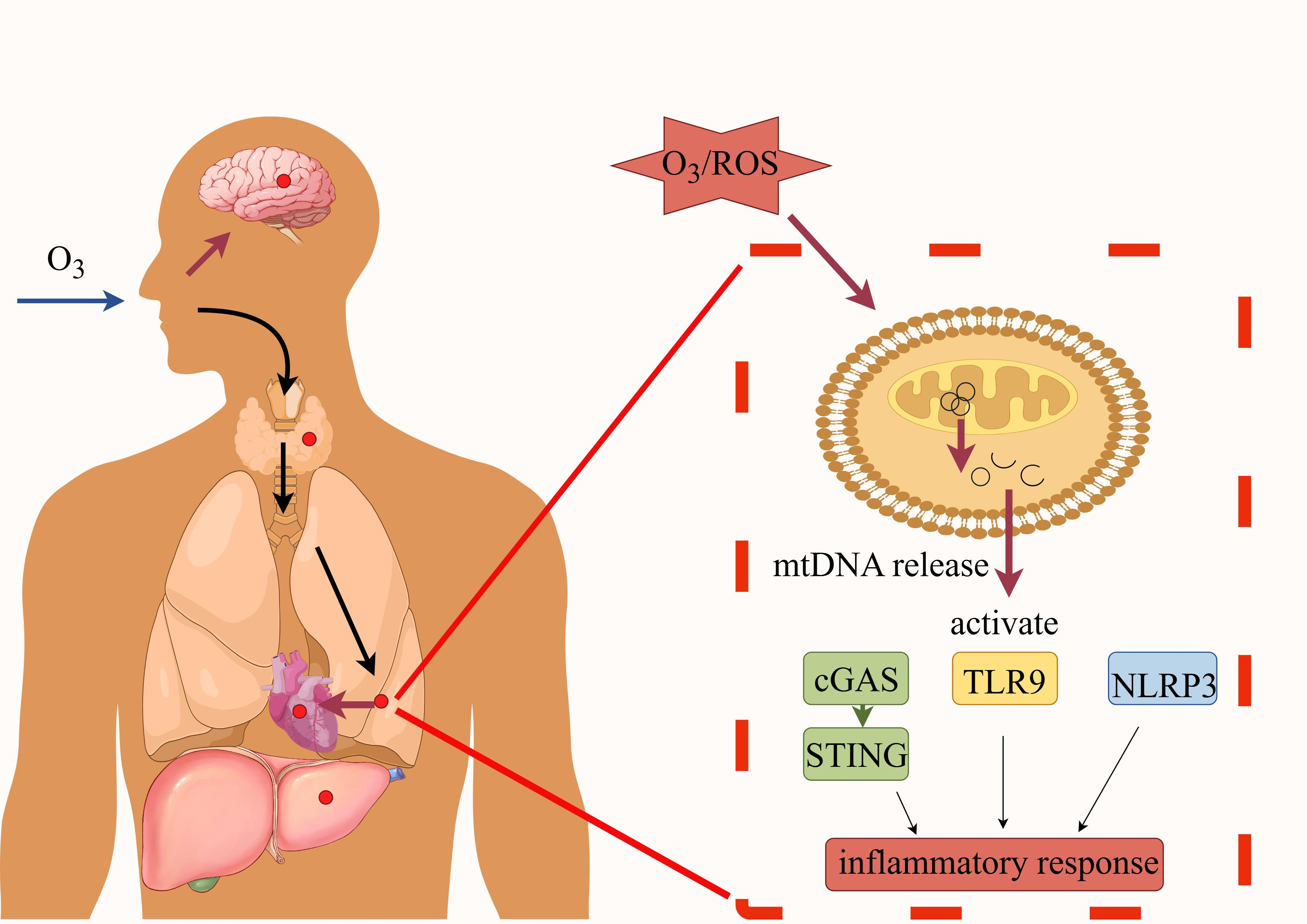
Advances in Elucidating the Mitochondrial DNA Mechanisms Underlying Ozone-Induced Inflammation

Highlights
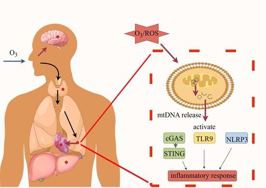
- Ozone exposure can induce the release of mitochondrial DNA (mtDNA) from mitochondria.
- The role of mtDNA in ozone exposure was reviewed.
- The mechanisms of innate immune response mediated by mtDNA were summarized.
- The results highlight the critical inductive role of mitochondrial DNA (mtDNA) in ozone exposure.
- They emphasize the urgent need for further experiments to elucidate the specific pathways activated downstream of mtDNA under ozone exposure conditions.
Abstract
1. Introduction
2. The Relationship Between Ozone Exposure and Mitochondrial Function
2.1. Health Effects of Ozone
2.2. The Effect of Ozone Exposure on Mitochondrial Function
3. Structure and Function of mtDNA
3.1. Structure and Function of Mitochondria
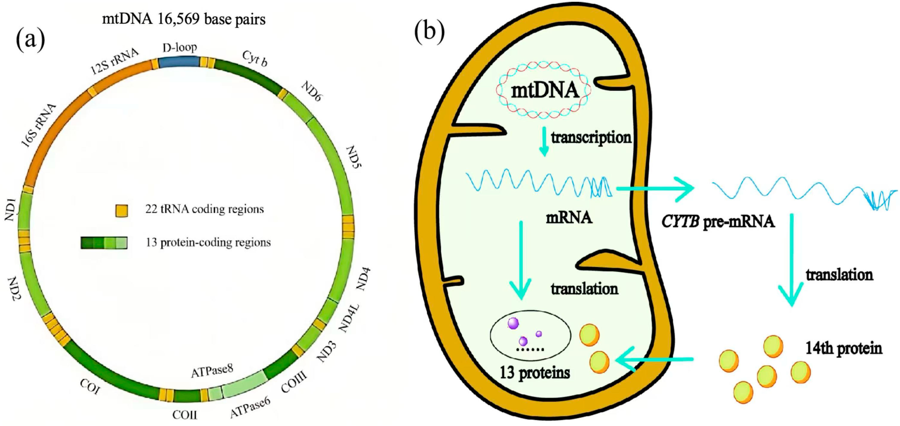
3.2. Structure of mtDNA
3.3. Functional Characteristics of Intracellular mtDNA
3.3.1. Copy Number and Abundance of mtDNA
3.3.2. mtDNA and Mitochondria Heteroplasmy
3.3.3. mtDNA and Mitochondrial Dynamics
4. Role of mtDNA in Ozone Exposure
4.1. Mechanisms of mtDNA Damage in Ozone Exposure
4.2. Source of mtDNA
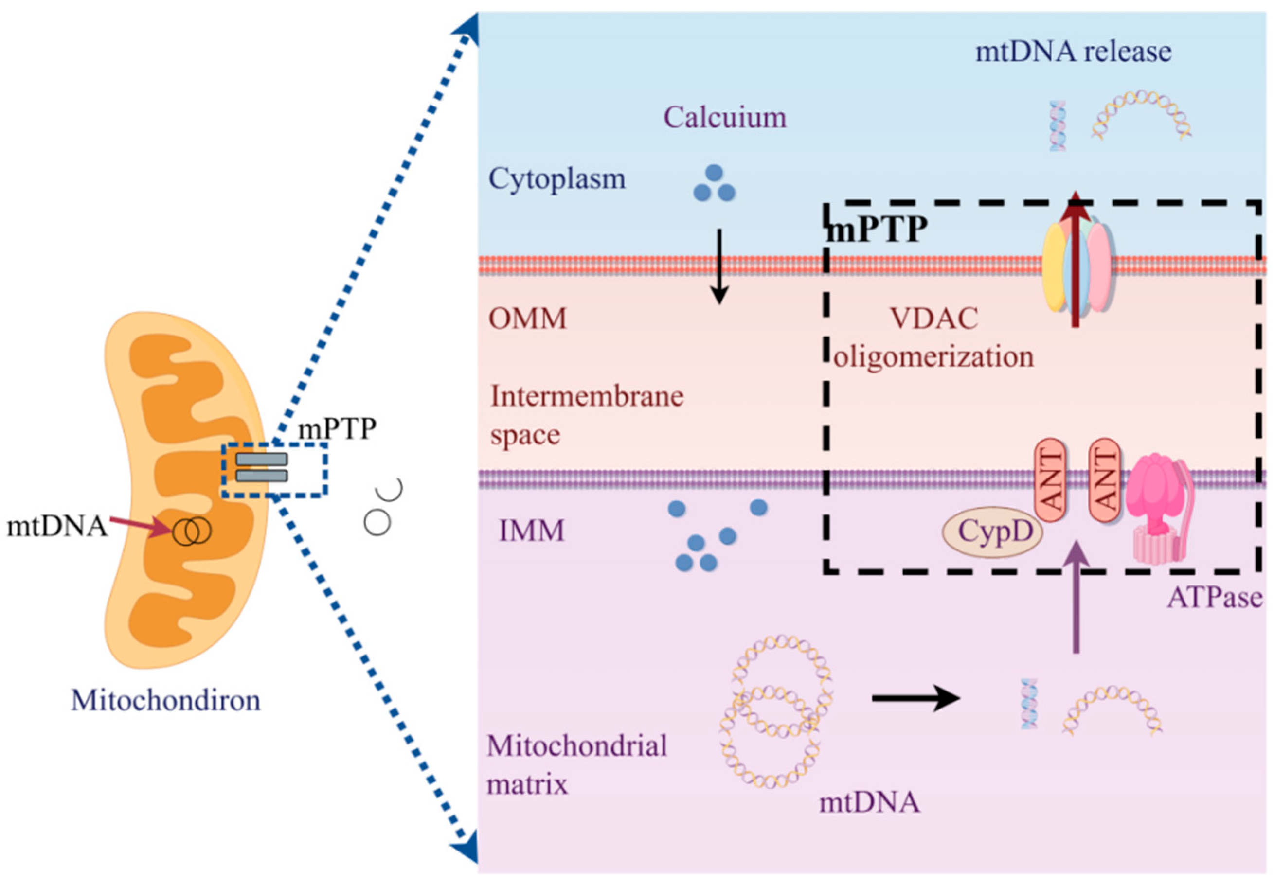
4.2.1. mtDNA Release Mediated by mPTP
4.2.2. mtDNA Release Mediated by BAK/BAX
4.2.3. mtDNA Release Mediated by Abnormal Mitochondrial Dynamics
4.2.4. mtDNA Release Mediated by Mitochondrial-Derived Vesicles (MDVs)
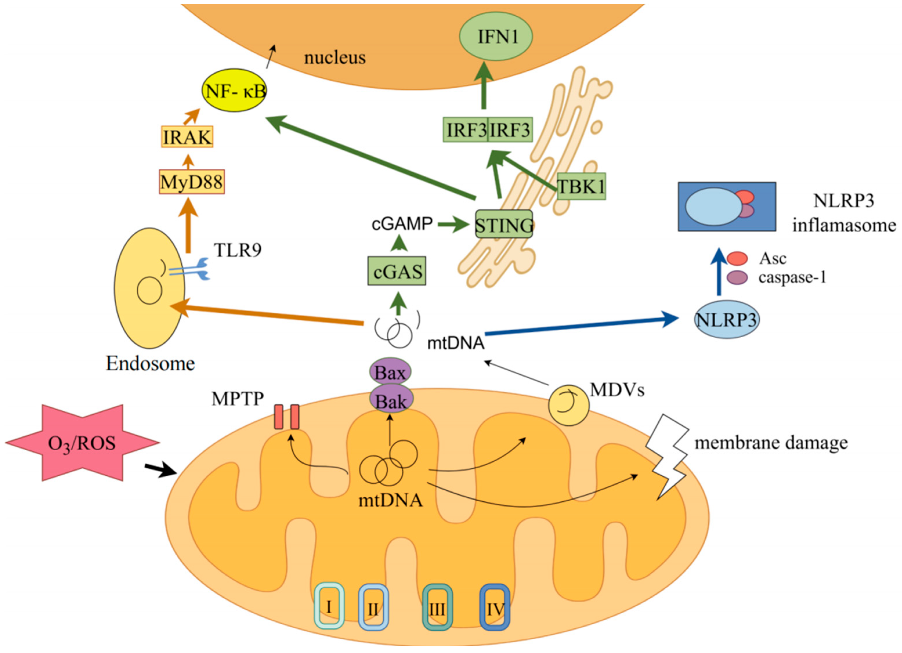
5. Released mtDNA Induces Inflammatory Response
5.1. cGAS-STING Pathway
5.2. TLR9/4-MyD88-NF-κB Pathway
5.3. NLRP3-ASC-Caspase1 Pathway
5.4. The Regulatory Relationship Among Ozone Exposure, mtDNA, and Inflammatory Response
6. Conclusions
Author Contributions
Funding
Institutional Review Board Statement
Informed Consent Statement
Data Availability Statement
Conflicts of Interest
References
- Global burden of 87 risk factors in 204 countries and territories, 1990–2019: A systematic analysis for the Global Burden of Disease Study 2019. Lancet 2020, 396, 1223–1249. [CrossRef]
- GBD 2021 Risk Factors Collaborators. Global burden and strength of evidence for 88 risk factors in 204 countries and 811 subnational locations, 1990–2021: A systematic analysis for the Global Burden of Disease Study 2021. Lancet 2024, 403, 2162–2203. [Google Scholar] [CrossRef]
- WHO. Guidelines Approved by the Guidelines Review Committee. In Who Global Air Quality Guidelines: Particulate Matter (PM2.5 and PM10), Ozone, Nitrogen Dioxide, Sulfur Dioxide and Carbon Monoxide; World Health Organization: Geneva, Switzerland, 2021. [Google Scholar]
- Zhang, J.; Wei, Y.; Fang, Z. Ozone Pollution: A Major Health Hazard Worldwide. Front. Immunol. 2019, 10, 2518. [Google Scholar] [CrossRef]
- Sicard, P.; Agathokleous, E.; Anenberg, S.C.; De Marco, A.; Paoletti, E.; Calatayud, V. Trends in urban air pollution over the last two decades: A global perspective. Sci. Total Environ. 2023, 858, 160064. [Google Scholar] [CrossRef]
- Wu, T.; Li, Z.; Wei, Y. Advances in understanding mechanisms underlying mitochondrial structure and function damage by ozone. Sci. Total Environ. 2023, 861, 160589. [Google Scholar] [CrossRef]
- Li, J.; Song, H.; Luo, T.; Cao, Y.; Zhang, L.; Zhao, Q.; Li, Z.; Hu, X.; Gu, J.; Tian, S. Exposure to O3 and NO2 on the interfacial chemistry of the pulmonary surfactant and the mechanism of lung oxidative damage. Chemosphere 2024, 362, 142669. [Google Scholar] [CrossRef] [PubMed]
- Zhu, L.; Fang, J.; Yao, Y.; Yang, Z.; Wu, J.; Ma, Z.; Liu, R.; Zhan, Y.; Ding, Z.; Zhang, Y. Long-term ambient ozone exposure and incident cardiovascular diseases: National cohort evidence in China. J. Hazard. Mater. 2024, 471, 134158. [Google Scholar] [CrossRef]
- Enweasor, C.; Flayer, C.H.; Haczku, A. Ozone-Induced Oxidative Stress, Neutrophilic Airway Inflammation, and Glucocorticoid Resistance in Asthma. Front. Immunol. 2021, 12, 631092. [Google Scholar] [CrossRef]
- Koch, R.E.; Josefson, C.C.; Hill, G.E. Mitochondrial function, ornamentation, and immunocompetence. Biol. Rev. 2016, 92, 1459–1474. [Google Scholar] [CrossRef] [PubMed]
- Gallo, G.; Rubattu, S.; Volpe, M. Mitochondrial Dysfunction in Heart Failure: From Pathophysiological Mechanisms to Therapeutic Opportunities. Int. J. Mol. Sci. 2024, 25, 2667. [Google Scholar] [CrossRef] [PubMed]
- Qin, X.; Li, H.; Zhao, H.; Fang, L.; Wang, X. Enhancing healthy aging with small molecules: A mitochondrial perspective. Med. Res. Rev. 2024, 44, 1904–1922. [Google Scholar] [CrossRef]
- Hu, Z.; Yang, L.; Zhang, M.; Tang, H.; Huang, Y.; Su, Y.; Ding, Y.; Li, C.; Wang, M.; Zhou, Y.; et al. A novel protein CYTB-187AA encoded by the mitochondrial gene CYTB modulates mammalian early development. Cell Metab. 2024, 36, 1586–1597.e7. [Google Scholar] [CrossRef] [PubMed]
- Xu, Y.; Chu, C.; Shi, Z.; Zhang, J. The role of hepatocyte mitochondrial DNA in liver injury. Biomed. Pharmacother. 2023, 168, 115692. [Google Scholar] [CrossRef]
- Tian, L.; Li, N.; Li, K.; Tan, Y.; Han, J.; Lin, B.; Lai, W.; Liu, H.; Shi, Y.; Xi, Z.; et al. Ambient ozone exposure induces ROS related-mitophagy and pyroptosis via NLRP3 inflammasome activation in rat lung cells. Ecotoxicol. Environ. Saf. 2022, 240, 113663. [Google Scholar] [CrossRef]
- Lenin, R.R.; Koh, Y.H.; Zhang, Z.; Yeo, Y.Z.; Parikh, B.H.; Seah, I.; Wong, W.; Su, X. Dysfunctional Autophagy, Proteostasis, and Mitochondria as a Prelude to Age-Related Macular Degeneration. Int. J. Mol. Sci. 2023, 24, 8763. [Google Scholar] [CrossRef]
- Garg, M.; Johri, S.; Chakraborty, K. Immunomodulatory role of mitochondrial DAMPs: A missing link in pathology? FEBS J. 2022, 290, 4395–4418. [Google Scholar] [CrossRef] [PubMed]
- Gando, S.; Levi, M.; Toh, C.-H. Trauma-induced innate immune activation and disseminated intravascular coagulation. J. Thromb. Haemost. 2024, 22, 337–351. [Google Scholar] [CrossRef]
- Coppotelli, G.; Ross, J. Mitochondria in Ageing and Diseases: The Super Trouper of the Cell. Int. J. Mol. Sci. 2016, 17, 711. [Google Scholar] [CrossRef] [PubMed]
- Fan, K.; Dong, N.; Fang, M.; Xiang, Z.; Zheng, L.; Wang, M.; Shi, Y.; Tan, G.; Li, C.; Xue, Y. Ozone exposure affects corneal epithelial fate by promoting mtDNA leakage and cGAS/STING activation. J. Hazard. Mater. 2024, 465, 133219. [Google Scholar] [CrossRef] [PubMed]
- Yu, S.; Zhang, M.; Zhu, J.; Yang, X.; Bigambo, F.M.; Snijders, A.M.; Wang, X.; Hu, W.; Lv, W.; Xia, Y. The effect of ambient ozone exposure on three types of diabetes: A meta-analysis. Environ. Health 2023, 22, 32. [Google Scholar] [CrossRef]
- Zhu, L.; Yuan, Y.; Mayvaneh, F.; Sun, H.; Zhang, Y.; Hu, C. Maternal ozone exposure lowers infant’s birthweight: A nationwide cohort of over 4 million livebirths in Iran. Ecotoxicol. Environ. Saf. 2024, 283, 116840. [Google Scholar] [CrossRef]
- Du, X.; Niu, Y.; Wang, C.; Wang, W.; Liu, C.; Meng, X.; Chu, C.; Chen, R.; Kan, H. Ozone exposure and blood transcriptome: A randomized, controlled, crossover trial among healthy adults. Environ. Int. 2022, 163, 107242. [Google Scholar] [CrossRef]
- Tovar, A.; Crouse, W.L.; Smith, G.J.; Thomas, J.M.; Keith, B.P.; McFadden, K.M.; Moran, T.P.; Furey, T.S.; Kelada, S.N.P. Integrative analysis reveals mouse strain-dependent responses to acute ozone exposure associated with airway macrophage transcriptional activity. Am. J. Physiol. Lung Cell. Mol. Physiol. 2022, 322, L33–L49. [Google Scholar]
- Singh, S.A.; Suresh, S.; Vellapandian, C. Ozone-induced neurotoxicity: In vitro and in vivo evidence. Ageing Res. Rev. 2023, 91, 102045. [Google Scholar] [CrossRef]
- Snow, S.J.; Henriquez, A.R.; Fisher, A.; Vallanat, B.; House, J.S.; Schladweiler, M.C.; Wood, C.E.; Kodavanti, U.P. Peripheral metabolic effects of ozone exposure in healthy and diabetic rats on normal or high-cholesterol diet. Toxicol. Appl. Pharmacol. 2021, 415, 115427. [Google Scholar] [CrossRef]
- Hülpüsch, C.; Rohayem, R.; Reiger, M.; Traidl-Hoffmann, C. Exploring the skin microbiome in atopic dermatitis pathogenesis and disease modification. J. Allergy Clin. Immunol. 2024, 154, 31–41. [Google Scholar] [CrossRef]
- Fuks, K.B.; Hüls, A.; Sugiri, D.; Altug, H.; Vierkötter, A.; Abramson, M.J.; Goebel, J.; Wagner, G.G.; Demuth, I.; Krutmann, J.; et al. Tropospheric ozone and skin aging: Results from two German cohort studies. Environ. Int. 2019, 124, 139–144. [Google Scholar] [CrossRef]
- Zannoni, N.; Lakey, P.S.J.; Won, Y.; Shiraiwa, M.; Rim, D.; Weschler, C.J.; Wang, N.; Ernle, L.; Li, M.; Bekö, G.; et al. The human oxidation field. Science 2022, 377, 1071–1077. [Google Scholar] [CrossRef] [PubMed]
- Que, L.G.; Stiles, J.V.; Sundy, J.S.; Foster, W.M. Pulmonary function, bronchial reactivity, and epithelial permeability are response phenotypes to ozone and develop differentially in healthy humans. J. Appl. Physiol. 2011, 111, 679–687. [Google Scholar] [CrossRef] [PubMed]
- Chen, C.; Li, T.; Sun, Q.; Shi, W.; He, M.Z.; Wang, J.; Liu, J.; Zhang, M.; Jiang, Q.; Wang, M.; et al. Short-term exposure to ozone and cause-specific mortality risks and thresholds in China: Evidence from nationally representative data, 2013–2018. Environ. Int. 2023, 171, 107666. [Google Scholar] [CrossRef] [PubMed]
- Ruan, Y.; Wang, Y.; Guo, J.; Man, T.; Hao, X.; Zhou, X.; Wang, C.; Deng, H.; Li, J.; Zou, Z.; et al. Maternal exposure to ambient ozone and fetal conotruncal heart defects in China: A multicenter cohort study. Ecotoxicol. Environ. Saf. 2024, 276, 116328. [Google Scholar] [CrossRef] [PubMed]
- Garcia, M.; Salazar, R.; Wilson, T.; Lucas, S.; Herbert, G.; Young, T.; Begay, J.; Denson, J.L.; Zychowski, K.; Ashley, R.; et al. Early Gestational Exposure to Inhaled Ozone Impairs Maternal Uterine Artery and Cardiac Function. Toxicol. Sci. 2021, 179, 121–134. [Google Scholar]
- Int Panis, L.; Provost, E.B.; Cox, B.; Louwies, T.; Laeremans, M.; Standaert, A.; Dons, E.; Holmstock, L.; Nawrot, T.; De Boever, P. Short-term air pollution exposure decreases lung function: A repeated measures study in healthy adults. Environ. Health 2017, 16, 60. [Google Scholar] [CrossRef]
- Wang, C.; Lin, J.; Niu, Y.; Wang, W.; Wen, J.; Lv, L.; Liu, C.; Du, X.; Zhang, Q.; Chen, B.; et al. Impact of ozone exposure on heart rate variability and stress hormones: A randomized-crossover study. J. Hazard. Mater. 2022, 421, 126750. [Google Scholar] [CrossRef]
- Hiraiwa, K.; van Eeden, S.F. Contribution of Lung Macrophages to the Inflammatory Responses Induced by Exposure to Air Pollutants. Mediat. Inflamm. 2013, 2013, 619523. [Google Scholar] [CrossRef]
- Xu, Z.; Wang, W.; Liu, Q.; Li, Z.; Lei, L.; Ren, L.; Deng, F.; Guo, X.; Wu, S. Association between gaseous air pollutants and biomarkers of systemic inflammation: A systematic review and meta-analysis. Environ. Pollut. 2022, 292, 118336. [Google Scholar] [CrossRef] [PubMed]
- Li, F.; Xu, M.; Wang, M.; Wang, L.; Wang, H.; Zhang, H.; Chen, Y.; Gong, J.; Zhang, J.; Adcock, I.M.; et al. Roles of mitochondrial ROS and NLRP3 inflammasome in multiple ozone-induced lung inflammation and emphysema. Respir. Res. 2018, 19, 230. [Google Scholar] [CrossRef]
- Liu, C.; Xiang, Y.; Liu, H.-j.; Gao, G.; Howard, S.T.; Zhu, X.-l.; Qin, X.-q. Involvement of integrin β4 in ozone stress-induced airway hyperresponsiveness. Biochem. Biophys. Res. Commun. 2010, 397, 290–295. [Google Scholar] [CrossRef]
- Michaudel, C.; Fauconnier, L.; Julé, Y.; Ryffel, B. Functional and morphological differences of the lung upon acute and chronic ozone exposure in mice. Sci. Rep. 2018, 8, 10611. [Google Scholar] [CrossRef]
- Nan, N.; Liu, Y.; Yan, Z.; Zhang, Y.; Li, S.; Zhang, J.; Qin, G.; Sang, N. Ozone induced multigenerational glucose and lipid metabolism disorders in Drosophila. Sci. Total Environ. 2024, 951, 175477. [Google Scholar] [CrossRef] [PubMed]
- Wiegman, C.H.; Michaeloudes, C.; Haji, G.; Narang, P.; Clarke, C.J.; Russell, K.E.; Bao, W.; Pavlidis, S.; Barnes, P.J.; Kanerva, J.; et al. Oxidative stress–induced mitochondrial dysfunction drives inflammation and airway smooth muscle remodeling in patients with chronic obstructive pulmonary disease. J. Allergy Clin. Immunol. 2015, 136, 769–780. [Google Scholar] [CrossRef]
- Tripathi, A.; Bartosh, A.; Whitehead, C.; Pillai, A. Activation of cell-free mtDNA-TLR9 signaling mediates chronic stress-induced social behavior deficits. Mol. Psychiatry 2023, 28, 4198–4211. [Google Scholar] [CrossRef]
- Li, Z.; Su, Q.; Xu, R.; Peng, J.; Zhu, X.; Wei, Y. Influence of different concentrations of ozone on the alteration of mitochondrial DNA copy numbers in human peripheral blood. Sci. Total Environ. 2023, 873, 162282. [Google Scholar] [CrossRef]
- Herrera, A.S.; Del, C.A.E.M.; Md Ashraf, G.; Zamyatnin, A.A.; Aliev, G. Beyond mitochondria, what would be the energy source of the cell? Cent. Nerv. Syst. Agents Med. Chem. 2015, 15, 32–41. [Google Scholar] [CrossRef] [PubMed]
- Mannella, C.A. Structure and dynamics of the mitochondrial inner membrane cristae. Biochim. Biophys. Acta 2006, 1763, 542–548. [Google Scholar] [CrossRef]
- Picard, M.; Shirihai, O.S. Mitochondrial signal transduction in cellular homeostasis. Cell Metab. 2022, 34, 1620–1653. [Google Scholar] [CrossRef]
- Sharma, P.; Sampath, H. Mitochondrial DNA Integrity: Role in Health and Disease. Cells 2019, 8, 100. [Google Scholar] [CrossRef] [PubMed]
- Perkins, G.A.; Renken, C.; Martone, M.; Young, S.; Ellisman, M.; Frey, T. Electron tomography of neuronal mitochondria: Three-dimensional structure and organization of cristae and membrane contacts. J. Struct. Biol. 1997, 119, 260–272. [Google Scholar] [CrossRef]
- Brand, M.D.; Nicholls, D.G. Assessing mitochondrial dysfunction in cells. Biochem. J. 2011, 435, 297–312. [Google Scholar] [CrossRef]
- Xia, Y.; Niu, Y.; Cai, J.; Liu, C.; Meng, X.; Chen, R.; Kan, H. Acute effects of personal ozone exposure on biomarkers of inflammation, oxidative stress, and mitochondrial oxidative damage. China CDC Wkly. 2021, 3, 987–992. [Google Scholar] [CrossRef] [PubMed]
- Anderson, S.; Bankier, A.T.; Barrell, B.G.; de Bruijn, M.H.; Coulson, A.R.; Drouin, J.; Eperon, I.C.; Nierlich, D.P.; Roe, B.A.; Sanger, F.; et al. Sequence and organization of the human mitochondrial genome. Nature 1981, 290, 457–465. [Google Scholar] [CrossRef] [PubMed]
- Kasamatsu, H.; Vinograd, J. Replication of circular DNA in eukaryotic cells. Annu. Rev. Biochem. 1974, 43, 695–719. [Google Scholar] [CrossRef]
- Fisher, R.P.; Clayton, D.A. Purification and characterization of human mitochondrial transcription factor 1. Mol. Cell. Biol. 1988, 8, 3496–3509. [Google Scholar]
- Falkenberg, M.; Larsson, N.G.; Gustafsson, C.M. Replication and Transcription of Human Mitochondrial DNA. Annu. Rev. Biochem. 2024, 93, 47–77. [Google Scholar] [CrossRef]
- Mercer, T.R.; Neph, S.; Dinger, M.E.; Crawford, J.; Smith, M.A.; Shearwood, A.-M.J.; Haugen, E.; Bracken, C.P.; Rackham, O.; Stamatoyannopoulos, J.A.; et al. The Human Mitochondrial Transcriptome. Cell 2011, 146, 645–658. [Google Scholar] [CrossRef]
- Garcia, I.; Jones, E.; Ramos, M.; Innis-Whitehouse, W.; Gilkerson, R. The little big genome: The organization of mitochondrial DNA. Front. Biosci. 2017, 22, 710–721. [Google Scholar] [CrossRef]
- Dong, Y.; Yoshitomi, T.; Hu, J.-F.; Cui, J. Long noncoding RNAs coordinate functions between mitochondria and the nucleus. Epigenetics Chromatin 2017, 10, 41. [Google Scholar] [CrossRef]
- Lobos-González, L.; Bustos, R.; Campos, A.; Silva, V.; Silva, V.; Jeldes, E.; Salomon, C.; Varas-Godoy, M.; Cáceres-Verschae, A.; Duran, E.; et al. Exosomes released upon mitochondrial ASncmtRNA knockdown reduce tumorigenic properties of malignant breast cancer cells. Sci. Rep. 2020, 10, 343. [Google Scholar] [CrossRef]
- Fitzpatrick, C.; Bendek, M.F.; Briones, M.; Farfán, N.; Silva, V.A.; Nardocci, G.; Montecino, M.; Boland, A.; Deleuze, J.-F.; Villegas, J.; et al. Mitochondrial ncRNA targeting induces cell cycle arrest and tumor growth inhibition of MDA-MB-231 breast cancer cells through reduction of key cell cycle progression factors. Cell Death Dis. 2019, 10, 423. [Google Scholar] [CrossRef] [PubMed]
- Barbalata, T.; Niculescu, L.S.; Stancu, C.S.; Pinet, F.; Sima, A.V. Elevated Levels of Circulating lncRNAs LIPCAR and MALAT1 Predict an Unfavorable Outcome in Acute Coronary Syndrome Patients. Int. J. Mol. Sci. 2023, 24, 12076. [Google Scholar] [CrossRef] [PubMed]
- Zhang, X.; Yuan, S.; Liu, J.; Tang, Y.; Wang, Y.; Zhan, J.; Fan, J.; Nie, X.; Zhao, Y.; Wen, Z.; et al. Overexpression of cytosolic long noncoding RNA cytb protects against pressure-overload-induced heart failure via sponging microRNA-103-3p. Mol. Ther. Nucleic Acids 2022, 27, 1127–1145. [Google Scholar] [CrossRef]
- Fernández-Vizarra, E.; Enríquez, J.A.; Pérez-Martos, A.; Montoya, J.; Fernández-Silva, P. Tissue-specific differences in mitochondrial activity and biogenesis. Mitochondrion 2011, 11, 207–213. [Google Scholar] [CrossRef] [PubMed]
- Naue, J.; Xavier, C.; Hörer, S.; Parson, W.; Lutz-Bonengel, S. Assessment of mitochondrial DNA copy number variation relative to nuclear DNA quantity between different tissues. Mitochondrion 2024, 74, 101823. [Google Scholar]
- Duran, H.E.; Simsek-Duran, F.; Oehninger, S.C.; Jones, H.W.; Castora, F.J. The association of reproductive senescence with mitochondrial quantity, function, and DNA integrity in human oocytes at different stages of maturation. Fertil. Steril. 2011, 96, 384–388. [Google Scholar] [CrossRef]
- Igoillo-Esteve, M.; Oliveira, A.F.; Cosentino, C.; Fantuzzi, F.; Demarez, C.; Toivonen, S.; Hu, A.; Chintawar, S.; Lopes, M.; Pachera, N.; et al. Exenatide induces frataxin expression and improves mitochondrial function in Friedreich ataxia. JCI Insight 2020, 5, 134221. [Google Scholar] [CrossRef]
- Gupta, R.; Kanai, M.; Durham, T.J.; Tsuo, K.; McCoy, J.G.; Kotrys, A.V.; Zhou, W.; Chinnery, P.F.; Karczewski, K.J.; Calvo, S.E.; et al. Nuclear genetic control of mtDNA copy number and heteroplasmy in humans. Nature 2023, 620, 839–848. [Google Scholar] [CrossRef] [PubMed]
- Tan, B.G.; Gustafsson, C.M.; Falkenberg, M. Mechanisms and regulation of human mitochondrial transcription. Nat. Rev. Mol. Cell Biol. 2023, 25, 119–132. [Google Scholar] [CrossRef] [PubMed]
- Enríquez, J.A.; Fernández-Silva, P.; Pérez-Martos, A.; López-Pérez, M.J.; Montoya, J. The Synthesis of mRNA in Isolated Mitochondria can be Maintained for Several Hours and is Inhibited by High Levels of ATP. Eur. J. Biochem. 2004, 237, 601–610. [Google Scholar] [CrossRef]
- Wiese, M.; Bannister, A.J. Two genomes, one cell: Mitochondrial-nuclear coordination via epigenetic pathways. Mol. Metab. 2020, 38, 100942. [Google Scholar] [CrossRef]
- Guarnieri, J.W.; Haltom, J.A.; Albrecht, Y.E.S.; Lie, T.; Olali, A.Z.; Widjaja, G.A.; Ranshing, S.S.; Angelin, A.; Murdock, D.; Wallace, D.C. SARS-CoV-2 mitochondrial metabolic and epigenomic reprogramming in COVID-19. Pharmacol. Res. 2024, 204, 107170. [Google Scholar]
- Ding, X.; Fang, T.; Pang, X.; Pan, X.; Tong, A.; Lin, Z.; Zheng, S.; Zheng, N. Mitochondrial DNA abnormalities and metabolic syndrome. Front. Cell Dev. Biol. 2023, 11, 1153174. [Google Scholar] [CrossRef]
- Malik, A.N.; Czajka, A. Is mitochondrial DNA content a potential biomarker of mitochondrial dysfunction? Mitochondrion 2013, 13, 481–492. [Google Scholar] [CrossRef]
- Venegas, V.; Wang, J.; Dimmock, D.; Wong, L.J. Real-Time Quantitative PCR Analysis of Mitochondrial DNA Content. Curr. Protoc. Hum. Genet. 2011, 68, 19.7.1–19.7.12. [Google Scholar] [CrossRef]
- Qin, P.; Qin, T.; Liang, L.; Li, X.; Jiang, B.; Wang, X.; Ma, J.; Hu, F.; Zhang, M.; Hu, D. The role of mitochondrial DNA copy number in cardiometabolic disease: A bidirectional two-sample mendelian randomization study. Cardiovasc. Diabetol. 2024, 23, 45. [Google Scholar] [CrossRef]
- Cerantonio, A.; Citrigno, L.; Greco, B.M.; De Benedittis, S.; Passarino, G.; Maletta, R.; Qualtieri, A.; Montesanto, A.; Spadafora, P.; Cavalcanti, F. The Role of Mitochondrial Copy Number in Neurodegenerative Diseases: Present Insights and Future Directions. Int. J. Mol. Sci. 2024, 25, 6062. [Google Scholar] [CrossRef]
- Low, H.C.; Chilian, W.M.; Ratnam, W.; Karupaiah, T.; Md Noh, M.F.; Mansor, F.; Ng, Z.X.; Pung, Y.F. Changes in Mitochondrial Epigenome in Type 2 Diabetes Mellitus. Br. J. Biomed. Sci. 2023, 80, 10884. [Google Scholar] [CrossRef] [PubMed]
- Chatterjee, D.; Das, P.; Chakrabarti, O. Mitochondrial Epigenetics Regulating Inflammation in Cancer and Aging. Front. Cell Dev. Biol. 2022, 10, 929708. [Google Scholar] [CrossRef] [PubMed]
- Borah, S.; Mishra, R.; Dey, S.; Suchanti, S.; Bhowmick, N.A.; Giri, B.; Haldar, S. Prognostic value of circulating mitochondrial DNA in prostate cancer and underlying mechanism. Mitochondrion 2023, 71, 40–49. [Google Scholar] [CrossRef]
- Cheng, Q.; Liu, Q.Q.; Lu, C. A state-of-the-science review of using mitochondrial DNA copy number as a biomarker for environmental exposure. Environ. Pollut. 2024, 346, 123642. [Google Scholar] [CrossRef]
- Árnadóttir, E.R.; Moore, K.H.S.; Guðmundsdóttir, V.B.; Ebenesersdóttir, S.S.; Guity, K.; Jónsson, H.; Stefánsson, K.; Helgason, A. The rate and nature of mitochondrial DNA mutations in human pedigrees. Cell 2024, 187, 3904–3918.e8. [Google Scholar] [CrossRef]
- Pereira, C.V.; Gitschlag, B.L.; Patel, M.R. Cellular mechanisms of mtDNA heteroplasmy dynamics. Crit. Rev. Biochem. Mol. Biol. 2021, 56, 510–525. [Google Scholar] [CrossRef] [PubMed]
- Zhou, Y.; Jin, Y.; Wu, T.; Wang, Y.; Dong, Y.; Chen, P.; Hu, C.; Pan, N.; Ye, C.; Shen, L.; et al. New insights on mitochondrial heteroplasmy observed in ovarian diseases. J. Adv. Res. 2023, 65, 211–226. [Google Scholar] [CrossRef]
- Otten, A.B.C.; Sallevelt, S.C.E.H.; Carling, P.J.; Dreesen, J.C.F.M.; Drüsedau, M.; Spierts, S.; Paulussen, A.D.C.; de Die-Smulders, C.E.M.; Herbert, M.; Chinnery, P.F.; et al. Mutation-specific effects in germline transmission of pathogenic mtDNA variants. Hum. Reprod. 2018, 33, 1331–1341. [Google Scholar] [CrossRef]
- Wilhelm, L.P.; Ganley, I.G. Mitochondria and peroxisomes: Partners in autophagy. Autophagy 2022, 19, 2162–2163. [Google Scholar] [CrossRef] [PubMed]
- Reyes, A.; Gissi, C.; Pesole, G.; Saccone, C. Asymmetrical directional mutation pressure in the mitochondrial genome of mammals. Mol. Biol. Evol. 1998, 15, 957–966. [Google Scholar] [CrossRef] [PubMed]
- Song, N.; Mei, S.; Wang, X.; Hu, G.; Lu, M. Focusing on mitochondria in the brain: From biology to therapeutics. Transl. Neurodegener. 2024, 13, 23. [Google Scholar] [CrossRef]
- Song, S.; Pursell, Z.F.; Copeland, W.C.; Longley, M.J.; Kunkel, T.A.; Mathews, C.K. DNA precursor asymmetries in mammalian tissue mitochondria and possible contribution to mutagenesis through reduced replication fidelity. Proc. Natl. Acad. Sci. USA 2005, 102, 4990–4995. [Google Scholar] [CrossRef]
- Roussou, R.; Metzler, D.; Padovani, F.; Thoma, F.; Schwarz, R.; Shraiman, B.; Schmoller, K.M.; Osman, C. Real-time assessment of mitochondrial DNA heteroplasmy dynamics at the single-cell level. EMBO J. 2024, 43, 5340–5359. [Google Scholar] [CrossRef]
- Kozal, J.S.; Jayasundara, N.; Massarsky, A.; Lindberg, C.D.; Oliveri, A.N.; Cooper, E.M.; Levin, E.D.; Meyer, J.N.; Giulio, R.T.D. Mitochondrial dysfunction and oxidative stress contribute to cross-generational toxicity of benzo(a)pyrene in Danio rerio. Aquat. Toxicol. 2023, 263, 106658. [Google Scholar] [CrossRef]
- Hägg, S.; Jylhävä, J.; Wang, Y.; Czene, K.; Grassmann, F. Deciphering the genetic and epidemiological landscape of mitochondrial DNA abundance. Hum. Genet. 2020, 140, 849–861. [Google Scholar] [CrossRef]
- Lareau, C.A.; Ludwig, L.S.; Muus, C.; Gohil, S.H.; Zhao, T.; Chiang, Z.; Pelka, K.; Verboon, J.M.; Luo, W.; Christian, E.; et al. Massively parallel single-cell mitochondrial DNA genotyping and chromatin profiling. Nat. Biotechnol. 2020, 39, 451–461. [Google Scholar] [CrossRef]
- Picard, M.; McManus, M.J.; Gray, J.D.; Nasca, C.; Moffat, C.; Kopinski, P.K.; Seifert, E.L.; McEwen, B.S.; Wallace, D.C. Mitochondrial functions modulate neuroendocrine, metabolic, inflammatory, and transcriptional responses to acute psychological stress. Proc. Natl. Acad. Sci. USA 2015, 112, E6614–E6623. [Google Scholar] [CrossRef]
- Bazzani, V.; Equisoain Redin, M.; McHale, J.; Perrone, L.; Vascotto, C. Mitochondrial DNA Repair in Neurodegenerative Diseases and Ageing. Int. J. Mol. Sci. 2022, 23, 11391. [Google Scholar] [CrossRef]
- Caielli, S.; Athale, S.; Domic, B.; Murat, E.; Chandra, M.; Banchereau, R.; Baisch, J.; Phelps, K.; Clayton, S.; Gong, M.; et al. Oxidized mitochondrial nucleoids released by neutrophils drive type I interferon production in human lupus. J. Exp. Med. 2016, 213, 697–713. [Google Scholar] [CrossRef] [PubMed]
- Zhang, Z.; Meng, P.; Han, Y.; Shen, C.; Li, B.; Hakim, M.A.; Zhang, X.; Lu, Q.; Rong, M.; Lai, R. Mitochondrial DNA-LL-37 Complex Promotes Atherosclerosis by Escaping from Autophagic Recognition. Immunity 2015, 43, 1137–1147. [Google Scholar] [CrossRef]
- Yu, Y.; Xu, L.; Qi, L.; Wang, C.; Xu, N.; Liu, S.; Li, S.; Tian, H.; Liu, W.; Xu, Y.; et al. ABT737 induces mitochondrial pathway apoptosis and mitophagy by regulating DRP1-dependent mitochondrial fission in human ovarian cancer cells. Biomed. Pharmacother. 2017, 96, 22–29. [Google Scholar] [CrossRef] [PubMed]
- Goglia, I.; Węglarz-Tomczak, E.; Gioia, C.; Liu, Y.; Virtuoso, A.; Bonanomi, M.; Gaglio, D.; Salmistraro, N.; De Luca, C.; Papa, M.; et al. Fusion–fission–mitophagy cycling and metabolic reprogramming coordinate nerve growth factor (NGF)-dependent neuronal differentiation. FEBS J. 2024, 291, 2811–2835. [Google Scholar] [CrossRef] [PubMed]
- Zhu, J.; Wang, Q.; Zheng, Z.; Ma, L.; Guo, J.; Shi, H.; Ying, R.; Gao, B.; Chen, S.; Yu, S.; et al. MiR-181a protects the heart against myocardial infarction by regulating mitochondrial fission via targeting programmed cell death protein 4. Sci. Rep. 2024, 14, 6638. [Google Scholar]
- Rangaraju, V.; Lewis, T.L., Jr.; Hirabayashi, Y.; Bergami, M.; Motori, E.; Cartoni, R.; Kwon, S.K.; Courchet, J. Pleiotropic Mitochondria: The Influence of Mitochondria on Neuronal Development and Disease. J. Neurosci. 2019, 39, 8200–8208. [Google Scholar] [CrossRef]
- Kim, M.; Gorelick, A.N.; Vàzquez-García, I.; Williams, M.J.; Salehi, S.; Shi, H.; Weiner, A.C.; Ceglia, N.; Funnell, T.; Park, T.; et al. Single-cell mtDNA dynamics in tumors is driven by coregulation of nuclear and mitochondrial genomes. Nat. Genet. 2024, 56, 889–899. [Google Scholar] [CrossRef]
- Bartho, L.A.; Fisher, J.J.; Cuffe, J.S.M.; Perkins, A.V. Mitochondrial transformations in the aging human placenta. Am. J. Physiol. -Endocrinol. Metab. 2020, 319, E981–E994. [Google Scholar] [CrossRef]
- Tabara, L.C.; Burr, S.P.; Frison, M.; Chowdhury, S.R.; Paupe, V.; Nie, Y.; Johnson, M.; Villar-Azpillaga, J.; Viegas, F.; Segawa, M.; et al. MTFP1 controls mitochondrial fusion to regulate inner membrane quality control and maintain mtDNA levels. Cell 2024, 187, 3619–3637.e27. [Google Scholar] [CrossRef]
- Chen, Z.; Wang, Z.H.; Zhang, G.; Bleck, C.K.E.; Chung, D.J.; Madison, G.P.; Lindberg, E.; Combs, C.; Balaban, R.S.; Xu, H. Mitochondrial DNA segregation and replication restrict the transmission of detrimental mutation. J. Cell Biol. 2020, 219, e201905160. [Google Scholar] [CrossRef]
- Banarase, T.A.; Sammeta, S.S.; Wankhede, N.L.; Mangrulkar, S.V.; Rahangdale, S.R.; Aglawe, M.M.; Taksande, B.G.; Upaganlawar, A.B.; Umekar, M.J.; Kale, M.B. Mitophagy regulation in aging and neurodegenerative disease. Biophys. Rev. 2023, 15, 239–255. [Google Scholar] [CrossRef]
- Bozi, L.H.M.; Bechara, L.R.G.; dos Santos, A.F.; Campos, J.C. Mitochondrial-derived vesicles: A new player in cardiac mitochondrial quality control. J. Physiol. 2016, 594, 6077–6078. [Google Scholar] [CrossRef]
- Meyer, J.N.; Leuthner, T.C.; Luz, A.L. Mitochondrial fusion, fission, and mitochondrial toxicity. Toxicology 2017, 391, 42–53. [Google Scholar] [CrossRef]
- Lin, J.; Epel, E. Stress and telomere shortening: Insights from cellular mechanisms. Ageing Res. Rev. 2022, 73, 101507. [Google Scholar] [CrossRef]
- Chatterjee, A.; Mambo, E.; Sidransky, D. Mitochondrial DNA mutations in human cancer. Oncogene 2006, 25, 4663–4674. [Google Scholar] [CrossRef] [PubMed]
- Neel, D.V.; Basu, H.; Gunner, G.; Bergstresser, M.D.; Giadone, R.M.; Chung, H.; Miao, R.; Chou, V.; Brody, E.; Jiang, X.; et al. Gasdermin-E mediates mitochondrial damage in axons and neurodegeneration. Neuron 2023, 111, 1222–1240.e9. [Google Scholar] [CrossRef] [PubMed]
- Jiang, Y.; Chen, J.; Guo, L.; Lan, Y.; Li, G.; Liu, Q.; Li, H.; Deng, F.; Guo, X.; Wu, S. Short-term effects of ambient gaseous air pollution on blood platelet mitochondrial DNA methylation and myocardial ischemia. Environ. Int. 2024, 185, 108533. [Google Scholar] [CrossRef] [PubMed]
- Li, R.; Chen, G.; Pan, M.; Hou, X.; Kang, N.; Chen, R.; Yuchi, Y.; Liao, W.; Liu, X.; Mao, Z.; et al. Adverse associations of long-term exposure to ambient ozone with molecular biomarkers of aging alleviated by residential greenness in rural Chinese adults. Environ. Int. 2022, 169, 107496. [Google Scholar] [CrossRef]
- Fu, J.; Leroy, P.; Tham, A.; Wong, H.; Tenney, R.; Chen, C.; Stiner, R.; Balmes, J.R.; Paquet, A.C.; Arjomandi, M. Inflammatory and Repair Pathways Induced in Human Bronchoalveolar Lavage Cells with Ozone Inhalation. PLoS ONE 2015, 10, e0127283. [Google Scholar]
- Starkov, A.A. The Role of Mitochondria in Reactive Oxygen Species Metabolism and Signaling. Ann. N. Y. Acad. Sci. 2008, 1147, 37–52. [Google Scholar] [CrossRef]
- Chao, M.-R.; Evans, M.D.; Hu, C.-W.; Ji, Y.; Møller, P.; Rossner, P.; Cooke, M.S. Biomarkers of nucleic acid oxidation—A summary state-of-the-art. Redox Biol. 2021, 42, 107872. [Google Scholar] [CrossRef]
- Maga, G.; Villani, G.; Crespan, E.; Wimmer, U.; Ferrari, E.; Bertocci, B.; Hübscher, U. 8-oxo-guanine bypass by human DNA polymerases in the presence of auxiliary proteins. Nature 2007, 447, 606–608. [Google Scholar] [CrossRef] [PubMed]
- Barker, G.F.; Manzo, N.D.; Cotich, K.L.; Shone, R.K.; Waxman, A.B. DNA Damage Induced by Hyperoxia. Am. J. Respir. Cell Mol. Biol. 2006, 35, 277–288. [Google Scholar] [CrossRef][Green Version]
- Hori, A.; Yoshida, M.; Shibata, T.; Ling, F. Reactive oxygen species regulate DNA copy number in isolated yeast mitochondria by triggering recombination-mediated replication. Nucleic Acids Res. 2009, 37, 749–761. [Google Scholar] [CrossRef]
- Furda, A.M.; Marrangoni, A.M.; Lokshin, A.; Van Houten, B. Oxidants and not alkylating agents induce rapid mtDNA loss and mitochondrial dysfunction. DNA Repair 2012, 11, 684–692. [Google Scholar] [CrossRef]
- Zhang, Q.; Raoof, M.; Chen, Y.; Sumi, Y.; Sursal, T.; Junger, W.; Brohi, K.; Itagaki, K.; Hauser, C.J. Circulating mitochondrial DAMPs cause inflammatory responses to injury. Nature 2010, 464, 104–107. [Google Scholar] [CrossRef] [PubMed]
- Chuang, G.C.; Yang, Z.; Westbrook, D.G.; Pompilius, M.; Ballinger, C.A.; White, C.R.; Krzywanski, D.M.; Postlethwait, E.M.; Ballinger, S.W. Pulmonary ozone exposure induces vascular dysfunction, mitochondrial damage, and atherogenesis. Am. J. Physiol. -Lung Cell. Mol. Physiol. 2009, 297, L209–L216. [Google Scholar] [CrossRef]
- Long, G.; Gong, R.; Wang, Q.; Zhang, D.; Huang, C. Role of released mitochondrial DNA in acute lung injury. Front. Immunol. 2022, 13, 973089. [Google Scholar] [CrossRef]
- Gao, X.; Xu, H.; Shang, J.; Yuan, L.; Zhang, Y.; Wang, L.; Zhang, W.; Luan, X.; Hu, G.; Chu, H.; et al. Ozonized carbon black induces mitochondrial dysfunction and DNA damage. Environ. Toxicol. 2017, 32, 944–955. [Google Scholar] [CrossRef]
- Bauer, T.M.; Murphy, E. Role of Mitochondrial Calcium and the Permeability Transition Pore in Regulating Cell Death. Circ. Res. 2020, 126, 280–293. [Google Scholar] [CrossRef]
- Panel, M.; Ghaleh, B.; Morin, D. Mitochondria and aging: A role for the mitochondrial transition pore? Aging Cell 2018, 17, e12793. [Google Scholar] [CrossRef] [PubMed]
- Madreiter-Sokolowski, C.T.; Thomas, C.; Ristow, M. Interrelation between ROS and Ca2+ in aging and age-related diseases. Redox Biol. 2020, 36, 101678. [Google Scholar]
- Alevriadou, B.R.; Patel, A.; Noble, M.; Ghosh, S.; Gohil, V.M.; Stathopulos, P.B.; Madesh, M. Molecular nature and physiological role of the mitochondrial calcium uniporter channel. Am. J. Physiol. Cell Physiol. 2021, 320, C465–C482. [Google Scholar]
- Banoth, B.; Cassel, S.L. Mitochondria in innate immune signaling. Transl. Res. 2018, 202, 52–68. [Google Scholar] [CrossRef]
- Xian, H.; Watari, K.; Sanchez-Lopez, E.; Offenberger, J.; Onyuru, J.; Sampath, H.; Ying, W.; Hoffman, H.M.; Shadel, G.S.; Karin, M. Oxidized DNA fragments exit mitochondria via mPTP- and VDAC-dependent channels to activate NLRP3 inflammasome and interferon signaling. Immunity 2022, 55, 1370–1385.e8. [Google Scholar] [CrossRef] [PubMed]
- Eirin, A.; Saad, A.; Tang, H.; Herrmann, S.M.; Woollard, J.R.; Lerman, A.; Textor, S.C.; Lerman, L.O. Urinary Mitochondrial DNA Copy Number Identifies Chronic Renal Injury in Hypertensive Patients. Hypertension 2016, 68, 401–410. [Google Scholar] [CrossRef] [PubMed]
- Izzo, V.; Bravo-San Pedro, J.M.; Sica, V.; Kroemer, G.; Galluzzi, L. Mitochondrial Permeability Transition: New Findings and Persisting Uncertainties. Trends Cell Biol. 2016, 26, 655–667. [Google Scholar] [CrossRef]
- Li, K.; Li, Y.; Shelton, J.M.; Richardson, J.A.; Spencer, E.; Chen, Z.J.; Wang, X.; Williams, R.S. Cytochrome c deficiency causes embryonic lethality and attenuates stress-induced apoptosis. Cell 2000, 101, 389–399. [Google Scholar] [CrossRef] [PubMed]
- Bleicken, S.; Jeschke, G.; Stegmueller, C.; Salvador-Gallego, R.; García-Sáez, A.J.; Bordignon, E. Structural Model of Active Bax at the Membrane. Mol. Cell 2014, 56, 496–505. [Google Scholar] [CrossRef] [PubMed]
- Qian, S.; Wang, W.; Yang, L.; Huang, H.W. Structure of transmembrane pore induced by Bax-derived peptide: Evidence for lipidic pores. Proc. Natl. Acad. Sci. USA 2008, 105, 17379–17383. [Google Scholar] [CrossRef]
- Riley, J.S.; Quarato, G.; Cloix, C.; Lopez, J.; O’Prey, J.; Pearson, M.; Chapman, J.; Sesaki, H.; Carlin, L.M.; Passos, J.F.; et al. Mitochondrial inner membrane permeabilisation enables mtDNArelease during apoptosis. EMBO J. 2018, 37, e99238. [Google Scholar] [PubMed]
- McArthur, K.; Whitehead, L.W.; Heddleston, J.M.; Li, L.; Padman, B.S.; Oorschot, V.; Geoghegan, N.D.; Chappaz, S.; Davidson, S.; San Chin, H.; et al. BAK/BAX macropores facilitate mitochondrial herniation and mtDNA efflux during apoptosis. Science 2018, 359, eaao6047. [Google Scholar]
- Tian, L.; Chu, N.; Yang, H.; Yan, J.; Lin, B.; Zhang, W.; Li, K.; Lai, W.; Bian, L.; Liu, H.; et al. Acute ozone exposure can cause cardiotoxicity: Mitochondria play an important role in mediating myocardial apoptosis. Chemosphere 2021, 268, 128838. [Google Scholar] [CrossRef]
- Fiévez, L.; Kirschvink, N.; Dogné, S.; Jaspar, F.; Merville, M.P.; Bours, V.; Lekeux, P.; Bureau, F. Impaired accumulation of granulocytes in the lung during ozone adaptation. Free Radic. Biol. Med. 2001, 31, 633–641. [Google Scholar]
- Maekawa, H.; Inoue, T.; Ouchi, H.; Jao, T.-M.; Inoue, R.; Nishi, H.; Fujii, R.; Ishidate, F.; Tanaka, T.; Tanaka, Y.; et al. Mitochondrial Damage Causes Inflammation via cGAS-STING Signaling in Acute Kidney Injury. Cell Rep. 2019, 29, 1261–1273.e6. [Google Scholar]
- Lood, C.; Blanco, L.P.; Purmalek, M.M.; Carmona-Rivera, C.; De Ravin, S.S.; Smith, C.K.; Malech, H.L.; Ledbetter, J.A.; Elkon, K.B.; Kaplan, M.J. Neutrophil extracellular traps enriched in oxidized mitochondrial DNA are interferogenic and contribute to lupus-like disease. Nat. Med. 2016, 22, 146–153. [Google Scholar] [CrossRef]
- Brillo, V.; Chieregato, L.; Leanza, L.; Muccioli, S.; Costa, R. Mitochondrial Dynamics, ROS, and Cell Signaling: A Blended Overview. Life 2021, 11, 332. [Google Scholar] [CrossRef]
- Wiegman, C.H.; Li, F.; Ryffel, B.; Togbe, D.; Chung, K.F. Oxidative Stress in Ozone-Induced Chronic Lung Inflammation and Emphysema: A Facet of Chronic Obstructive Pulmonary Disease. Front. Immunol. 2020, 11, 1957. [Google Scholar] [CrossRef]
- Weng, J.; Liu, Q.; Li, C.; Feng, Y.; Chang, Q.; Xie, M.; Wang, X.; Li, M.; Zhang, H.; Mao, R.; et al. TRPA1-PI3K/Akt-OPA1-ferroptosis axis in ozone-induced bronchial epithelial cell and lung injury. Sci. Total Environ. 2024, 918, 170668. [Google Scholar] [CrossRef]
- Pohjoismäki, J.L.O.; Boettger, T.; Liu, Z.; Goffart, S.; Szibor, M.; Braun, T. Oxidative stress during mitochondrial biogenesis compromises mtDNA integrity in growing hearts and induces a global DNA repair response. Nucleic Acids Res. 2012, 40, 6595–6607. [Google Scholar] [CrossRef]
- Shu, L.; Hu, C.; Xu, M.; Yu, J.; He, H.; Lin, J.; Sha, H.; Lu, B.; Engelender, S.; Guan, M.; et al. ATAD3B is a mitophagy receptor mediating clearance of oxidative stress-induced damaged mitochondrial DNA. EMBO J. 2021, 40, e106283. [Google Scholar]
- Zhou, X.; Wang, J.; Yu, L.; Qiao, G.; Qin, D.; Yuen-Kwan Law, B.; Ren, F.; Wu, J.; Wu, A. Mitophagy and cGAS–STING crosstalk in neuroinflammation. Acta Pharm. Sin. B 2024, 14, 3327–3361. [Google Scholar] [CrossRef] [PubMed]
- König, T.; McBride, H.M. Mitochondrial-derived vesicles in metabolism, disease, and aging. Cell Metab. 2024, 36, 21–35. [Google Scholar] [CrossRef]
- Shutt, T.E.; McBride, H.M. Staying cool in difficult times: Mitochondrial dynamics, quality control and the stress response. Biochim. Biophys. Acta (BBA) Mol. Cell Res. 2013, 1833, 417–424. [Google Scholar] [CrossRef] [PubMed]
- Cadete, V.J.J.; Deschênes, S.; Cuillerier, A.; Brisebois, F.; Sugiura, A.; Vincent, A.; Turnbull, D.; Picard, M.; McBride, H.M.; Burelle, Y. Formation of mitochondrial-derived vesicles is an active and physiologically relevant mitochondrial quality control process in the cardiac system. J. Physiol. 2016, 594, 5343–5362. [Google Scholar] [CrossRef]
- Szczesny, B.; Marcatti, M.; Ahmad, A.; Montalbano, M.; Brunyánszki, A.; Bibli, S.-I.; Papapetropoulos, A.; Szabo, C. Mitochondrial DNA damage and subsequent activation of Z-DNA binding protein 1 links oxidative stress to inflammation in epithelial cells. Sci. Rep. 2018, 8, 914. [Google Scholar] [CrossRef]
- Soubannier, V.; McLelland, G.-L.; Zunino, R.; Braschi, E.; Rippstein, P.; Fon, E.A.; McBride, H.M. A Vesicular Transport Pathway Shuttles Cargo from Mitochondria to Lysosomes. Curr. Biol. 2012, 22, 135–141. [Google Scholar] [CrossRef]
- Vähäheikkilä, M.; Peltomaa, T.; Róg, T.; Vazdar, M.; Pöyry, S.; Vattulainen, I. How cardiolipin peroxidation alters the properties of the inner mitochondrial membrane? Chem. Phys. Lipids 2018, 214, 15–23. [Google Scholar] [CrossRef]
- Ren, M.; Phoon, C.K.L.; Schlame, M. Metabolism and function of mitochondrial cardiolipin. Prog. Lipid Res. 2014, 55, 1–16. [Google Scholar] [CrossRef]
- McLelland, G.-L.; Soubannier, V.; Chen, C.X.; McBride, H.M.; Fon, E.A. Parkin and PINK1 function in a vesicular trafficking pathway regulating mitochondrial quality control. EMBO J. 2014, 33, 282–295. [Google Scholar] [CrossRef]
- Newman, L.E.; Weiser Novak, S.; Rojas, G.R.; Tadepalle, N.; Schiavon, C.R.; Grotjahn, D.A.; Towers, C.G.; Tremblay, M.-È.; Donnelly, M.P.; Ghosh, S.; et al. Mitochondrial DNA replication stress triggers a pro-inflammatory endosomal pathway of nucleoid disposal. Nat. Cell Biol. 2024, 26, 194–206. [Google Scholar] [CrossRef]
- Raval, P.K.; Martin, W.F.; Gould, S.B. Mitochondrial evolution: Gene shuffling, endosymbiosis, and signaling. Sci. Adv. 2023, 9, eadj4493. [Google Scholar] [CrossRef] [PubMed]
- Madreiter-Sokolowski, C.T.; Ramadani-Muja, J.; Ziomek, G.; Burgstaller, S.; Bischof, H.; Koshenov, Z.; Gottschalk, B.; Malli, R.; Graier, W.F. Tracking intra- and inter-organelle signaling of mitochondria. FEBS J. 2019, 286, 4378–4401. [Google Scholar] [CrossRef] [PubMed]
- Meyer, A.; Laverny, G.; Bernardi, L.; Charles, A.L.; Alsaleh, G.; Pottecher, J.; Sibilia, J.; Geny, B. Mitochondria: An Organelle of Bacterial Origin Controlling Inflammation. Front. Immunol. 2018, 9, 536. [Google Scholar] [CrossRef]
- Cai, X.; Chiu, Y.-H.; Chen, Z.J. The cGAS-cGAMP-STING Pathway of Cytosolic DNA Sensing and Signaling. Mol. Cell 2014, 54, 289–296. [Google Scholar] [PubMed]
- Victorelli, S.; Salmonowicz, H.; Chapman, J.; Martini, H.; Vizioli, M.G.; Riley, J.S.; Cloix, C.; Hall-Younger, E.; Machado Espindola-Netto, J.; Jurk, D.; et al. Apoptotic stress causes mtDNA release during senescence and drives the SASP. Nature 2023, 622, 627–636. [Google Scholar] [CrossRef]
- VanPortfliet, J.J.; Chute, C.; Lei, Y.; Shutt, T.E.; West, A.P. Mitochondrial DNA release and sensing in innate immune responses. Hum. Mol. Genet. 2024, 33, R80–R91. [Google Scholar] [CrossRef]
- Sun, L.; Wu, J.; Du, F.; Chen, X.; Chen, Z.J. Cyclic GMP-AMP Synthase Is a Cytosolic DNA Sensor That Activates the Type I Interferon Pathway. Science 2013, 339, 786–791. [Google Scholar] [CrossRef]
- Kranzusch, P.J.; Lee, A.S.-Y.; Berger, J.M.; Doudna, J.A. Structure of Human cGAS Reveals a Conserved Family of Second-Messenger Enzymes in Innate Immunity. Cell Rep. 2013, 3, 1362–1368. [Google Scholar] [CrossRef] [PubMed]
- Zhang, X.; Wu, J.; Du, F.; Xu, H.; Sun, L.; Chen, Z.; Brautigam, C.A.; Zhang, X.; Chen, Z.J. The Cytosolic DNA Sensor cGAS Forms an Oligomeric Complex with DNA and Undergoes Switch-like Conformational Changes in the Activation Loop. Cell Rep. 2014, 6, 421–430. [Google Scholar]
- Kranzusch, P.J. cGAS and CD-NTase enzymes: Structure, mechanism, and evolution. Curr. Opin. Struct. Biol. 2019, 59, 178–187. [Google Scholar] [CrossRef]
- Yum, S.; Li, M.; Fang, Y.; Chen, Z.J. TBK1 recruitment to STING activates both IRF3 and NF-κB that mediate immune defense against tumors and viral infections. Proc. Natl. Acad. Sci. USA 2021, 118, e2100225118. [Google Scholar] [CrossRef]
- Ding, Y.-y.; Xu, F.; Wang, Y.-f.; Han, L.-l.; Huang, S.-q.; Zhao, S.; Ma, L.-l.; Zhang, T.-h.; Zhao, W.-j.; Chen, X.-d. Electroacupuncture alleviates postoperative pain through inhibiting neuroinflammation via stimulator of interferon genes/type-1 interferon pathway. J. Integr. Med. 2023, 21, 496–508. [Google Scholar] [CrossRef]
- Li, T.; Chen, Z.J. The cGAS–cGAMP–STING pathway connects DNA damage to inflammation, senescence, and cancer. J. Exp. Med. 2018, 215, 1287–1299. [Google Scholar] [CrossRef]
- Rivas-Arancibia, S.; Rodríguez-Martínez, E.; Valdés-Fuentes, M.; Miranda-Martínez, A.; Hernández-Orozco, E.; Reséndiz-Ramos, C. Changes in SOD and NF-κB Levels in Substantia Nigra and the Intestine through Oxidative Stress Effects in a Wistar Rat Model of Ozone Pollution. Antioxidants 2024, 13, 536. [Google Scholar] [CrossRef] [PubMed]
- Krieg, A.M.; Yi, A.K.; Matson, S.; Waldschmidt, T.J.; Bishop, G.A.; Teasdale, R.; Koretzky, G.A.; Klinman, D.M. CpG motifs in bacterial DNA trigger direct B-cell activation. Nature 1995, 374, 546–549. [Google Scholar] [CrossRef] [PubMed]
- Xie, F.; Kitagawa, Y.; Ogata, H.; Yasuhara, S.; You, Z.; Jeevendra Martyn, J.A. Morphine induces inflammatory responses via both TLR4 and cGAS-STING signaling pathways. Cytokine 2024, 183, 156737. [Google Scholar] [CrossRef]
- Tresse, E.; Marturia-Navarro, J.; Sew, W.Q.G.; Cisquella-Serra, M.; Jaberi, E.; Riera-Ponsati, L.; Fauerby, N.; Hu, E.; Kretz, O.; Aznar, S.; et al. Mitochondrial DNA damage triggers spread of Parkinson’s disease-like pathology. Mol. Psychiatry 2023, 28, 4902–4914. [Google Scholar] [CrossRef] [PubMed]
- Fitzgerald, K.A.; Kagan, J.C. Toll-like Receptors and the Control of Immunity. Cell 2020, 180, 1044–1066. [Google Scholar] [CrossRef] [PubMed]
- Yang, S.; Lu, H.; Wang, J.; Qi, X.; Liu, X.; Zhang, X. The Effect of Toll-Like Receptor 4 on Macrophage Cytokines During Endotoxin Induced Uveitis. Int. J. Mol. Sci. 2012, 13, 7508–7520. [Google Scholar] [CrossRef]
- Leonardi, R.; Perrotta, R.E.; Loreto, C.; Musumeci, G.; Crimi, S.; Dos Santos, J.N.; Rusu, M.C.; Bufo, P.; Barbato, E.; Pannone, G. Toll-like receptor 4 expression in the epithelium of inflammatory periapical lesions. An immunohistochemical study. Eur. J. Histochem. 2015, 59, 2547. [Google Scholar] [CrossRef] [PubMed]
- Okamoto, M.; Furuichi, S.; Nishioka, Y.; Oshikawa, T.; Tano, T.; Ahmed, S.U.; Takeda, K.; Akira, S.; Ryoma, Y.; Moriya, Y.; et al. Expression of toll-like receptor 4 on dendritic cells is significant for anticancer effect of dendritic cell-based immunotherapy in combination with an active component of OK-432, a streptococcal preparation. Cancer Res. 2004, 64, 5461–5470. [Google Scholar] [CrossRef]
- Kou, M.; Wang, L. Surface toll-like receptor 9 on immune cells and its immunomodulatory effect. Front. Immunol. 2023, 14, 1259989. [Google Scholar] [CrossRef]
- Guillerey, C.; Mouriès, J.; Polo, G.; Doyen, N.; Law, H.K.W.; Chan, S.; Kastner, P.; Leclerc, C.; Dadaglio, G. Pivotal role of plasmacytoid dendritic cells in inflammation and NK-cell responses after TLR9 triggering in mice. Blood 2012, 120, 90–99. [Google Scholar] [CrossRef]
- McKelvey, K.J.; Highton, J.; Hessian, P.A. Cell-specific expression of TLR9 isoforms in inflammation. J. Autoimmun. 2011, 36, 76–86. [Google Scholar] [CrossRef]
- Duan, T.; Feng, Y.; Du, Y.; Xing, C.; Chu, J.; Ou, J.; Liu, X.; Zhu, M.; Qian, C.; Yin, B.; et al. USP3 plays a critical role in the induction of innate immune tolerance. EMBO Rep. 2023, 24, e57828. [Google Scholar] [CrossRef]
- Deepe, G.; Wang, J.P.; Lee, C.K.; Akalin, A.; Finberg, R.W.; Levitz, S.M. Contributions of the MyD88-Dependent Receptors IL-18R, IL-1R, and TLR9 to Host Defenses following Pulmonary Challenge with Cryptococcus neoformans. PLoS ONE 2011, 6, e26232. [Google Scholar]
- Loures, F.V.; Pina, A.; Felonato, M.; Feriotti, C.; de Araújo, E.F.; Calich, V.L.G.; Deepe, G.S. MyD88 Signaling Is Required for Efficient Innate and Adaptive Immune Responses to Paracoccidioides brasiliensis Infection. Infect. Immun. 2011, 79, 2470–2480. [Google Scholar] [CrossRef]
- Iliev, D.B.; Sobhkhez, M.; Fremmerlid, K.; Jørgensen, J.B. MyD88 Interacts with Interferon Regulatory Factor (IRF) 3 and IRF7 in Atlantic Salmon (Salmo salar). J. Biol. Chem. 2011, 286, 42715–42724. [Google Scholar] [CrossRef]
- Zhou, J.; Sun, T.; Jin, S.; Guo, Z.; Cui, J. Dual Feedforward Loops Modulate Type I Interferon Responses and Induce Selective Gene Expression during TLR4 Activation. iScience 2020, 23, 100881. [Google Scholar] [CrossRef] [PubMed]
- Vallance, T.M.; Zeuner, M.-T.; Williams, H.F.; Widera, D.; Vaiyapuri, S. Toll-Like Receptor 4 Signalling and Its Impact on Platelet Function, Thrombosis, and Haemostasis. Mediat. Inflamm. 2017, 2017, 9605894. [Google Scholar] [CrossRef]
- Theiner, G.; Rößner, S.; Dalpke, A.; Bode, K.; Berger, T.; Gessner, A.; Lutz, M.B. TLR9 cooperates with TLR4 to increase IL-12 release by murine dendritic cells. Mol. Immunol. 2008, 45, 244–252. [Google Scholar] [CrossRef]
- Sharif, H.; Wang, L.; Wang, W.L.; Magupalli, V.G.; Andreeva, L.; Qiao, Q.; Hauenstein, A.V.; Wu, Z.; Núñez, G.; Mao, Y.; et al. Structural mechanism for NEK7-licensed activation of NLRP3 inflammasome. Nature 2019, 570, 338–343. [Google Scholar] [CrossRef]
- Martinon, F.; Burns, K.; Tschopp, J. The inflammasome: A molecular platform triggering activation of inflammatory caspases and processing of proIL-beta. Mol. Cell 2002, 10, 417–426. [Google Scholar] [CrossRef]
- Thornberry, N.A.; Bull, H.G.; Calaycay, J.R.; Chapman, K.T.; Howard, A.D.; Kostura, M.J.; Miller, D.K.; Molineaux, S.M.; Weidner, J.R.; Aunins, J.; et al. A novel heterodimeric cysteine protease is required for interleukin-1 beta processing in monocytes. Nature 1992, 356, 768–774. [Google Scholar] [CrossRef] [PubMed]
- Lin, H.-B.; Wei, G.-S.; Li, F.-X.; Guo, W.-J.; Hong, P.; Weng, Y.-Q.; Zhang, Q.-Q.; Xu, S.-Y.; Liang, W.-B.; You, Z.-J.; et al. Macrophage–NLRP3 Inflammasome Activation Exacerbates Cardiac Dysfunction after Ischemic Stroke in a Mouse Model of Diabetes. Neurosci. Bull. 2020, 36, 1035–1045. [Google Scholar] [CrossRef] [PubMed]
- Fernandez, M.V.; Miller, E.; Krammer, F.; Gopal, R.; Greenbaum, B.D.; Bhardwaj, N. Ion efflux and influenza infection trigger NLRP3 inflammasome signaling in human dendritic cells. J. Leukoc. Biol. 2016, 99, 723–734. [Google Scholar] [CrossRef]
- Huang, Y.; Hua, M.; Cui, X. Fungal β-Glucan Activates the NLRP3 Inflammasome in Human Bronchial Epithelial Cells Through ROS Production. Inflammation 2017, 41, 164–173. [Google Scholar] [CrossRef]
- Swanson, K.V.; Deng, M.; Ting, J.P.Y. The NLRP3 inflammasome: Molecular activation and regulation to therapeutics. Nat. Rev. Immunol. 2019, 19, 477–489. [Google Scholar] [CrossRef]
- Zhou, R.; Yazdi, A.S.; Menu, P.; Tschopp, J. A role for mitochondria in NLRP3 inflammasome activation. Nature 2010, 469, 221–225. [Google Scholar] [CrossRef] [PubMed]
- Nakahira, K.; Haspel, J.A.; Rathinam, V.A.K.; Lee, S.-J.; Dolinay, T.; Lam, H.C.; Englert, J.A.; Rabinovitch, M.; Cernadas, M.; Kim, H.P.; et al. Autophagy proteins regulate innate immune responses by inhibiting the release of mitochondrial DNA mediated by the NALP3 inflammasome. Nat. Immunol. 2010, 12, 222–230. [Google Scholar] [CrossRef]
- Zhong, Z.; Liang, S.; Sanchez-Lopez, E.; He, F.; Shalapour, S.; Lin, X.-j.; Wong, J.; Ding, S.; Seki, E.; Schnabl, B.; et al. New mitochondrial DNA synthesis enables NLRP3 inflammasome activation. Nature 2018, 560, 198–203. [Google Scholar] [CrossRef]
- Chen, L.; Mao, L.-s.; Xue, J.-Y.; Jian, Y.-H.; Deng, Z.-W.; Mazhar, M.; Zou, Y.; Liu, P.; Chen, M.-T.; Luo, G.; et al. Myocardial ischemia-reperfusion injury: The balance mechanism between mitophagy and NLRP3 inflammasome. Life Sci. 2024, 355, 122998. [Google Scholar] [CrossRef] [PubMed]
- Xu, M.; Wang, L.; Wang, M.; Wang, H.; Zhang, H.; Chen, Y.; Wang, X.; Gong, J.; Zhang, J.; Adcock, I.M.; et al. Mitochondrial ROS and NLRP3 inflammasome in acute ozone-induced murine model of airway inflammation and bronchial hyperresponsiveness. Free Radic. Res. 2019, 53, 780–790. [Google Scholar] [CrossRef] [PubMed]
- Tian, L.; Yan, J.; Li, K.; Zhang, W.; Lin, B.; Lai, W.; Bian, L.; Liu, H.; Xi, Z.; Liu, X. Ozone exposure promotes pyroptosis in rat lungs via the TLR2/4-NF-κB-NLRP3 signaling pathway. Toxicology 2021, 450, 152668. [Google Scholar] [CrossRef]
- Chen, Z.; Behrendt, R.; Wild, L.; Schlee, M.; Bode, C. Cytosolic nucleic acid sensing as driver of critical illness: Mechanisms and advances in therapy. Signal Transduct. Target. Ther. 2025, 10, 90. [Google Scholar] [CrossRef]
- Jiménez-Loygorri, J.I.; Villarejo-Zori, B.; Viedma-Poyatos, Á.; Zapata-Muñoz, J.; Benítez-Fernández, R.; Frutos-Lisón, M.D.; Tomás-Barberán, F.A.; Espín, J.C.; Area-Gómez, E.; Gomez-Duran, A.; et al. Mitophagy curtails cytosolic mtDNA-dependent activation of cGAS/STING inflammation during aging. Nat. Commun. 2024, 15, 830. [Google Scholar] [CrossRef]
- Shimada, K.; Crother, T.R.; Karlin, J.; Dagvadorj, J.; Chiba, N.; Chen, S.; Ramanujan, V.K.; Wolf, A.J.; Vergnes, L.; Ojcius, D.M.; et al. Oxidized mitochondrial DNA activates the NLRP3 inflammasome during apoptosis. Immunity 2012, 36, 401–414. [Google Scholar] [CrossRef] [PubMed]



Disclaimer/Publisher’s Note: The statements, opinions and data contained in all publications are solely those of the individual author(s) and contributor(s) and not of MDPI and/or the editor(s). MDPI and/or the editor(s) disclaim responsibility for any injury to people or property resulting from any ideas, methods, instructions or products referred to in the content. |
© 2026 by the authors. Licensee MDPI, Basel, Switzerland. This article is an open access article distributed under the terms and conditions of the Creative Commons Attribution (CC BY) license.
Share and Cite
Chen, Q.; Liu, H.; Zhou, J.; Wei, Y.; He, L. Advances in Elucidating the Mitochondrial DNA Mechanisms Underlying Ozone-Induced Inflammation. Toxics 2026, 14, 248. https://doi.org/10.3390/toxics14030248
Chen Q, Liu H, Zhou J, Wei Y, He L. Advances in Elucidating the Mitochondrial DNA Mechanisms Underlying Ozone-Induced Inflammation. Toxics. 2026; 14(3):248. https://doi.org/10.3390/toxics14030248
Chicago/Turabian StyleChen, Qianhui, Hao Liu, Junhe Zhou, Yongjie Wei, and Lingyan He. 2026. "Advances in Elucidating the Mitochondrial DNA Mechanisms Underlying Ozone-Induced Inflammation" Toxics 14, no. 3: 248. https://doi.org/10.3390/toxics14030248
APA StyleChen, Q., Liu, H., Zhou, J., Wei, Y., & He, L. (2026). Advances in Elucidating the Mitochondrial DNA Mechanisms Underlying Ozone-Induced Inflammation. Toxics, 14(3), 248. https://doi.org/10.3390/toxics14030248