Article

Effects of Gossypol Exposure on Ovarian Reserve Function: Comprehensive Risk Assessment Based on TRAEC Strategy

1
Center of Reproduction and Genetics, State Key Laboratory of Reproductive Medicine and Offspring Health, the Affiliated Suzhou Hospital of Nanjing Medical University, Suzhou Municipal Hospital, Gusu School, Nanjing Medical University, Suzhou 215000, China
2
State Key Laboratory of Reproductive Medicine and Offspring Health, Nanjing Medical University, Nanjing 210000, China
3
The Affiliated Wuxi Center for Disease Control and Prevention of Nanjing Medical University, Wuxi Center for Disease Control and Prevention, Wuxi Medical Center, Nanjing Medical University, Wuxi 214000, China
*
Authors to whom correspondence should be addressed.
Toxics 2025, 13(9), 763; https://doi.org/10.3390/toxics13090763
Submission received: 4 August 2025 / Revised: 26 August 2025 / Accepted: 5 September 2025 / Published: 9 September 2025

Highlights

What are the main findings?
  • TRAEC assessment classifies gossypol as moderate ovarian toxicant (score 4.68).
  • Gossypol depletes follicles and impairs oocyte maturation, especially in young mice.
  • Gossypol-induced ovarian damage shows limited recovery after 30-day withdrawal.
  • Study integrates in vivo, in vitro and omics evidence for robust risk evaluation.
What is the implication of the main finding?
  • These findings demonstrate that gossypol exerts moderate but partially irreversible reproductive toxicity, underscoring potential risks for ovarian health.
  • The results highlight the necessity for cautious clinical application of gossypol and the importance of further investigations into safer therapeutic alternatives.

Abstract

This study evaluated the reproductive toxicity and reversibility of gossypol exposure in female Institute of Cancer Research (ICR) mice using the Targeted Risk Assessment of Environmental Chemicals (TRAEC) framework. Mice treated with gossypol (20 mg/kg/day, 30 days) showed reduced body weight (35.90 ± 3.19 g vs. 30.26 ± 0.91 g, p < 0.001), depletion of primordial follicles (46.2 ± 4.8 vs. 27.5 ± 3.6, p < 0.01), and impaired oocyte maturation, with polar body extrusion decreasing from 65.9% to 22.6% at 60 μM (p < 0.0001). In the human granulosa-like tumor cell line (KGN), apoptosis increased to 91.1% at 20 μg/mL compared with 11.46% in controls (p < 0.0001). Proteomic profiling identified 151 differentially expressed proteins, enriched in steroidogenesis, immune regulation, and mitochondrial metabolism. After one-month withdrawal, partial morphological recovery was observed, but endocrine function remained impaired. The TRAEC risk score of 4.68 classified gossypol as a moderate reproductive toxicant. These findings indicate that gossypol damages ovarian reserve and oocyte competence, with only partial reversibility, highlighting the need for caution in its clinical use.

Graphical Abstract

1. Introduction

Gossypol is a naturally occurring polyphenolic compound primarily derived from the seeds, roots, and stems of Gossypium hirsutum L., and is abundantly present in cottonseed oil and its byproducts, such as cottonseed meal [1,2]. Cottonseed meal, as a high-quality protein source, is widely used in livestock and poultry production in China [3,4]. As a result, humans may be indirectly exposed to gossypol through the consumption of animal products—including meat, eggs, and milk—derived from animals fed with cottonseed-based feed [3].
In addition to its agricultural relevance, gossypol has garnered attention for its broad pharmacological activities, including antiviral, antioxidant, and antitumor effects. Clinically, it has been used to manage gynecological conditions such as uterine fibroids, endometriosis, and dysfunctional uterine bleeding [2]. Furthermore, it acts as a natural inhibitor of oncogenic proteins like APE1 and members of the BCL-2 family, showing antineoplastic activity against various cancers, including breast, ovarian, and prostate malignancies [1,2]. Consequently, human exposure may also occur via its oral administration or therapeutic use. Pharmacokinetic studies have shown that gossypol is primarily eliminated through the gastrointestinal tract, with minimal urinary excretion, but it exhibits uneven tissue distribution, accumulating most in the liver, followed by bile, the kidneys, and the spleen [4,5].
Despite its therapeutic potential, gossypol poses significant toxicological risks. It has been shown to suppress lymphocyte proliferation, impair macrophage function, and disrupt immune responses. Additionally, it exerts neurotoxic and hepatotoxic effects, and its acute exposure has been associated with mortality in animal models [3,5,6]. Of particular concern is its reproductive toxicity. Historical epidemiological reports from China described a decade-long absence of childbirths in certain regions during the 1930s–1940s, later attributed to the dietary substitution of soybean oil with crude cottonseed oil due to economic hardship. By the 1960s, residents in several rural areas exhibited symptoms such as fatigue and burning sensations in the extremities—termed “burning fever”—followed by reproductive dysfunctions including amenorrhea in women and impotence in men, suggesting a possible link between gossypol exposure and reproductive impairment [6].
Experimental animal studies have corroborated the reproductive toxicity of gossypol in females, demonstrating impaired folliculogenesis, reduced viable follicle counts, and increased follicular atresia, resulting in disrupted estrous cycles, embryogenesis, and pregnancy outcomes [7,8]. In vitro studies further confirmed its cytotoxicity toward secondary oocytes and bovine embryos [9,10]. However, inconsistencies in gossypol study results remain. For example, Jimenez et al. observed no significant morphological changes in preantral follicles treated with gossypol, although its high doses induced the upregulation of estrogen receptors, implying potential disruption of intra-follicular steroidogenic signaling [11]. These conflicting findings highlight the need for a systematic and multidisciplinary evaluation of gossypol’s reproductive toxicity. Additionally, it is also unclear whether ovarian function can be restored following gossypol exposure.
Age is a critical factor influencing ovarian susceptibility to toxicants [12]. While the reproductive stage is characterized by active follicular development, reproductive senescence involves follicle depletion, hormonal imbalance, and reduced fertility [13]. The age-dependent effects of gossypol on ovarian function remain poorly understood. Moreover, previous reports of irreversible azoospermia in males following gossypol exposure raise concerns about its potential for permanent reproductive damage [14]. To address these issues, this study employed two exposure models—reproductive period and reproductive senescence—to examine stage-specific effects and assess the extent and reversibility of ovarian dysfunction caused by gossypol.
To ensure a systematic and evidence-based risk evaluation, this study adopted the Targeted Risk Assessment of Environmental Chemicals (TRAEC) framework. The TRAEC framework integrates four key dimensions—reliability scores, risk intensity scores, correlation scores, and integrity scores—and stratifies health risks into three levels: low (0–4), moderate (4–8), and high (8–10) [15,16,17]. By combining epidemiological data, in vivo and in vitro experiments, and mechanistic insights, the TRAEC framework offers a structured, quantitative approach for assessing reproductive risks of environmental chemicals such as gossypol, providing a robust foundation for risk mitigation in reproductive health contexts.
The objectives of our study are to investigate the risk of gossypol exposure on ovarian function, the differences in its effects between the reproductive and perimenopausal periods, the mechanisms underlying gossypol’s impact on ovarian function, and the reversibility of ovarian function recovery following gossypol exposure. We aim to provide experimental and theoretical foundations for the prevention and control of ovarian diseases associated with gossypol exposure.

2. Materials and Methods

2.1. Risk Assessment of Gossypol-Induced Ovarian Reserve Decline Based on the TRAEC Strategy

In this study, the Targeted Risk Assessment of Environmental Chemicals (TRAEC) strategy was employed to systematically evaluate the potential risk of gossypol exposure on ovarian reserve function. This assessment was conducted in accordance with the TRAEC framework, which comprises the following sequential steps: (1) identification of the core scientific question; (2) integration of evidence derived from targeted literature searches and in-house data, encompassing epidemiological, in vivo, and in vitro studies; and (3) comprehensive evaluation of the compiled evidence across four key dimensions—study reliability (rigor of design and execution), exposure-outcome correlation (strength of association), risk intensity (alignment with the research hypothesis), and evidence integrity (completeness and consistency of evidence). These criteria were quantitatively scored to assess the overall strength of evidence and classify the level of risk associated with gossypol-induced ovarian dysfunction.

2.2. Literature Retrieval and Screening

A systematic literature search was conducted using PubMed, Web of Science, and the China National Knowledge Infrastructure (CNKI) databases, covering all publications available up to 1 April 2025. The search strategy utilized the following keywords: “gossypol” AND (“ovary” OR “oocyte” OR “granulosa cell” OR “luteal cell”). Inclusion criteria covered in vivo, in vitro, cohort, or case–control studies reporting quantitative reproductive outcomes related to gossypol exposure. Studies lacking original data or relevant endpoints were excluded. Eligible studies were scored using the TRAEC framework.

2.3. Experimental Animals

Female ICR mice aged 6 and 40 weeks (n = 40 per group) were purchased from the Animal Center of Nanjing Medical University. Female Institute of Cancer Research (ICR) mice were obtained from the Laboratory Animal Center and housed in a Specific Pathogen-Free (SPF) environment with controlled temperature (22 ± 2 °C) and humidity (55 ± 5%) and a 12 h light/dark cycle. After a 1-week acclimation, experiments were conducted following ethical approval (ethics approval number: 2406007).

2.4. Experimental Design

Eighty female ICR mice were divided by age (6 and 40 weeks) and randomly assigned to control or gossypol groups (n = 20/group). Gossypol acetate (Shanghai YuanYe Bio, Shanghai, China) was given via oral gavage (20 mg/kg/day) for 30 days; controls received corn oil. Fifteen mice per group were euthanized one day post-treatment for acute toxicity evaluation. Five mice per group were monitored for 30 days to assess their recovery.
Mice were anesthetized with avertin (20 μL/g; Dalian Meilun Biotechnology, Dalian, China). Blood was collected via enucleation, and both ovaries were harvested. One ovary was fixed in 4% paraformaldehyde, paraffin-embedded, sectioned (5 μm), and H&E-stained for histology. The other ovary and serum were stored at −80 °C for molecular analyses.

2.5. Follicle Counting

H&E-stained serial ovarian sections were examined using a Nikon Ni-E microscope. Follicles were categorized as follows: primordial follicles—oocytes surrounded by a single layer of flattened granulosa cells; primary follicles—oocytes enclosed by a single layer of cuboidal granulosa cells; secondary follicles—two or more granulosa cell layers and zona pellucida; antral follicles—presence of an antral cavity and cumulus mass; and atretic follicles—degenerated oocyte, disrupted granulosa layers, or detachment. Every third section was analyzed; only follicles with visible nucleoli were counted to prevent duplication.

2.6. ELISA for Hormone Quantification

Serum AMH and FSH levels were measured using ELISA kits (Jianglai Bio, Shanghai, China). Assays were performed per manufacturer instructions, including serial dilution, standard/sample addition to 96-well plates, incubation with HRP-conjugated antibodies, substrate reaction, and OD detection at 450 nm using a BioTek Synergy2 reader (BioTek Instruments, Inc., Winooski, VT, USA). Hormone concentrations were calculated from standard curves (R2 ≥ 0.98). All samples were assayed in duplicate.

2.7. Oocyte Culture and Maturation

A 50 mM gossypol stock was prepared by dissolving 0.029 g of gossypol acetate in 1 mL of DMSO. Its working concentrations (0, 30, 60, and 120 μM) were made by adding 0, 0.6, 1.2, and 2.4 μL of the stock solution to 1 mL of M16 medium (Sigma-Aldrich, St. Louis, MO, USA), pre-equilibrated at 37 °C in 5% CO2. Ovaries from six 6-week-old ICR mice were collected after cervical dislocation. Germinal vesicle (GV)-stage oocytes were isolated under a stereomicroscope (Olympus SZX16, Tokyo, Japan) and cultured (40–50 per group) in media with respective gossypol concentrations. GVBD and PBE rates were assessed at 4 h and 14 h, respectively.

2.8. Immunofluorescence of Oocytes

GV-stage oocytes were collected from five 6-week-old ICR mice and treated with 0, 30, 60, or 120 μM gossypol to assess cytoskeletal and mitochondrial alterations.
Spindle assembly and Chromosome alignment: After 9 h of culture, oocytes were fixed (4% paraformaldehyde), permeabilized (0.5% Triton X-100), and blocked. Spindles were stained with anti-α-tubulin antibody (Thermo Fisher Scientific, Waltham, MA, USA), and nuclei were stained with Hoechst 33,342 (KeyGen BioTECH, Nanjing, China). Images were captured using a Zeiss LSM700 confocal microscope.
Mitochondrial distribution: After 8 h, oocytes were incubated with MitoTracker Red CMXRos (Thermo Fisher Scientific, Waltham, MA, USA) for 1 h, then fixed and imaged to assess mitochondrial localization.

2.9. KGN Cell Proliferation Assay (EdU)

KGN cells (from Prof. Ran Huo, Nanjing Medical University) were cultured in DMEM (Gibco, Carlsbad, CA, USA) with 10% FBS (NEWZERUM, Christchurch, New Zealand), 100 U/mL penicillin, and 100 μg/mL streptomycin (Invitrogen, Carlsbad, CA, USA) at 37 °C and 5% CO2.
A 40 μg/mL gossypol stock was prepared (0.002 g gossypol in 100 μL DMSO, diluted in 50 mL medium). Its working concentrations (0, 5, 10, and 20 μg/mL) were applied to cells in 24-well plates for 24 h. Proliferation was assessed using an EdU kit (Beyotime, Shanghai, China) following standard protocols: EdU incubation (2 h), fixation, permeabilization, click reaction, Hoechst 33,342 counterstaining (KeyGen BioTECH, Nanjing, China), and imaging on a Nikon Ti2-U microscope. EdU-positive nuclei were quantified to calculate proliferation rates.

2.10. KGN Cell Apoptosis Assay (TUNEL)

TUNEL assays were performed under the same gossypol treatment concentrations and duration as the EdU assay. After 24-h treatment, apoptosis was detected using the TUNEL Cell Apoptosis Kit (Beyotime, China). Cells were fixed in 4% paraformaldehyde, permeabilized with PBS, incubated with TUNEL reaction mix, and imaged via fluorescence microscopy. The proportion of TUNEL-positive cells was used as an index of apoptosis.

2.11. Proteomic Analysis of Ovarian Tissue

Ovaries from three control and three gossypol-treated 6-week-old mice were analyzed by data-independent acquisition (DIA) mass spectrometry (Majorbio Bio-Pharm, Shanghai, China). Total proteins were extracted and trypsin-digested, and the obtained peptides were analyzed using LC-MS/MS with an Astral high-resolution mass spectrometer (400–1200 m/z, DIA mode). Data were processed via Spectronaut using project-specific or library-free workflows. Peptide FDR was controlled at <1%. Differentially expressed proteins (DEPs) were defined as proteins with a fold change of ≥1.5 or ≤0.67 with p < 0.05. GO and KEGG analyses were performed for functional annotation.

2.12. Statistical Analysis

Data were analyzed using GraphPad Prism 10.0 (GraphPad Software, San Diego, CA, USA) and presented as the mean ± SD. Student’s t-test was used for two-group comparisons; one-way ANOVA with Tukey’s post hoc test for multiple groups. Mann–Whitney U test was applied to non-parametric data. p < 0.05 was considered statistically significant.

3. Results

3.1. Identification of the Scientific Question: Does Gossypol Exposure Affect Ovarian Reserve Function?

Gossypol has recently gained attention for its potent antitumor properties, making it a promising candidate for cancer therapy. Initially developed as a male contraceptive, its clinical use was discontinued due to concerns about irreversible infertility. However, data on its reproductive toxicity in female germ cells remain scarce. Thus, this study aims to address the following critical question: does gossypol exposure impair ovarian reserve function in females?

3.2. Evidence Collection and Integration

3.2.1. Literature Search and Screening

Following a predefined strategy, 250 articles related to gossypol exposure and ovarian function were retrieved from PubMed, Web of Science, and CNKI. After removing 63 duplicates, 187 articles were screened based on inclusion and exclusion criteria. Full-text review led to the exclusion of 10 additional articles due to insufficient data or methodological flaws. Ultimately, 36 studies were included in the TRAEC-based systematic risk assessment (Figure 1).

3.2.2. Overview of Included Studies

Key characteristics of the 36 included studies are summarized in Table 1 and Table 2.
Although gossypol has been studied for gynecological conditions such as endometriosis, uterine fibroids, and abnormal bleeding [7], no epidemiological studies specifically examined its impact on ovarian reserve. Among the included studies, two studies combined in vivo and in vitro experiments, resulting in 38 independent datasets: 17 in vivo animal studies and 21 in vitro cellular studies.

3.2.3. Summary of In Vivo Studies

Seventeen in vivo studies were identified, utilizing various animal models including mice, rats, cows, ewes, hamsters, bats, and ducks. Specifically, six studies used rats; four used cows; two used mice; two used ewes; and one each used hamsters, bats, and ducks (Table 1). All studies evaluated the structural and functional effects of gossypol on ovarian tissue.

3.2.4. Summary of In Vitro Studies

A total of 21 in vitro studies were included, employing diverse cell and tissue types (Table 2), including granulosa cells (n = 6); luteal cells (n = 6); oocytes (n = 4); whole ovarian tissue (n = 2); and one study each on theca cells, ovarian cells, isolated follicles, and oocyte–cumulus complexes. Collectively, these studies provide a comprehensive view of gossypol’s impact on ovarian cell morphology, function, hormone synthesis, and developmental competence, forming a robust evidence base for risk assessment.

3.3. Gossypol Exposure Induces Decline in Ovarian Reserve Function

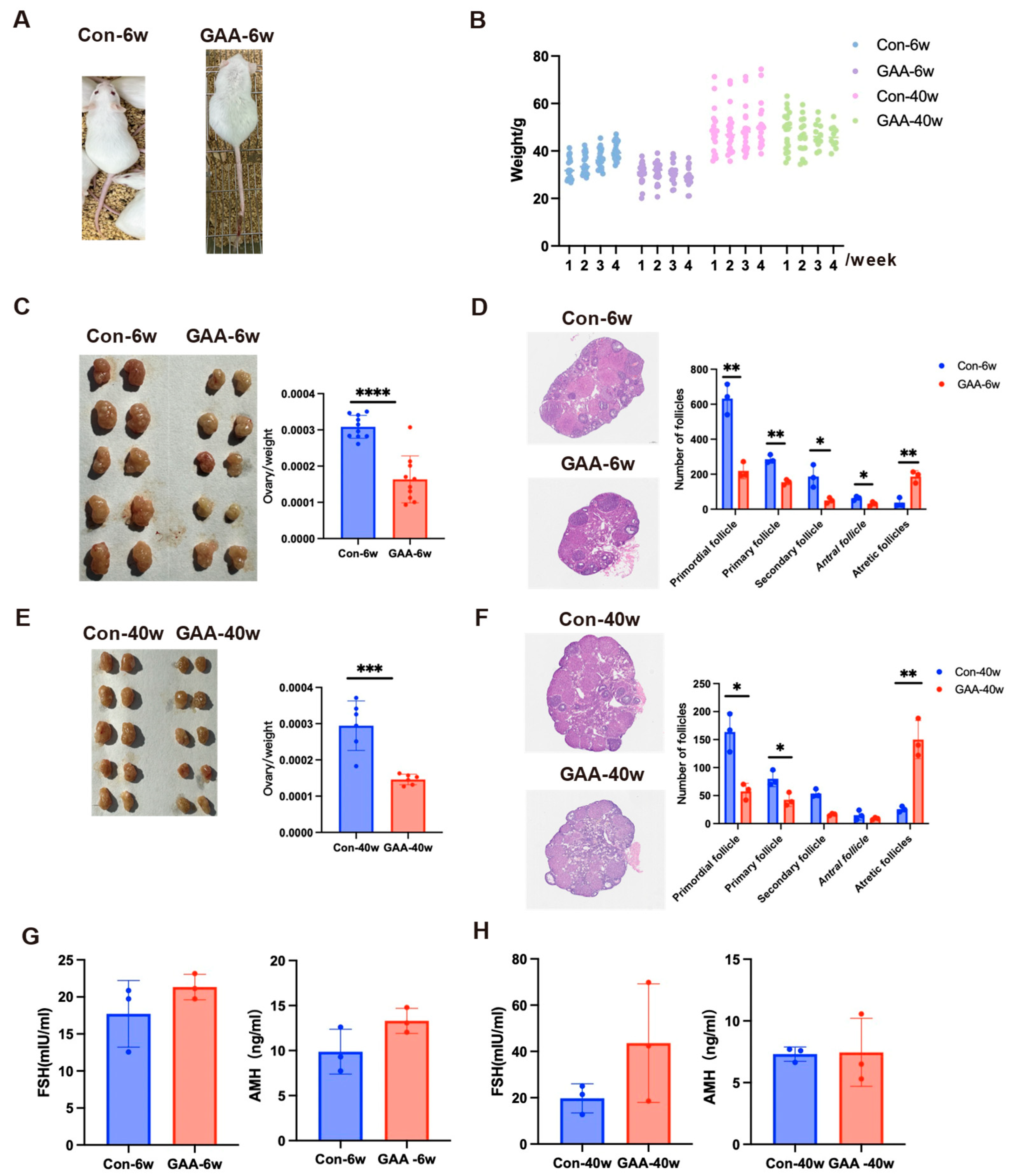
To evaluate the effects of gossypol on ovarian reserve at different reproductive stages, female mice aged 6 weeks (young group) and 40 weeks (aged group) were orally administered gossypol daily for 30 days. Post-exposure, 6-week-old mice exhibited visible clinical symptoms, including sparse, coarse fur and hemorrhagic signs (petechiae and ecchymosis) on the tail, which were absent in controls (Figure 2A). Both age groups showed significant body weight reduction compared with their respective controls (6-week-old: control 35.90 ± 3.19 g vs. gossypol 30.26 ± 0.91 g; 40-week-old: control 49.17 ± 1.20 g vs. gossypol 46.88 ± 1.25 g) (Figure 2B). Ovarian volume and ovary-to-body weight ratios were significantly decreased following treatment, with more pronounced changes in the young group (p < 0.0001) and significant reductions in the aged group (p < 0.001) (Figure 2C,E). Histopathological analysis using H&E staining revealed marked disruption of ovarian architecture in gossypol-treated mice (Figure 2D,F). Ovaries from control animals displayed intact histology, with follicles at all developmental stages, abundant primordial follicles, and minimal atresia. In contrast, gossypol-treated ovaries exhibited reduced total follicle counts, depletion of primordial and primary follicles, increased atresia, and partially collapsed antral follicles with oocyte degeneration. Quantitative follicle analysis confirmed significant reductions in the numbers of primordial (p < 0.01), primary (p < 0.01), and secondary follicles (p < 0.05) in 6-week-old mice. Antral follicle counts also declined (p < 0.05), while atretic follicles significantly increased (p < 0.01). In the 40-week-old group, similar trends were observed, with decreased numbers of primordial and primary follicles (p < 0.05), and a 5-fold increase in the atretic follicle ratio (p < 0.01) compared with controls. To further assess endocrine function, serum levels of anti-Müllerian hormone (AMH) and follicle-stimulating hormone (FSH) were measured (Figure 2G,H). In both age groups, AMH and FSH levels showed a mild upward trend after gossypol treatment; however, these changes were not statistically significant (p > 0.05).

3.4. Effects of Gossypol on Mouse Oocytes and KGN Cells

3.4.1. Impairment of Oocyte Maturation

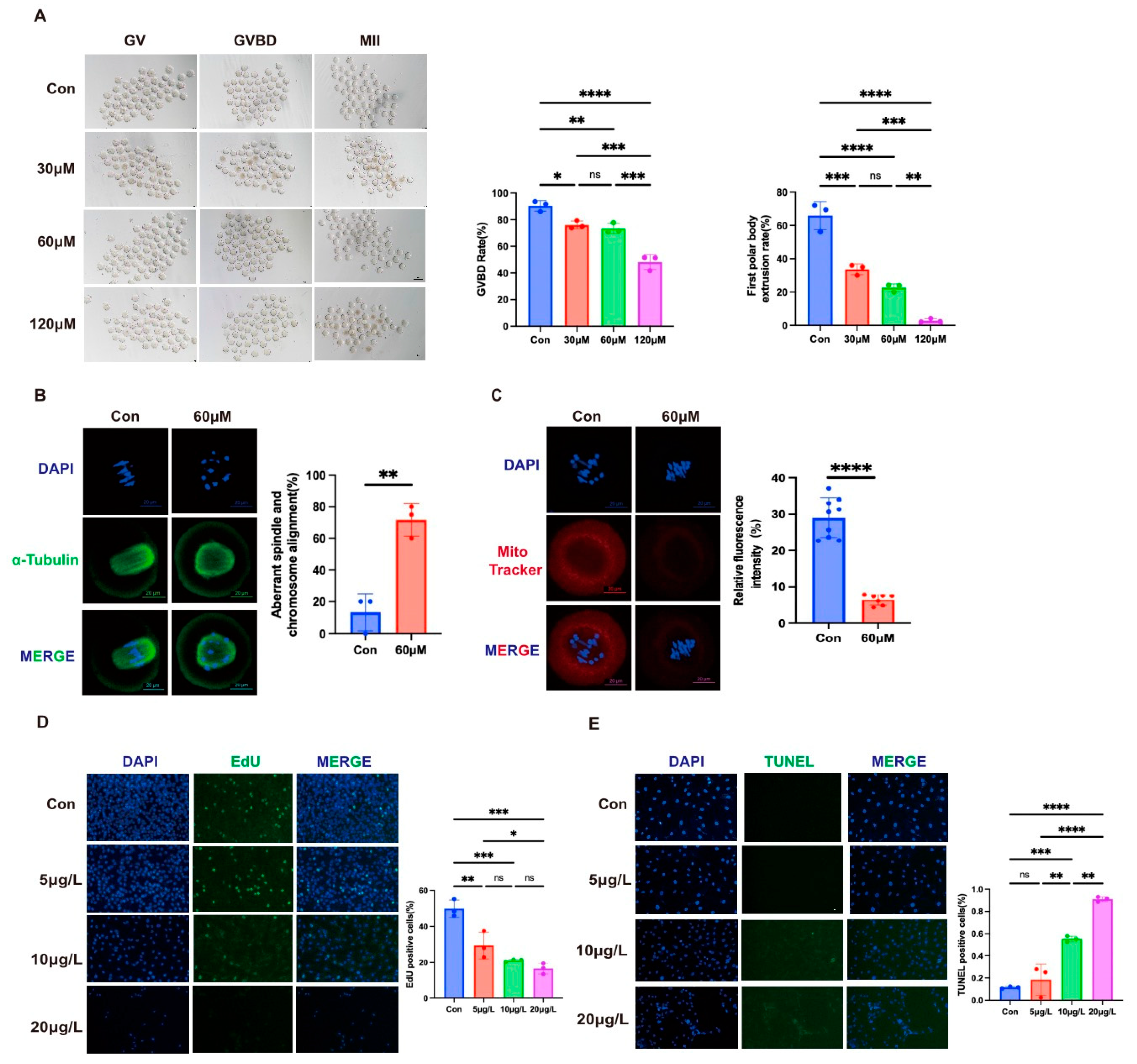
Germinal vesicle breakdown (GVBD) and first polar body extrusion (PBE) rates were assessed after 4 and 14 h of in vitro culture, respectively (Figure 3A). At 4 h, control oocytes showed a GVBD rate of 90.39 ± 3.91%, whereas gossypol treatment (30, 60, and 120 μM) caused a significant, dose-dependent reduction to 76.07 ± 2.95%, 73.46 ± 3.76%, and 48.22 ± 5.68% (p < 0.05). At 14 h, PBE rates declined from 65.88 ± 8.44% in controls to 33.67 ± 3.22% (30 μM, p < 0.001), 22.62 ± 2.28% (60 μM, p < 0.0001), and 2.86 ± 1.17% (120 μM, p < 0.001), indicating substantial inhibition of maturation. Based on these findings, 60 μM was the chosen gossypol concentration for subsequent mechanistic studies.

3.4.2. Disruption of Spindle Assembly and Mitochondrial Function

After 9 h of culture, control oocytes exhibited barrel-shaped spindles with correct chromosome alignment (Figure 3B). In contrast, 60 μM of gossypol significantly increased spindle abnormalities and misaligned chromosomes (71.6 ± 10.41% vs. 13.33 ± 11.55%, p < 0.01). Additionally, MitoTracker staining showed a dramatic reduction in mitochondrial activity (6.4 ± 1.43% vs. 28.93 ± 5.41% in controls, p < 0.0001) (Figure 3C). This suggests that gossypol disrupts both spindle formation and mitochondrial function, thereby impairing cytoplasmic maturation and oocyte competence.

3.4.3. Inhibition of KGN Cell Proliferation and Induction of Apoptosis

EdU and TUNEL assays demonstrated that gossypol impairs proliferation and induces apoptosis in human granulosa-like tumor (KGN) cells. EdU incorporation decreased with increasing gossypol concentrations: 29.41 ± 7.42% at 5 μg/L (p < 0.01), 21.02 ± 0.68% at 10 μg/L, and 16.48 ± 3.06% at 20 μg/L (both p < 0.001) (Figure 3D). TUNEL staining revealed a dose-dependent increase in apoptosis: 18.50 ± 14.04% (5 μg/L, p > 0.05), 55.12 ± 2.70% (10 μg/L, p < 0.001), and 91.06 ± 2.23% (20 μg/L, p < 0.0001) compared with the control (11.46 ± 9.75%) (Figure 3E). Together, these results indicate that gossypol exerts direct cytotoxic effects on ovarian granulosa cells by inhibiting proliferation and promoting apoptosis.

3.5. Integrated Risk Assessment of Gossypol Exposure on Ovarian Reserve Function Based on the TRAEC Strategy

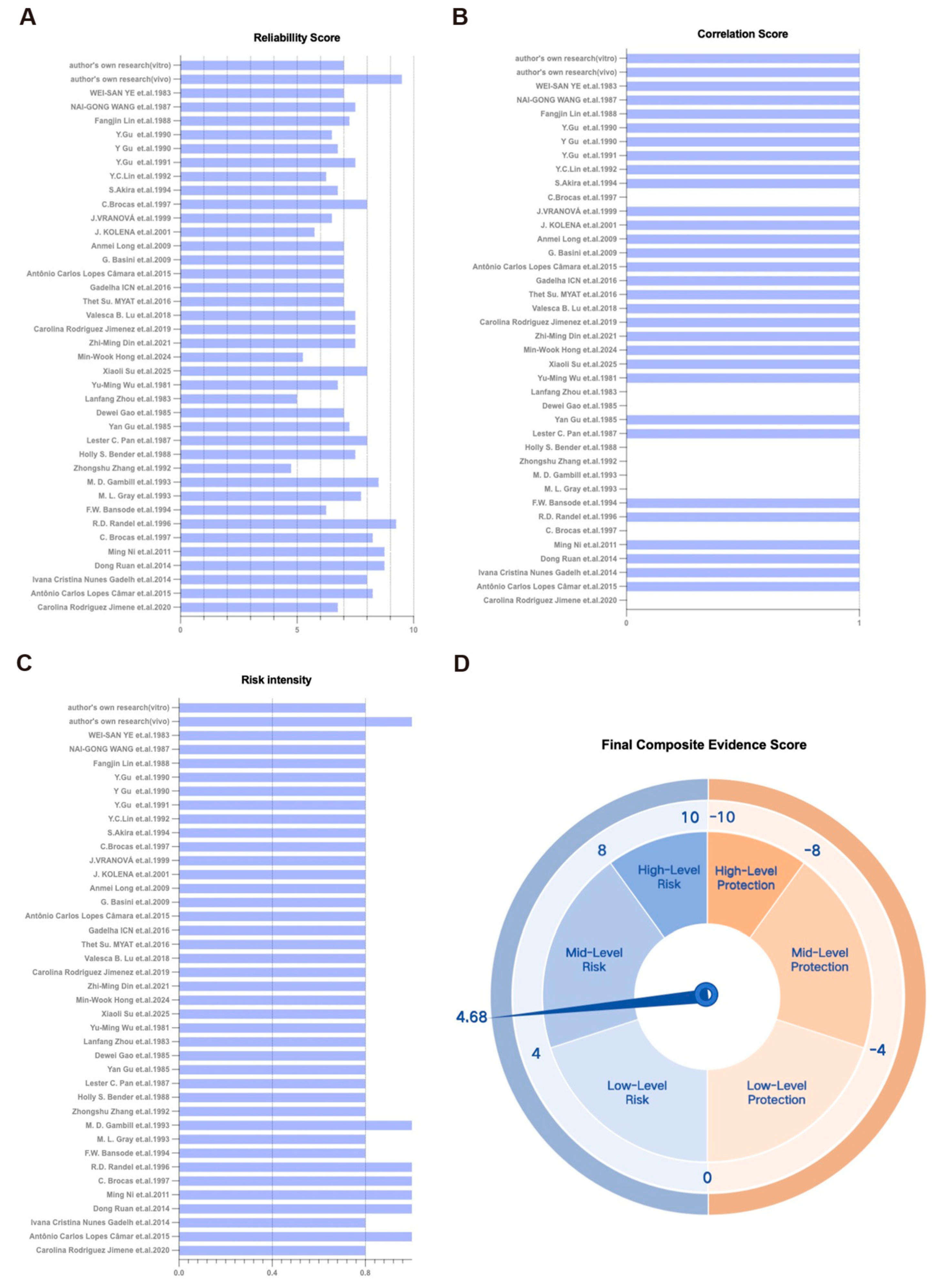
In this study, we applied a multi-evidence-based Targeted Risk Assessment of Environmental Chemicals (TRAEC) strategy to systematically evaluate the potential reproductive toxicity of gossypol on ovarian reserve function. The assessment focused on four core dimensions: reliability scores, risk intensity scores, correlation scores, and integrity scores. Each study was independently scored by two reviewers, with discrepancies resolved via discussion involving a third reviewer. This two-tiered review process enhances objectivity and scientific validity. Reliability was rated on a 1–10 scale (Figure 4A). Correlation scores are summarized in Figure 4B. Risk intensity (0–1 scale) indicates the degree to which findings support the study hypothesis (Figure 4C). Evidence count reflects the total number of studies across all dimensions. Integrity was adjusted to reflect the exclusion of epidemiological studies by redistributing weight between in vivo and in vitro evidence. The overall TRAEC-derived risk assessment yielded a composite score of 4.68 (Figure 4D), indicating moderate risk (score range: 4–8). No protective effects were observed.
In summary, this integrative TRAEC analysis classifies gossypol exposure as a moderate reproductive risk, due to its significant evidence of ovarian reserve impairment. These findings underscore the need for cautious use of gossypol in clinical settings and warrant further targeted investigations.

3.6. Proteomic Analysis of Ovarian Tissue Following Gossypol Exposure

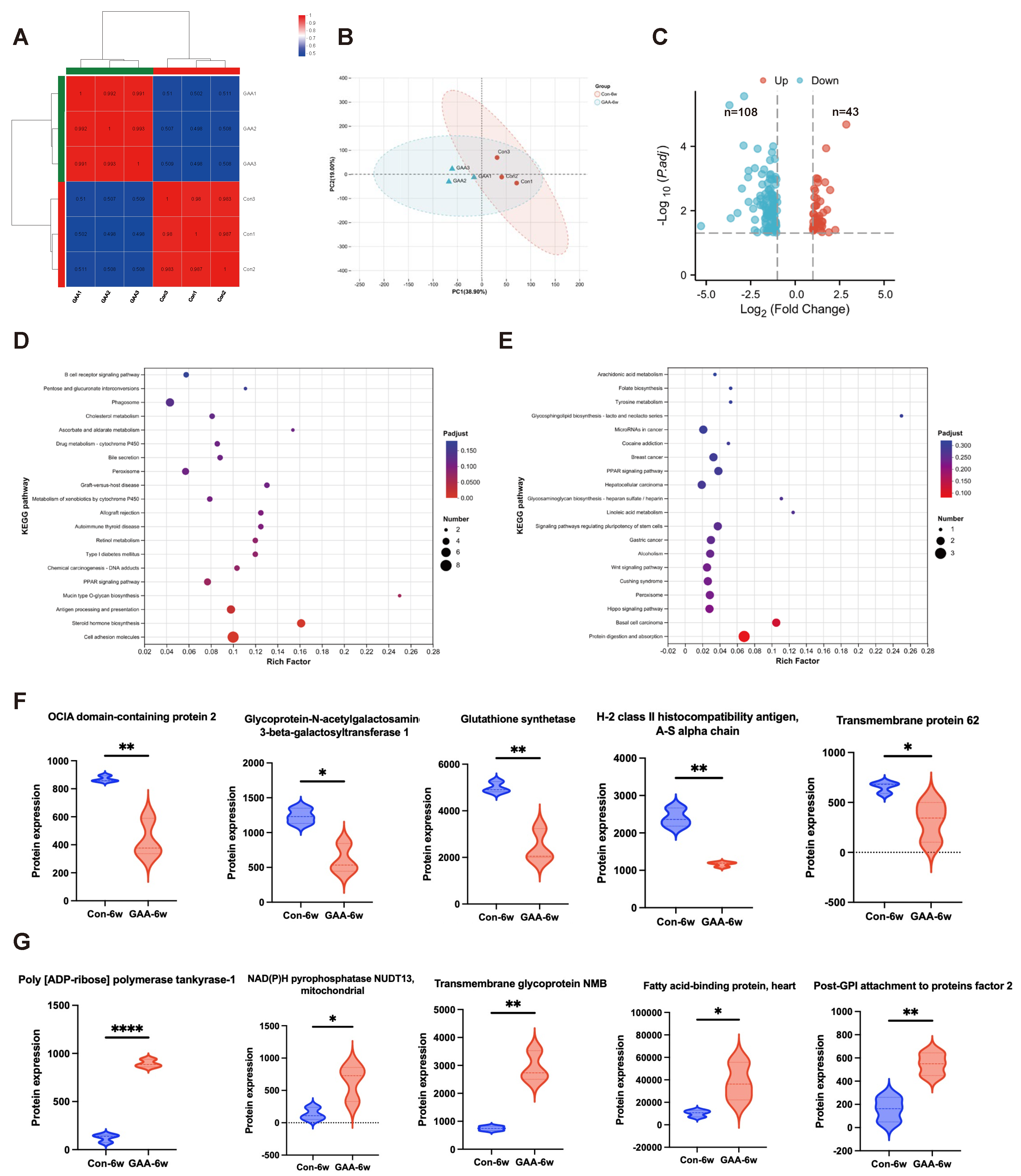
To elucidate molecular mechanisms underlying gossypol-induced ovarian dysfunction, we performed a comprehensive proteomic comparison between ovaries from 6-week-old female mice treated with gossypol and age-matched controls (Figure 5). Mass spectrometry detected 8328 proteins, with reliable quantification of 8121 proteins across all samples. Sample correlation analysis demonstrated high intra-group reproducibility (R2 > 0.98) and clear inter-group discrimination (R2 < 0.5), consistent with principal component analysis (PCA) results (Figure 5A,B). Differential expression analysis revealed 151 significantly altered proteins following gossypol treatment, with 43 upregulated and 108 downregulated proteins (fold change ≥ 2 or ≤0.5; p < 0.05) (Figure 5C). KEGG pathway enrichment indicated that downregulated proteins primarily participate in cell adhesion, steroid hormone biosynthesis, and antigen processing/presentation pathways (Figure 5D), suggesting that gossypol disrupts ovarian endocrine, immune, and structural functions. Conversely, upregulated proteins were enriched in pathways related to protein digestion/absorption and basal cell carcinoma (Figure 5E), possibly reflecting compensatory stress responses. Figure 5F highlights the five most significantly downregulated proteins, associated with immune regulation, cell adhesion, and metabolism. Notably, glutathione synthetase (GSS)—a key enzyme in glutathione, cysteine, and methionine metabolism—suggests oxidative stress as a contributor to ovarian toxicity. Figure 5G presents the top five upregulated proteins, including the mitochondria-localized NAD(P)H pyrophosphatase NUDT13 and fatty acid-binding protein 3 (FABP3), the latter linked to PPAR signaling, indicating disruptions in lipid metabolism and energy homeostasis.
Overall, proteomic profiling indicates that gossypol exposure induces extensive alterations in critical signaling pathways—steroidogenesis, immune regulation, mitochondrial metabolism, and cell adhesion—thereby impairing ovarian cellular function and depleting ovarian reserve capacity.

3.7. Assessment of Reversibility of Gossypol-Induced Ovarian Reserve Decline

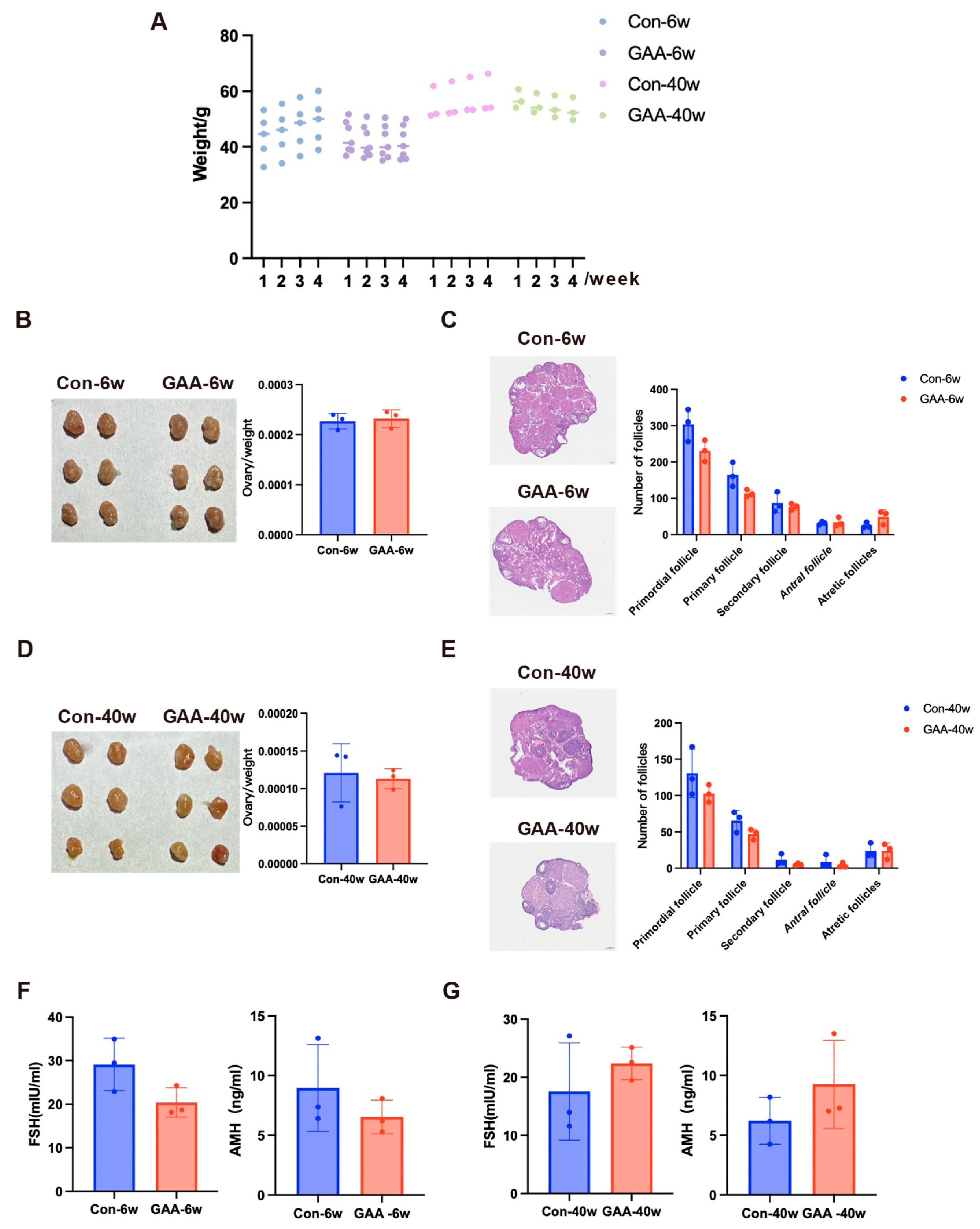
To evaluate the potential reversibility of gossypol-induced ovarian damage, subsets of 6-week-old and 40-week-old female mice underwent a 30-day recovery period following cessation of gossypol treatment. Throughout recovery, body weights in treated mice remained slightly lower than their age-matched controls (6 weeks: control 46.38 ± 2.39 g vs. gossypol 42.14 ± 0.83 g; 40 weeks: control 56.57 ± 1.37 g vs. gossypol 54.91 ± 1.64 g), with no further decline, suggesting systemic stabilization (Figure 6A). After one month of withdrawal, ovarian volume and ovary-to-body weight ratios did not differ significantly between treated and control mice in either age group (p > 0.05), indicating partial morphological recovery (Figure 6B,E). Histological evaluation showed modest increases in primordial and primary follicles and a reduction in atretic follicles in both groups compared with post-treatment levels. However, none of these follicular metrics returned to control baselines, and differences remained statistically non-significant (p > 0.05), indicating incomplete structural restoration (Figure 6C,F). Endocrine assessments revealed slight post-withdrawal increases in serum AMH and FSH, but these did not reach control levels or statistical significance (Figure 6G). Consequently, current data are insufficient to conclude that gossypol causes sustained endocrine disruption, warranting further investigation.
Within a one-month recovery period, gossypol-induced ovarian damage appears only partially reversible. Although ovarian structure and follicular composition showed modest improvement, endocrine and functional recovery was not significant. These results suggest that gossypol may exert lasting effects on ovarian reserve, and further research is needed to determine the extent and reversibility of its reproductive endocrine impact.

4. Discussion

This study systematically evaluated the effects of gossypol exposure on ovarian reserve function and its reversibility in female mice, elucidating its potential reproductive toxicity and endocrine-disrupting effects. We quantitatively assessed the reproductive toxicity of gossypol utilizing the TRAEC strategy that integrates multiple lines of evidence from epidemiological, in vivo, and in vitro studies.
Despite numerous studies reporting the reproductive effects of gossypol, results have been inconsistent [8,9,11,18,19,20,21,22,23,24,25,26,27,28,29,30,31,32,33,34,35,36,37,38,39,40,41,42,43,44,45,46,47,48,49,50], and the overall reproductive risk has not been clearly quantified. We adopted the TRAEC strategy to address this gap, offering a comprehensive, systematic, and quantitative risk assessment. Based on 36 eligible studies, we determined a moderate reproductive risk level (risk score: 4.68) associated with gossypol exposure. This integrative approach overcomes the limitations of relying on a single type of evidence, providing a more accurate toxicity profile for environmental chemicals. Notably, no epidemiological studies in human populations were identified, despite gossypol acetate’s clinical use for treating uterine fibroids and endometriosis [51,52], and its potential as an anticancer agent [1,53,54,55,56]. These findings highlight the need for future population-based investigations to determine the potential reproductive impact of gossypol in humans.
Our findings demonstrate that gossypol exposure significantly impairs ovarian function in female mice. Macroscopically, treated mice exhibited reduced body weight and smaller ovarian volume, suggesting inhibited overall growth and ovarian development. Previous studies support this observation, with Luz et al. (2018) reporting follicular atresia rates of 65–88% in rats, mice, and goats following seven days of gossypol treatment, and another study observing up to 79.4% atretic follicles in sheep [19]. Our study also showed that the number of growing follicles at all stages decreased after gossypol exposure, while the number of atretic follicles increased. In isolated oocyte experiments, 60 μM gossypol disrupted spindle formation and mitochondrial membrane potential, leading to a 63.7% decrease in polar body extrusion and increased oxidative stress [34,36]. These studies are consistent with our research findings. Granulosa cell studies indicate that gossypol inhibits proliferation and dysregulates steroidogenic enzymes (CYP11A1, HSD3B1, CYP17A1), reducing estradiol-17β and progesterone production, while leaving LH receptor expression unchanged [35,37,39]. Additionally, estrogen receptor α expression was altered [11]. Additionally, we also found that gossypol exposure induces apoptosis in granulosa cells. Together, these results suggest that gossypol impairs ovarian function through compound mechanisms involving follicle depletion, hormone disruption, and oocyte maturation failure.
Previous studies have not reported on the effects of gossypol on ovarian function in different age groups. Our research findings indicate that gossypol exposure led to notable structural damage within the ovarian tissue and significantly depleted the follicular reserve in both young and aged mice. Notably, the ovarian damage was considerably more severe in the 6-week-old group, suggesting that ovaries during the reproductive age may be more susceptible to gossypol-induced toxicity. This highlights a critical high-risk window that warrants further investigation to better understand the underlying mechanisms, potentially leading to the development of preventive measures or targeted therapies to mitigate the adverse effects of gossypol on ovarian health. Advances in omics technologies have deepened our understanding of gossypol’s ovarian effects.
Additionally, the development of omics technologies assists in elucidating the mechanisms of ovarian damage caused by gossypol. Metabolomic and redox proteomic studies in SKOV3 granulosa cells [56] and RNA-seq in primary granulosa cells [35] have been conducted, although whole-ovary omics remains unexplored. The cellular complexity of ovarian tissue—including oocytes, granulosa, and immune, stromal, and endothelial cells [57]—warrants comprehensive proteomic analysis. Our proteomic profiling revealed that gossypol disrupted pathways related to protein metabolism and carcinogenesis (upregulated), while downregulating pathways involved in cell adhesion, steroidogenesis, and antigen presentation. Notably, immune-related proteins such as OCIA2, H-2 class II antigen α-chain, and GALNT1 were significantly reduced [58,59,60], implicating immune dysregulation in ovarian impairment—an established factor in primary ovarian insufficiency [61,62]. Additionally, proteins involved in mitochondrial function and metabolism (NUDT13, FABP3) were altered, and glutathione synthetase—a key enzyme in redox homeostasis—was downregulated [63,64,65]. These findings align with prior evidence of gossypol’s impact on germ cell metabolism and redox balance [34,36].
We synthesized the existing literature on gossypol’s effects on female fertility and identified multiple mechanisms through which it impairs ovarian function. Gossypol disrupts oocyte development by impairing both nuclear and cytoplasmic processes—including spindle assembly and mitochondrial function [34,36]. It may also disturb the balance between granulosa and theca cell proliferation and apoptosis. Additionally, gossypol interferes with endocrine regulation by altering sex hormone production and disrupting estrogen receptor expression [35,37,39]. Moreover, it impairs cellular energy metabolism, increases ROS generation and lipid peroxidation, damages cellular ultrastructure, and induces apoptosis—particularly targeting mitochondria [34,35,36]. Our findings further suggest an immunoregulatory component to its action, potentially involving disruption of the hypothalamic–pituitary–ovarian (HPO) axis, leading to follicular atresia and endocrine dysfunction.
Historically, gossypol was evaluated as a male contraceptive [66], but concerns about irreversible fertility effects limited its use [14,67]. A multicenter study of 151 men treated for up to 16 weeks revealed that, one year post-treatment, only 10 of 24 participants in the 10 mg/day group exhibited recovered sperm counts, while 8 of 43 remained azoospermic [14]. A mouse study by Wang et al. found that, although short-term gossypol exposure caused reproductive and renal toxicity, these effects reversed following withdrawal [68]. In contrast, our study—which spanned over one complete mouse follicular cycle (17–19 days) [69] during both exposure and recovery—demonstrated only partial restoration of follicle numbers and hormone levels after one month, indicating that gossypol-induced ovarian damage may be partially irreversible. This may stem from structural ovarian damage (e.g., follicle depletion and increased apoptosis) that is not readily repairable and sustained hormonal imbalances hinting at long-term endocrine disturbance. These findings underscore the importance of fertility protection in women receiving gossypol acetate for gynecological or oncological conditions.
However, our study still has the following limitations: First, as this study was conducted using a murine model, its applicability to humans may be limited. Future studies should monitor fertility outcomes in women exposed to gossypol to evaluate both short- and long-term reproductive effects. Second, although proteomic analysis revealed multiple disrupted pathways, the interactions among these pathways and their specific molecular mechanisms require further investigation. Advanced approaches such as gene-knockout or overexpression models may help elucidate the roles of key regulatory molecules. Third, the recovery phase was limited to one month; extending this period could yield a more comprehensive understanding of the reversibility of gossypol-induced ovarian impairment.

5. Conclusions

This study applied an innovative environmental chemical risk assessment framework (i.e., TRAEC) to evaluate the effects of gossypol exposure on ovarian reserve by integrating published evidence with novel experimental data. The results demonstrate that gossypol poses a moderate reproductive risk, as it reduces ovarian reserve in female mice, impairs oocyte maturation, and disrupts pathways involved in hormone production and antigen processing. In vitro assays further confirmed that gossypol inhibits granulosa cell proliferation and induces apoptosis. Although partial recovery was observed one month after exposure cessation, residual functional impairment and endocrine alterations persisted, indicating potentially irreversible reproductive effects. Future studies should focus on human-based investigations to clarify gossypol’s long-term impact on ovarian health and explore alternative compounds—such as dextrorotary (+) -gossypol—that may offer therapeutic benefits without compromising fertility [70].

Author Contributions

X.S.: Writing—original draft, Visualization, Methodology, Formal analysis, Conceptualization. J.Y.: Writing—original draft, Visualization, Methodology, Formal analysis, Conceptualization. X.M.: Writing—original draft, Visualization, Methodology, Formal analysis, Conceptualization. Y.Z.: Writing—review and editing. R.H.: Writing—review and editing, Supervision, Funding acquisition, Conceptualization. Q.M.: Writing—review and editing, Supervision, Funding acquisition, Conceptualization. All authors have read and agreed to the published version of the manuscript.

Funding

This work was supported by Development Program of China (2022YFC2702900) and Suzhou Municipal Health Commission (GSWS2023106). This research did not receive any specific grant from funding agencies in the public, commercial, or not-for-profit sectors.

Institutional Review Board Statement

The animal study protocol was approved by the Institutional Animal Care and Use Committee (IACUC) of Nanjing Medical University (Protocol #2406007).

Informed Consent Statement

Not applicable.

Data Availability Statement

Data is contained within the article.

Acknowledgments

We sincerely thank the anonymous reviewers for their insightful and constructive comments on this manuscript. Special thanks are extended to all members of our research team, as well as to Professors Ran Huo, for their steadfast support and collaborative efforts throughout this study. We also acknowledge Majorbio Bio-Pharm Technology Co., Ltd. (https://www.majorbio.com, accessed on 16 July 2024.) for providing professional support in sequencing and bioinformatics analysis. Finally, we gratefully acknowledge the Suzhou Municipal Health Commission for providing financial support for this work.

Conflicts of Interest

The authors declare that they have no known competing financial interests or personal relationships that could have appeared to influence the work reported in this paper.

References

  1. Pal, D.; Sahu, P.; Sethi, G.; Wallace, C.E.; Bishayee, A. Gossypol and Its Natural Derivatives: Multitargeted Phytochemicals as Potential Drug Candidates for Oncologic Diseases. Pharmaceutics 2022, 14, 2624. [Google Scholar] [CrossRef]
  2. Paunovic, D.; Rajkovic, J.; Novakovic, R.; Grujic-Milanovic, J.; Mekky, R.H.; Popa, D.; Calina, D.; Sharifi-Rad, J. The potential roles of gossypol as anticancer agent: Advances and future directions. Chin. Med. 2023, 18, 163. [Google Scholar] [CrossRef] [PubMed]
  3. Hu, B. Risk Assessment of Free Gossypol Residues in Mutton Sheep. Master’s Thesis, Xinjiang Agricultural University, Urumqi, China, 2016. [Google Scholar]
  4. Lü, Y.; Wang, X.; Zhao, Q.; Zhang, J. Research progress on safe limits of gossypol in feed and its residues in livestock products. Chin. Agric. Bull. 2010, 26, 1–5. [Google Scholar]
  5. Zhou, W.; Zheng, W.; Gao, W.; Zuo, X.; Mao, Q.; Hu, B.; Shi, S. Research progress on risk assessment of free gossypol in livestock and poultry products. Herbivores 2014, 5, 12–15. [Google Scholar] [CrossRef]
  6. Randel, R.D.; Chase, C.C., Jr.; Wyse, S.J. Effects of gossypol and cottonseed products on reproduction of mammals. J. Anim. Sci. 1992, 70, 1628–1638. [Google Scholar] [CrossRef]
  7. Gadelha, I.C.; Fonseca, N.B.; Oloris, S.C.; Melo, M.M.; Soto-Blanco, B. Gossypol toxicity from cottonseed products. Sci. World J. 2014, 2014, 231635. [Google Scholar] [CrossRef]
  8. Luz, V.B.; Gadelha, I.C.N.; Cordeiro, L.A.V.; Melo, M.M.; Soto-Blanco, B. In vitro study of gossypol’s ovarian toxicity to rodents and goats. Toxicon Off. J. Int. Soc. Toxinol. 2018, 145, 56–60. [Google Scholar] [CrossRef] [PubMed]
  9. Brocas, C.; Rivera, R.M.; Paula-Lopes, F.F.; McDowell, L.R.; Calhoun, M.C.; Staples, C.R.; Wilkinson, N.S.; Boning, A.J.; Chenoweth, P.J.; Hansen, P.J. Deleterious actions of gossypol on bovine spermatozoa, oocytes, and embryos. Biol. Reprod. 1997, 57, 901–907. [Google Scholar] [CrossRef] [PubMed][Green Version]
  10. Hernández-Cerón, J.; Jousan, F.D.; Soto, P.; Hansen, P.J. Timing of inhibitory actions of gossypol on cultured bovine embryos. J. Dairy Sci. 2005, 88, 922–928. [Google Scholar] [CrossRef]
  11. Jimenez, C.R.; Moretti, D.B.; Corrêa, P.S.; da Costa, R.L.D.; Mui, T.S.; Machado-Neto, R.; Louvandini, H. Morphological-metric, ultrastructural and immunohistochemical effects of gossypol on cultured granulosa cells and oocytes of ewes using MOEPF. Anim. Reprod. Sci. 2019, 201, 22–31. [Google Scholar] [CrossRef]
  12. Ding, T.; Yan, W.; Zhou, T.; Shen, W.; Wang, T.; Li, M.; Zhou, S.; Wu, M.; Dai, J.; Huang, K.; et al. Endocrine disrupting chemicals impact on ovarian aging: Evidence from epidemiological and experimental evidence. Environ. Pollut. (Barking Essex 1987) 2022, 305, 119269. [Google Scholar] [CrossRef]
  13. Wu, C.; Chen, D.; Stout, M.B.; Wu, M.; Wang, S. Hallmarks of ovarian aging. Trends Endocrinol. Metab. TEM 2025, 36, 418–439. [Google Scholar] [CrossRef]
  14. Coutinho, E.M.; Athayde, C.; Atta, G.; Gu, Z.P.; Chen, Z.W.; Sang, G.W.; Emuveyan, E.; Adekunle, A.O.; Mati, J.; Otubu, J.; et al. Gossypol blood levels and inhibition of spermatogenesis in men taking gossypol as a contraceptive. A multicenter, international, dose-finding study. Contraception 2000, 61, 61–67. [Google Scholar] [CrossRef] [PubMed]
  15. Wan, D.; Liu, Y.; Chang, Q.; Liu, Z.; Wang, Q.; Niu, R.; Gao, B.; Guan, Q.; Xia, Y. Micro/Nanoplastic Exposure on Placental Health and Adverse Pregnancy Risks: Novel Assessment System Based upon Targeted Risk Assessment Environmental Chemicals Strategy. Toxics 2024, 12, 553. [Google Scholar] [CrossRef] [PubMed]
  16. Ning, J.; Ding, C.; Xu, H.; Liu, Z.; Guan, Q.; Xia, Y.; Xu, Q. Effect of per- and polyfluoroalkyl substances on neurodevelopment: Evidence-based risk assessment in the TRAEC strategy context. Environ. Int. 2024, 191, 109003. [Google Scholar] [CrossRef] [PubMed]
  17. Zhao, S.S.; Xu, Q.Q.; Su, Y.; Yin, Z.; Guan, Q.; Liu, Z.; Song, L.; Shen, R. Risk assessment of pyridaben exposure on neurodevelopment of offspring by using TRAEC strategy. Sustain. Horiz. 2025, 13, 100135. [Google Scholar] [CrossRef]
  18. Jimenez, C.R.; Moretti, D.B.; da Silva, T.P.; Corrêa, P.S.; da Costa, R.L.D.; Siu, T.M.; Louvandini, H. Cottonseed (gossypol) intake during gestation and lactation does affect the ovarian population in ewes and lambs? Res. Vet. Sci. 2021, 135, 557–567. [Google Scholar] [CrossRef]
  19. Câmara, A.C.; Gadelha, I.C.; Borges, P.A.; de Paiva, S.A.; Melo, M.M.; Soto-Blanco, B. Toxicity of Gossypol from Cottonseed Cake to Sheep Ovarian Follicles. PLoS ONE 2015, 10, e0143708. [Google Scholar] [CrossRef]
  20. Gadelha, I.C.; de Macedo, M.F.; Oloris, S.C.; Melo, M.M.; Soto-Blanco, B. Gossypol promotes degeneration of ovarian follicles in rats. Sci. World J. 2014, 2014, 986184. [Google Scholar] [CrossRef]
  21. Ruan, D.; Lin, Y.; Zhang, H.; Chen, W.; Wang, S. Effects of dietary cottonseed meal levels on egg production performance, egg quality, plasma biochemical parameters, ovarian morphology, and gossypol residues in laying ducks at peak production. Chin. J. Anim. Nutr. 2014, 26, 353–362. [Google Scholar]
  22. Ni, M.; Yuan, Z.; Li, R.; Wu, J.; Yuan, L.; Wen, L.; Yuan, H. Effect of gossypol acetate on apoptosis of luteal cells in mice. In Proceedings of the 7th National Conference on Internal Medicine of Livestock 2011; Chinese Association of Animal Science and Veterinary Medicine: Beijing, China, 2011; Volume 1, pp. 390–397, College of Veterinary Medicine, Hunan Agricultural University. [Google Scholar]
  23. Randel, R.D.; Willard, S.T.; Wyse, S.J.; French, L.N. Effects of diets containing free gossypol on follicular development, embryo recovery and corpus luteum function in Brangus heifers treated with bFSH. Theriogenology 1996, 45, 911–922. [Google Scholar] [CrossRef]
  24. Bansode, F.W. Genotoxic effects of gossypol acetate on the ovary of Rhinopoma kinneari wroughton (Microchiroptera: Mammalia). Contraception 1994, 49, 601–607. [Google Scholar] [CrossRef]
  25. Gray, M.L.; Greene, L.W.; Williams, G.L. Effects of dietary gossypol consumption on metabolic homeostasis and reproductive endocrine function in beef heifers and cows. J. Anim. Sci. 1993, 71, 3052–3059. [Google Scholar] [CrossRef] [PubMed]
  26. Gambill, M.D.; Humphrey, W.D. Effects of diets containing gossypol on ovarian histology, function and fertility in prepubertal beef heifers. Theriogenology 1993, 40, 585–593. [Google Scholar] [CrossRef] [PubMed]
  27. Zhang, Z.; Wang, R.; Shen, Q. Study on drug-induced aneuploidy of germ cells and early embryos in mice. Reprod. Contracept. 1992, 81, 34–39. [Google Scholar]
  28. Bender, H.S.; Saunders, G.K.; Misra, H.P. A histopathologic study of the effects of gossypol on the female rat. Contraception 1988, 38, 585–592. [Google Scholar] [CrossRef] [PubMed]
  29. Pan, L.C.; Nadakavukaren, M.J.; Jensen, D.R. Effects of ingested gossypol on the ultrastructure of rat ovarian follicles. Cell Tissue Res. 1987, 250, 215–220. [Google Scholar] [CrossRef]
  30. Gu, Y.; Anderson, N.O. Effects of gossypol on the estrous cycle and ovarian weight in the rat. Contraception 1985, 32, 491–496. [Google Scholar] [CrossRef]
  31. Gao, D.; Zhang, M.; Li, C.; Yuan, Q. Study on the toxicological effects of gossypol acetate on pregnant rats. Chin. Pharm. J. 1985, 8, 464–467. [Google Scholar]
  32. Zhou, L.; Lei, H. Effects of gossypol acetate on uterus and ovary. Acta Pharm. Sin. 1984, 3, 220–223. [Google Scholar] [CrossRef]
  33. Wu, Y.M.; Chappel, S.C.; Flickinger, G.L. Effects of gossypol on pituitary-ovarian endocrine function, ovulation and fertility in female hamsters. Contraception 1981, 24, 259–268. [Google Scholar] [CrossRef]
  34. Su, X.; He, Y.; Li, H.; Yu, T.; Sun, Q.; Chen, M.; Zhang, B.; Wang, W.; Ju, S.; Li, Q. Melatonin protects porcine oocytes from gossypol-induced meiosis defects via regulation of SIRT1-mediated mitophagy. Food Chem. Toxicol. Int. J. Publ. Br. Ind. Biol. Res. Assoc. 2025, 195, 115122. [Google Scholar] [CrossRef]
  35. Hong, M.W.; Kim, H.; Choi, S.Y.; Sharma, N.; Lee, S.J. Effect of Gossypol on Gene Expression in Swine Granulosa Cells. Toxins 2024, 16, 436. [Google Scholar] [CrossRef] [PubMed]
  36. Ding, Z.M.; Chen, Y.W.; Wang, Y.S.; Ahmad, M.J.; Yang, S.J.; Duan, Z.Q.; Liu, M.; Yang, C.X.; Xiong, J.J.; Liang, A.X.; et al. Gossypol exposure induces mitochondrial dysfunction and oxidative stress during mouse oocyte in vitro maturation. Chem.-Biol. Interact. 2021, 348, 109642. [Google Scholar] [CrossRef]
  37. Myat, T.S.; Tetsuka, M. Gossypol inhibits LH-induced steroidogenesis in bovine theca cells. Anim. Sci. J. 2017, 88, 63–71. [Google Scholar] [CrossRef] [PubMed]
  38. Gadelha, I.C.N.; Lima, M.A.; Melo, M.M.; Soto-Blanco, B. Gossypol promotes the degeneration of chicken ovarian follicles in vitro. Rev. Bras. De Ciência Avícola [Braz. J. Poult. Sci.] 2016, 18, 505–510. [Google Scholar] [CrossRef]
  39. Basini, G.; Bussolati, S.; Baioni, L.; Grasselli, F. Gossypol, a polyphenolic aldehyde from cotton plant, interferes with swine granulosa cell function. Domest. Anim. Endocrinol. 2009, 37, 30–36. [Google Scholar] [CrossRef]
  40. Long, A.; Wu, J.; Yuan, L.; Yuan, H. Effects of gossypol on proliferation and apoptosis of primary cultured luteal cells in pigs. Chin. J. Vet. Sci. 2009, 29, 1303–1306. [Google Scholar] [CrossRef]
  41. Kolena, J.; Vrsanská, S.; Nagyová, E.; Jezová, M. Gossypol inhibits follicle-stimulating hormone-and epidermal growth factor-stimulated expansion of oocyte-cumulus complexes from porcine preovulatory follicles. Physiol. Res. 2001, 50, 627–630. [Google Scholar] [CrossRef]
  42. Vranová, J.; Jezová, M.; Scsuková, S.; Kolena, J. Inhibitory effect of gossypol on basal and luteinization factor-stimulated progesterone synthesis in porcine granulosa cells. Physiol. Res. 1999, 48, 119–128. [Google Scholar]
  43. Akira, A.; Ohmura, H.; Uzumcu, M.; Araki, T.; Lin, Y.C. Gossypol inhibits aromatase activity in cultured porcine granulosa cells. Theriogenology 1994, 41, 1489–1497. [Google Scholar] [CrossRef]
  44. Lin, Y.C.; Gu, Y.; Brueggemeier, R.W.; Rikihisa, Y. Binding of 3H-gossypol in organelles of cultured bovine luteal cells. Life Sci. 1992, 50, 443–447. [Google Scholar] [CrossRef] [PubMed]
  45. Gu, Y.; Li, P.K.; Lin, Y.C.; Rikihisa, Y.; Brueggemeier, R.W. Gossypolone suppresses progesterone synthesis in bovine luteal cells. J. Steroid Biochem. Mol. Biol. 1991, 38, 709–715. [Google Scholar] [CrossRef] [PubMed]
  46. Gu, Y.; Chang, C.J.; Rikihisa, Y.; Lin, Y.C. Inhibitory effect of gossypol on human chorionic gonadotropin (hCG)-induced progesterone secretion in cultured bovine luteal cells. Life Sci. 1990, 47, 407–414. [Google Scholar] [CrossRef] [PubMed]
  47. Gu, Y.; Lin, Y.C.; Rikihisa, Y. Inhibitory effect of gossypol on steroidogenic pathways in cultured bovine luteal cells. Biochem. Biophys. Res. Commun. 1990, 169, 455–461. [Google Scholar] [CrossRef]
  48. Lin, J.; Zheng, H. In vitro observation of the effects of gossypol acetate on estradiol and progesterone production by granulosa cells of mature rat follicles. Reprod. Contracept. 1988, 4, 59–64. [Google Scholar]
  49. Wang, N.G.; Guan, M.Z.; Lei, H.P. Effects of gossypol acetic acid on rat luteal cells in vitro. J. Ethnopharmacol. 1987, 20, 45–51. [Google Scholar] [CrossRef]
  50. Ye, W.S.; Liang, J.C.; Hsu, T.C. Toxicity of a male contraceptive, gossypol, in mammalian cell cultures. In Vitro 1983, 19, 53–57. [Google Scholar] [CrossRef]
  51. Zhang, Y.; Wu, H.; Diao, L. Feasibility analysis of compound gossypol acetate tablets combined with levonorgestrel intrauterine system in the treatment of adenomyosis after lesion excision. Chin. J. Ration. Drug Use Explor. 2021, 18, 69–72. [Google Scholar]
  52. Jiang, R.; Chen, Z.; Ni, M.; Li, X.; Ying, H.; Fen, J.; Wan, D.; Peng, C.; Zhou, W.; Gu, L. A traditional gynecological medicine inhibits ovarian cancer progression and eliminates cancer stem cells via the LRPPRC-OXPHOS axis. J. Transl. Med. 2023, 21, 504. [Google Scholar] [CrossRef]
  53. Hsieh, Y.S.; Yu, C.H.; Chu, S.C.; Lin, C.Y.; Chen, P.N. Gossypol Inhibits Metastasis of Lung Cell Carcinoma by Reversing Epithelial to Mesenchymal Transition and Suppressing Proteases Activity. Environ. Toxicol. 2024, 39, 5209–5221. [Google Scholar] [CrossRef]
  54. Xue, Q.; Wang, W.; Liu, J.; Wang, D.; Zhang, T.; Shen, T.; Liu, X.; Wang, X.; Shao, X.; Zhou, W.; et al. LRPPRC confers enhanced oxidative phosphorylation metabolism in triple-negative breast cancer and represents a therapeutic target. J. Transl. Med. 2025, 23, 372. [Google Scholar] [CrossRef] [PubMed]
  55. Li, G.; Shi, S.; Tan, J.; He, L.; Liu, Q.; Fang, F.; Fu, H.; Zhong, M.; Mai, Z.; Sun, R.; et al. Highly Efficient Synergistic Chemotherapy and Magnetic Resonance Imaging for Targeted Ovarian Cancer Therapy Using Hyaluronic Acid-Coated Coordination Polymer Nanoparticles. Adv. Sci. 2024, 11, e2309464. [Google Scholar] [CrossRef] [PubMed]
  56. Wang, J.; Jin, L.; Li, X.; Deng, H.; Chen, Y.; Lian, Q.; Ge, R.; Deng, H. Gossypol induces apoptosis in ovarian cancer cells through oxidative stress. Mol. Biosyst. 2013, 9, 1489–1497. [Google Scholar] [CrossRef] [PubMed]
  57. Kinnear, H.M.; Tomaszewski, C.E.; Chang, A.L.; Moravek, M.B.; Xu, M.; Padmanabhan, V.; Shikanov, A. The ovarian stroma as a new frontier. Reproduction 2020, 160, R25–R39. [Google Scholar] [CrossRef]
  58. Chojnacka, K.J.; Elancheliyan, P.; Mussulini, B.H.M.; Mohanraj, K.; Callegari, S.; Gosk, A.; Banach, T.; Góral, T.; Szczepanowska, K.; Rehling, P.; et al. Ovarian carcinoma immunoreactive antigen-like protein 2 (OCIAD2) is a novel complex III-specific assembly factor in mitochondria. Mol. Biol. Cell 2022, 33, ar29. [Google Scholar] [CrossRef] [PubMed]
  59. Poluektov, Y.; George, M.; Daftarian, P.; Delcommenne, M.C. Assessment of SARS-CoV-2 specific CD4(+) and CD8 (+) T cell responses using MHC class I and II tetramers. Vaccine 2021, 39, 2110–2116. [Google Scholar] [CrossRef]
  60. Sun, X.; Zhan, M.; Sun, X.; Liu, W.; Meng, X. C1GALT1 in health and disease. Oncol. Lett. 2021, 22, 589. [Google Scholar] [CrossRef]
  61. Qi, L.; Li, Y.; Zhang, L.; Li, S.; Zhang, X.; Li, W.; Qin, J.; Chen, X.; Ji, Y.; Xue, Z.; et al. Immune and oxidative stress disorder in ovulation-dysfunction women revealed by single-cell transcriptome. Front. Immunol. 2023, 14, 1297484. [Google Scholar] [CrossRef]
  62. Zhang, C.; Yu, D.; Mei, Y.; Liu, S.; Shao, H.; Sun, Q.; Lu, Q.; Hu, J.; Gu, H. Single-cell RNA sequencing of peripheral blood reveals immune cell dysfunction in premature ovarian insufficiency. Front. Endocrinol. 2023, 14, 1129657. [Google Scholar] [CrossRef]
  63. Abdelraheim, S.R.; Spiller, D.G.; McLennan, A.G. Mouse Nudt13 is a Mitochondrial Nudix Hydrolase with NAD(P)H Pyrophosphohydrolase Activity. Protein J. 2017, 36, 425–432. [Google Scholar] [CrossRef]
  64. Shand, H.; Patra, S.; Ghorai, S. Fatty acid binding protein as a new age biomarker. Clin. Chim. Acta Int. J. Clin. Chem. 2025, 565, 120029. [Google Scholar] [CrossRef]
  65. Lapenna, D. Glutathione and glutathione-dependent enzymes: From biochemistry to gerontology and successful aging. Ageing Res. Rev. 2023, 92, 102066. [Google Scholar] [CrossRef] [PubMed]
  66. Qian, S.Z.; Wang, Z.G. Gossypol: A potential antifertility agent for males. Annu. Rev. Pharmacol. Toxicol. 1984, 24, 329–360. [Google Scholar] [CrossRef]
  67. Waites, G.M.; Wang, C.; Griffin, P.D. Gossypol: Reasons for its failure to be accepted as a safe, reversible male antifertility drug. Int. J. Androl. 1998, 21, 8–12. [Google Scholar] [CrossRef]
  68. Wang, H.; Piao, Z.; Ma, H.; Cao, L.; Liu, J.; Wu, J. Short-term exposure to gossypol causes reversible reproductive toxicity and nephrotoxicity in mice. Nan Fang Yi Ke Da Xue Xue Bao [J. South. Med. Univ.] 2023, 43, 251–256. [Google Scholar] [CrossRef]
  69. Hoage, T.R.; Cameron, I.L. Folliculogenesis in the ovary of the mature mouse: A radioautographic study. Anat. Rec. 1976, 184, 699–709. [Google Scholar] [CrossRef] [PubMed]
  70. Lin, J.L.; Fang, X.; Li, J.X.; Chen, Z.W.; Wu, W.K.; Guo, X.X.; Liu, N.J.; Huang, J.F.; Chen, F.Y.; Wang, L.J.; et al. Dirigent gene editing of gossypol enantiomers for toxicity-depleted cotton seeds. Nat. Plants 2023, 9, 605–615. [Google Scholar] [CrossRef] [PubMed]
Figure 1. Flow diagram of selecting articles for risk assessment.
Toxics 13 00763 g001
Figure 2. Effects of gossypol on ovarian reserve function in female mice. (A) Gross morphological comparison of 6-week-old mice treated with gossypol (20 mg/kg/day for 30 days) and age-matched controls, showing sparse fur and tail hemorrhagic signs in treated animals. (B) Body weight changes in 6-week-old control (n = 20) and gossypol-treated (n = 17) mice, and in 40-week-old control (n = 20) and gossypol-treated (n = 10) mice. (C) Representative images of ovarian morphology in 6-week-old control and gossypol-treated mice (n = 10 per group). (D) Hematoxylin and eosin (H&E) staining of ovarian sections from 6-week-old mice, with quantification of follicle numbers at different developmental stages (primordial, primary, secondary, antral, and atretic). (E) Representative ovarian morphology in 40-week-old control and gossypol-treated mice (n = 10 per group). (F) H&E staining of ovarian tissue from 40-week-old mice, with corresponding follicle counts at each stage. (G) Serum concentrations of follicle-stimulating hormone (FSH) and anti-Müllerian hormone (AMH) in 6-week-old control and gossypol-treated mice measured using ELISA (n = 3 per group). (H) Serum FSH and AMH concentrations in 40-week-old mice after gossypol exposure compared with controls (n = 3 per group). Data are presented as mean ± SD. Statistical comparisons were performed using Student’s t-test. **** p < 0.0001, *** p < 0.001, ** p < 0.01, * p < 0.05, and ns = not significant.
Toxics 13 00763 g002
Figure 3. Effects of gossypol exposure on mouse oocytes and KGN cells. (A) Germinal vesicle breakdown (GVBD, assessed at 4 h) and first polar body extrusion (PBE, assessed at 14 h) in mouse oocytes cultured with increasing concentrations of gossypol (30, 60, and 120 μM) compared with untreated controls. Scale bar: 100 μm. (B) Representative confocal images of spindle morphology and chromosome alignment in oocytes cultured for 9 h with or without 60 μM gossypol. Quantification of spindle/chromosome abnormalities is shown. Scale bar: 20 μm. (C) Mitochondrial distribution and fluorescence intensity in oocytes cultured for 9 h with or without 60 μM gossypol, detected using MitoTracker Red staining. Scale bar: 20 μm. (D) Proliferation of KGN cells treated with gossypol (5, 10, and 20 μg/mL) for 24 h, measured by EdU incorporation assay. Quantitative analysis indicates dose-dependent inhibition of cell proliferation. Scale bar = 250 μm. (E) Apoptosis of KGN cells exposed to gossypol (5, 10, and 20 μg/mL) for 24 h, evaluated by TUNEL staining. A concentration-dependent increase in apoptosis was observed. Scale bar: 100 μm. All experiments were performed in triplicate. Data are expressed as mean ± SD. Statistical analysis was performed using one-way ANOVA. **** p < 0.0001, *** p < 0.001, ** p < 0.01, * p < 0.05, and ns—not significant.
Toxics 13 00763 g003
Figure 4. The TRAEC-based scoring results of gossypol exposure on ovarian reserve function [8,9,11,18,19,20,21,22,23,24,25,26,27,28,29,30,31,32,33,34,35,36,37,38,39,40,41,42,43,44,45,46,47,48,49,50]. (A) Reliability scores of the included studies, reflecting experimental design quality and methodological rigor. (B) Correlation scores assessing the strength of association between gossypol exposure and reproductive outcomes. (C) Risk intensity scores, indicating the degree to which individual studies supported the hypothesis of gossypol-induced ovarian dysfunction. (D) Composite evidence scores derived from integration of all four TRAEC dimensions (reliability, correlation, risk intensity, and integrity). The final analysis included both published studies (summarized in Table 1 and Table 2) and our original experimental data. Each study was independently evaluated and scored by two researchers, with discrepancies resolved through discussion with a third reviewer. Data are presented as mean values for each scoring dimension, ensuring consistency with the TRAEC framework for environmental chemical risk assessment.
Toxics 13 00763 g004
Figure 5. Proteomic profiling of ovarian tissue following gossypol exposure in mice. (A) Heatmap showing sample-to-sample correlation of quantified protein expression profiles, indicating high intra-group reproducibility and clear inter-group separation. (B) Principal component analysis (PCA) demonstrating distinct clustering between control and gossypol-treated ovarian tissue samples, confirming overall proteomic differences. Triangles represent the gossypol-exposed group, and circles represent the control group. (C) Volcano plot of differentially expressed proteins (DEPs) between gossypol-treated and control groups. DEPs were defined as proteins with fold change ≥ 2 or ≤0.5 and p < 0.05. (D) KEGG pathway enrichment analysis of significantly downregulated proteins, highlighting pathways involved in cell adhesion, steroid hormone biosynthesis, and antigen processing/presentation. (E) KEGG pathway enrichment analysis of significantly upregulated proteins, including pathways related to protein digestion/absorption and cellular stress responses. (F) The five most significantly downregulated proteins, mainly associated with immune regulation, cell adhesion, and metabolism. (G) The five most significantly upregulated proteins, including those involved in mitochondrial metabolism and lipid signaling. Proteomic analysis was performed using data-independent acquisition (DIA) mass spectrometry, with <1% false discovery rate for peptide identification. Data are expressed as mean values of three biological replicates per group. Statistical comparisons were conducted using t-tests. **** p < 0.0001, ** p < 0.01, and * p < 0.05.
Toxics 13 00763 g005
Figure 6. Reversibility of gossypol-induced impairment in ovarian reserve function following drug withdrawal. (A) Body weight changes in 6-week-old control (n = 5) and gossypol-treated (n = 7) mice, and in 40-week-old control (n = 3) and gossypol-treated (n = 3) mice, one month after cessation of gossypol administration (20 mg/kg/day for 30 days). (B) Representative ovarian morphology in 6-week-old control and gossypol-treated mice after one month of recovery (n = 6 per group). (C) Hematoxylin and eosin (H&E) staining of ovarian sections in 6-week-old mice after recovery, with quantitative analysis of follicle counts at different developmental stages (primordial, primary, secondary, antral, and atretic follicles). (D) Representative ovarian morphology in 40-week-old control and gossypol-treated mice after one month of withdrawal (n = 6 per group). (E) H&E staining and follicle count analysis in 40-week-old mice after recovery, showing partial but incomplete restoration of follicular populations. (F) Serum concentrations of follicle-stimulating hormone (FSH) and anti-Müllerian hormone (AMH) in 6-week-old control and gossypol-treated mice one month after drug cessation, measured by ELISA (n = 3 per group). (G) Serum FSH and AMH concentrations in 40-week-old mice after one month of withdrawal compared with age-matched controls (n = 3 per group). All data are presented as mean ± SD. Statistical analysis was performed using Student’s t-test. A partial morphological improvement was observed in both age groups, whereas endocrine parameters did not fully return to baseline. p < 0.05 was considered statistically significant.
Toxics 13 00763 g006
Table 1. In vivo studies on the effects of gossypol on ovarian function in mammals.
IDStudyLocationSpeciesExposure PeriodAgeType of
Gossypol
Exposure DoseExposure MethodsOutcomes
1Jimenez et al., 2021 [18]BrazilSanta Inês ewes10 months/Cottonseed/FeedingCottonseed had no effect on maternal–offspring follicular dynamics but may alter the future steroidogenic response in offspring of dams exposed to cottonseeds during their reproductive period.
2Câmara et al., 2015 [19]BrazilSanta Inês crossbred ewes63 days/Cottonseed cakeFeed was offered at 1.5% of the animal’s body weight.FeedingGossypol-fed sheep exhibited a significantly reduced proportion of viable ovarian follicles (20.6%) and an increased proportion of atretic follicles (79.4%).
3Gadelha et al., 2014 [20]BrazilWistar rats15 days60 to 70 days oldGossypol acetic acid (Fluka, G4382)25 mg gossypol/kg/daySubcutaneous injectionGossypol reduced the number of viable follicles and altered hormone levels, thereby disrupting the estrous cycle.
4Ruan et al., 2014 [21]ChinaFujian Longyan mountain hemp duck3 months19 weeksCottonseed meal14.82, 28.03, 32.81, 39.18, and 50.16 mg/kg free gossypolFeedingDominant follicle integrity was significantly compromised at a dietary free gossypol level of 28.03 mg/kg, characterized by deformation, rupture, partial dissolution, and hemorrhagic spots on follicular vascular walls.
5Ni et al., 2011 [22]ChinaKunming mice24 h, 48 h25 to 30 days oldGossypol acetic acid1.6 and 8.40 mg/L, 0.5 mLIntraperitoneal injectionGossypol can effectively induce apoptosis in mouse luteal cells.
6Brocas et al., 1997 [9]USANonlactating Holstein cow9 weeks/Cottonseed meal6.84 g free gossypol/dayFeedingNo significant differences (p > 0.10) were observed between cottonseed meal-fed and control cows regarding oocyte yield, cleavage rate, or blastocyst development.
7Randel et al., 1996 [23]USABrangus heifers65 days2 years oldCottonseed meal; whole cottonseed5 g free gossypol/day; 15 g free gossypol/dayFeedingOvarian and stromal weights, as well as total follicle counts per heifer, did not differ among the three treatment groups. However, CSM heifers had fewer follicles >5 mm compared with WCS and control heifers.
8Bansode, 1994 [24]IndiaRhinopoma kinneari2 days, 4 days, 6 days, 8 days/Gossypol acetate10 mg/dayFeedingGossypol acetate exerts cytotoxic and antifolliculogenic effects, inducing oocyte and follicle degeneration and inhibiting their development.
9Gray et al., 1993 [25]USAPostpubertal beef heifers; mature cows62 days;
33 weeks
/Cottonseed meal and whole cottonseeds0, 0.5, 2.5, 5, 10, and 20 g free
gossypol/day;
20 mg free gossypol/kg/day
FeedingThe gossypol levels used in these studies are unlikely to impair reproductive performance in beef heifers or cows.
10Gambill and Humphrey, 1993 [26]USACrossbred beef heifers64 days/Cottonseed meal and whole cottonseeds6.1 g free gossypol/day; 13.7 g free gossypol/dayFeedingOvarian metrics, corpus luteum characteristics, and follicle number and size were comparable across treatments.
11Zhang et al., 1992 [27]ChinaICR mice/8–13 weeksGossypol acetic acid5, 20, and 50 mg/kg/Gossypol acetate at varying doses did not affect oocyte aneuploidy rates in mice.
12Bender et al., 1988 [28]USASprague Dawley rats30 days/Gossypol acetic acid40 and 60 mg gossypol/kg/dayIntragastric administrationDespite fewer estrous cycles, gossypol-treated rats showed no histopathological changes in their ovaries, uterus, or vagina.
13Pan et al., 1987 [29]USARats60 days/Gossypol acetic acid20 mg gossypol/kg/dayOrallyChanges included increased ooplasmic lysosomes and underdeveloped smooth endoplasmic reticulum in granulosa cells.
14Gu and Anderson, 1985 [30]USALong–Evans strain rats15 days;
20 days
/Gossypol acetate10 mg gossypol/kg/day; 1, 5, and 10 mg gossypol/kg/daySubcutaneous injectionAt male-effective doses, gossypol halted estrous cycles and significantly reduced ovarian weight.
15Gao et al., 1985 [31]ChinaRatsDay 1–5 of
gestation
/Gossypol100, 81, 66, 53, 43, 35, 28, and 23 mg gossypol/kg/dayIntragastric administrationOvarian tissues showed no significant changes; corpus luteum and follicles at all stages appeared normal across all doses.
16Zhou and Lei, 1984 [32]ChinaWistar ratsSix times a week for 8 consecutive weeks/Gossypol acetic acid30 mg/kg gossypolIntragastric administrationOvarian morphology did not differ significantly between gossypol and control groups.
17Wu et al., 1981 [33]USAHamsters76 days;
40 days;
20 days
/Gossypol5 mg gossypol/kg/day;
10 mg gossypol/kg/day;
10 and 20 mg gossypol/kg/day
Intragastric administrationGossypol altered pituitary and ovarian hormones in proestrus and estrus but did not affect estrous length, ovulation count, or pregnancy rate.
Table 2. In vitro studies on the effects of gossypol on ovarian function in mammals.
IDStudyLocationExposure
Period
Cell TypeCell SourceType of GossypolExposure ConcentrationOutcomes
1Su et al., 2025 [34]China44 hOocytesPorcine
ovaries
Gossypol
(purity >99%, MedChemExpress, Princeton, NJ, USA)
10, 20, and 40 μM gossypolGossypol impaired porcine oocyte maturation in vitro by reducing PB1 extrusion, inhibiting cumulus expansion, and disrupting meiosis.
2Hong et al., 2024 [35]Korea72 hGranulosa cellsSwine ovaries/6.25 and 12.5 μM gossypolGossypol is cytotoxic to porcine granulosa cells, inhibiting proliferation and impairing oocyte maturation.
3Ding et al., 2021 [36]China2 h; 8 h; 14 hOocytesKunming mouse ovaries (3–4 weeks old)Gossypol
(Yirui Biotech, Hangzhou, China)
20, 40, and 60 μM gossypolGossypol impairs polar body extrusion, disrupts spindle structure, induces mitochondrial dysfunction and oxidative stress, and triggers early apoptosis.
4Jimenez et al., 2019 [11]Brazil24 h; 96 hGranulosa cells and oocytesSanta Inês ewe ovaries
(1-year-old)
Gossypol acetic acid (G4382, Sigma-Aldrich, São Paulo, Brazil)5, 10, and 20 μg/mL gossypolGossypol impairs granulosa cell development and preantral follicle integrity in sheep.
5Luz et al., 2018 [8]Brazil24 h; 7 daysOvariesRat, mouse, and goat ovariesGossypol acetic acid (G4382, Fluka, Buchs, Switzerland)5, 10, and 20 μg/mL gossypolGossypol may directly impair follicular maturation and female fertility.
6Myat and Tetsuka, 2017 [37]Japen24 h at 1 day and 7 daysTheca cellsBovine ovariesGossypol
(Sigma-Aldrich, St. Louis, MO, USA)
0.2, 1, 5, and 25 μg/mL gossypolGossypol inhibits thecal steroidogenesis by downregulating steroidogenic enzyme genes without impacting cell viability in cattle.
7Gadelha et al., 2016 [38]Brazil24 h; 7 daysOvarian folliclesAdult Bantam chicken ovariesGossypol acetic acid (G4382, Fluka, Buchs, Switzerland)5, 10, and 20 μg/mL gossypolGossypol increased atresia across all follicular stages in cultured chicken ovaries, indicating impaired follicle viability and maturation.
8Câmara et al., 2015 [19]Brazil24 h; 7 daysovariesSanta Inês ewe ovaries (3–5 years old)Gossypol acetic acid
(G4382, Fluka, Buchs, Switzerland)
5, 10, and 20 μg/mL gossypolGossypol in cottonseed directly induces follicular atresia in sheep.
9Basini et al., 2009 [39]Italy48 hGranulosa cellLarge White cross-bred gilt swine ovariesGossypol (Sigma-Aldrich, St. Louis, MO, USA)5 and 25 μg/mL gossypolGossypol markedly impairs porcine granulosa cell proliferation, steroidogenesis, and angiogenesis.
10Long et al., 2009 [40]China24 h; 48 hLuteal cellsLandrace–Yorkshire
hybrid sow corpora lutea
Gossypol (Zhejiang Institute of Light Industry, Hangzhou, China)0.4, 2, 10, and 50 mg/L gossypolGossypol dose-dependently inhibits luteal cell proliferation and induces apoptosis, with effects increasing over time at certain concentrations.
11Kolena et al., 2001 [41]Czech Republic Oocyte–cumulus complexesPorcine
ovaries
/10−4, 10−5, and 10−6 M gossypolGossypol suppresses FSH- and EGF-induced OCC expansion, reduces progesterone secretion, and decreases EGF receptor levels in granulosa cells.
12Vranová et al., 1999 [42]Slovak Republic3 daysGranulosa cellsPorcine small folliclesGossypol (Sigma Chemical Company, St. Louis, MO, USA)10−5, 5 × 10⁻⁵, 10−4, and 5 × 10⁻⁴ M gossypolGossypol inhibits large follicles or conditioned media from stimulating progesterone production in cultured small follicles.
13Brocas et al. (1997) [9]USA OocytesCow ovariesGossypol (Sigma Chemical Company, St. Louis, MO, USA; Lot 93H4014)2.5, 5, and 10 μg/mL gossypolGossypol addition during in vitro maturation did not affect oocyte cleavage rate (p > 0.10).
14Akira et al., 1994 [43]USA48 hGranulosa cellsPorcine ovaries /1–4 μM gossypolGossypol inhibits FSH-induced aromatase activity in cultured porcine granulosa cells.
15Lin et al., 1992 [44]USA3 hLuteal cellsBovine3H-gossypol acetic acid (prepared in-house following Stipanovic’s method)2.15 and 3.4 μM 3H-gossypol acetic acid (3H-gossypol)The cell membrane showed the highest gossypol binding, with most localized in particulate fractions.
16Gu et al., 1991 [45]USA3 hLuteal cellsBovine ovaries containing corpora luteaGossypol acetic acid (Sigma-Aldrich, St. Louis, MO, USA) 4.25, 8.5, 12.75,
and 17.00 μM gossypol or gossypolone
Gossypol inhibits both 3β-hydroxysteroid and cytochrome P450scc enzyme activities.
17Gu et al., 1990a [46]USA3 hLuteal cellsDairy cow corpora luteaGossypol acetic acid (Sigma-Aldrich, St. Louis, MO, USA)10, 20, and 40 μg/mL gossypol acetic acidGossypol dose-dependently inhibits hCG- and forskolin-induced progesterone secretion and intracellular cAMP formation.
18Gu et al., 1990b [47]USA3 hLuteal cellsBovine ovaries containing corpora luteaGossypol (Sigma-Aldrich, St. Louis, MO, USA)4.25, 8.5, 17, and 34 μM gossypol;
170 μM gossypol
Gossypol suppresses progesterone synthesis in bovine luteal cells by inhibiting steroidogenic enzymes.
19Lin and Zheng, 1988 [48]China4 h; 24 hGranulosa cellsRats (30-day-old)Gossypol (Shanghai Oil No. 2 Factory, Shanghai, China)33, 44, and 55 μg/50
μL gossypol
Gossypol selectively inhibits progesterone synthesis in granulosa cells without significantly affecting aromatase activity.
20Wang et al., 1987 [49]China3 hLuteal cellsWistar ratsGossypol acetic acid (Sigma-Aldrich, St. Louis, MO, USA)10, 20, and 30 μg/mL gossypolGAA inhibits luteal steroidogenesis by suppressing gonadotropin-stimulated cAMP formation via adenylate cyclase inhibition.
21Ye et al., 1983 [50]USA24 h; 48 hOvary cellsChinese hamsterGossypol acetate (Sigma-Aldrich, St. Louis, MO, USA)5, 10, 50, and 100 μg/mL gossypolGossypol dose-dependently reduces survival and mitotic index of Chinese hamster ovary cells.
Disclaimer/Publisher’s Note: The statements, opinions and data contained in all publications are solely those of the individual author(s) and contributor(s) and not of MDPI and/or the editor(s). MDPI and/or the editor(s) disclaim responsibility for any injury to people or property resulting from any ideas, methods, instructions or products referred to in the content.