Article

Prenatal Exposure to Imidacloprid Affects Cognition and Anxiety-Related Behaviors in Male and Female CD-1 Mice

by
Colin Lee
1,
Jessica Quito
1,
Truman Poteat
1,
Vasiliki E. Mourikes
1,
Jodi A. Flaws
1 and
Megan M. Mahoney
1,2,*
1
Comparative Biosciences, College of Veterinary Medicine, University of Illinois Urbana Champaign, Urbana, IL 61801, USA
2
Neuroscience Program, University of Illinois Urbana Champaign, Urbana, IL 61801, USA
*
Author to whom correspondence should be addressed.
Toxics 2025, 13(11), 918; https://doi.org/10.3390/toxics13110918
Submission received: 6 October 2025 / Revised: 23 October 2025 / Accepted: 25 October 2025 / Published: 27 October 2025
(This article belongs to the Section Neurotoxicity)

Abstract

Neonicotinoid pesticides, including imidacloprid (IMI), are widely used in agriculture and as household insecticides. IMI displays strong affinity for insect nicotinic acetylcholine receptors (nAChRs); however, neonicotinoids still partially bind to mammalian nAChRs. Relatively little is known about how neonicotinoid exposure alters learning, memory or mood, even though nAChRs play a role in these mechanisms. We tested the hypothesis that developmental exposure to IMI impairs performance on memory tasks, and anxiety- and depressive-like behavior. We orally dosed pregnant CD-1 mice from gestation day 10 to birth with vehicle or IMI at 0.5 mg/kg/day or 5.7 mg/kg/day. When exposed animals were adults, we examined cognitive and emotional behaviors and we examined the effect of IMI on α7 and α4 nAChR subunit mRNA expression using qPCR. For both sexes, IMI exposure was associated with impaired striatal-dependent procedural learning task and hippocampal-dependent spatial learning but had no effect on hippocampal-dependent working memory. Males, but not females, displayed increased anxiety-like behavior, with low dose subjects displaying more pronounced effects, suggesting a non-linear dose response. In males, we found lower α7 subunit mRNA expression in the hippocampus and amygdala and lower α4 mRNA expression in the striatum compared to controls. Thus, exposure to IMI during a critical period is associated with disruptions to cognitive and anxiety-like behaviors. Additionally, in males, IMI exposure is associated with reduced expression of nAChR subunits in relevant brain regions.

1. Introduction

Neonicotinoids are pesticides that are derivatives of nicotine, and act as ligands for nicotinic acetylcholine receptors [1]. Multiple neonicotinoids are used throughout the world including acetamiprid, clothianidin, thiamethoxam, dinotefuran, and imidacloprid (IMI). These display strong affinity for insect nicotinic acetylcholine receptors (nAChRs) [2], and through this binding can cause either fatal neuroexcitation or paralysis. Meanwhile, neonicotinoids display poor affinity for vertebrate nAChRs [3], and thus have been considered relatively safe pesticides. Neonicotinoids are used heavily, with application on an estimated 79–100% of domestic corn and 34–44% of domestic soybeans in 2011 [4], and they are used in most topical flea and tick medicines [5]. Unsurprisingly, this leads to environmental contamination, as neonicotinoids are widely found in soil, dust, produce, and groundwater [6,7,8,9]. Strikingly, neonicotinoid concentrations in urban streams can be as high or higher than streams near agricultural areas [10,11], underscoring the contribution of topical pesticides and residues on food to environmental contamination [12]. Additionally, conventional wastewater treatment methods do little to remove neonicotinoids [13,14], so they are prevalent in finished drinking water [7,9]. In humans, these pesticides are found in child, adolescent, and adult samples including urine, breastmilk and blood [15,16,17,18,19].
Despite their presumed safety for mammalian exposure, a growing body of evidence demonstrates toxic effects of neonicotinoid pesticides [20,21]. Specifically, in mice and rats, IMI causes oxidative stress [22,23], central nervous system and liver damage [24,25], male and female reproductive toxicity [26,27,28], hormone changes [26,27,29,30,31], and impaired immune function [25,32]. In mice, developmental exposure to IMI is associated with changes in neurogenesis and microglia activity in the brain [23,33]. Additionally, IMI has greater effects on mammalian receptors, including human receptors, than initially thought [34]. IMI can still bind to and activate mammalian nAChRs [35], and IMI metabolites can be far more potent than their parent compounds, with agonist abilities comparable to that of nicotine [34]. nAChRs strongly regulate learning, memory, anxiety, and attention, and chronic nAChR activation by nicotine significantly alters these behaviors and nAChR function [36,37], so similar activation by neonicotinoids may cause profound cognitive and behavioral dysfunction.
Consistent with its widespread environmental contamination, IMI is found in samples from pregnant women [38,39,40], indicating prenatal exposure in humans. This period is critical for normal brain development, and perturbations during it can cause lasting behavioral consequences. Prenatal exposure to numerous drugs and environmental toxicants [41,42], including nicotine, adversely affects adult cognition. Even without adult exposure, prenatal nicotine exposure causes reductions in memory and acetylcholine levels [43], hippocampal glutamatergic signaling [44], α7 nAChR subunit expression [45], and striatal dopamine release [46]. Studies have investigated behavioral effects of prenatal IMI exposure, revealing several sensorimotor deficits [23,47], procedural memory task deficits [48] and a reduction in contextual memory in a fear conditioning task [41]. Almost no work has examined the effects of prenatal IMI exposure on mood. However, mice exposed to other neonicotinoids can have an increase in anxiety-like behavior [49,50,51,52], although this result is not always found [53]. Further, a relatively small amount of work on neonicotinoids has examined sex differences [52,54] despite the fact that in humans, urinary concentrations of neonicotinoids differ in men compared to women making this an area needing investigation [55,56]. Our work will extend these studies by examining how developmental exposure to IMI impacts anxiety-like behavior and sex differences in responsiveness.
This study examined the effects of prenatal exposure to IMI, the most commonly used neonicotinoid, on adult cognitive and mood behaviors. We tested the hypothesis that prenatal IMI exposure would be associated with impaired hippocampal- and striatal-dependent learning in a sex specific manner via altered nAChR mRNA expression. Our work focuses solely on the gestational period in contrast to similar studies [41]. We demonstrate impairments in spatial and procedural learning and increases in anxiety-like behaviors. Additionally, we find that IMI affects expression of α7 and α4 nAChR subunit expression, adding to our understanding about the potential mechanisms underlying behavioral impairment.

2. Materials and Methods

2.1. Animals and Dosing

Adult CD-1 mice were acquired from Charles River Laboratories (Boston, MA, USA). Animals were group housed at 25 °C in conventional polystyrene cages on a 12 h light/12 h dark cycle. Single males were paired with two females each, and females were checked daily for sperm plugs. Once sperm plugs were detected females were housed singly. Oral dosing began 10 days after plug formation (corresponding to gestational day 10) and continued daily until birth. Dams were randomly assigned to be given one of three doses: vehicle (corn oil with 2% DMSO, Sigma Aldritch, St. Louis, MO, USA), low dose (0.5 mg IMI/kg BW/day in vehicle, IMI from Sigma Aldrich), and high dose (5.7 mg IMI/kg BW/day in vehicle). The lower dose of IMI is within human exposure limits, and has been shown to have reproductive toxicity [30], and the higher dose is the NOAEL (no observable adverse effects limit) for mammals [17,57].
We did not measure IMI in the brain or serum of dosed dams or exposed mice as its disposition following dosing is known. In rats and mice, oral dosing with this pesticide leads to detectable IMI in the brain and urine [58,59]. IMI is detectable in mouse embryos 1 h following dosing of the pregnant mouse dam [59], and in the brains of pups nursing from dams exposed daily to IMI [33]. However, IMI is not found in the bodies of pups exposed to IMI on gestation day 9 and collected at birth [59], or in the brains of 70-day-old animals that were exposed during gestation and nursing [33]. Thus, IMI crosses the placenta but does not accumulate in tissues [59]. Based on these studies, we assume IMI was not present in the brains of our adult animals prenatally exposed to IMI.
Dams were dosed daily by gently pipetting the solution into the cheek area of the animal. Pups were weaned 21 days after birth. Only 1 male and 1 female were used from each litter for a given task. Sample sizes in behavioral tasks ranged from 4 to 12 per group and previous research shows this is appropriate for detecting differences in behavior. Sample sizes in the qPCR experiment ranged from 3 to 8. Littermates were used in different tasks. Different mice were used on each task so that mice were naïve to testing. When prenatally exposed animals were adults (>60 days of age) we tested them on a battery of behavioral tasks: the Barnes maze, rewarded alternation on the T-maze, Elevated Plus maze, Light-Dark transition test, tail suspension test, and Y-maze. All tests were done in the light phase of the light/dark cycle at 25 °C. We randomized the order of animals being tested in each experiment; researchers were not blinded during testing. All but rewarded alternation on the T-maze were analyzed with Ethovision XT-16 software (Noldus Information Technology, Wageningen, The Netherlands). All experiments with animals were approved by the Institutional Animal Care and Use Committee at the University of Illinois and adhered to ARRIVE’s guidelines for reporting animal research.

2.2. Barnes Maze (Spatial Learning)

The Barnes maze apparatus consisted of a circular platform (90 cm diameter) with 20 evenly spaced holes, elevated 90 cm off the ground. The maze was enclosed by a black folding screen (50 cm away) with 3 large, monochromatic shapes spaced evenly along the screen, and floodlights spaced evenly above the screens (illumination of approximately 3000 lux on the maze). For each mouse, one hole was randomly selected as the goal hole, and an escape hatch was attached underneath the hole. Mice were given apparatus habituation on day 1 and trained for days 1–4 (day 1 included both habituation and training), followed by a probe trial that occurred on day 5. For habituation, mice were placed in the escape hatch for 1 min, then moved to the maze center, and given up to 5 min to locate and enter the escape hatch; if mice did not enter the hatch in this time, they were guided to it. Training consisted of 4 trials per day. For training, mice were placed in the maze center and given up to 3 min to enter the escape hole. If they failed to enter it, they were gently guided to it and moved into the hatch. Loud white noise (90 db) was played while mice were on the maze and turned off as soon as they entered the hatch-whether by themselves or after guiding and mice remained in the hatch for 15 s. For the probe trial, the hatch was removed, and mice were allowed to explore the maze for 60 s. First hole poked, time to locate the site of the hatch and distance traveled before locating the site of the hatch were recorded. For analysis, tracking was terminated once mice located the escape hatch, as mice would occasionally locate the escape hatch but choose not to enter it, a phenomenon reported by [60].

2.3. T-Maze (Procedural Learning)

The maze structure had a start arm (39 cm L × 10 cm W × 20 cm H) that included a 9 cm entry zone and 2 choice arms (30 cm L × 10 cm W). Guillotine doors allowed the entry zone and choice arms to be closed off, and a 17 cm long central partition separated the choice arms. Testing was done in an illuminated room (250–300 lux). Mice were trained according to published protocols [61,62] with 1 day of habituation and 3 days of training prior to a testing day. The day prior to habituation, mice were food restricted overnight. The following day, mice were allowed to run freely through the maze for 3 min (repeated 4 times), with continuous access to food reward (50% sweetened condensed milk [SCM]) pipetted at the end of both goal arms. Each training day consisted of 11 trials, beginning with a single forced trial, with only 1 (randomly selected) goal arm open, containing 40 μL SCM pipetted at the end of the arm. In trials 2–11, both arms were open, and mice were rewarded with SCM if they selected the opposite arm as before. If they selected the incorrect arm, they were confined to the arm for the duration normally required to consume the SCM. After each trial mice were picked up and returned to the start arm. On day 5 mice were tested: there was no forced trial as in training, but otherwise the protocol was the same. Performance for a training day or testing day was quantified as the percentage of correct choices for the final 10 trials, and then normalized to the first day of training.

2.4. Elevated Plus Maze (Anxiety-like Behavior)

The elevated plus maze (EPM) used a plus-shaped structure elevated 50 cm off the ground, with 15 cm high walls on 2 arms and no walls on 2 arms. The arms were 35 cm-long. Mice were placed in the maze center facing an open arm and allowed to move freely for 5 min. This was done in an illuminated room (250–300). Total number of entries and time spent in open and closed arms were calculated. Increased preference for closed arms indicated greater anxiety-like behavior.

2.5. Light-Dark Transition Test (Anxiety-like Behavior)

The light–dark transition test used a box split between a brightly illuminated (250 lux) open chamber (28 × 28 × 31 cm) and an enclosed dark (<10 lux) chamber (14 × 14 × 31 cm) with a doorway in between. Mice were placed in the dark half and allowed to move freely through the box for 10 min. The time spent in the open half and latency to enter the open half were recorded. Increased time spent in the dark portion of the box was considered greater anxiety-like behavior.

2.6. Tail Suspension Test (Depression-like Behavior)

This test measured depressive-like behavior in mice [63]. Animals were habituated to the testing room for 1 h. Then, they were removed from their home cage and they were suspended in the air by taping the end of their tail to the edge of a shelf. This was done in a lit room (250–300 lux). Animals were recorded for 6 min and we scored the amount of time animals were immobile as a marker of depressive-like behavior.

2.7. Y-Maze (Working Memory)

The Y-maze consisted of 3 equal-sized arms (20 cm L × 7 cm W × 15 cm H) arranged in a Y shape. Mice were placed in the center of the maze and allowed to run freely through it for 10 min. We scored the last 8 min of the test. Entries into each arm were recorded, and the frequency of spontaneous alternations, defined as successive entry into each of the 3 arms without doubling back, was quantified. This test was run in a lit room (250–300 lux).

2.8. mRNA Expression of α4 and α7 Receptor Subunits of nAChRs in the Brain

We used quantitative PCR (qPCR) to analyze the mRNA expression levels of α4 and α7 receptor subunits of nAChRs. RNA transcripts from the hippocampus, striatum, and amygdala were analyzed because these brain areas regulate spatial memory, procedural memory, and emotion and anxiety, respectively. The animals that were run on the Barnes maze were used for these experiments. Mice (7–8 months old) were euthanized with isoflurane and decapitated. Brains were removed and flash frozen on dry ice, then stored at −80 °C (males: n = 3 control, 5 low dose, 7 high dose; females: n = 4 control, 6 low dose, 8 high dose). For tissue collection, brains were allowed to partially thaw, cut into 1 mm sections, and then bilateral punches were collected from the hippocampus (0.5 mm), striatum (1.0 mm), and amygdala (0.25 mm). Immediately following collection, RNA was extracted using a Qiagen RNeasy Mini Kit (Qiagen, Germantown, MD, USA), according to manufacturer instructions. RNA was eluted in RNAse-free water, stored at −20° C. Next, the concentration of RNA was quantified using a Thermo Scientific NanoDrop One Microvolume UV-Vis Spectrophotometer (Thermo Fisher Scientific, Waltham, MA, USA), and 200 ng RNA was reverse transcribed to cDNA with iScript Reverse Transcriptase (Bio-Rad Laboratories, Hercules, CA, USA) via a Biometra Thermocycler (Avantor, Radnor, PA, USA). Finally, qPCR was performed in 10 μL reaction volumes, containing cDNA derived from 10 ng total RNA, 0.75 pmol/μL of each forward and reverse primers, and 6.25 μL SsoFast EvaGreen dye (Bio-Rad Laboratories). Forward and reverse primers for α4 receptor subunit were ATGTCACCTCCATCCGCATC and TGACTGCAAAGTCCCCGTC, respectively (Integrated DNA Technologies, Coralville, IA, USA). Forward and reverse primers for α7 receptor subunit were CCGTGTACTTCTCCCTGAGC and TTCACTCCGGGGTACTCAGA, respectively [64]. The housekeeping gene used to normalize expression was β-actin and the forward and reverse primers were GGGCACAGTGTGGGTGAC and CTGGCACCACACCTTCTAC, respectively. Quantification was performed using the CFX96 Real-Time Detection System (Bio-Rad Laboratories, Hercules, CA, USA) and CFX Manager 3.1 Software. The qPCR protocol began with incubation at 95 °C for 5 min. This was followed by 40 cycles at 95 °C for 10 s, at 60 °C for 10 s, and at 72 °C for 10 s. Melting from 65 °C to 95 °C was followed by extension at 72 °C for 2 min. Melting temperature graphs, standard curves, and threshold cycle (Ct) values were acquired for each gene. We calculated fold change in the low- and high-exposed groups relative to the control group using the Pfaffl method which accounts for differences in primer efficiencies [65].

2.9. Statistics

Statistical analysis was performed with GraphPad Prism 10.6.1. Data were assessed for normality before analysis. Further, outliers were identified using ROUT analysis and removed (Q = 1%). Data were considered significant when p < 0.05. Two-way repeated measures ANOVAs with day of training and dose as the independent variables were followed with Dunnett’s multiple comparisons test to assess performance on the Barnes maze. Probe trial data were not normally distributed so they were analyzed with the non-parametric Kruskal–Wallis test. We used mixed effects models with Tukey’s post-test on the T-maze data. One-way ANOVAs with Tukey’s post-tests were used for the EPM, light–dark transition test, Y-maze, and Tail Suspension Test using dose as the independent variable. The qPCR data was analyzed with the Pfaffle method to generate relative fold change values of the treatment groups compared to the controls while accounting for differences in primer efficiencies [65]. These values were analyzed using an ANOVA when data were normally distributed or a Kruskal–Wallis test when data were not. We also analyzed the data with Cohen’s D analyses to determine effect sizes [66,67]. Males and females were analyzed in separate analyses.

3. Results

3.1. IMI Exposure Impairs Spatial Learning in Both Males and Females

For males on the Barnes maze (Figure 1A; F [2.68, 80.5] = 3.56; p = 0.02; two-way repeated measures ANOVA with Dunnett’s multiple comparisons tests), low-dose IMI exposure significantly increased the latency to locate the goal hole on training days 1 (82.1 s vs. 148.3 s; p = 0.003) and 3 (63.1 s vs. 138.9 s; p = 0.0001) compared to controls. Latency was not affected for the high-dose group. For females (Figure 1B), high-dose exposure significantly increased latency on day 2 high (110.4 s) vs. controls (54.8; p = 0.02; Dunnett’s multiple comparisons test), and on day 3, latency to locate the goal hole was significantly longer for both high (75.3 s; p = 0.02) and low (69.1 s; p = 0.04) IMI-exposed subjects compared to controls (24.5 s). IMI exposure did not significantly affect the probe trial in males (H(2) = 2.028, p = 0.36) or females (H(2) = 1.12, p = 0.57, Kruskal–Wallis test) compared to controls (Supplementary Figure S1).

3.2. Both IMI-Exposed Males and Females Displayed Impairments in Procedural Learning

IMI exposure reduced rewarded alternation in both males and females on the T-maze compared to controls. Interestingly, males appeared more sensitive to IMI than females as low and high IMI exposures had an impact compared to only high exposure in females. Specifically, in males (Figure 2A; F (2,22) = 3.50; Mixed-effects model with Tukey’s post-test), controls performed significantly better than both low- (p = 0.007) and high- (p = 0.029) dosed groups on day 3 of training and the testing day (con vs. low, p = 0.048, con vs. high, p = 0.026). In females (Figure 2B; F (4,30) = 2.73; mixed-effects model with Dunnett’s multiple comparisons test), high-dose individuals (78.5 ± 15.5% of baseline) performed worse than controls on the testing day (171.8 ± 16.6%; p = 0.003).

3.3. Prenatal IMI Increased Anxiety-like Behavior in Males but Not Females

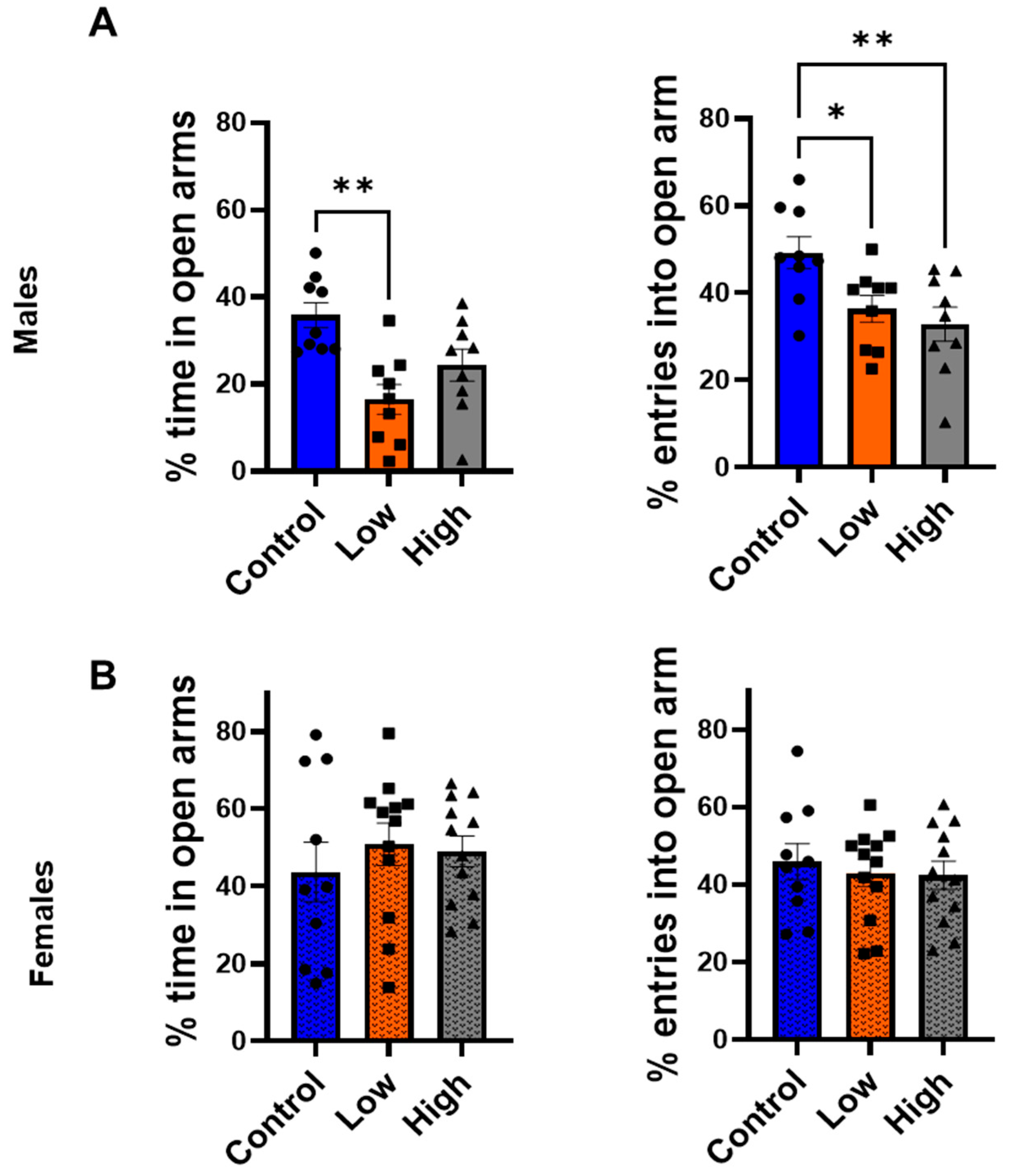
Males displayed increased anxiety-like behavior in both the EPM and light/dark transition test, with greater effects observed in low-dose individuals compared to controls. In the EPM (Figure 3A), both high- (32.9 ± 3.9%, p = 0.009) and low-dose (36.4 ± 3.0%, p = 0.04) subjects made significantly fewer percentage of entries into open arms than controls (49.2 ± 3.7%; F (2,24) = 5.87; one-way ANOVA with Tukey’s post-tests), but only low-dose males (16.4 ± 3.4%) spent significantly less time than controls for the percent of total time spent in in the open arms (36.8 ± 2.8%, p = 0.001; F (2,24) = 8.48; one-way ANOVA with Tukey’s post-tests). No significant differences were detected in females in the EPM (Figure 3B) (F (2,31) = 0.2212, p = 0.8).
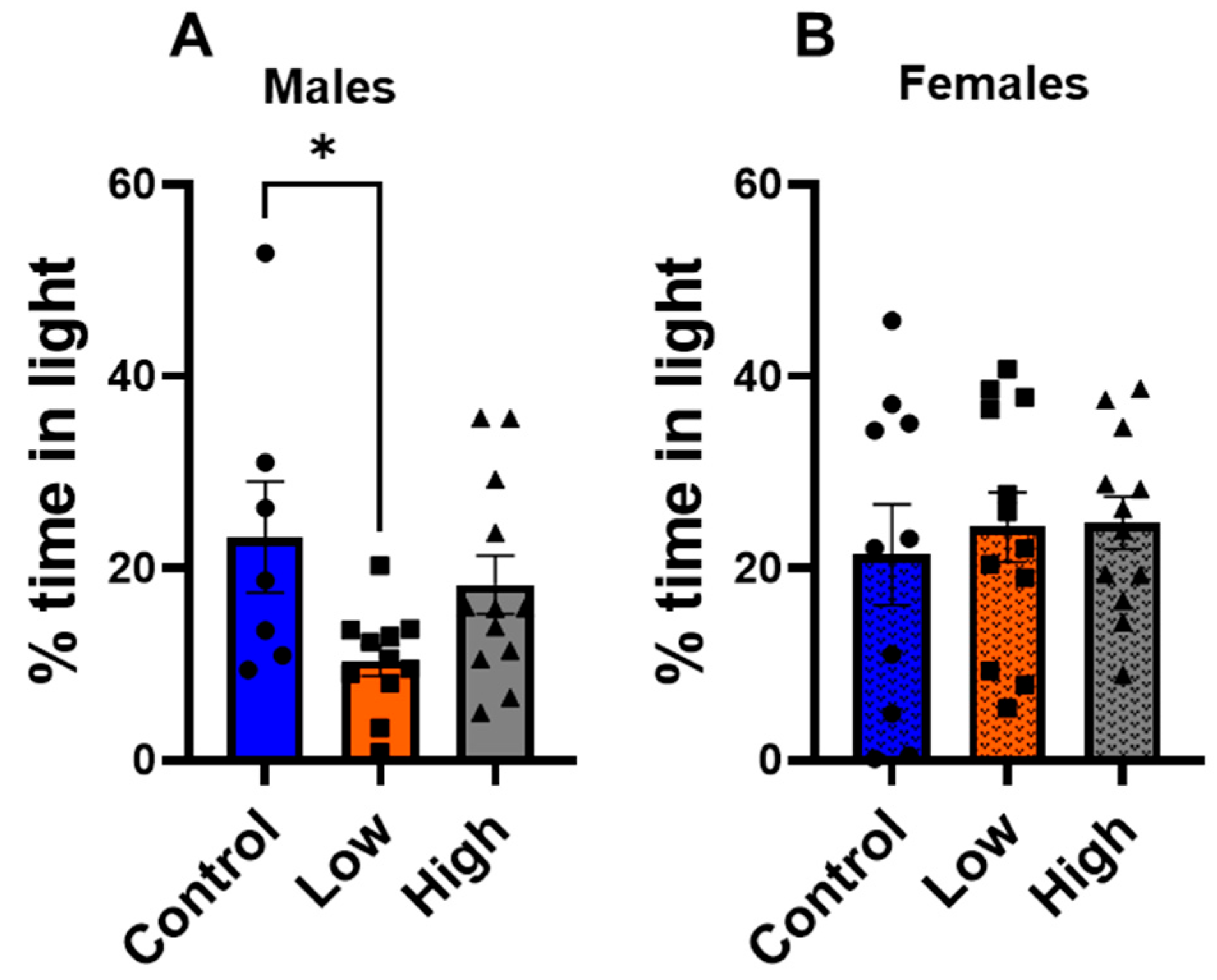
In the light/dark transition test (Figure 4), low-dose males (10.4 ± 1.6%) spent significantly less time in the illuminated half of the box than controls (25.3 ± 5.8%; F (2,27) = 3.59; p = 0.04; one-way ANOVA with Tukey’s post-test), but high-dose males were not statistically different from controls (18.3 ± 3.0%; p = 0.57). Again, IMI-exposed females did not differ from controls in this test (Figure 4B) (F (2,31) = 0.2068, p = 0.8).

3.4. Prenatal IMI Did Not Affect Depression-like Behavior in Either Sex

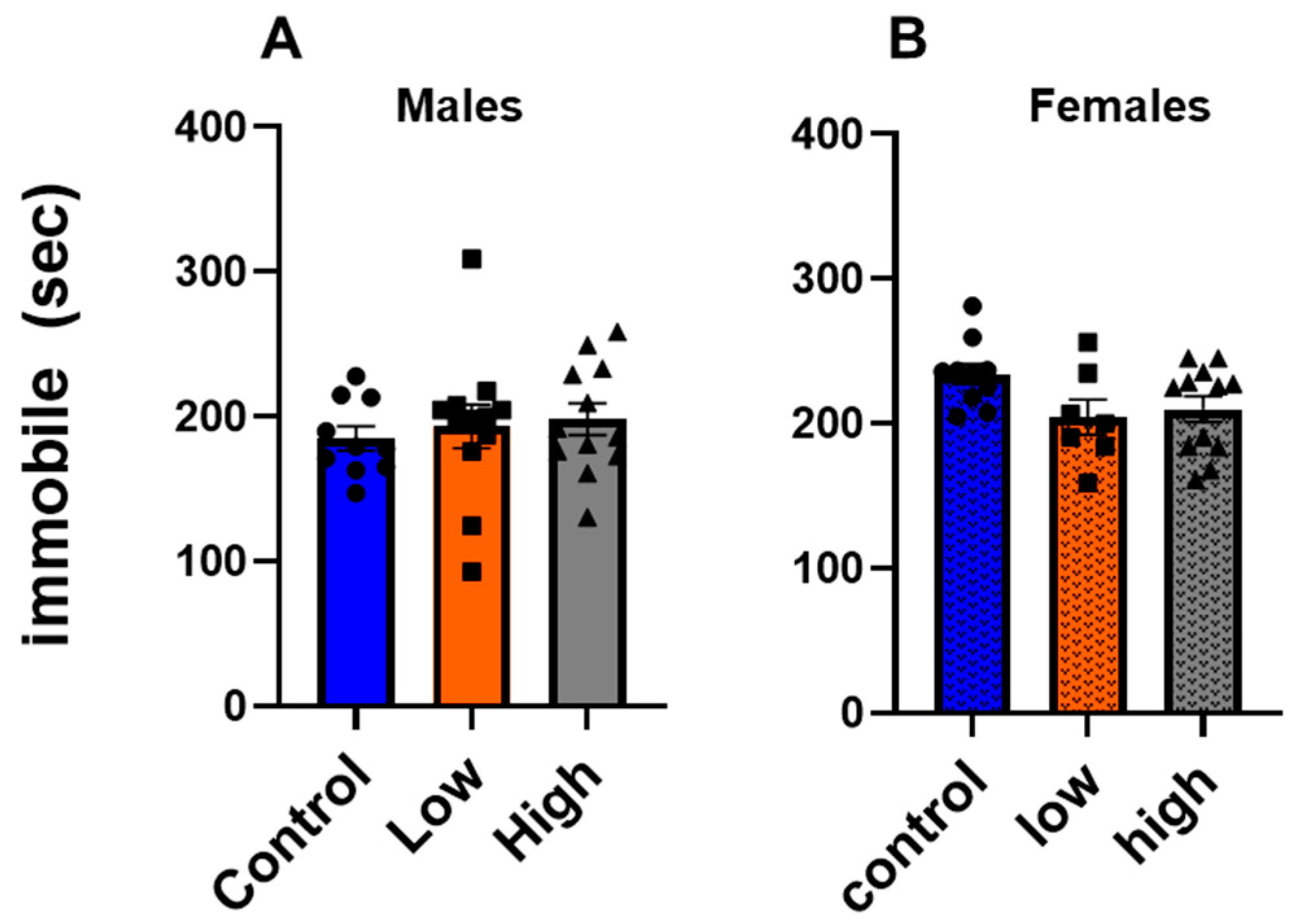
In the tail suspension test (Figure 5), we did not find any effect of prenatal IMI exposure in either males (F (2,31) = 0.2915, p = 0.74) or females (F (2,26) = 2.824; p = 0.077, one-way ANOVA with Tukey’s post-tests).

3.5. Prenatal IMI Did Not Influence Working Memory in Either Sex

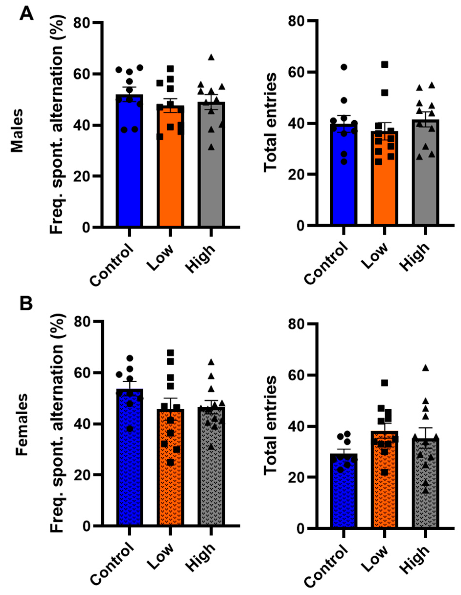
In the Y-maze, IMI exposure did not significantly alter the frequency of spontaneous alternations in either males (Figure 6A; F (2,28) = 0.60; p = 0.56; one-way ANOVA) or females (Figure 6B; F [(2,30) = 1.60; p = 0.22; one-way ANOVA). Additionally, IMI exposure did not significantly affect the number of entries for either males (Figure 6A; F (2,28) = 0.35; p = 0.71; one-way ANOVA) or females (Figure 6B; F (2,30) = 1.95; p = 0.22; one-way ANOVA) compared to controls.

3.6. Prenatal IMI Exposure Altered α7 and α4 nAChR Expression in Males but Not Females

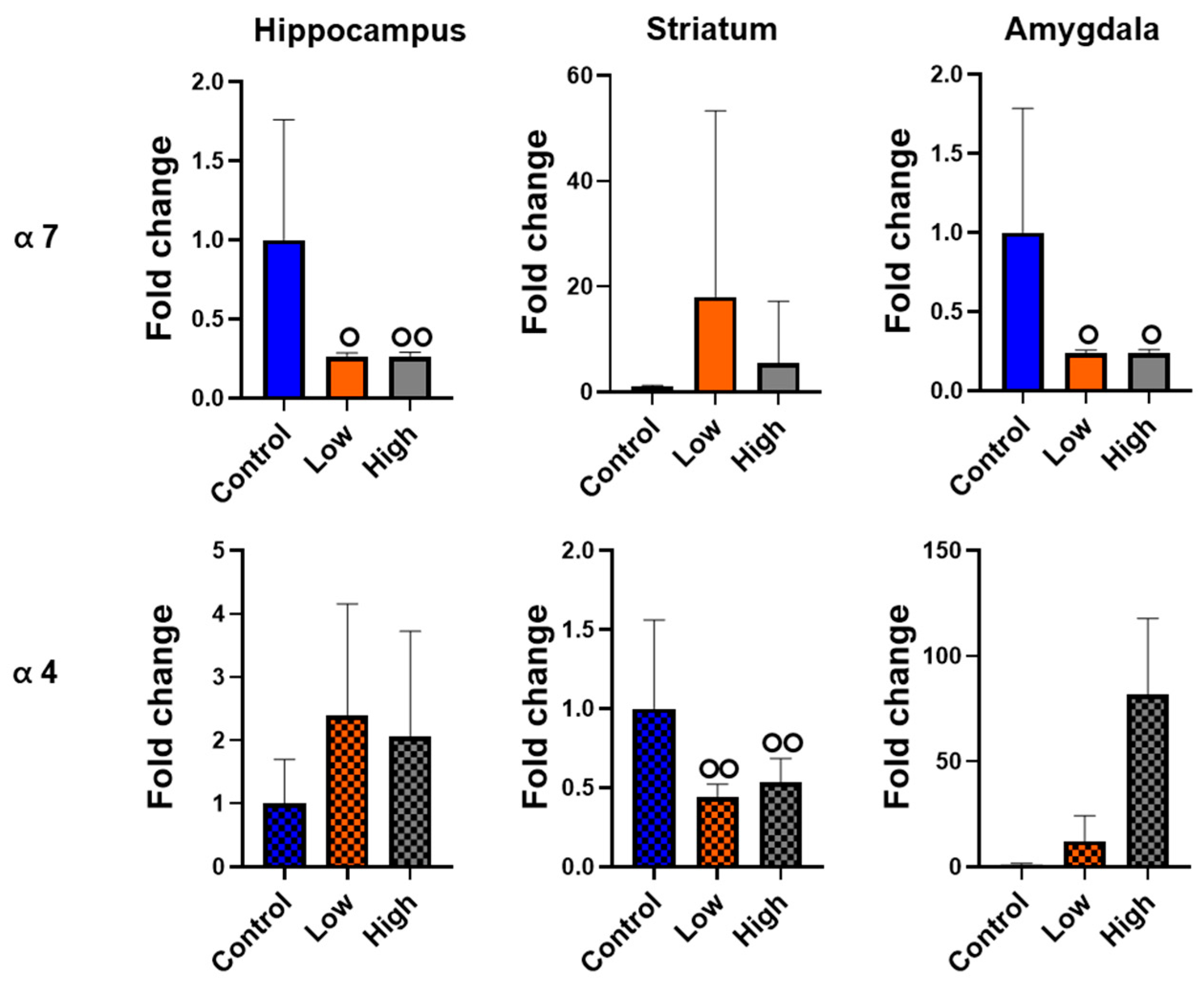
We performed qPCR for α7 or α4 nAChR subunits in the hippocampus, striatum, and amygdala. For males, we did not find a statistical difference in any comparison using ANOVA. However, Cohen’s D effect size analysis revealed hippocampal α7 expression had both large (control vs. high dose; Cohen’s D = 0.86) and medium (control vs. low dose; Cohen’s D = 0.65) effect sizes, striatal α4 expression had large effect sizes for both prenatal IMI doses (Cohen’s Ds: control vs. low dose = 1.62; control vs. high dose = 1.13), and amygdala α7 expression had medium effect sizes for both doses (Cohen’s Ds: control vs. low dose = 0.59; control vs. high dose = 0.78; Figure 7). IMI exposure did not significant differences in the expression of either receptor in the brains of females when we analyzed the data with an ANOVA or with Cohen’s D effect size analysis (Figure 8).

4. Discussion

This study examined how prenatal exposure to relatively low doses of IMI affects anxiety- and depression-like behaviors, procedural memory, working memory, and spatial memory in male and female mice. We found spatial memory and procedural memory were impacted by prenatal IMI in both males and females (Figure 1 and Figure 2). Working memory, measured by the spontaneous alternation, was not altered by IMI (Figure 6) nor was depression-like behavior (Figure 5) compared to controls. However, anxiety-like behavior was impacted by prenatal IMI exposure, but only in males (Figure 3 and Figure 4). Interestingly, IMI affected nAChR expression in the brains of male but not female mice (Figure 7 and Figure 8), suggesting a potential mechanism underlying the observed behavioral differences, although this needs additional investigation. Previous studies have demonstrated that prenatal neonicotinoid exposure impairs sensorimotor processes [47,68,69], and our work demonstrates IMI effects on more demanding cognitive tasks. Additionally, we demonstrate that these effects of IMI can be produced solely from in utero exposure, demonstrating powerful lifelong consequences that occur from IMI exposure during a critical period of development.
Prenatal IMI exposure impairs multiple forms of learning as measured by decreased acquisition of spatial memory in the hippocampal-dependent Barnes maze. For both sexes, prenatal IMI exposure increased the time to locate the entry hole for 2 of the first 3 training days, but individuals had successfully learned the task by day 4. We did not see an effect of IMI exposure on all days of training, possibly due to individual variation in learning acquisition in a cognitively challenging task. No significant differences were observed in the probe trial, indicating that hippocampal-dependent short term reference memory was not significantly affected by IMI exposure [60]. Our data are consistent with reports that other neonicotinoids impact memory. Male, but not female mice exposed to clothianidin as adults spent an increased amount of time learning the location of an escape hole in the Barnes Maze task compared to controls indicating dysfunctions in memory [52]. Additionally, adult male rats exposed to the neonicotinoid acetamiprid (20 mg and 40 mg/kg/day) for 28 days spent more time finding the escape platform in the Morris Water maze when compared to females [70]. Our work uses a prenatal exposure paradigm and thus suggests neonicotinoid exposure at multiple ages can cause negative effects on memory.
Although IMI exposure affected hippocampal dependent spatial learning in the Barnes maze (Figure 1), it did not affect spontaneous alternation on the Y-maze, a hippocampal-dependent working memory task (Figure 6). This difference may reflect the lesser cognitive demand of the Y-maze task, as it does not require learning. This lack of an effect in the Y-maze is observed in other studies with neonicotinoids [33,71]. Although, our Y-maze results differ from those of Mudgal et al. [72] who found that exposure to IMI for 28 days in adult animals significantly reduced the frequency of spontaneous alternations in male Swiss albino mice. However, Mudgal et al. used a much higher concentration of IMI than the current study (45 mg/kg BW/day), and their control mice performed spontaneous alternations at a much higher rate (close to 80% vs. 52.1 ± 3.0% [males] and 53.8 ± 2.9% [females]), so it is unclear if the differences between our experiments reflect the difference between adult and prenatal exposure windows, dose, or differences in experimental details such as mouse strain.
In the striatum-dependent T-maze task, IMI exposure impaired performance for both sexes, but males appeared more strongly affected than females (Figure 2) as IMI-exposed males displayed impairments in both training and on the testing day, whereas females only showed impairments on the testing day. In contrast, in the Barnes maze, IMI-exposed subjects eventually reached the same level of competency as controls but needed more training than the controls to do so. Providing extra training on the T-maze would allow us to see if a similar pattern holds true for procedural learning and provide greater resolution into how IMI impairs learning.
Males were also more affected by prenatal IMI exposure in tests of anxiety-like behavior. In both the EPM (Figure 3) and light-dark transition task (Figure 4) males exposed to IMI had an increase in anxiety-like behavior compared to controls. This was not observed in females and is consistent with the findings of Kubo et al. [52]. A single dose of clothianidin (5 mg/kg and 50 mg/kg) increased anxiety-like behaviors as measured by reduced exploration in the Open Field test and reduced time and entries into open arms in the EPM in males but not in females [52]. We cannot determine from our data how this sex difference is produced, but it may involve differences in dopaminergic signaling in the amygdala. Male mice have significantly more dopaminergic boutons in the basolateral amygdala than female mice [73], and nAChR activation enhances dopamine release in the amygdala [74], so males may experience greater IMI-induced dysregulation of this signaling than females. The cholinergic system also plays a role in modulating anxiety and there are sex differences with respect to nAChR expression and acetylcholine release, thus this system is another potential target for IMI [75,76,77]. Alternatively, recent studies suggest that male rats generally demonstrate greater anxiety-like behavior than females on the EPM [78], so it may be that our experiments using females did not adequately capture intergroup differences in anxiety. Finally, another potential mechanism could be sex differences in detoxifying enzymes. The P450 enzymes play a role in neonicotinoid resistance in insects [79,80] and there are known sex differences in the expression of specific enzymes in house flies [81] and rodents [82]. For example, the brains of male rats express higher mRNA for Cype2c11, Cyp2c13, Cyp1a1, and Cyp4a3 compared to females, and this may be a mechanism underlying the outcomes in the current study [82].
Our EPM and light–dark transition data also suggest IMI follows a non-linear dose response curve with respect to male anxiety-like behaviors. Only the low-dose IMI group displayed differences on the light-dark transition task (Figure 4), and the low-dose IMI group displayed differences on both parameters in the EPM (Figure 3), whereas the high-dose IMI group only displayed differences for percent of entries into open arms. It is well documented that environmental chemical exposure can produce U-shaped dose–response curves with low doses having different effects than relatively high doses [83,84]. We hypothesize that this non-linear dose effect occurs with respect to IMI, but this requires additional testing. Further, chemicals including IMI exert different effects in different tissues, cell types, and brain regions [26,58,85].
We found reduced mRNA transcript expression of the nAChR α7 in the hippocampus and amygdala of males, and a reduction in α4 transcript in the striatum of males. The reduced expression of these receptors in the male tissues mirrors the effects produced by prenatal nicotine exposure [45,86], and may reflect homeostatic responses to excessive activation of nAChRs in the prenatal period. This reduced expression may be partially responsible for the learning deficits in males. However, our data are correlational, and further mechanistic studies are needed. The relationship between α7 expression in the amygdala and increased anxiety-like behavior is less clear, however, as reductions in cholinergic signaling typically reduce these behaviors [87,88]. However, these data do not necessarily indicate a lack of nAChR-related effects on cognition. Receptor expression is only one component of neurotransmission. Endogenous acetylcholine release, currents evoked by acetylcholine binding, cholinesterase activity, intrinsic excitability of nAChR-expressing neurons, and postsynaptic neurotransmitter release can all be altered by IMI exposure [71,89]. Changes to any of these could cause increased anxiety-like behavior in males and could also influence procedural and spatial learning.
The lack of effects of IMI on female neural α7 and α4 subunit expression are puzzling given that females demonstrated some impairments in the learning tasks. One explanation is that IMI alters other nAChRs; whereas α7 and α4β2 are heavily implicated in learning and memory, other isoforms of the receptors exist [90]. Additionally, IMI may affect the plasticity of nAChR expression (i.e., the ability to change nAChR expression as an individual learns a task). IMI may affect cognition by mechanisms such as altering the endocrine system indirectly leading to changes in cognition. Additionally, numerous other pesticides have multiple pharmacological effects beyond their intended effects. For example, organophosphates, designed to prevent cholinergic degradation at the synapse, also enhance protein kinase A activity [91,92]. Modeling studies show a single toxicant can have strong affinity for dozens of proteins in a single organ [93], so it would not be surprising if a similar phenomenon occurs with IMI exposure in the brain. However, it is important to note the small sample size in the qPCR studies, and we did not quantify the receptor proteins. Other limitations in our work include the potential for dams to alter their maternal care or have heightened stress in response to treatment with IMI.
Future work could investigate the impact of IMI exposure on other candidates such as detoxifying P450 enzymes [82] and markers of neuronal proliferation [71], or mechanistic studies of receptor pharmacology and assays of neurotransmitters. Ultimately, it is likely that the effects of IMI derive from some combination of the above hypotheses and dysregulation of nAChR-mediated signaling [71]. Determining these mechanisms will be essential, as this insight will be necessary to develop therapies to combat IMI’s behavioral effects.

5. Conclusions

This study provides additional evidence that prenatal exposure to IMI at relatively low doses can impact anxiety-like behavior and cognition and that sex differences exist in response to prenatal IMI exposures. The effects of neonicotinoids on nAChR-mediated signaling require additional exploration to identify mechanistic insight into these effects within the brain.

Supplementary Materials

The following supporting information can be downloaded at: https://www.mdpi.com/article/10.3390/toxics13110918/s1, Figure S1: Spatial memory performance on the probe trial for the Barnes maze (mean +SEM) for males and females. Animals were prenatally exposed to vehicle (Control) or IMI at either a low (0.5 mg/kg/day) or high (5.7 mg/kg/day) dose. Animals were tested on the Barnes maze for 4 days followed by the probe trial on the 5th day. Data represent the duration of seconds for the subject to locate the goal hole. Symbols represent individual animals and circles, squares and triangles represent control, low, and high dose animals, respectively. Sample sizes were 10–12 for males and 9–12 for females.

Author Contributions

Conceptualization, C.L., M.M.M. and V.E.M.; methodology, C.L., V.E.M. and M.M.M.; validation, C.L. and M.M.M.; formal analysis, M.M.M., C.L., J.Q. and T.P.; investigation, C.L., J.Q. and T.P.; resources, M.M.M.; data curation, C.L., T.P. and J.Q.; writing—original draft preparation, C.L. and M.M.M.; writing—review and editing, C.L., J.Q., T.P., V.E.M., J.A.F. and M.M.M.; visualization, C.L. and M.M.M.; supervision, M.M.M. and J.A.F.; project administration, M.M.M. and C.L.; funding acquisition, M.M.M., J.A.F. and V.E.M. All authors have read and agreed to the published version of the manuscript.

Funding

This research was funded by NIH T32 ES007326 to C.L., NIH R25 ES025059 to J.Q., NIH F30 ES033914 to V.E.M., NIH R21 ES036520 to J.A.F., and University of Illinois RB22093 and NIH R03 ES035189 to M.M.M.

Institutional Review Board Statement

The animal study protocol was approved by the Institutional Animal Care and Use Committee (protocol 22157, approved September 2022).

Informed Consent Statement

Not applicable.

Data Availability Statement

All relevant data of this study are given in the manuscript. Additional data will be provided upon request.

Acknowledgments

The authors would like to thank Ella Gusinde, Nirmal Nathan, Fatima Hussein, Kendall McFarlin, and Aneta Dzielska for help with behavioral analyses and Mary Laws for assistance with RT-PCR.

Conflicts of Interest

The authors declare no conflicts of interest. The funders had no role in the design of the study; in the collection, analyses, or interpretation of data; in the writing of the manuscript; or in the decision to publish the results.

Abbreviations

The following abbreviations are used in this manuscript:
EPMElevated Plus Maze
IMIImidacloprid
nAChRNicotinic Acetylcholine Receptors
NOAELNo Observable Adverse Effects Level
SCMSweetened Condensed Milk

References

  1. Tomizawa, M.; Casida, J.E. Neonicotinoid insecticide toxicology: Mechanisms of selective action. Annu. Rev. Pharmacol. Toxicol. 2005, 45, 247–268. [Google Scholar] [CrossRef]
  2. Tan, J.; Galligan, J.J.; Hollingworth, R.M. Agonist actions of neonicotinoids on nicotinic acetylcholine receptors expressed by cockroach neurons. Neurotoxicology 2007, 28, 829–842. [Google Scholar] [CrossRef]
  3. Yamamoto, I.; Yabuta, G.; Tomizawa, M.; Saito, T.; Miyamoto, T.; Kagabu, S. Molecular Mechanism for Selective Toxicity of Nicotinoids and Neonicotinoids. J. Pestic. Sci. 1995, 20, 33–40. [Google Scholar] [CrossRef]
  4. Douglas, M.R.; Tooker, J.F. Large-scale deployment of seed treatments has driven rapid increase in use of neonicotinoid insecticides and preemptive pest management in US field crops. Environ. Sci. Technol. 2015, 49, 5088–5097. [Google Scholar] [CrossRef] [PubMed]
  5. Perkins, R.; Goulson, D. To flea or not to flea: Survey of UK companion animal ectoparasiticide usage and activities affecting pathways to the environment. PeerJ 2023, 11, e15561. [Google Scholar] [CrossRef] [PubMed]
  6. Covert, S.A.; Shoda, M.E.; Stackpoole, S.M.; Stone, W.W. Pesticide mixtures show potential toxicity to aquatic life in US streams, water years 2013–2017. Sci. Total Environ. 2020, 745, 141285. [Google Scholar] [CrossRef]
  7. Wan, Y.; Han, Q.; Wang, Y.; He, Z. Five degradates of imidacloprid in source water, treated water, and tap water in Wuhan, central China. Sci. Total Environ. 2020, 741, 140227. [Google Scholar] [CrossRef]
  8. Hladik, M.L.; Main, A.R.; Goulson, D. Environmental Risks and Challenges Associated with Neonicotinoid Insecticides. Environ. Sci. Technol. 2018, 52, 3329–3335. [Google Scholar] [CrossRef]
  9. Craddock, H.A.; Huang, D.; Turner, P.C.; Quiros-Alcala, L.; Payne-Sturges, D.C. Trends in neonicotinoid pesticide residues in food and water in the United States, 1999–2015. Environ. Health 2019, 18, 7. [Google Scholar] [CrossRef] [PubMed]
  10. Batikian, C.M.; Lu, A.; Watanabe, K.; Pitt, J.; Gersberg, R.M. Temporal pattern in levels of the neonicotinoid insecticide, imidacloprid, in an urban stream. Chemosphere 2019, 223, 83–90. [Google Scholar] [CrossRef]
  11. Struger, J.; Grabuski, J.; Cagampan, S.; Sverko, E.; McGoldrick, D.; Marvin, C.H. Factors influencing the occurrence and distribution of neonicotinoid insecticides in surface waters of southern Ontario, Canada. Chemosphere 2017, 169, 516–523. [Google Scholar] [CrossRef]
  12. Perkins, R.; Barron, L.; Glauser, G.; Whitehead, M.; Woodward, G.; Goulson, D. Down-the-drain pathways for fipronil and imidacloprid applied as spot-on parasiticides to dogs: Estimating aquatic pollution. Sci. Total Environ. 2024, 917, 170175. [Google Scholar] [CrossRef] [PubMed]
  13. Sadaria, A.M.; Sutton, R.; Moran, K.D.; Teerlink, J.; Brown, J.V.; Halden, R.U. Passage of fiproles and imidacloprid from urban pest control uses through wastewater treatment plants in northern California, USA. Environ. Toxicol. Chem. 2017, 36, 1473–1482. [Google Scholar] [CrossRef]
  14. Sadaria, A.M.; Supowit, S.D.; Halden, R.U. Mass Balance Assessment for Six Neonicotinoid Insecticides During Conventional Wastewater and Wetland Treatment: Nationwide Reconnaissance in United States Wastewater. Environ. Sci. Technol. 2016, 50, 6199–6206. [Google Scholar] [CrossRef]
  15. Baker, S.E.; Serafim, A.B.; Morales-Agudelo, P.; Vidal, M.; Calafat, A.M.; Ospina, M. Quantification of DEET and neonicotinoid pesticide biomarkers in human urine by online solid-phase extraction high-performance liquid chromatography-tandem mass spectrometry. Anal. Bioanal. Chem. 2019, 411, 669–678. [Google Scholar] [CrossRef]
  16. Ospina, M.; Wong, L.-Y.; Baker, S.E.; Serafim, A.B.; Morales-Agudelo, P.; Calafat, A.M. Exposure to neonicotinoid insecticides in the U.S. general population: Data from the 2015–2016 national health and nutrition examination survey. Environ. Res. 2019, 176, 108555. [Google Scholar] [CrossRef]
  17. Zhang, D.; Lu, S. Human exposure to neonicotinoids and the associated health risks: A review. Environ. Int. 2022, 163, 107201. [Google Scholar] [CrossRef]
  18. Laubscher, B.; Diezi, M.; Renella, R.; Mitchell, E.A.D.; Aebi, A.; Mulot, M.; Glauser, G. Multiple neonicotinoids in children’s cerebro-spinal fluid, plasma, and urine. Environ. Health 2022, 21, 10. [Google Scholar] [CrossRef] [PubMed]
  19. Zhang, H.; Wang, Y.; Zhu, H.; Lu, S.; Wang, Y.; Xue, J.; Zhang, T.; Kannan, K.; Sun, H. Infantile Internal and External Exposure to Neonicotinoid Insecticides: A Comparison of Levels across Various Sources. Environ. Sci. Technol. 2023, 57, 5358–5367. [Google Scholar] [CrossRef] [PubMed]
  20. Botnaru, A.A.; Lupu, A.; Morariu, P.C.; Jitareanu, A.; Nedelcu, A.H.; Morariu, B.A.; Anton, E.; Di Gioia, M.L.; Lupu, V.V.; Dragostin, O.M.; et al. Neurotoxic Effects of Pesticides: Implications for Neurodegenerative and Neurobehavioral Disorders. J. Xenobiot. 2025, 15, 83. [Google Scholar] [CrossRef]
  21. Sass, J.B.; Donley, N.; Freese, W. Neonicotinoid pesticides: Evidence of developmental neurotoxicity from regulatory rodent studies. Front. Toxicol. 2024, 6, 1438890. [Google Scholar] [CrossRef]
  22. Wang, X.; Anadon, A.; Wu, Q.H.; Qiao, F.; Ares, I.; Martinez-Larranaga, M.R.; Yuan, Z.H.; Martinez, M.A. Mechanism of Neonicotinoid Toxicity: Impact on Oxidative Stress and Metabolism. Annu. Rev. Pharmacol. Toxicol. 2018, 58, 471–507. [Google Scholar] [CrossRef]
  23. Zou, X.; Ebizuka, Y.; Sakamaki, Y.; Shobudani, M.; Tang, Q.; Luo, M.; Kobayashi, M.; Kigata, T.; Shibutani, M. Progressive motor dysfunction and loss of cerebellar Purkinje and granule cells in rat offspring after maternal exposure to imidacloprid. Toxicology 2025, 518, 154246. [Google Scholar] [CrossRef] [PubMed]
  24. Yan, S.; Meng, Z.Y.; Tian, S.N.; Teng, M.M.; Yan, J.; Jia, M.; Li, R.S.; Zhou, Z.Q.; Zhu, W.T. Neonicotinoid insecticides exposure cause amino acid metabolism disorders, lipid accumulation and oxidative stress in ICR mice. Chemosphere 2020, 246, 125661. [Google Scholar] [CrossRef]
  25. Duzguner, V.; Erdogan, S. Acute oxidant and inflammatory effects of imidacloprid on the mammalian central nervous system and liver in rats. Pestic. Biochem. Physiol. 2010, 97, 13–18. [Google Scholar] [CrossRef]
  26. Kapoor, U.; Srivastava, M.K.; Srivastava, L.P. Toxicological impact of technical imidacloprid on ovarian morphology, hormones and antioxidant enzymes in female rats. Food Chem. Toxicol. 2011, 49, 3086–3089. [Google Scholar] [CrossRef]
  27. Sardar, A.; David, M.; Jahan, S.; Afsar, T.; Ahmad, A.; Ullah, A.; Almajwal, A.; Shafique, H.; Razak, S. Determination of biochemical and histopathological changes on testicular and epididymis tissues induced by exposure to insecticide Imidacloprid during postnatal development in rats. BMC Pharmacol. Toxicol. 2023, 24, 68. [Google Scholar] [CrossRef] [PubMed]
  28. Mourikes, V.E.; Santacruz-Marquez, R.; Deviney, A.; Laws, M.J.; Ulanov, A.V.; La Frano, M.R.; Flaws, J.A. Ovarian antral follicles metabolize imidacloprid in vitro. Toxicol. Sci. 2023, 196, 229–237. [Google Scholar] [CrossRef]
  29. Thompson, D.A.; Lehmler, H.J.; Kolpin, D.W.; Hladik, M.L.; Vargo, J.D.; Schilling, K.E.; LeFevre, G.H.; Peeples, T.L.; Poch, M.C.; LaDuca, L.E.; et al. A critical review on the potential impacts of neonicotinoid insecticide use: Current knowledge of environmental fate, toxicity, and implications for human health. Environ. Sci. Process. Impacts 2020, 22, 1315–1346. [Google Scholar] [CrossRef]
  30. Bal, R.; Turk, G.; Tuzcu, M.; Yilmaz, O.; Kuloglu, T.; Gundogdu, R.; Gur, S.; Agca, A.; Ulas, M.; Cambay, Z.; et al. Assessment of imidacloprid toxicity on reproductive organ system of adult male rats. J. Environ. Sci. Health Part B 2012, 47, 434–444. [Google Scholar] [CrossRef]
  31. Mourikes, V.E.; Lee, C.; Oladosu, J.I.; Deviney, A.; Stubblefield, W.; Laws, M.J.; Mahoney, M.; Flaws, J.A. The effects of imidacloprid exposure on the mouse ovary in vivo. Reprod. Toxicol. 2025, 137, 109045. [Google Scholar] [CrossRef]
  32. Badgujar, P.C.; Jain, S.K.; Singh, A.; Punia, J.S.; Gupta, R.P.; Chandratre, G.A. Immunotoxic effects of imidacloprid following 28 days of oral exposure in BALB/c mice. Environ. Toxicol. Pharmacol. 2013, 35, 408–418. [Google Scholar] [CrossRef]
  33. Namba, K.; Tominaga, T.; Ishihara, Y. Decreases in the number of microglia and neural circuit dysfunction elicited by developmental exposure to neonicotinoid pesticides in mice. Environ. Toxicol. 2024, 39, 3944–3955. [Google Scholar] [CrossRef]
  34. Loser, D.; Grillberger, K.; Hinojosa, M.G.; Blum, J.; Haufe, Y.; Danker, T.; Johansson, Y.; Möller, C.; Nicke, A.; Bennekou, S.H.; et al. Acute effects of the imidacloprid metabolite desnitro-imidacloprid on human nACh receptors relevant for neuronal signaling. Arch. Toxicol. 2021, 95, 3695–3716. [Google Scholar] [CrossRef]
  35. Kimura-Kuroda, J.; Komuta, Y.; Kuroda, Y.; Hayashi, M.; Kawano, H. Nicotine-Like Effects of the Neonicotinoid Insecticides Acetamiprid and Imidacloprid on Cerebellar Neurons from Neonatal Rats. PLoS ONE 2012, 7, e32432. [Google Scholar] [CrossRef] [PubMed]
  36. Davis, J.A.; Gould, T.J. Hippocampal nAChRs mediate nicotine withdrawal-related learning deficits. Eur. Neuropsychopharmacol. 2009, 19, 551–561. [Google Scholar] [CrossRef] [PubMed]
  37. Fujii, S.; Ji, Z.; Sumikawa, K. Inactivation of alpha7 ACh receptors and activation of non-alpha7 ACh receptors both contribute to long term potentiation induction in the hippocampal CA1 region. Neurosci. Lett. 2000, 286, 134–138. [Google Scholar] [CrossRef]
  38. Mahai, G.; Wan, Y.; Xia, W.; Wang, A.; Qian, X.; Li, Y.; He, Z.; Li, Y.; Xu, S. Exposure assessment of neonicotinoid insecticides and their metabolites in Chinese women during pregnancy: A longitudinal study. Sci. Total Environ. 2022, 818, 151806. [Google Scholar] [CrossRef] [PubMed]
  39. Buckley, J.P.; Kuiper, J.R.; Bennett, D.H.; Barrett, E.S.; Bastain, T.; Breton, C.V.; Chinthakindi, S.; Dunlop, A.L.; Farzan, S.F.; Herbstman, J.B.; et al. Exposure to Contemporary and Emerging Chemicals in Commerce among Pregnant Women in the United States: The Environmental influences on Child Health Outcome (ECHO) Program. Environ. Sci. Technol. 2022, 56, 6560–6573. [Google Scholar] [CrossRef]
  40. Pan, C.; Yu, J.; Yao, Q.; Lin, N.; Lu, Z.; Zhang, Y.; Zhao, S.; Wang, Z.; Lei, X.; Tian, Y.; et al. Prenatal neonicotinoid insecticides Exposure, oxidative Stress, and birth outcomes. Environ. Int. 2022, 163, 107180. [Google Scholar] [CrossRef]
  41. Saito, H.; Furukawa, Y.; Sasaki, T.; Kitajima, S.; Kanno, J.; Tanemura, K. Behavioral effects of adult male mice induced by low-level acetamiprid, imidacloprid, and nicotine exposure in early-life. Front. Neurosci. 2023, 17, 1239808. [Google Scholar] [CrossRef] [PubMed]
  42. Raja, G.L.; Subhashree, K.D.; Kantayya, K.E. In utero exposure to endocrine disruptors and developmental neurotoxicity: Implications for behavioural and neurological disorders in adult life. Environ. Res. 2022, 203, 111829. [Google Scholar] [CrossRef]
  43. Gavini, K.; Yang, E.; Parameshwaran, K. Developmental nicotine exposure impairs memory and reduces acetylcholine levels in the hippocampus of mice. Brain Res. Bull. 2021, 176, 1–7. [Google Scholar] [CrossRef]
  44. Wang, H.; Davila-Garcia, M.I.; Yarl, W.; Gondre-Lewis, M.C. Gestational nicotine exposure regulates expression of AMPA and NMDA receptors and their signaling apparatus in developing and adult rat hippocampus. Neuroscience 2011, 188, 168–181. [Google Scholar] [CrossRef]
  45. Li, J.; Bo, L.; Zhang, P.; Gao, Q.; Li, L.; Tang, J.; Wu, C.; Li, D.; Xiao, J.; Chen, J.; et al. Exposure to nicotine during pregnancy and altered learning and memory in the rat offspring. Nicotine Tob. Res. 2015, 17, 661–666. [Google Scholar] [CrossRef]
  46. Gold, A.B.; Keller, A.B.; Perry, D.C. Prenatal exposure of rats to nicotine causes persistent alterations of nicotinic cholinergic receptors. Brain Res. 2009, 1250, 88–100. [Google Scholar] [CrossRef] [PubMed]
  47. Abou-Donia, M.B.; Goldstein, L.B.; Bullman, S.; Tu, T.; Khan, W.A.; Dechkovskaia, A.M.; Abdel-Rahman, A.A. Imidacloprid Induces Neurobehavioral Deficits and Increases Expression of Glial Fibrillary Acidic Protein in the Motor Cortex and Hippocampus in Offspring Rats Following in Utero Exposure. J. Toxicol. Environ. Health Part A 2008, 71, 119–130. [Google Scholar] [CrossRef]
  48. Bhaskar, R.; Mishra, A.K.; Mohanty, B. Neonatal Exposure to Endocrine Disrupting Chemicals Impairs Learning Behaviour by Disrupting Hippocampal Organization in Male Swiss Albino Mice. Basic Clin. Pharmacol. Toxicol. 2017, 121, 44–52. [Google Scholar] [CrossRef]
  49. Hirano, T.; Yanai, S.; Takada, T.; Yoneda, N.; Omotehara, T.; Kubota, N.; Minami, K.; Yamamoto, A.; Mantani, Y.; Yokoyama, T.; et al. NOAEL-dose of a neonicotinoid pesticide, clothianidin, acutely induce anxiety-related behavior with human-audible vocalizations in male mice in a novel environment. Toxicol. Lett. 2018, 282, 57–63. [Google Scholar] [CrossRef]
  50. Hirai, A.; Sugio, S.; Nimako, C.; Nakayama, S.M.M.; Kato, K.; Takahashi, K.; Arizono, K.; Hirano, T.; Hoshi, N.; Fujioka, K.; et al. Ca2+ imaging with two-photon microscopy to detect the disruption of brain function in mice administered neonicotinoid insecticides. Sci. Rep. 2022, 12, 5114. [Google Scholar] [CrossRef] [PubMed]
  51. Shoda, A.; Murata, M.; Kimura, M.; Hara, Y.; Yonoichi, S.; Ishida, Y.; Mantani, Y.; Yokoyama, T.; Hirano, T.; Ikenaka, Y.; et al. Transgenerational effects of developmental neurotoxicity induced by exposure to a no-observed-adverse-effect level (NOAEL) of neonicotinoid pesticide clothianidin. J. Vet. Med. Sci. 2023, 85, 1023–1029. [Google Scholar] [CrossRef]
  52. Kubo, S.; Hirano, T.; Miyata, Y.; Ohno, S.; Onaru, K.; Ikenaka, Y.; Nakayama, S.M.M.; Ishizuka, M.; Mantani, Y.; Yokoyama, T.; et al. Sex-specific behavioral effects of acute exposure to the neonicotinoid clothianidin in mice. Toxicol. Appl. Pharmacol. 2022, 456, 116283. [Google Scholar] [CrossRef]
  53. Sano, K.; Isobe, T.; Yang, J.; Win-Shwe, T.T.; Yoshikane, M.; Nakayama, S.F.; Kawashima, T.; Suzuki, G.; Hashimoto, S.; Nohara, K.; et al. In utero and Lactational Exposure to Acetamiprid Induces Abnormalities in Socio-Sexual and Anxiety-Related Behaviors of Male Mice. Front. Neurosci. 2016, 10, 228. [Google Scholar] [CrossRef]
  54. Kaku, K.; Sasaki, T.; Hara, K.; Tanemura, K. A single dose of clothianidin exposure induces varying sex-specific behavioral changes in adulthood depending on the developmental stage of its administration. J. Toxicol. Sci. 2024, 49, 301–311. [Google Scholar] [CrossRef] [PubMed]
  55. Zhang, T.; Song, S.; Bai, X.; He, Y.; Zhang, B.; Gui, M.; Kannan, K.; Lu, S.; Huang, Y.; Sun, H. A nationwide survey of urinary concentrations of neonicotinoid insecticides in China. Environ. Int. 2019, 132, 105114. [Google Scholar] [CrossRef]
  56. Godbole, A.M.; Moonie, S.; Coughenour, C.; Zhang, C.; Chen, A.; Vuong, A.M. Exploratory analysis of the associations between neonicotinoids and measures of adiposity among US adults: NHANES 2015–2016. Chemosphere 2022, 300, 134450. [Google Scholar] [CrossRef]
  57. Arfat, Y.; Mahmood, N.; Tahir, M.U.; Rashid, M.; Anjum, S.; Zhao, F.; Li, D.J.; Sun, Y.L.; Hu, L.; Zhihao, C.; et al. Effect of imidacloprid on hepatotoxicity and nephrotoxicity in male albino mice. Toxicol. Rep. 2014, 1, 554–561. [Google Scholar] [CrossRef] [PubMed]
  58. Kapoor, U.; Srivastava, M.K.; Trivedi, P.; Garg, V.; Srivastava, L.P. Disposition and acute toxicity of imidacloprid in female rats after single exposure. Food Chem. Toxicol. 2014, 68, 190–195. [Google Scholar] [CrossRef]
  59. Passoni, A.; Mariani, A.; Comolli, D.; Fanelli, R.; Davoli, E.; De Paola, M.; Bagnati, R. An integrated approach, based on mass spectrometry, for the assessment of imidacloprid metabolism and penetration into mouse brain and fetus after oral treatment. Toxicology 2021, 462, 152935. [Google Scholar] [CrossRef] [PubMed]
  60. Gawel, K.; Gibula, E.; Marszalek-Grabska, M.; Filarowska, J.; Kotlinska, J.H. Assessment of spatial learning and memory in the Barnes maze task in rodents—Methodological consideration. Naunyn-Schmiedeberg’s Arch. Pharmacol. 2019, 392, 1–18. [Google Scholar] [CrossRef]
  61. Deacon, R.M.J.; Rawlins, J.N.P. T-maze alternation in the rodent. Nat. Protoc. 2006, 1, 7–12. [Google Scholar] [CrossRef]
  62. Murithi, C.K.; Kabaru, J.M.; Patel, N.B. Catha edulis Forsk (khat) reduces spontaneous and rewarded alternation in female mice. IBRO Rep. 2020, 9, 270–275. [Google Scholar] [CrossRef]
  63. Can, A.; Dao, D.T.; Terrillion, C.E.; Piantadosi, S.C.; Bhat, S.; Gould, T.D. The Tail Suspension Test. J. Vis. Exp. 2011, 59, e3769. [Google Scholar] [CrossRef] [PubMed]
  64. Mourikes, V.E.; Santacruz-Marquez, R.; Deviney, A.; Neff, A.; Laws, M.J.; Flaws, J.A. Neonicotinoids differentially modulate nicotinic acetylcholine receptors in immature and antral follicles in the mouse ovary. Biol. Reprod. 2024, 111, 472–482. [Google Scholar] [CrossRef] [PubMed]
  65. Pfaffl, M.W. A new mathematical model for relative quantification in real-time RT-PCR. Nucleic Acids Res. 2001, 29, e45. [Google Scholar] [CrossRef]
  66. Sullivan, G.M.; Feinn, R. Using Effect Size—Or Why the P Value Is Not Enough. J. Grad. Med. Educ. 2012, 4, 279–282. [Google Scholar] [CrossRef]
  67. Cohen, J. A Power Primer. Psychol. Bull. 1992, 112, 155–159. [Google Scholar] [CrossRef] [PubMed]
  68. Tanaka, T. Reproductive and neurobehavioral effects of clothianidin administered to mice in the diet. Birth Defects Res. Part B Dev. Reprod. Toxicol. 2012, 95, 151–159. [Google Scholar] [CrossRef]
  69. Tanaka, T. Effects of maternal clothianidin exposure on behavioral development in F(1) generation mice. Toxicol. Ind. Health 2012, 28, 697–707. [Google Scholar] [CrossRef]
  70. Shamsi, M.; Soodi, M.; Shahbazi, S.; Omidi, A. Effect of Acetamiprid on spatial memory and hippocampal glutamatergic system. Environ. Sci. Pollut. Res. 2021, 28, 27933–27941. [Google Scholar] [CrossRef]
  71. Zou, X.; Tang, Q.; Ojiro, R.; Ozawa, S.; Shobudani, M.; Sakamaki, Y.; Ebizuka, Y.; Jin, M.; Yoshida, T.; Shibutani, M. Increased spontaneous activity and progressive suppression of adult neurogenesis in the hippocampus of rat offspring after maternal exposure to imidacloprid. Chem.-Biol. Interact. 2024, 399, 111145. [Google Scholar] [CrossRef]
  72. Mudgal, R.; Sharma, S.; Singh, S.; Ravichandiran, V. The neuroprotective effect of ascorbic acid against imidacloprid-induced neurotoxicity and the role of HO-1 in mice. Front. Neurol. 2023, 14, 1130575. [Google Scholar] [CrossRef]
  73. Manion, M.T.C.; Glasper, E.R.; Wang, K.H. A sex difference in mouse dopaminergic projections from the midbrain to basolateral amygdala. Biol. Sex Differ. 2022, 13, 75. [Google Scholar] [CrossRef]
  74. Palotai, M.; Bagosi, Z.; Jaszberenyi, M.; Csabafi, K.; Dochnal, R.; Manczinger, M.; Telegdy, G.; Szabo, G. Ghrelin and nicotine stimulate equally the dopamine release in the rat amygdala. Neurochem. Res. 2013, 38, 1989–1995. [Google Scholar] [CrossRef]
  75. Moen, J.K.; Lee, A.M. Sex Differences in the Nicotinic Acetylcholine Receptor System of Rodents: Impacts on Nicotine and Alcohol Reward Behaviors. Front. Neurosci. 2021, 15, 745783. [Google Scholar] [CrossRef] [PubMed]
  76. Mitsushima, D.; Takase, K.; Funabashi, T.; Kimura, F. Gonadal Steroids Maintain 24 h Acetylcholine Release in the Hippocampus: Organizational and Activational Effects in Behaving Rats. J. Neurosci. 2009, 29, 3808–3815. [Google Scholar] [CrossRef] [PubMed]
  77. Takase, K.; Kimura, F.; Yagami, T.; Mitsushima, D. Sex-specific 24-h acetylcholine release profile in the medial prefrontal cortex: Simultaneous measurement of spontaneous locomotor activity in behaving rats. Neuroscience 2009, 159, 7–15. [Google Scholar] [CrossRef]
  78. Knight, P.; Chellian, R.; Wilson, R.; Behnood-Rod, A.; Panunzio, S.; Bruijnzeel, A.W. Sex differences in the elevated plus-maze test and large open field test in adult Wistar rats. Pharmacol. Biochem. Behav. 2021, 204, 173168. [Google Scholar] [CrossRef] [PubMed]
  79. Puinean, A.M.; Foster, S.P.; Oliphant, L.; Denholm, I.; Field, L.M.; Millar, N.S.; Williamson, M.S.; Bass, C. Amplification of a Cytochrome P450 Gene Is Associated with Resistance to Neonicotinoid Insecticides in the Aphid Myzus persicae. PLoS Genet. 2010, 6, e1000999. [Google Scholar] [CrossRef]
  80. Zoh, M.G.; Gaude, T.; Prud’homme, S.M.; Riaz, M.A.; David, J.P.; Reynaud, S. Molecular bases of P450-mediated resistance to the neonicotinoid insecticide imidacloprid in the mosquito. Aquat. Toxicol. 2021, 236, 105860. [Google Scholar] [CrossRef]
  81. Højland, D.H.; Vagn Jensen, K.M.; Kristensen, M. A comparative study of P450 gene expression in field and laboratory Musca domestica L. strains. Pest Manag. Sci. 2014, 70, 1237–1242. [Google Scholar] [CrossRef]
  82. Gerges, S.H.; El-Kadi, A.O.S. Sexual Dimorphism in the Expression of Cytochrome P450 Enzymes in Rat Heart, Liver, Kidney, Lung, Brain, and Small Intestine. Drug Metab. Dispos. 2023, 51, 81–94. [Google Scholar] [CrossRef] [PubMed]
  83. Vandenberg, L.N.; Colborn, T.; Hayes, T.B.; Heindel, J.J.; Jacobs, D.R., Jr.; Lee, D.H.; Shioda, T.; Soto, A.M.; vom Saal, F.S.; Welshons, W.V.; et al. Hormones and endocrine-disrupting chemicals: Low-dose effects and nonmonotonic dose responses. Endocr. Rev. 2012, 33, 378–455. [Google Scholar] [CrossRef]
  84. Hill, C.E.; Myers, J.P.; Vandenberg, L.N. Nonmonotonic Dose-Response Curves Occur in Dose Ranges That Are Relevant to Regulatory Decision-Making. Dose-Response 2018, 16, 1559325818798282. [Google Scholar] [CrossRef] [PubMed]
  85. Hirai, A.; Yamazaki, R.; Kobayashi, A.; Kimura, T.; Nomiyama, K.; Shimma, S.; Nakayama, S.M.M.; Ishizuka, M.; Ikenaka, Y. Detection of Changes in Monoamine Neurotransmitters by the Neonicotinoid Pesticide Imidacloprid Using Mass Spectrometry. Toxics 2022, 10, 696. [Google Scholar] [CrossRef]
  86. Nunes-Freitas, A.L.; Ribeiro-Carvalho, A.; Lima, C.S.; Dutra-Tavares, A.C.; Manhaes, A.C.; Lisboa, P.C.; Oliveira, E.; Gaspar de Moura, E.; Filgueiras, C.C.; Abreu-Villaca, Y. Nicotine exposure during the third trimester equivalent of human gestation: Time course of effects on the central cholinergic system of rats. Toxicol. Sci. 2011, 123, 144–154. [Google Scholar] [CrossRef]
  87. Pang, X.; Liu, L.; Ngolab, J.; Zhao-Shea, R.; McIntosh, J.M.; Gardner, P.D.; Tapper, A.R. Habenula cholinergic neurons regulate anxiety during nicotine withdrawal via nicotinic acetylcholine receptors. Neuropharmacology 2016, 107, 294–304. [Google Scholar] [CrossRef]
  88. Kutlu, M.G.; Gould, T.J. Nicotine modulation of fear memories and anxiety: Implications for learning and anxiety disorders. Biochem. Pharmacol. 2015, 97, 498–511. [Google Scholar] [CrossRef] [PubMed]
  89. Picciotto, M.R.; Higley, M.J.; Mineur, Y.S. Acetylcholine as a Neuromodulator: Cholinergic Signaling Shapes Nervous System Function and Behavior. Neuron 2012, 76, 116–129. [Google Scholar] [CrossRef]
  90. Ho, T.N.T.; Abraham, N.; Lewis, R.J. Structure-Function of Neuronal Nicotinic Acetylcholine Receptor Inhibitors Derived from Natural Toxins. Front. Neurosci. 2020, 14, 609005. [Google Scholar] [CrossRef]
  91. Torres-Altoro, M.I.; Mathur, B.N.; Drerup, J.M.; Thomas, R.; Lovinger, D.M.; O’Callaghan, J.P.; Bibb, J.A. Organophosphates dysregulate dopamine signaling, glutamatergic neurotransmission, and induce neuronal injury markers in striatum. J. Neurochem. 2011, 119, 303–313. [Google Scholar] [CrossRef] [PubMed]
  92. Tsai, C.Y.; Wu, C.H.; Chan, S.H.; Chang, A.Y. Muscarinic receptor-independent activation of cyclic adenosine monophosphate-dependent protein kinase in rostral ventrolateral medulla underlies the sympathoexcitatory phase of cardiovascular responses during mevinphos intoxication in the rat. Shock 2007, 27, 559–564. [Google Scholar] [CrossRef] [PubMed]
  93. Montes-Grajales, D.; Olivero-Verbel, J. Structure-based Identification of Endocrine Disrupting Pesticides Targeting Breast Cancer Proteins. Toxicology 2020, 439, 152459. [Google Scholar] [CrossRef] [PubMed]
Figure 1. Spatial memory performance on the Barnes maze (mean ± SEM) for (A) males and (B) females. Animals were prenatally exposed to vehicle (Control) or IMI at either a low (0.5 mg/kg/day) or high (5.7 mg/kg/day) dose. Animals were given 4 trials/day for 4 days. Data represent the duration of seconds for the subject to locate the goal hole. * p < 0.05, ** p < 0.01, and *** p < 0.001. Symbols represent individual animals and circles, squares, and triangles represent control, low, and high dose animals, respectively. Sample sizes were 10–12 for males and 9–12 for females.
Toxics 13 00918 g001
Figure 2. Procedural memory performance using forced alternation in the T-maze (mean ± SEM) for (A) males and (B) females. Animals were prenatally exposed to vehicle (Control) or IMI at either a low (0.5 mg/kg/day) or high (5.7 mg/kg/day) dose. Animals were given a forced choice, followed by 10 trials/day for 3 days and on testing day they were given 10 trials without a forced choice. Data are plotted as the frequency of correct alternations relative to Day 1. * p < 0.05, ** p < 0.01. Symbols represent individual animals and circles, squares, and triangles represent control, low, and high dose animals, respectively. Sample sizes were 9–10 for males and 4–9 for females.
Toxics 13 00918 g002
Figure 3. Anxiety-like behavior was tested using Elevated plus maze (EPM) (mean ± SEM) for (A) males and (B) females. We measured percent of time spent in the open arms and percent of entries into open arms. * p < 0.05, ** p < 0.01. Symbols represent individual animals and circles, squares, and triangles represent control, low, and high dose animals, respectively. Sample sizes were 9–10 for males and 10–12 for females.
Toxics 13 00918 g003
Figure 4. Anxiety-like behavior was tested in the light–dark transition test (mean ± SEM) for (A) males and (B) females. We measured percent of time spent in the light portion of the box. * p < 0.05. Symbols represent individual animals and circles, squares, and triangles represent control, low, and high dose animals, respectively. Sample sizes were 10–12 for males and 7–12 for females.
Toxics 13 00918 g004
Figure 5. Depression-like behavior was tested in the tail suspension test for (A) males and (B) females. Data (average + SEM) is plotted as duration of time spent immobile (sec). Symbols represent individual animals and circles, squares, and triangles represent control, low, and high dose animals, respectively. Sample sizes were 10–12 for males and 7–12 for females.
Toxics 13 00918 g005
Figure 6. Working memory performance tested in the Y-maze (mean ± SEM) for (A) males and (B) females. Animals were prenatally exposed to vehicle (Control) or IMI at either a low (0.5 mg/kg/day) or high (5.7 mg/kg/day) dose. The percent of correct spontaneous alternation and the total number of entries were calculated for the last 8 min of a 10 min test. Symbols represent individual animals and circles, squares, and triangles represent control, low, and high dose animals, respectively. Sample sizes were 10–11 for males and 9–13 for females.
Toxics 13 00918 g006
Figure 7. The relative expression of the α-7 subunit (top) and α-4 subunit (bottom) of nAChR in the hippocampus, striatum, and amygdala of male mice. Data are represented as mean +SEM and are expressed relative to controls. Data were analyzed with Cohen’s D; one circle indicates a medium effect and 2 circles indicate a relatively large effect size compared to controls. Sample sizes: n = 3 control, 5 low dose, 7 high dose.
Toxics 13 00918 g007
Figure 8. The relative expression of the α-7 subunit (top) and α-4 subunit (bottom) of nAChR in the hippocampus, striatum, and amygdala of female mice. Data are represented as mean +SEM and are expressed relative to controls. Data were analyzed with Cohen’s D. Sample sizes: n = 4 control, 6 low dose, 8 high dose.
Toxics 13 00918 g008
Disclaimer/Publisher’s Note: The statements, opinions and data contained in all publications are solely those of the individual author(s) and contributor(s) and not of MDPI and/or the editor(s). MDPI and/or the editor(s) disclaim responsibility for any injury to people or property resulting from any ideas, methods, instructions or products referred to in the content.