

Exploration of the Pathogenesis of Chronic Obstructive Pulmonary Disease Caused by Smoking—Based on Bioinformatics Analysis and In Vitro Experimental Evidence


Abstract
:1. Introduction
2. Materials and Methods
2.1. Data Retrieval
2.2. Screening of COPD-Related Differentially Expressed miRNA and Prediction of Target Genes
2.3. Analysis of Functional Enrichment of Target Genes Regulated by COPD-Related miRNAs
2.4. Construction of Interaction Network of Target Gene Proteins Regulated by COPD-Related miRNAs
2.5. Experimental Validation of Target Genes Regulated by COPD-Related miRNAs
2.5.1. Cell Culture
2.5.2. Construction of COPD Cell Model
2.5.3. Determination of mRNA Expression Levels
2.5.4. Determination of the Protein Contents
2.6. Statistical Analysis
3. Results
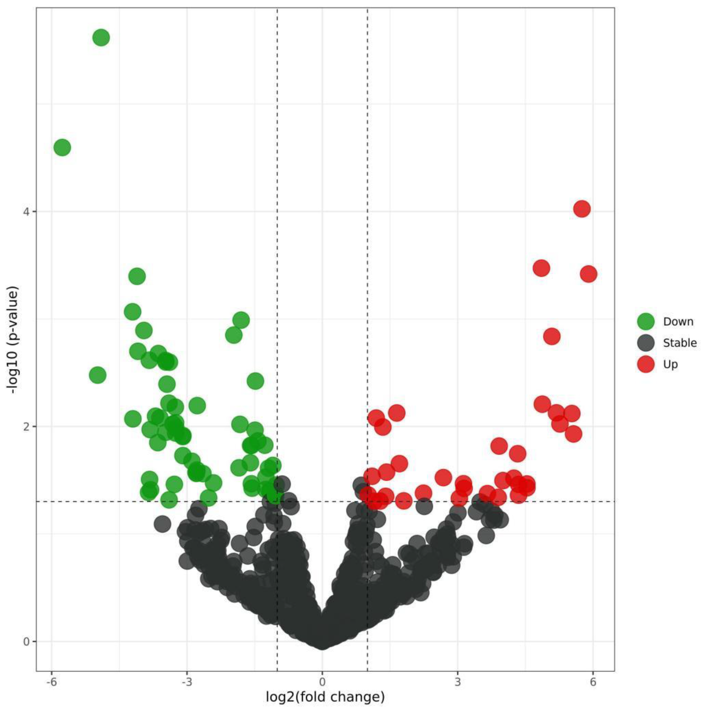
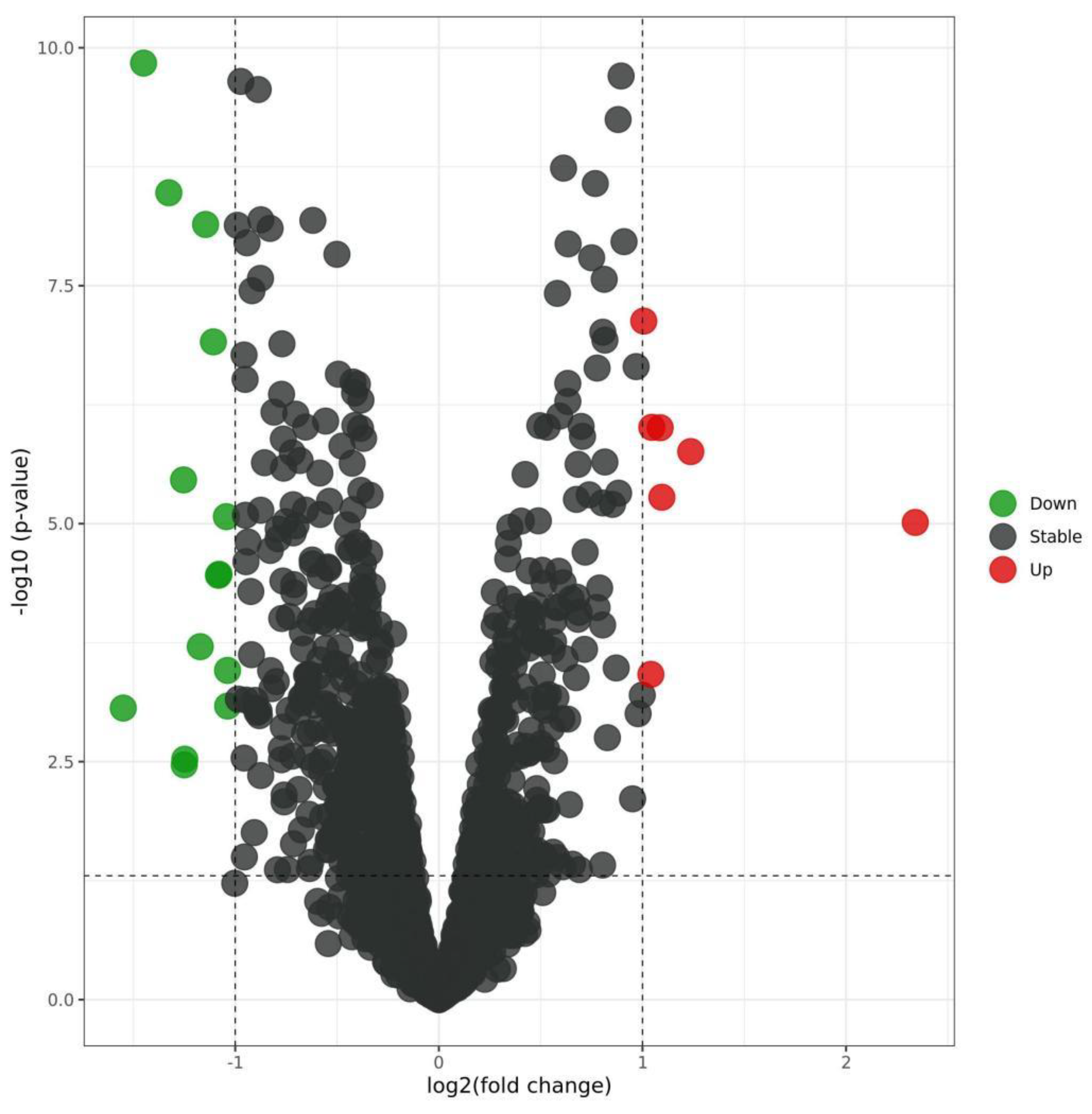
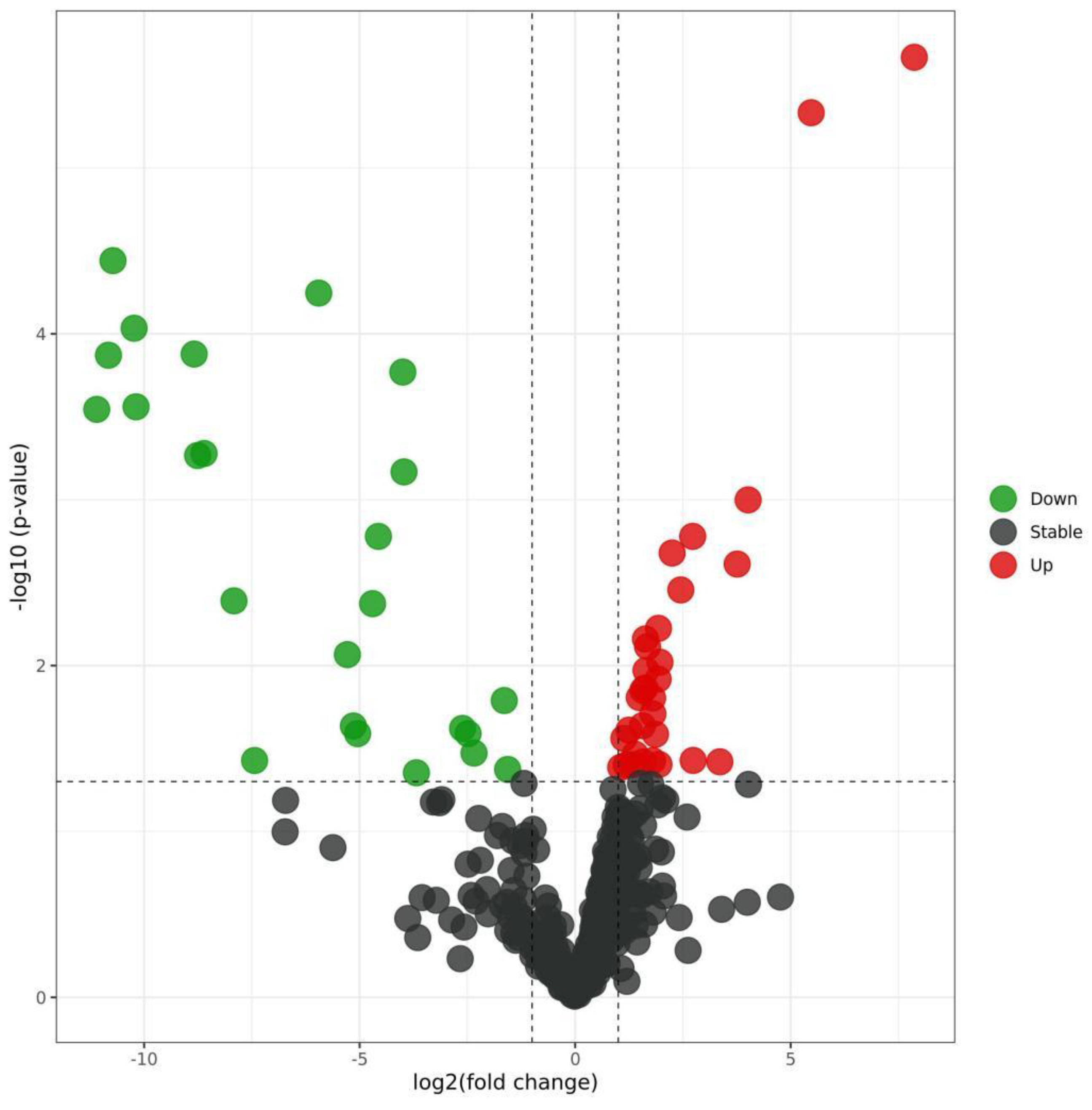
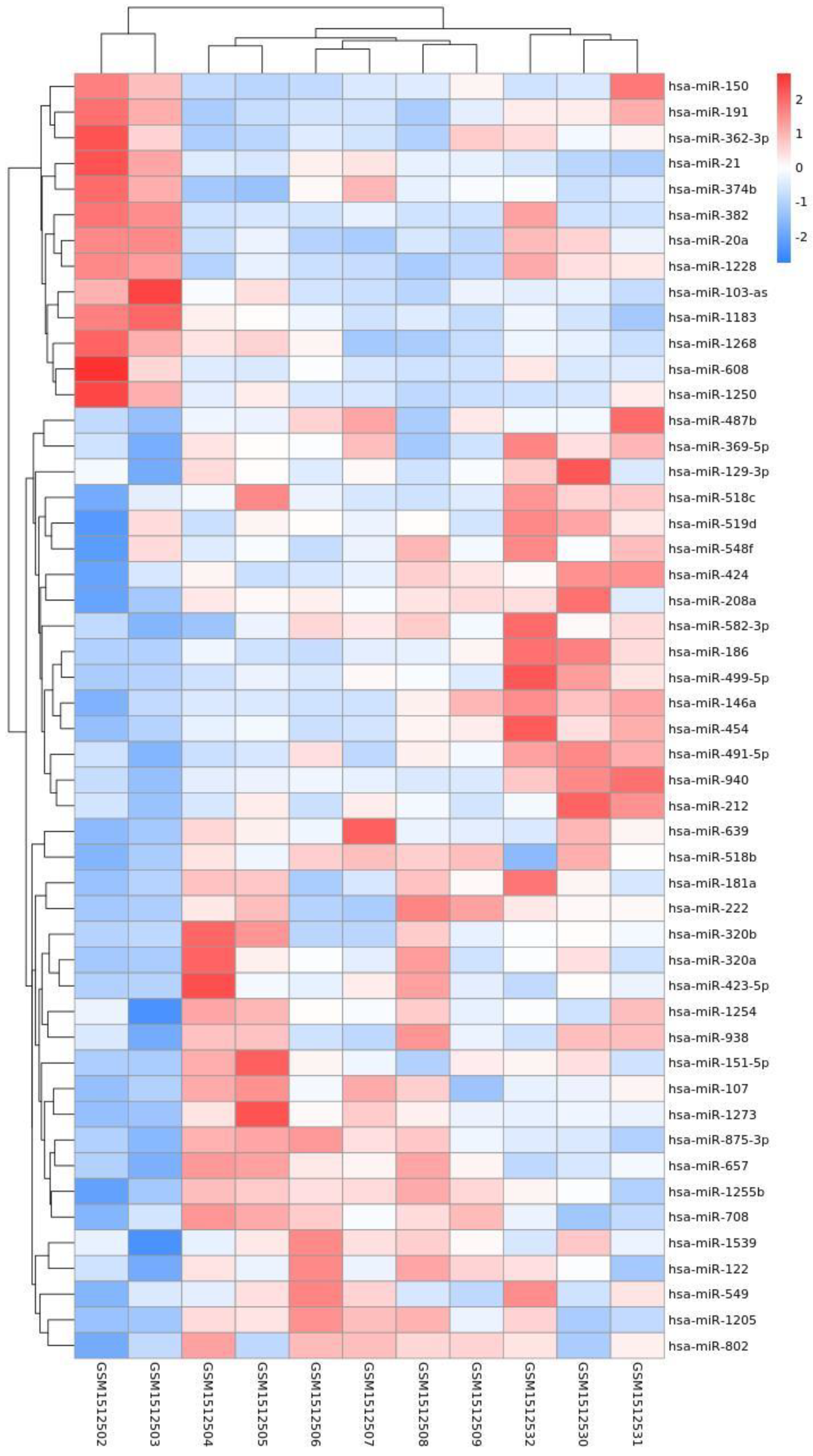
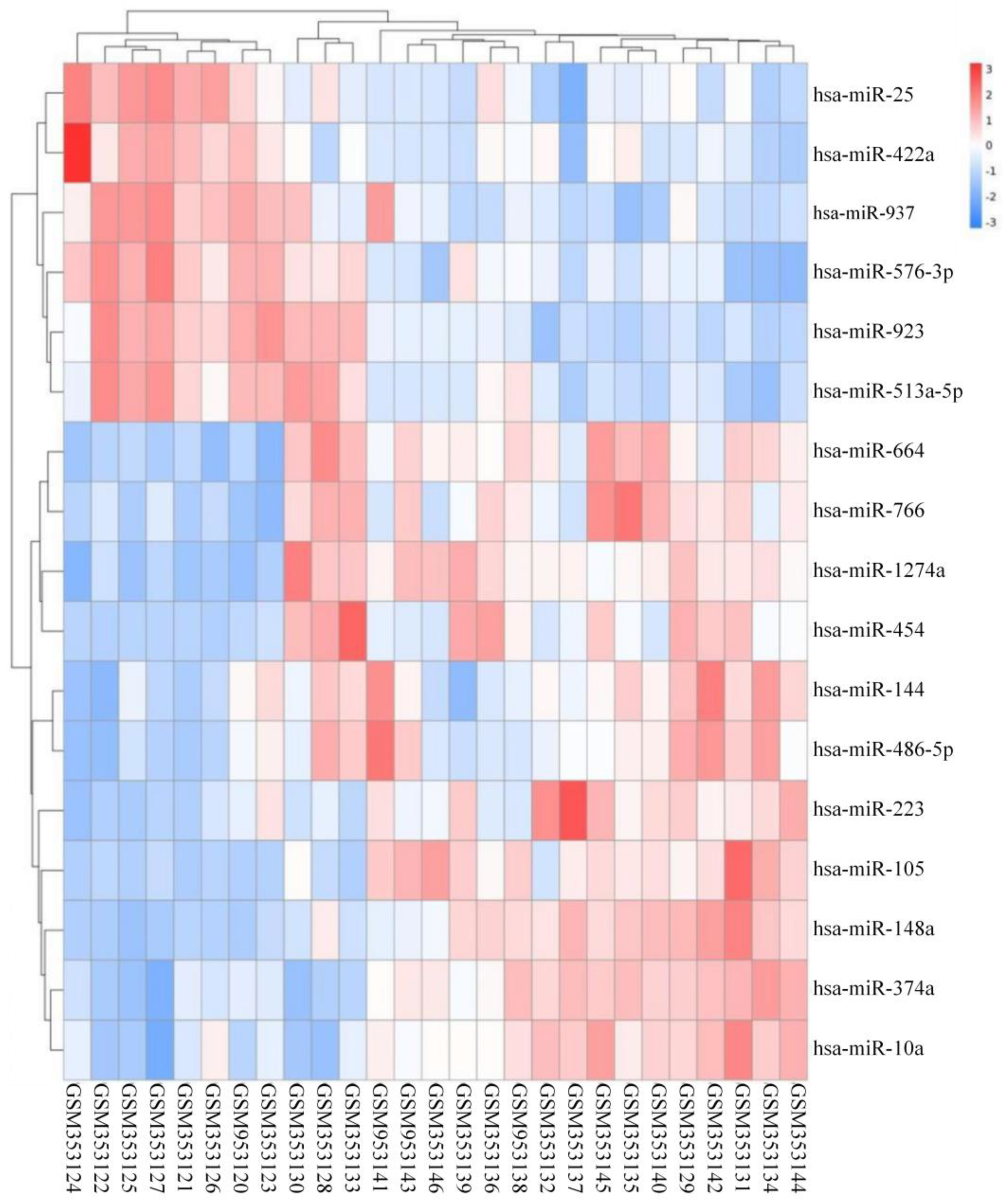
3.1. Differentially Expressed miRNAs in COPD
3.2. Screening of COPD-Related Differentially Expressed miRNA and Prediction of Target Genes
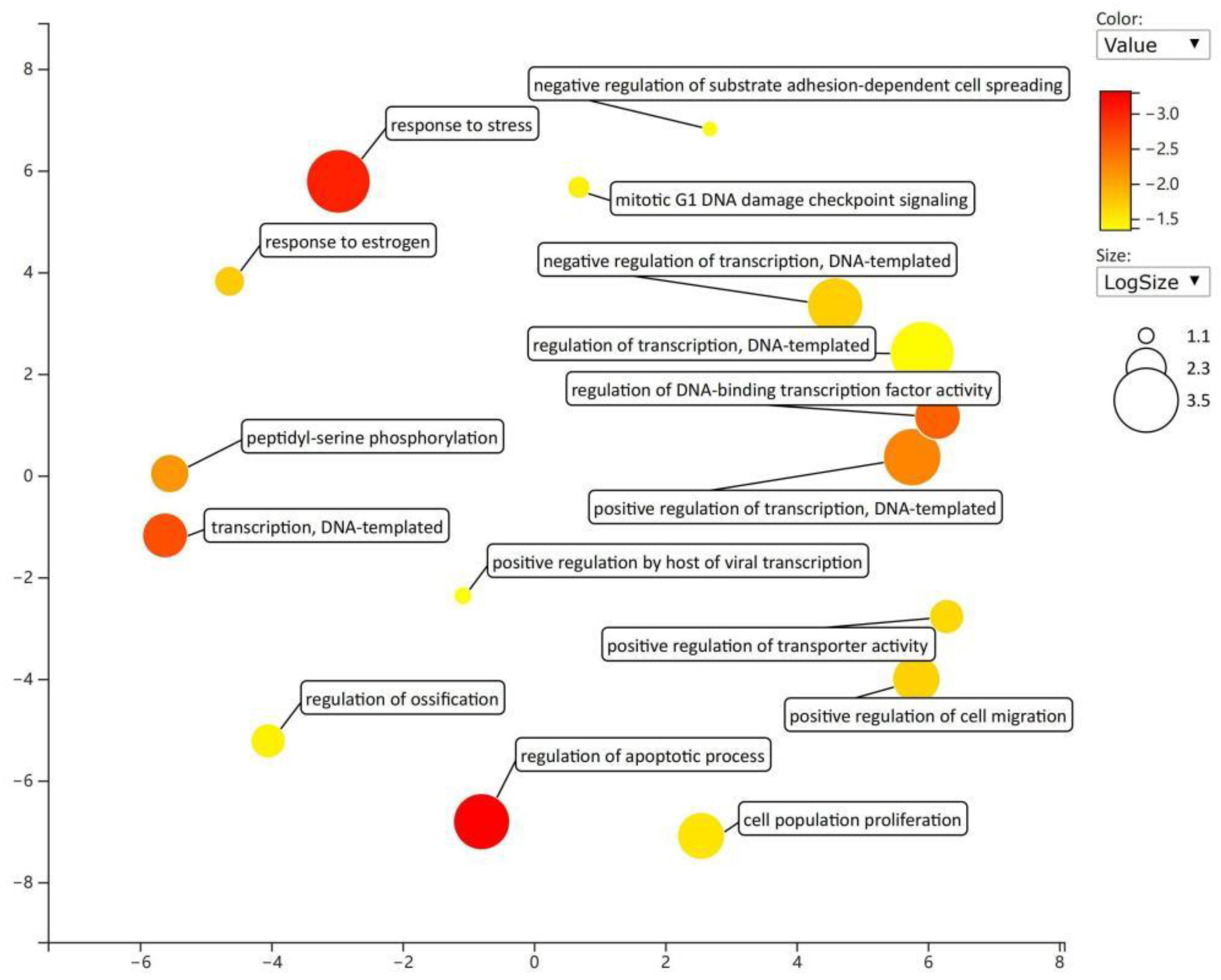
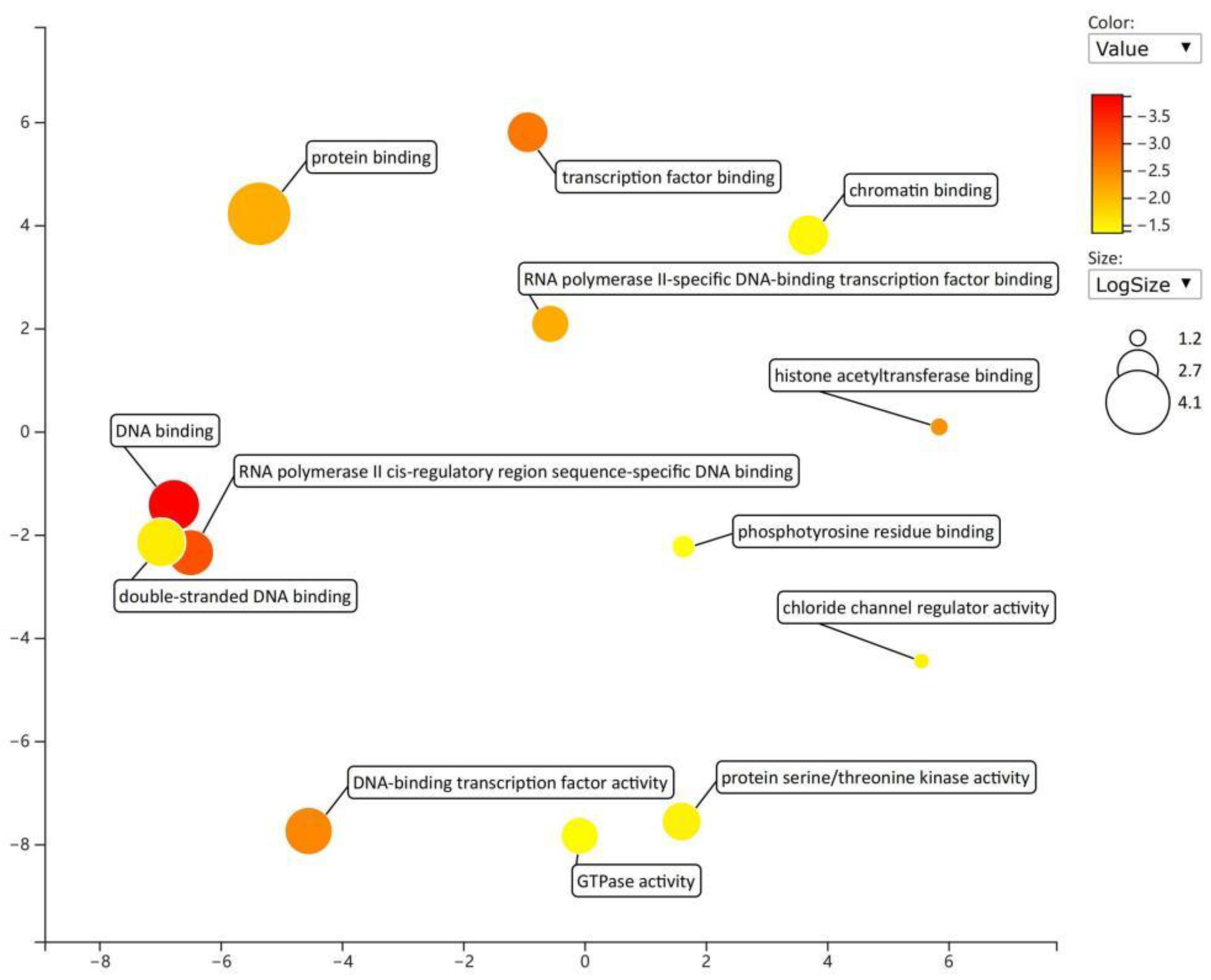
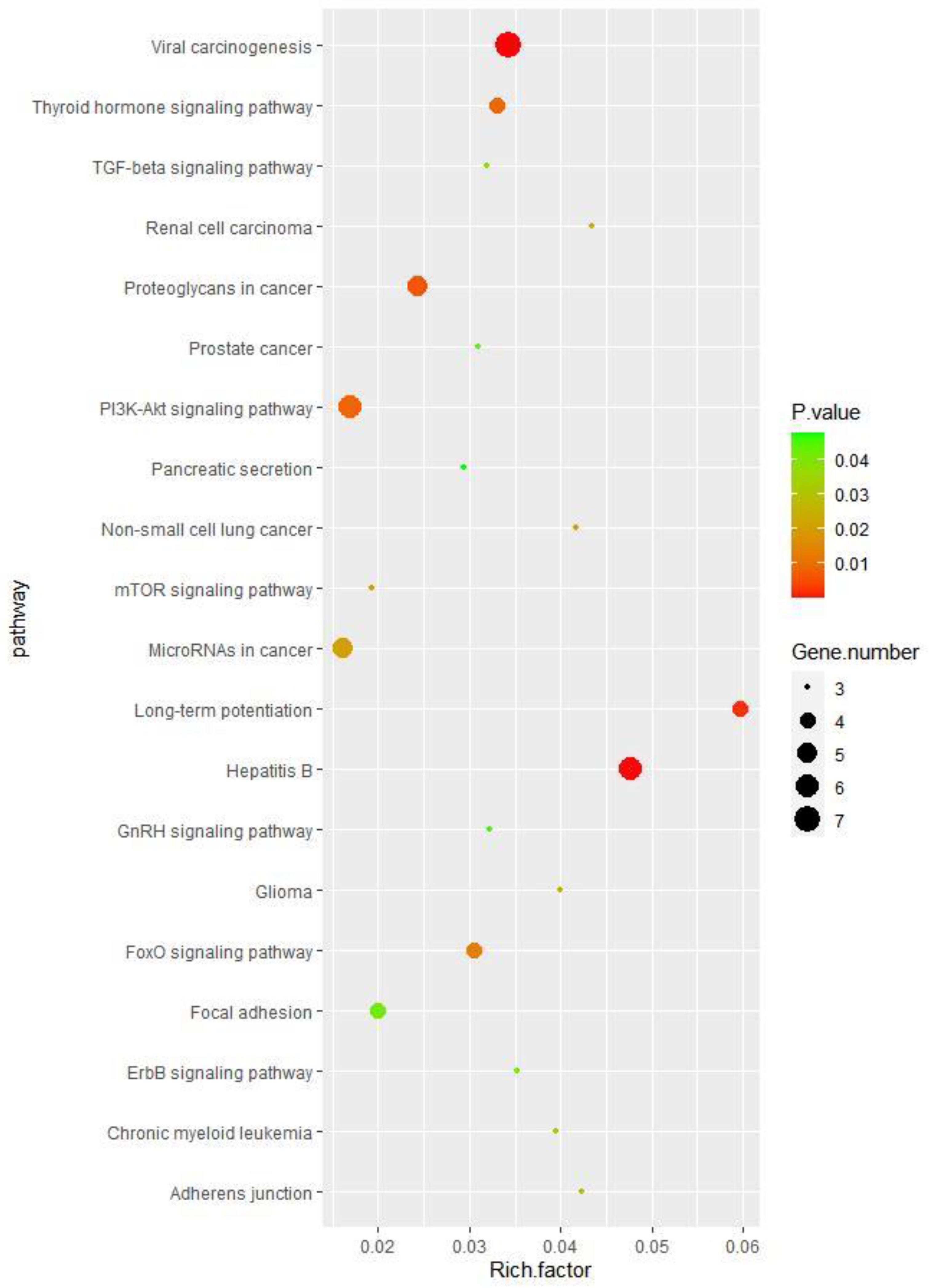
3.3. Functional Analysis Results of Target Genes with Differentially Expressed miRNAs Related to COPD
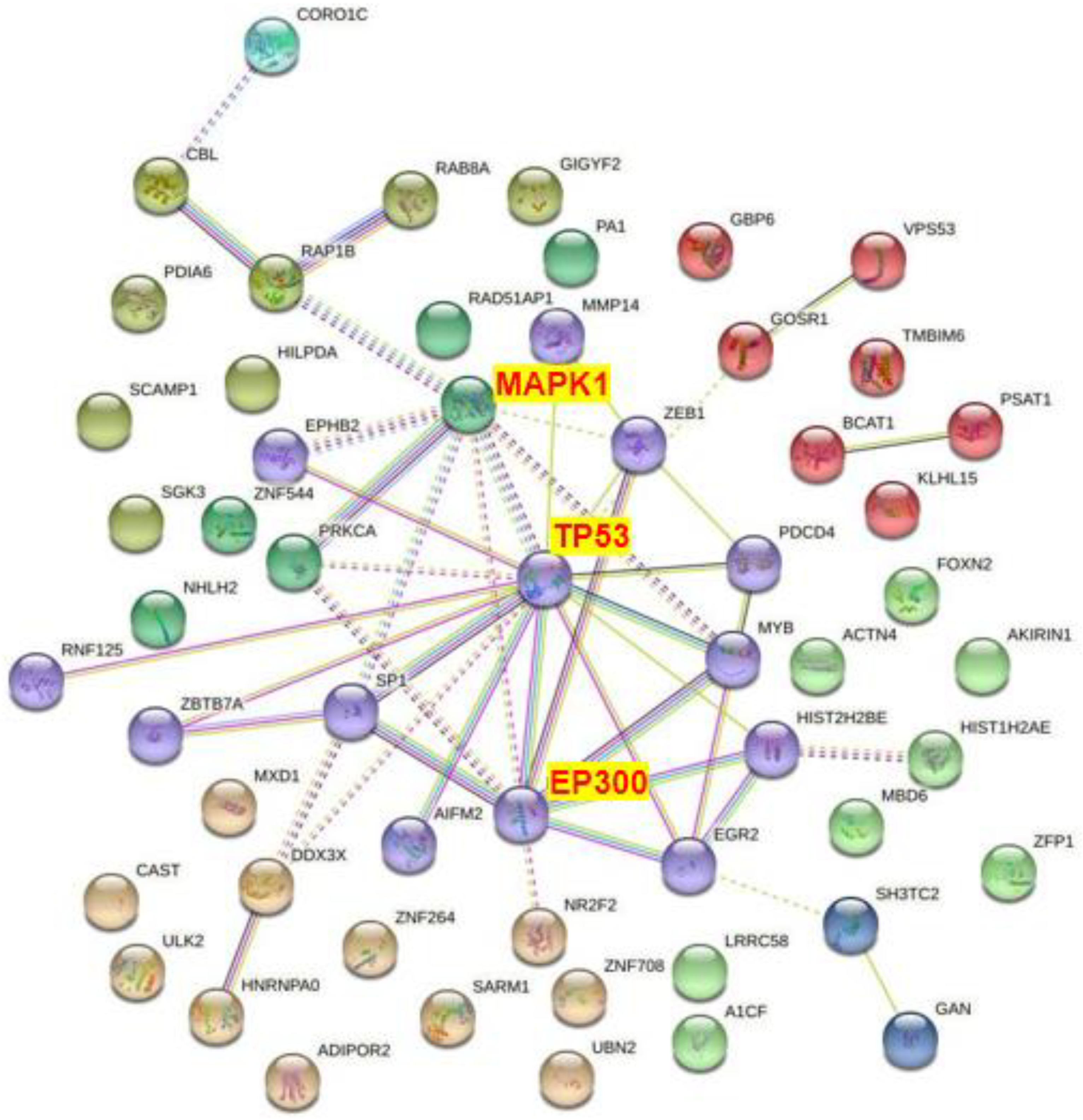
3.4. PPI Network Diagram of COPD-Related miRNA Target Genes with Differential Expression
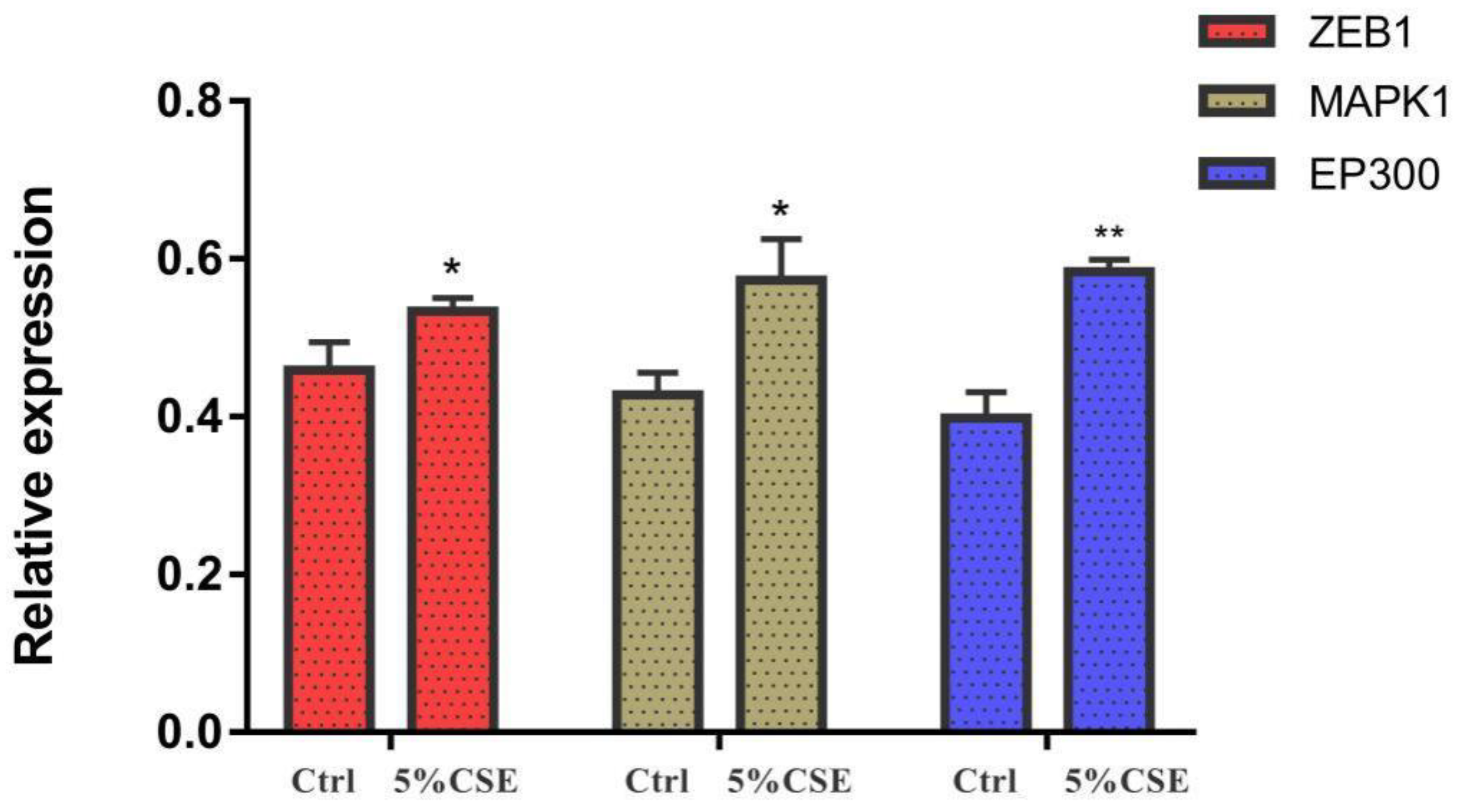
3.5. Real-Time PCR Results
3.6. ELISA Results
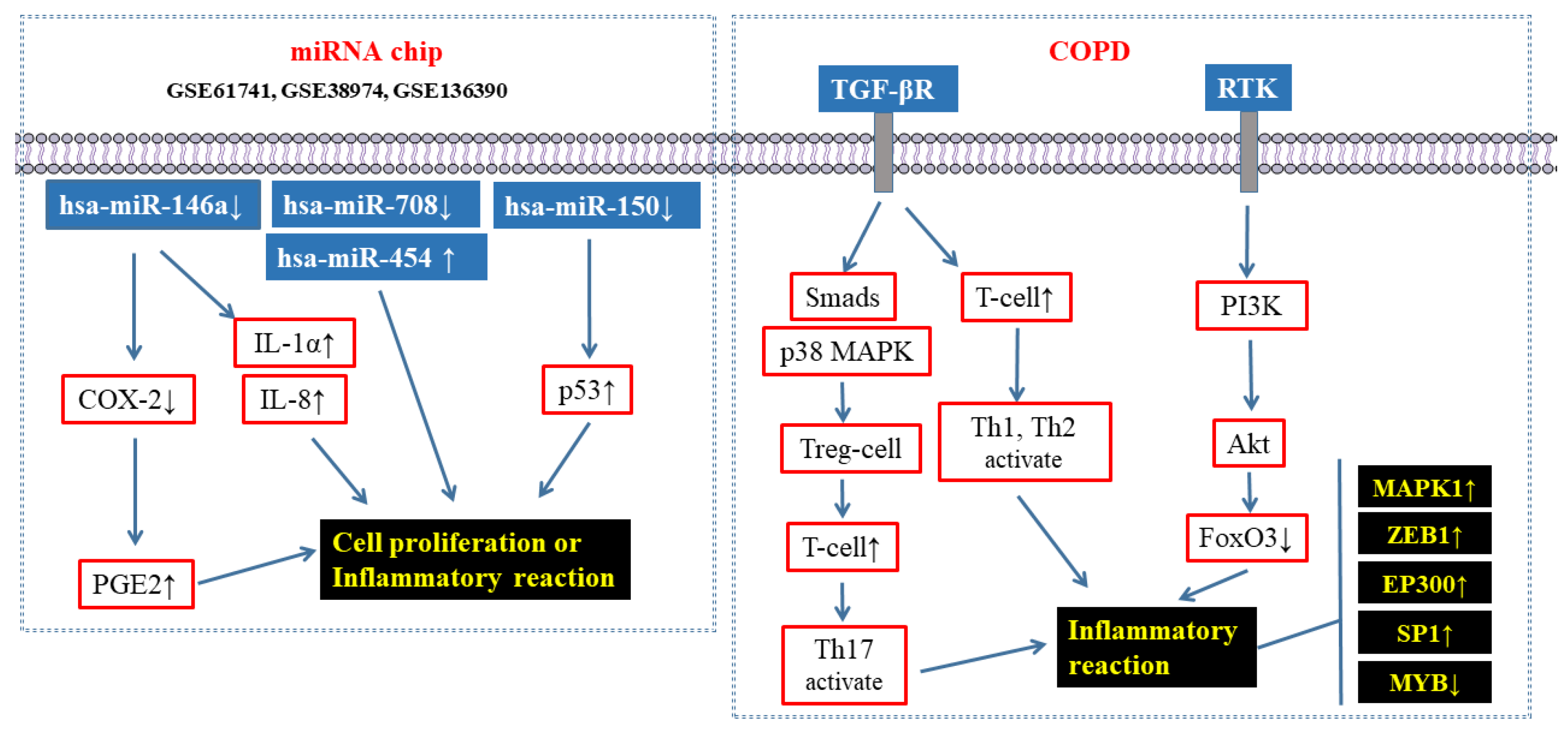
4. Discussion
4.1. The Combination of Bioinformatics and Molecular Biology May Be the Key to Opening the “Door” of Understanding the Pathological Process of COPD
4.2. Bioinformatics Analysis Showed That Several Molecules Were Closely Related to the Pathological Process of COPD
4.3. The Confirmatory Experimental Results Were Relatively Consistent with the Bioinformatics Analysis Results
5. Conclusions
Supplementary Materials
Author Contributions
Funding
Institutional Review Board Statement
Informed Consent Statement
Data Availability Statement
Conflicts of Interest
References
- Soleimani, F.; Dobaradaran, S.; De-La-Torre, G.E.; Schmidt, T.C.; Saeedi, R. Content of toxic components of cigarette, cigarette smoke vs cigarette butts: A comprehensive systematic review. Sci. Total Environ. 2022, 813, 152667. [Google Scholar] [CrossRef] [PubMed]
- Gerlach, G.; Braun, M.; Dröge, J.; Groneberg, D.A. Do budget cigarettes emit more particles? An aerosol spectrometric comparison of particulate matter concentrations between private-label cigarettes and more expensive brand-name cigarettes. Int. J. Environ. Res. Public Health 2022, 19, 5920. [Google Scholar] [CrossRef] [PubMed]
- Van Deusen, A.; Hyland, A.; Travers, M.J.; Wang, C.; Higbee, C.; King, B.A.; Alford, T.; Cummings, K.M. Secondhand smoke and particulate matter exposure in the home. Nicotine Tob. Res. 2009, 11, 635–641. [Google Scholar] [CrossRef] [PubMed]
- Chan, K.H.; Wright, N.; Xiao, D.; Guo, Y.; Chen, Y.; Du, H.; Yang, L.; Millwood, I.Y.; Pei, P.; Wang, J.; et al. Tobacco smoking and risks of more than 470 diseases in China: A prospective cohort study. Lancet Public Health 2022, 7, e1014–e1026. [Google Scholar] [CrossRef] [PubMed]
- Decramer, M.; Janssens, W.; Miravitlles, M. Chronic obstructive pulmonary disease. Lancet 2012, 379, 1341–1351. [Google Scholar] [CrossRef] [PubMed]
- Esteller, M. Non-coding RNAs in human disease. Nat. Rev. Genet. 2011, 12, 861–874. [Google Scholar] [CrossRef] [PubMed]
- Ouyang, C.; Wang, W.; Wu, D.; Wang, W.; Ye, X.; Yang, Q. Analysis of serum exosome microRNAs in the rat model of chronic obstructive pulmonary disease. Am. J. Transl. Res. 2023, 15, 138–150. [Google Scholar]
- Ding, Y.; Tang, S.; Zhou, Z.; Wei, H.; Yang, W. Plasma miR-150-5p as a Biomarker for Chronic Obstructive Pulmonary Disease. Int. J. Chron. Obstruct. Pulmon. Dis. 2023, 18, 399–406. [Google Scholar] [CrossRef]
- van Nijnatten, J.; Brandsma, C.A.; Steiling, K.; Hiemstra, P.S.; Timens, W.; van den Berge, M.; Faiz, A. High miR203a-3p and miR-375 expression in the airways of smokers with and without COPD. Sci. Rep. 2022, 12, 5610. [Google Scholar] [CrossRef]
- Fang, L.; Wang, X.; Zhang, M.; Khan, P.; Tamm, M.; Roth, M. MicroRNA-101-3p Suppresses mTOR and Causes Mitochondrial Fragmentation and Cell Degeneration in COPD. Can. Respir. J. 2022, 2022, 5933324. [Google Scholar] [CrossRef]
- Zhu, M.; Ye, M.; Wang, J.; Ye, L.; Jin, M. Construction of Potential miRNA-mRNA Regulatory Network in COPD Plasma by Bioinformatics Analysis. Int. J. Chron. Obstruct. Pulmon. Dis. 2020, 15, 2135–2145. [Google Scholar] [CrossRef] [PubMed]
- Shen, Y.; Lu, H.; Song, G. MiR-221-3p and miR-92a-3p enhances smoking-induced inflammation in COPD. J. Clin. Lab. Anal. 2021, 35, e23857. [Google Scholar] [CrossRef] [PubMed]
- Agarwal, V.; Bell, G.W.; Nam, J.W.; Bartel, D.P. Predicting effective microRNA target sites in mammalian mRNAs. eLife 2015, 4, e05005. [Google Scholar] [CrossRef] [PubMed]
- Liu, W.; Wang, X. Prediction of functional microRNA targets by integrative modeling of microRNA binding and target expression data. Genome Biol. 2019, 20, 18. [Google Scholar] [CrossRef] [PubMed]
- Huang, D.W.; Sherman, B.T.; Lempicki, R.A. Bioinformatics enrichment tools: Paths toward the comprehensive functional analysis of large gene lists. Nucleic Acids Res. 2009, 37, 1–13. [Google Scholar] [CrossRef] [PubMed]
- Szklarczyk, D.; Morris, J.H.; Cook, H.; Kuhn, M.; Wyder, S.; Simonovic, M.; Santos, A.; Doncheva, N.T.; Roth, A.; Bork, P.; et al. The STRING database in 2017: Quality-controlled protein-protein association networks, made broadly accessible. Nucleic Acids Res. 2017, 45, D362–D368. [Google Scholar] [CrossRef] [PubMed]
- Kode, A.; Rajendrasozhan, S.; Caito, S.; Yang, S.-R.; Megson, I.L.; Rahman, I. Resveratrol induces glutathione synthesis by activation of Nrf2 and protects against cigarette smoke-mediated oxidative stress in human lung epithelial cells. Am. J. Physiol. Lung Cell. Mol. Physiol. 2008, 294, L478–L488. [Google Scholar] [CrossRef] [PubMed]
- Yang, S.R.; Chida, A.S.; Bauter, M.R.; Shafiq, N.; Seweryniak, K.; Maggirwar, S.B.; Kilty, I.; Rahman, I. Cigarette smoke induces proinflammatory cytokine release by activation of NF-kappaB and posttranslational modifications of histone deacetylase in macrophages. Am. J. Physiol. Lung Cell. Mol. Physiol. 2006, 291, L46–L57. [Google Scholar] [CrossRef]
- GBD 2019 Chronic Respiratory Diseases Collaborators. Global burden of chronic respiratory diseases and risk factors, 1990-2019: An update from the Global Burden of Disease Study 2019. EClinicalMedicine 2023, 59, 101936. [Google Scholar] [CrossRef]
- Holtjer, J.C.; Bloemsma, L.D.; Beijers, R.J.; Cornelissen, M.E.; Hilvering, B.; Houweling, L.; Vermeulen, R.C.; Downward, G.S.; der Zee, A.-H.M.-V. Identifying risk factors for COPD and adult-onset asthma: An umbrella review. Eur. Respir. Rev. 2023, 32, 230009. [Google Scholar] [CrossRef]
- Huang, Q.; Li, S.; Wan, J.; Nan, W.; He, B. Association between ethylene oxide exposure and prevalence of COPD: Evidence from NHANES 2013–2016. Sci. Total Environ. 2023, 885, 163871. [Google Scholar] [CrossRef] [PubMed]
- Barnes, P.J. COPD 2020: New directions needed. Am. J. Physiol. Lung Cell. Mol. Physiol. 2020, 319, L884–L886. [Google Scholar] [CrossRef] [PubMed]
- Kuhn, D.E.; Martin, M.M.; Feldman, D.S.; Terry, A.V.; Nuovo, G.J.; Elton, T.S. Experimental validation of miRNA targets. Methods 2008, 44, 47–54. [Google Scholar] [CrossRef] [PubMed]
- Wu, Y.; Li, Q.; Zhang, R.; Dai, X.; Chen, W.; Xing, D. Circulating microRNAs: Biomarkers of disease. Clin. Chim. Acta 2021, 516, 46–54. [Google Scholar] [CrossRef] [PubMed]
- Sato, T.; Liu, X.; Nelson, A.; Nakanishi, M.; Kanaji, N.; Wang, X.; Kim, M.; Li, Y.; Sun, J.; Michalski, J.; et al. Reduced miR-146a increases prostaglandin E₂ in chronic obstructive pulmonary disease fibroblasts. Am. J. Respir. Crit. Care Med. 2010, 182, 1020–1029. [Google Scholar] [CrossRef] [PubMed]
- Dang, X.; Qu, X.; Wang, W.; Liao, C.; Li, Y.; Zhang, X.; Xu, D.; Baglole, C.J.; Shang, D.; Chang, Y. Bioinformatic analysis of microRNA and mRNA Regulation in peripheral blood mononuclear cells of patients with chronic obstructive pulmonary disease. Respir. Res. 2017, 18, 4. [Google Scholar] [CrossRef] [PubMed]
- Sun, G.; Wang, R.; Li, M.; Zhou, S.; Zeng, D.; Xu, X.; Xu, R. Effect of a single nucleotide polymorphism in miR-146a on COX-2 protein expression and lung function in smokers with chronic obstructive pulmonary disease. Int. J. Chron. Obstruct. Pulmon. Dis. 2015, 10, 463–473. [Google Scholar] [CrossRef]
- Osei, E.T.; Florez-Sampedro, L.; Tasena, H.; Faiz, A.; Noordhoek, J.A.; Timens, W.; Postma, D.S.; Hackett, T.L.; Heijink, I.H.; Brandsma, C.A. miR-146a-5p plays an essential role in the aberrant epithelial-fibroblast cross-talk in COPD. Eur. Respir. J. 2017, 49, 1602538. [Google Scholar] [CrossRef]
- Keller, A.; Ludwig, N.; Fehlmann, T.; Kahraman, M.; Backes, C.; Kern, F.; Vogelmeier, C.F.; Diener, C.; Fischer, U.; Biertz, F.; et al. Low miR-150-5p and miR-320b Expression Predicts Reduced Survival of COPD Patients. Cells 2019, 8, 1162. [Google Scholar] [CrossRef]
- Xue, H.; Li, M.X. MicroRNA-150 protects against cigarette smoke-induced lung inflammation and airway epithelial cell apoptosis through repressing p53: MicroRNA-150 in CS-induced lung inflammation. Hum. Exp. Toxicol. 2018, 37, 920–928. [Google Scholar] [CrossRef]
- Liu, B.; Li, R.; Zhang, J.; Meng, C.; Zhang, J.; Song, X.; Lv, C. MicroRNA-708-3p as a potential therapeutic target via the ADAM17-GATA/STAT3 axis in idiopathic pulmonary fibrosis. Exp. Mol. Med. 2018, 50, e465. [Google Scholar] [CrossRef]
- Zhu, D.Y.; Li, X.N.; Qi, Y.; Liu, D.L.; Yang, Y.; Zhao, J.; Zhang, C.Y.; Wu, K.; Zhao, S. MiR-454 promotes the progression of human non-small cell lung cancer and directly targets PTEN. Biomed. Pharmacother. 2016, 81, 79–85. [Google Scholar] [CrossRef]
- Liu, X.; Qu, J.; Xue, W.; He, L.; Wang, J.; Xi, X.; Liu, X.; Yin, Y.; Qu, Y. Bioinformatics-based identification of potential microRNA biomarkers in frequent and non-frequent exacerbators of COPD. Int. J. Chron. Obstruct. Pulmon. Dis. 2018, 13, 1217–1228. [Google Scholar] [CrossRef] [PubMed]
- Aschner, Y.; Downey, G.P. Transforming growth factor-β: Master regulator of the respiratory system in health and disease. Am. J. Respir. Cell. Mol. Biol. 2016, 54, 647–655. [Google Scholar] [CrossRef] [PubMed]
- Mahmood, M.Q.; Reid, D.; Ward, C.; Muller, H.K.; Knight, D.A.; Sohal, S.S.; Walters, E.H. Transforming growth factor (TGF) β(1) and Smad signalling pathways: A likely key to EMT-associated COPD pathogenesis. Respirology 2017, 22, 133–140. [Google Scholar] [CrossRef] [PubMed]
- Lachapelle, P.; Li, M.; Douglass, J.; Stewart, A. Safer approaches to therapeutic modulation of TGF-β signaling for respiratory disease. Pharmacol. Ther. 2018, 187, 98–113. [Google Scholar] [CrossRef] [PubMed]
- Hasan, M.; Neumann, B.; Haupeltshofer, S.; Stahlke, S.; Claudio Fantini, M.; Angstwurm, K.; Bogdahn, U.; Kleiter, I. Activation of TGF-β-induced non-Smad signaling pathways during Th17 differentiation. Immunol. Cell Biol. 2015, 93, 662–672. [Google Scholar] [CrossRef]
- Travis, M.A.; Sheppard, D. TGF-β activation and function in immunity. Annu. Rev. Immunol. 2014, 32, 51–82. [Google Scholar] [CrossRef]
- Xie, Y.; He, Q.; Chen, H.; Lin, Z.; Xu, Y.; Yang, C. Crocin ameliorates chronic obstructive pulmonary disease-induced depression via PI3K/Akt mediated suppression of inflammation. Eur. J. Pharmacol. 2019, 862, 172640. [Google Scholar] [CrossRef]
- Sun, X.; Chen, L.; He, Z. PI3K/Akt-Nrf2 and Anti-Inflammation Effect of macrolides in chronic obstructive pulmonary disease. Curr. Drug Metab. 2019, 20, 301–304. [Google Scholar] [CrossRef]
- Hwang, J.-W.; Rajendrasozhan, S.; Yao, H.; Chung, S.; Sundar, I.K.; Huyck, H.L.; Pryhuber, G.S.; Kinnula, V.L.; Rahman, I. FOXO3 deficiency leads to increased susceptibility to cigarette smoke-induced inflammation, airspace enlargement, and chronic obstructive pulmonary disease. J. Immunol. 2011, 187, 987–998. [Google Scholar] [CrossRef] [PubMed]
- Sun, J.; Gu, X.; Wu, N.; Zhang, P.; Liu, Y.; Jiang, S. Human antigen R enhances the epithelial-mesenchymal transition via regulation of ZEB-1 in the human airway epithelium. Respir. Res. 2018, 19, 109. [Google Scholar] [CrossRef] [PubMed]
- Guan, R.; Wang, J.; Li, D.; Li, Z.; Liu, H.; Ding, M.; Cai, Z.; Liang, X.; Yang, Q.; Long, Z.; et al. Hydrogen sulfide inhibits cigarette smoke-induced inflammation and injury in alveolar epithelial cells by suppressing PHD2/HIF-1α/MAPK signaling pathway. Int. Immunopharmacol. 2020, 81, 105979. [Google Scholar] [CrossRef] [PubMed]
- Wu, D.; Yuan, Y.; Lin, Z.; Lai, T.; Chen, M.; Li, W.; Lv, Q.; Yuan, B.; Li, D.; Wu, B. Cigarette smoke extract induces placental growth factor release from human bronchial epithelial cells via ROS/MAPK (ERK-1/2)/Egr-1 axis. Int. J. Chron. Obstruct. Pulmon. Dis. 2016, 11, 3031–3042. [Google Scholar] [CrossRef]
- Rubio, K.; Singh, I.; Dobersch, S.; Sarvari, P.; Günther, S.; Cordero, J.; Mehta, A.; Wujak, L.; Cabrera-Fuentes, H.; Chao, C.-M.; et al. Inactivation of nuclear histone deacetylases by EP300 disrupts the MiCEE complex in idiopathic pulmonary fibrosis. Nat. Commun. 2019, 10, 2229. [Google Scholar] [CrossRef]


| Symb | Genbank Accession No. | Primer Sequences (5′-3′) |
|---|---|---|
| β-actin | NM_001101.5 | F: GACTACCTCATGAAGATCCTCACC R: TCTCCTTAATGTCACGCACGATT |
| EP300 | NM_001362843.2 | F: GTTCCTTCCTCAGACTCAGTTC R: CATTATAGGAGAGTTCACCGGG |
| MYB | NM_001130172.2 | F: GAAGCAGATTTTTCACCTAGCC R: CTAGGTTCTCCTGCAGGTTTAG |
| EGR2 | NM_000399.5 | F: CGAATCCACACTGGGCATAAG R: AAACTTTCGGCCACAGTAGTC |
| PRKCA | NM_002737.3 | F: GGTGAAGGACCACAAATTCATC R: CACCCGGACAAGAAAAAGTAAC |
| TP53 | NM_000546.6 | F: TTCCTGAAAACAACGTTCTGTC R: AACCATTGTTCAATATCGTCCG |
| MAPK1 | NM_002745.5 | F: ATGGTGTGCTCTGCTTATGATA R: TCTTTCATTTGCTCGATGGTTG |
| SP1 | NM_001251825.2 | F: TCACTCCATGGATGAAATGACA R: CAGAGGAGGAAGAGATGATCTG |
| ZEB1 | NM_001128128.3 | F: CAGGCAAAGTAAATATCCCTGC R: GGTAAAACTGGGGAGTTAGTCA |
Disclaimer/Publisher’s Note: The statements, opinions and data contained in all publications are solely those of the individual author(s) and contributor(s) and not of MDPI and/or the editor(s). MDPI and/or the editor(s) disclaim responsibility for any injury to people or property resulting from any ideas, methods, instructions or products referred to in the content. |
© 2023 by the authors. Licensee MDPI, Basel, Switzerland. This article is an open access article distributed under the terms and conditions of the Creative Commons Attribution (CC BY) license (https://creativecommons.org/licenses/by/4.0/).
Share and Cite
Zhang, Y.; Sheng, Y.; Gao, Y.; Lin, Y.; Cheng, B.; Li, H.; Zhang, L.; Xu, H. Exploration of the Pathogenesis of Chronic Obstructive Pulmonary Disease Caused by Smoking—Based on Bioinformatics Analysis and In Vitro Experimental Evidence. Toxics 2023, 11, 995. https://doi.org/10.3390/toxics11120995
Zhang Y, Sheng Y, Gao Y, Lin Y, Cheng B, Li H, Zhang L, Xu H. Exploration of the Pathogenesis of Chronic Obstructive Pulmonary Disease Caused by Smoking—Based on Bioinformatics Analysis and In Vitro Experimental Evidence. Toxics. 2023; 11(12):995. https://doi.org/10.3390/toxics11120995
Chicago/Turabian StyleZhang, Yingchi, Yuxin Sheng, Yanrong Gao, Yujia Lin, Bin Cheng, Hongmei Li, Ling Zhang, and Haiming Xu. 2023. "Exploration of the Pathogenesis of Chronic Obstructive Pulmonary Disease Caused by Smoking—Based on Bioinformatics Analysis and In Vitro Experimental Evidence" Toxics 11, no. 12: 995. https://doi.org/10.3390/toxics11120995
APA StyleZhang, Y., Sheng, Y., Gao, Y., Lin, Y., Cheng, B., Li, H., Zhang, L., & Xu, H. (2023). Exploration of the Pathogenesis of Chronic Obstructive Pulmonary Disease Caused by Smoking—Based on Bioinformatics Analysis and In Vitro Experimental Evidence. Toxics, 11(12), 995. https://doi.org/10.3390/toxics11120995