Article

Chronic Perigestational Exposure to Chlorpyrifos Induces Perturbations in Gut Bacteria and Glucose and Lipid Markers in Female Rats and Their Offspring

1
PeriTox UMR_I 01 Laboratory, University Center for Health Research, CURS-UPJV, Picardy Jules Verne University, CEDEX 1, 80054 Amiens, France
2
Laboratory of Cellular and Molecular Biology, University of the Brothers Mentouri Constantine 1, Constantine 2500, Algeria
3
Transformations & Agro-Ressources ULR7519, Institut Polytechnique UniLaSalle—Université d’Artois, 60026 Beauvais, France
4
Center for Human Biology, CHU Amiens-Picardie, 80000 Amiens, France
*
Author to whom correspondence should be addressed.
Toxics 2022, 10(3), 138; https://doi.org/10.3390/toxics10030138
Submission received: 24 February 2022 / Revised: 5 March 2022 / Accepted: 10 March 2022 / Published: 12 March 2022
(This article belongs to the Special Issue Developmental Exposure to Environmental Contaminants)

Abstract

:
An increasing burden of evidence is pointing toward pesticides as risk factors for chronic disorders such as obesity and type 2 diabetes, leading to metabolic syndrome. Our objective was to assess the impact of chlorpyrifos (CPF) on metabolic and bacteriologic markers. Female rats were exposed before and during gestation and during lactation to CPF (1 mg/kg/day). Outcomes such as weight, glucose and lipid profiles, as well as disturbances in selected gut bacterial levels, were measured in both the dams (at the end of the lactation period) and in their female offspring at early adulthood (60 days of age). The results show that the weight of CPF dams were lower compared to the other groups, accompanied by an imbalance in blood glucose and lipid markers, and selected gut bacteria. Intra-uterine growth retardation, as well as metabolic disturbances and perturbation of selected gut bacteria, were also observed in their offspring, indicating both a direct effect on the dams and an indirect effect of CPF on the female offspring. Co-treatment with inulin (a prebiotic) prevented some of the outcomes of the pesticide. Further investigations could help better understand if those perturbations mimic or potentiate nutritional risk factors for metabolic syndrome through high fat diet.

Graphical Abstract

1. Introduction

Chemical pollution of the environment by insecticides has become a global phenomenon [1]. They are defined as chemicals used to prevent and control pests, including vectors of human or animal diseases. They are used to control unwanted plant or animal species that interfere with agricultural products [2,3,4]. Pesticides can include herbicides, insecticides, fungicides, disinfectants and rodenticides [3]. According to the most recent statistics on agriculture, forestry and fisheries for the European Union, the total quantity of pesticides sold in Europe amounted to almost 360,000 tons with a significant use of fungicides and bactericides (44%), and herbicides (32%) [5].
The use of pesticides in both developing and developed countries has increased dramatically in recent times. They are widely used in agriculture to increase the yields, quality and appearance of products and reduce the need for agricultural labor [6]. Thus, the potential contamination of the environment by pesticides raises concerns for the public and regulators. In the process of reducing phytochemical products, France has implemented National Health and Environment Plans (PNSE1: 2004–2008, PNSE2: 2009–2013, PNSE3: 2015–2019, PNSE 4: 2020–2024), initiated by the law of 9 August 2004 relating to public health policy. They aim to study the health consequences of exposure to various environmental pollution and include, among other things, estimating the population’s exposure to pesticides, improving knowledge of pesticide exposure and the monitoring of occupational exposures. The Ecophyto plan was created in 2008 to reduce the use of phytosanitary products in France whilst maintaining economically efficient agriculture. Initially, the Ecophyto plan aimed to reduce the use of these substances by 50% by 2020. The target has been extended to 2025 in the face of implementation difficulties [7,8].
Some pesticides are considered endocrine disruptors [9]; therefore, daily exposure is likely to have serious and irreversible effects on the health of individuals [10]. Organophosphates (OP) represent the largest category used in the world and the most widespread due to their bioaccumulation in the environment [11,12,13,14], although their use in France is currently declining due to their consequences on animal and human health. Human exposure to these pesticides is mainly oral, by ingestion of pesticide residues in fruits and vegetables [15]. The extensive and indiscriminate use of OP pesticides in agriculture has been of major concern due to its potentially known or suspected harmful effects on humans. Among the most widely used OP is chlorpyrifos (CPF). Chlorpyrifos was first synthesized by German researchers in 1930 and first introduced in the United States in 1965 as a household insecticide by The Dow Chemical Company (Midland, MI, USA) [16,17]. According to the classification of the World Health Organization (WHO), chlorpyrifos is a class II pesticide of moderate toxicity [18]. CPF is a potent OP insecticide with low water solubility (80.9 mg/L) and a high adsorption coefficient in soil, has a longer half-life in soil (ranging from 65 to 360 days) [19], a wide spectrum of insecticidal action and relative safety compared to other organophosphates, which has led to its intensive use in agriculture. After the application of chlorpyrifos, less than 0.1% of the pesticides applied have a real impact on the intended target. The rest of the residue remains in the environment [16].
CPF use has been closely monitored in recent years, and while it is officially still authorized in 20 EU member states, renewal has been postponed pending further safety reports. The UK government adopted strict restrictions on the use of chlorpyrifos in 2016 due to new safety issues for human health. In the United States, the use of chlorpyrifos indoors has been banned since 2001. The ban process for agricultural use began in 2015in California, and Hawaii banned the sale and use of chlorpyrifos at the end of 2020 [20]. However, the situation remains unclear and CPF can still be found in a number of settings.
CPF is recognized as a neurotoxic agent due to its inhibitory effect on various cholinesterase (ChE) enzymes at the central nervous (CNS) and systemic level [21]. High levels of pesticide residues have been found in several human cohorts [22,23,24,25,26]. According to the United States Environmental Protection Agency (EPA), the no-observed-adverse-effect level (NOAEL) of CPF for acute dietary exposure for the inhibition of red blood cell cholinesterase is 0.5 mg/kg/day [27]. Exposure to CPF has been linked to significant alterations in metabolites that interfere with cellular energy production and amino acid metabolism [28,29,30]; it also acts on hormonal signaling [31,32] and metabolism [33,34], and leads to the disruption of glucose and lipid metabolisms [35,36,37,38], leading to weight gain [39,40,41,42] and increasing the risk of developing chronic non-communicable diseases.
Considering that the oral route is the main cause of human exposure, the impact of a pesticide on the digestive tract is of particular interest. Since it is the first physiological barrier to come into contact with ingested food contaminants, many studies began to focus on the impact of CPF on the gut barrier and gut microbiota [35,43,44,45,46]. The gut microbiota refers to billions of microorganisms residing in the intestine and has a mutualistic relationship with its host [47]. It has several functional roles and impacts on human physiology. It modulates the host’s nutrition through the production of some vitamins and the fermentation of non-digestible food components by the host, protects against pathogens [48] and drug metabolism, and influences intestinal epithelial homeostasis [49].
Exposure to CPF induced by disturbance is often characterized by a decrease in the number of beneficial microorganisms and a simultaneous increase in the number of potentially pathogenic microorganisms leading to dysbiosis [50,51,52,53]. In addition, CPF has been shown to increase intestinal permeability in rats [43,45] or in vitro [54,55], inducing a bacterial translocation which corresponds to the passage of viable bacteria of the gastrointestinal flora through the barrier of the intestinal mucosa (the lamina propria), to the mesenteric nodes, then to normally sterile internal organs such as the spleen and the liver [56].
Pregnancy is a very sensitive period of life where epigenetic marks can impact the long-term development of chronic disorders in the next generation. This is known as the concept of “developmental origin of health and disease” (DOHaD). Contamination of CPF during this period can lead to delayed maturation of the gut microbiota, affecting the bowel function [57]. This may contribute to the onset of obesity and type 2 diabetes (T2D) later in life [58].
Compelling evidence suggests that oral prebiotic supplementation improves these metabolic disorders [59,60]. Additionally, prebiotics are likely associated with increased bifidobacteria and lactobacilli, and the production of short chain fatty acids (SCFA), which are involved in modulating host metabolism [61]. They also strengthen the intestinal barrier, increase satiety by promoting intestinal hormones, improve glucose tolerance, and counteract fatty liver disease (lipogenesis) and insulin resistance [62].
The main objective of the study was to evaluate the effect of perigestional exposure to CPF on the metabolic regulations of dams and their female offspring in early adulthood. For this purpose, weight, lipid and glucose metabolism, levels of selected intestinal bacteria and bacterial translocation were assessed. The secondary objective was to study the protective effect of a prebiotic (inulin) on the same parameters.

2. Materials and Methods

2.1. Experimental Conditions

Chlorpyrifos (O,O-diethyl-O-(3,5,6-trichloro-2-pyridinyl)-phosphorothioate) with a purity of 99.8% was supplied by LGC Standards (Molsheim, France). Inulin was a kind gift from Cosucra (Belgium). Wistar rats were purchased from Janvier laboratories (Le Genest Saint Isle, France). Animal standard diet (Serlab3436, 3.1 kcal/g, Serlab, Montataire, France) was identical throughout the study.
The experiment was carried out according to the protocol approved by the Regional Directorate for Health, Animal Protection and the Environment (Amiens, France) and the Ministry of Research (reference number APAFIS # 8207-2016121322563594 v2 approved on 5 September 2017). All animals were treated in accordance with the EU Directive 2010/63.
Animals were housed in a NexGen Max cage system mounted on an EcoFlow rack system (Allentown Inc, Bussy Saint Georges, France) under constant conditions in a temperature- and air-controlled room (23 °C) with a 12-h light/dark cycle. The size of each group was set to four females for the reproduction protocol in order to reach a minimum of 6–10 female offspring for the passive impact analysis. After acclimation, sixteen 7-week old female Wistar rats (body weight (b.w.) 225 ± 4.9 g) were randomly assigned to four groups (n = 4/group) and housed two per cage.
Chlorpyrifos was dissolved in commercially available organic rapeseed oil at a concentration of 10 mg/mL. CPF solution or rapeseed oil only were administered daily (5 consecutive days followed by a 2-day break) by gavage at a dose of 1 mL/kg b.w. to the animals. This was equivalent to a final concentration of 1 mg/kg b.w. (the oral “no observed adverse effect level“, or NOAEL, for inhibition of cerebral cholinesterase activity in rats [63]) for the CPF groups. Chicory inulin was dissolved in water at a concentration of 10 g/L. Animals were given access to the inulin-enriched or regular drinking water ad libitum. The treatments associated with each group is detailed in Figure 1.
Females were fed a standard diet and the corresponding gavage and drinking water for four consecutive months before mating them throughout gestation and lactation. Male rats were housed under the same conditions as females and received a standard diet. Gestation was assessed daily after mating (two females per male) by the presence of sperm in vaginal smears. The dams were then housed individually until birth and with their pups until weaning. The day of parturition was considered postnatal day 1 (PND1). The average litter size was 6 pups per dam. At the time of weaning (PND21), the female offspring were separated from their mother and housed with their littermates. Male offspring were included in a separate project [64,65]. They were housed according to density requirement in the EU legislation and fed a standard diet until the end of the experiment on PND60. Food and water consumption were measured. Dams were weighted daily throughout the experiment. To determine whether or not exposure during gestation and lactation induced growth retardation, pups were weighed at birth (PND3), at weaning (PND21) and at PND60.
At the end of the experiment (PND21 for dams, PND60 for offspring), the rats were euthanized by intraperitoneal administration of sodium pentobarbital, and plasma as well as various intestinal segments (ileum, colon and cecum) and internal organs (spleen, liver, mesenteric fat tissue and gonadal fat tissue) were removed under sterile conditions.

2.2. Metabolic Perturbations

Approximately 3 mL of intracardiac blood was collected from each animal into a test tube and centrifuged at 1500× g for 10 min at 4 °C. The harvested serum samples were transferred to clean test tubes before analyses.
Standard spectrophotometric methods based on an automation program from the University Hospital of Amiens (KT-6400 analyzer (Genrui Biotech Inc., Shenzhen, China)) were used to measure the following serum parameters: Cholesterol, High Density Lipoprotein (HDL), Low Density Lipoprotein (LDL), triglycerides (TG), blood sugar.

2.3. Disruption of Key Bacteria

2.3.1. Concentrations of Selected Intestinal Bacteria and Bacterial Translocation

Organs were weighed, placed in a sterile stomacher bag and homogenized in Ringer’s saline solution (Bio-Rad, Marnes-la-Coquette, France) before serial 1/10 dilutions. Then, 1 mL of homogenate of intestinal segments and sterile organs was spread on different selective and non-selective media for qualitative and quantitative cultures of selected aerobic and anaerobic intestinal bacteria (Bactron Anaerobic, Sheldon Manufacturing, Cornelius, OR, USA) and incubated at 37 °C for 48 h to 4 days [51,52]. After incubation, bacteria were identified using standard microbiological techniques [45,50,66]. The isolated colonies grown on Petri dishes were counted using an automatic colony counter (Scan® 500, Interscience, St Nom la Bretèche, France) and expressed in log (CFU)/g of tissue.

2.3.2. Microbial Metabolites

Primary dilution samples for selected gut-bacteria analyses were used for short chain fatty acid (SCFA) measures. The supernatant was acidified to pH 2 using 25 µL of H2SO4 (2 M) and injected into a BP21 gas chromatography (GC) column (length: 30 m, inner diameter: 0.53 µm, film thickness: 1 µm) with an internal standard (4-hydroxy-4-methyl-2-pentanone). H2 was used as a carrier gas at a flow rate of 1.5 mL/min. The initial oven temperature was 135 °C and was held there for 6 min, then raised by 25 °C/min to 180 °C and held for 1 min, then increased by 25 °C/min to 230 °C, and finally maintained at 230 °C for 1 min. Glass liner ultra-inert was used for the split injection. The temperatures of the flame ionization detector and the injection port were 280 °C and 240 °C, respectively. The flow rates of H2 and air as makeup gas were 40 and 400 mL/min, respectively. The sample volume injected for the GC analysis (AutoSystem XL; PerkinElmer) was 1 µL and the run time for each analysis was approximately 10 min. The three main short-chain fatty acids (acetic acid, propionic acid, butyric acid) were identified as a function of the retention time of the different elution peaks. The quantification was obtained by comparison with a standard curve [67].

2.3.3. Serum Lipopolysaccharide (LPS)

Plasma LPS is a useful marker for the identification of increased intestinal permeability and thus of intestinal injury. Plasma assay was performed with the Rat Lipopolysaccharide ELISA kit according to the manufacturer instructions (#CSB E14247r, CliniSciences, Nanterre, France).

2.4. Statistical Analyses

Statistical analyses were performed with StatView software (version 5.0, Abacus Concepts Inc., Berkeley, CA, USA) and SPSS Statistics software (version 25.0). Data were analyzed using one-way and two-way ANOVA and KHI2 assay. The independence of endpoints measured in littermates was evaluated by one-way ANOVA for each parameter and treatment group to assess intra and intergroup effects. Except for body weight at PND3 and PND21 in control group, litter effect was not detected. Offspring were thus considered a valid experimental unit for the F1 impact analyses. In all analyses, the threshold of statistical significance was set at p < 0.05.

3. Results

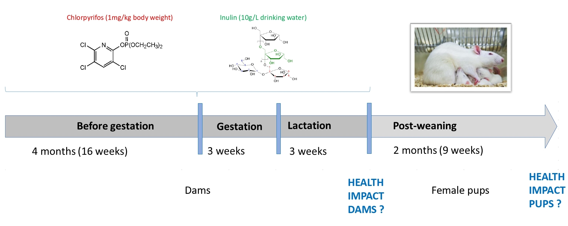
3.1. Animal Weight and Weight Gain

To mimic chronic exposure, we exposed dams for 4 months before gestation, as well as during gestation and lactation (Table 1). No significant differences for weight gain were observed in dams. At PND3 (Figure 2A) the pups from dams exposed to CPF alone (7.5 g) were significantly smaller than the corresponding inulin group (9.9 g for CPFI, p = 0.043). This was still significant at weaning (45.3 g for CPF vs. 55.2 g for C and 56.0 g for CPFI, p = 0.042 and p = 0.050, respectively, Figure 2B). No significant variation remained at early adulthood (PND60, Figure 2C). To complete these observations, no variation was observed in drinking and eating patterns either.

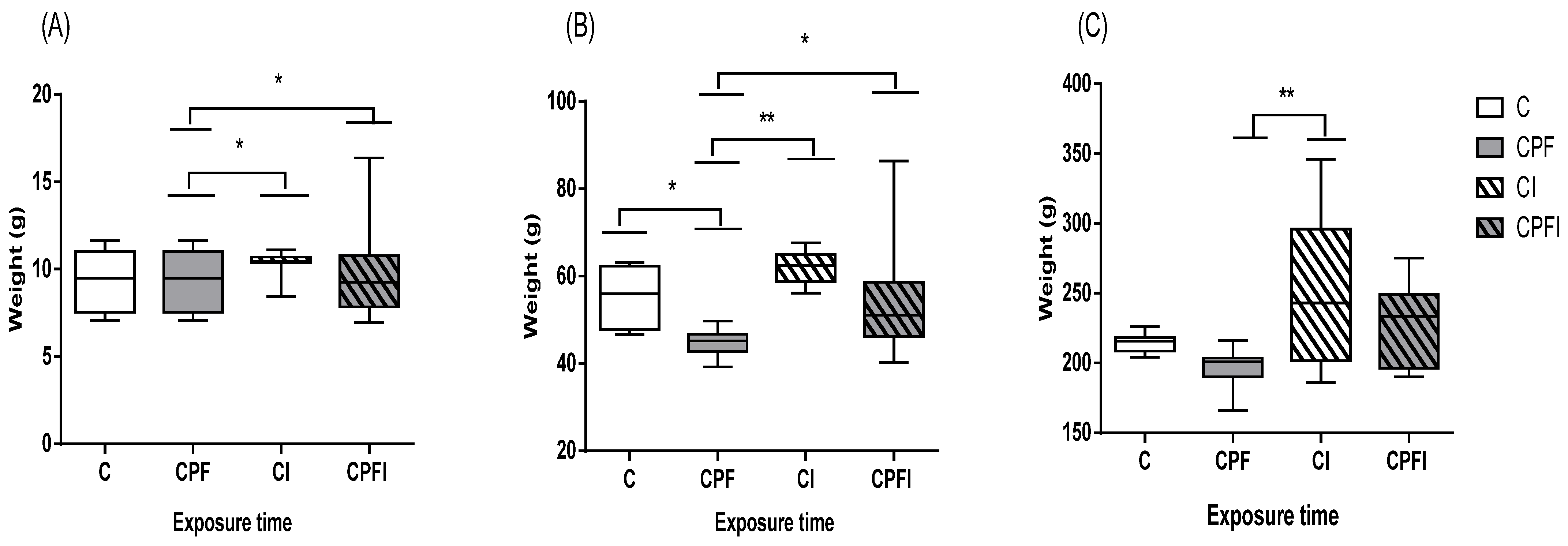
3.2. Metabolic Perturbations

In dams and offspring to PND60, chronic exposure to CPF significantly altered the glycemic and lipid profile in rats (Table 2 and Figure 3).
Blood fasting glucose was significantly higher in dams exposed to CPF compared to the control (p = 0.0001) and the corresponding inulin-fed animals (p = 0.004), but only versus control group in offspring (p = 0.002, Figure 3A). Following direct exposure to CPF in dams or indirectly via lactation, lipid balance was significantly disturbed. In dams treated with CPF the total cholesterol increased by 20% (p = 0.05) and 28% for triglycerides (p = 0.029), while HDL-cholesterol levels decreased by 33% (p = 0.048). Moreover, in female offspring, as shown in Figure 3B, the CPF effect at PND60 remained strong with total cholesterol increased by 16% (p = 0.012), triglycerides by up to 15% (p = 0.0001 and p = 0.009 against C and CPFI, respectively), and LDL levels by up to 27% (p = 0.001 and p = 0.002 against C and CPFI, respectively). Finally, HDL levels decreased up to 30% in plasma of CPF offspring (p = 0.0001 and p = 0.0001 against C and CPFI, respectively).

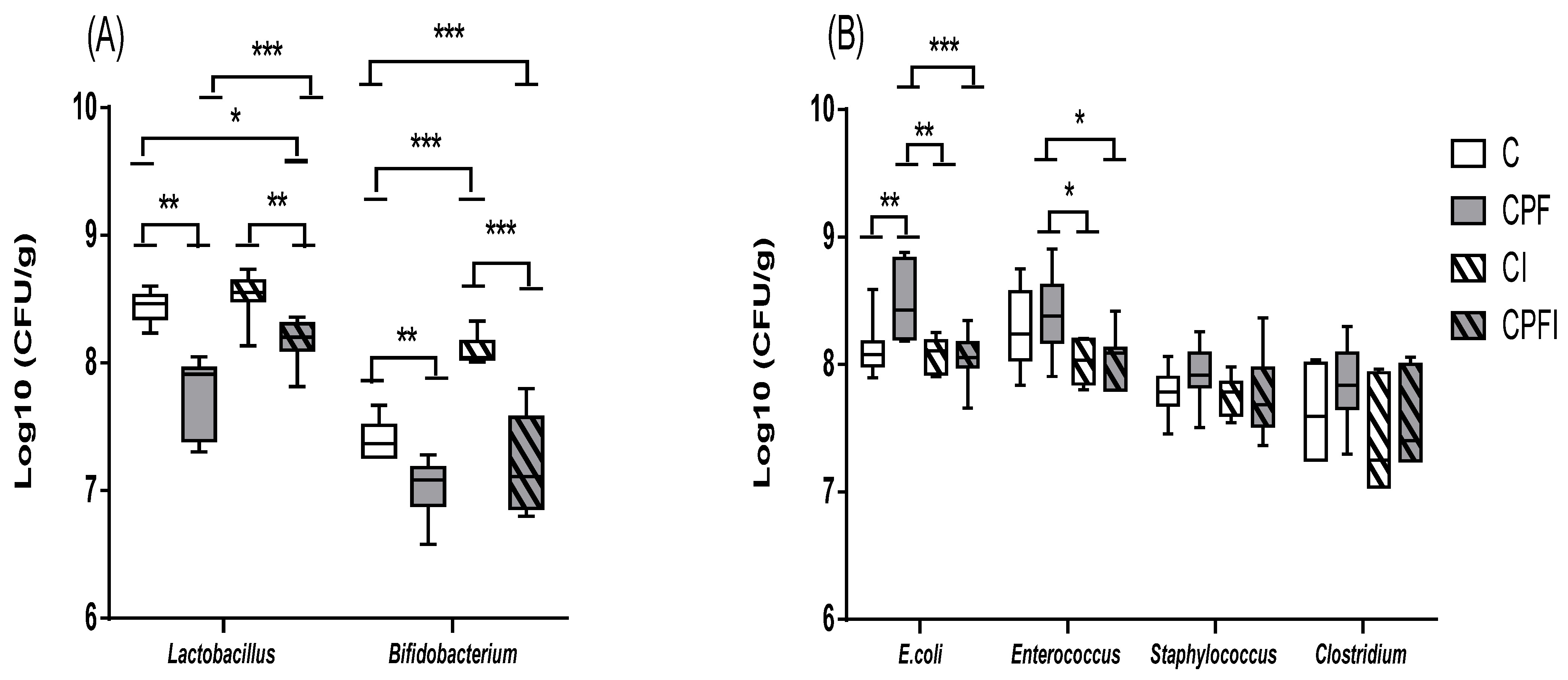
3.3. Disturbances of the Selected Intestinal Bacteria

3.3.1. Concentrations of Selected Intestinal Bacteria

The pesticide altered the levels of selected gut bacteria in the dams. With regards to the total flora (total aerobic flora p = 0.673, total anaerobic flora p = 0.673), no significant difference between the groups was observed. When investigating specific bacterial populations (Table 3 and Table 4), the concentration of Lactobacillus spp. decreases significantly by 1 log in the dams treated with CPF compared to the controls (p = 0.05). For Bifidobacterium spp., the results revealed no significant difference among the groups, but the level of Bifidobacterium decreased in the groups treated with CPF (0.3 log vs. control group). When considering potentially pathogenic flora, bacterial populations were more abundant for animals treated with CPF (0.4 log; p = 0.009 and 0.7 log; p = 0.024) against control group for E. coli and Enterococcus, respectively. Moreover, that the addition of the prebiotic decreased by 1 log the abundance of Enterococcus (p = 0.003). Clostridium population was increased in the CPF group (0.4 log vs. control) but the results showed no significant difference among the groups. In the offspring, no significant difference among the groups was observed for the total flora. The potentially beneficial flora, Lactobacillus spp. and Bifidobacterium, was significantly modulated in the different treatment groups (Figure 4A). Specifically, the level of Lactobacillus spp. decreased by 0.7 log with CPF against the control group (p = 0.0001) while inulin showed a protective 0.5 log increase in bacterial content for Lactobacillus (p = 0.0001). For potentially pathogenic flora (Figure 4B), E. coli was significantly more abundant (0.4 log) in rats treated with CPF (p = 0.001 vs. control group) and decreased by 0.5 log in the CPF group with inulin supplementation p = 0.0001 vs. CPF). Similarly, inulin reduced by 0.4 log the number of Enterococcus found in the CPF group (p = 0.022). The results of Staphylococcus and Clostridium showed no significant difference among the groups, but their abundance tended to increase with exposure to pesticides (0.2 log vs. control) and decrease with inulin supplementation (0.3 log vs. CPF only).

3.3.2. Bacterial Metabolites

Another consequence of dysbiosis is the impaired production of short chain fatty acids (SCFA), which are dependent on various factors in the host microbiota. The profiles of individual SCFA differed depending on the treatment. The presence of CPF alone increased the acetic acid content (2%) in the dams, but not significantly. In the female offspring the CPF significantly increased the ratio of acetic acid and inulin prevented it (p = 0.012; CPF = 71.3% versus CPFI = 53.7%). The presence of inulin increased the levels of propionic and butyric acid in dams and offspring; variations were not significant for dams for either SCFA. In offspring, propionic acid was decreased with CPF and recovered with inulin (CPFI = 15.3% versus C = 15.4% versus CPF = 12.6%). Butyric acid inhibition by CPF and recovery by inulin, however, was significant in the offspring (p = 0.008; CPFI = 27.4% versus CPF = 16.0%).

3.3.3. Bacterial Translocation

Overall, a large aerobic and anaerobic translocation to the spleen was observed in the CPF groups (33% of dams and 25% of offspring), compared to the control and control inulin group (0% of dams and offspring) and CPF inulin (50% of dams and 0% of offspring). The CPF versus control differences were statistically significant only in the female offspring (p = 0.046). These results were corroborated by serum LPS concentrations in dams which were two-fold higher for the CPF group compared to the control and inulin, but no significant difference was observed. This was not observed in the offspring.

4. Discussion

Overall, whether it is through direct or indirect exposure to OP, there are changes in body weight, disruption of lipid metabolism and glucose metabolism, and changes in levels of selected gut bacteria. However, how CPF disrupts these metabolisms is not fully understood.

4.1. Animal Weight and Weight Gain

Weight gain for the dams as well as birth weight have been investigated extensively with regards to risk factors of chronic disorders (obesity, type 2 diabetes), but also as an indicator of the toxic capacity during gestation due to the development of the offspring. Several in vivo studies have suggested that CPF treatment can increase or decrease body weight and birth weight, depending on the dose tested.
The results of the present study revealed that the body weight of the CPF-treated groups was significantly lower than that of the other groups after chronic exposure to low levels of CPF. This is consistent with reports by several authors [68,69,70,71,72]. Researchers have suggested that a decrease in body weight may be due to increased oxidative stress and the degeneration of lipids and proteins [39,73,74]. Exposure to the 1 mg/kg dose did not significantly affect maternal weight gain compared to other groups. And the developmental assessment of female offspring indicated that all CPF-treated groups had low body mass gain compared to other PND60 groups, which is consistent with previous findings [38] and other studies [37,68,75,76].

4.2. Metabolic Perturbations

In this study, blood glucose concentration was markedly increased in both dams and female offspring. Consistent with these results, OP have been shown to induce hyperglycemia [37,38,77,78,79,80]. An increasing number of researchers have begun to focus on the mechanism of exposure to OP and hyperglycemia while other studies have reported that OP do not affect metabolic indices [35,81,82].
Changes in lipid profile due to CPF administration were observed in this study, other studies also showed an increase in TG in agreement with the present results [41,83] while others have observed lower or unchanged TG levels [37,84].
CPF also disrupts serum TC levels. In our study, cholesterol levels were higher, and several studies are in agreement with our results [28,37,41,83,84]. They suggested that serum TC levels may be increased in the groups treated with CPF due to blockage of the hepatic bile ducts, resulting in decreased or stopped TC secretion in the duodenum [39,85].
The result of the present study also demonstrated the dyslipidemic effect exerted by chronic administration of low levels of CPF. This was evident by the increased LDL levels and decreased HDL levels recorded in the serum of CPF-exposed rats. Some authors have suggested that CPF causes liver damage and lipoprotein synthesis [28,41,83,84].
In our experiment, chronic exposure to low doses of CPF was also responsible for developmental alterations in male pups, characterized by low birth weight, decreased plasma growth factor (IGF1) levels, leptin and a small increase in insulinemia. These effects were still present at weaning but disappeared in early adulthood when the animals were no longer exposed [64].

4.3. Disturbances of the Selected Intestinal Bacteria

To summarize, levels of key bacteria were unstable in dams and offspring with an increase in potentially pathogenic flora to the detriment of potentially beneficial flora in rats treated with CPF. CPF exposure was associated with significant microbial perturbation, showing the influence of CPF exposure on a number of bacteria, with reduced abundance of Lactobacillus spp. and Bifidobacterium spp., and a higher level of Enterococcus spp., E. coli, Staphylococcus spp. and Clostridium spp. in rats treated with CPF. Similar results were shown in previous work and other studies [50,84,86,87,88,89]. These results demonstrate that exposure to CPF induces disturbances in the gut microbiota and that early gut microbiota disruption could lead to long-term effects.
Another consequence of dysbiosis is the altered production of short-chain fatty acids (SCFA), which depend on various factors in the host microbiota. Butyrate, propionate and acetate represent 90 to 95% of SCFA present in the colon [90,91]. They are produced by fermentation of dietary fibers and are often associated with the prevention of several pathologies linked to inflammation or oxidative stress [92]. These results are in agreement with previous results on CPF [38] and other results on the different pesticides carbendazim [93], permethrin [94] and propamocarb [95], which suggests that the perturbation of the intestinal microbiota following pesticide exposure altered the proportions of SCFA, disturbed the energetic homeostasis and elicited multiple tissue inflammatory responses.
The bacterial translocation was predominantly higher in the CPF groups than in the other groups. It is recognized that microbial dysbiosis exerts a strong influence on the intestinal permeability [45,96,97]. The translocation phenomenon could be explained by the increase in permeability observed after exposure to CPF [45,50]. CPF alone inhibits the expression of tight junction and structural genes, while inulin and CPF/inulin co-treatment tended to increase expression [54]. Neonatal translocation is completely normal and even essential for the maturation of the immune system [98], but its persistence after weaning could become pathogenic. It is, therefore, likely that the pesticide disrupted the tight junctions, while the mixture enhanced the activity of the tight junctions [54]. Higher LPS concentrations in the serum of dams is a marker of permeability as well as a risk factor for low grade inflammation [99]. These are also consistent with C-reactive protein (CRP) serum concentrations in dams following CPF exposure (data not shown) as a sign of inflammation.

4.4. Nutritional Prevention

Prebiotic dietary fibers are likely to selectively modulate the gut microbiota and exert positive health effects [100]. The prevention approach used in this study was based on the use of inulin, acting as a prebiotic to counteract the effects of CPF on the gut microbiota [38,54], which in turn affects intestinal functions, such as metabolism and the integrity of the intestine [101]. Co-treatment with inulin prevents dysbiosis as well as early markers of dysmetabolism in rats chronically exposed to CPF. However, we could not find any other work in the literature studying this particular prevention strategy to counter pesticides except for an article on zebrafish [102].
Present results indicated that inulin consumption improved the lipid and glycemic profiles, consistent with other studies. The mechanism by which inulin acts on glucose and lipid metabolism remains unclear. A number of hypotheses have been proposed regarding the effect of prebiotics on improving dysmetabolism. One mechanism included decreased absorption of cholesterol via intestinal epithelial cells [103]. Inulin is a soluble, viscous compound that increases the thickness of the unstirred layer of the small intestine, thereby inhibiting absorption of cholesterol [104]. Inulin does not bind to bile acid in the upper digestive tract; however, it can help soluble bile acids bind to bacteria or insoluble compounds, such as calcium phosphate, by lowering the pH of the cecum [105]. As a result, fecal excretion of bile acids increases cholesterol utilization to rebuild bile acid in the liver and decrease the concentration of circulating and hepatic cholesterol [106]. Also, inulin treatment acts on glucose metabolism by improving serum GLP-1 levels to suppress IL-6 secretion and production, and hepatic gluconeogenesis and resulted in moderation of insulin tolerance. These results indicate that intestine-liver crosstalk is the primary mechanism for moderating insulin resistance by inulin [106,107,108].
Specific changes in the composition of the gut microbiota occurred in the prebiotic group; inulin increased the number of Lactobacillus and Bifidobacterium but also reduced the number of Enterococcus spp., E. coli, Staphylococcus spp. and Clostridium spp. The combination of CPF and inulin reduced the number of enterococci, while the numbers of Bifidobacterium and Lactobacillus increased. Another potential mechanistic pathway may be behind the change in the composition of the gut microbiota after inulin supplementation leading to an increase in SCFA and decrease in cecal pH. SCFA are important for health because they improve energy metabolism in the liver and muscles and immune function in the large intestine [109,110,111,112].

4.5. Perigestational Modulation of CPF

In the general population, new parents tend to look more closely into the food given to their infants, trying to aim for organic, pesticide-free options, even though their own diet may not be modified dramatically. It is not uncommon either that the diet of prospecting parents does not differ from that of the global population [113,114,115]. With this in mind, we set up this model to expose the dams to treatments before mating and continue it up to weaning but discontinue treatment for the offspring.
Differences in the toxicity of CPF between fetus and mother have already been reported due to the lipophilic power of CPF, which can cross the placental barrier [28,68,76,116]. CPF and its metabolite DETP (diethyl thiophosphonate) have been studied and published in the literature and residues have been detected in newborns [116,117]. 3,5,6-trichloro-2-pyridinol (TCPy), a specific metabolite of ethyl and methyl chlorpyrifos, was also recently found in the hair collected at birth in 311 new mothers [118]. This is, therefore, a critical window of exposure to toxic environment xenobiotics through aerosol or oral intake. During gestation and early childhood, the internal organs are still undergoing a process of maturation. Previous work showed that the intestine and microbiota maturation were delayed following CPF treatment [50]. The objective of the work presented herein was to better understand whether these perturbations would still have consequences if treatment was interrupted after weaning.
These data, together with the knowledge that a number of chronic disorders such as obesity or type 2 diabetes can be triggered by in utero imprinting, suggest that it is likely that CPF may lead to resilient effects event after treatment was discontinued. The results presented here for the offspring clearly support this hypothesis. It would be interesting to see if there were some epigenetic modifications in target tissues. Unfortunately sampling conditions did not allow for genomic analyses.
In addition, birth weight is thought to be predictive of chronic disorders. A low birth weight, often observed in experiments with OP, tends to be associated with adult risks of obesity and metabolic syndrome, whilst a larger birth weight would more likely be associated with increased risks of type 2 diabetes. Data show that both glucose and lipid profiles are modified by CPF, more targeted analyses or long-term experiments would be required to clearly differentiate between the two pathologies as they encounter a number of overlapping features.

5. Conclusions

The study allowed us to gather new evidence of resilient and indirect impact on offspring of CPF in metabolic disorders as summarized in Table 5. Modulations in specific bacterial populations as well as glucose and lipid profiles suggest mechanisms of dysmetabolism typical of obesity and diabetes [58]. The large number of endpoints measured would require statistical partitioning among variables, and this is a bias that may need to be highlighted when reaching overall conclusions. The strength of this work lies in the duration of the exposure of dams to the pesticide and the measure of indirect impact to the offspring to better mimic real-life settings. Further investigation in the epigenetic imprinting that could be associated with transgenerational effects would be complementary to these findings. In addition, while cultured microbiology may seem to be outdated for microbiota analyses compared to shotgun metagenomics, it is of great interest to follow up on bacterial translocation and perturbation of the intestinal permeability for a complete picture of the intestinal ecosystem. Finally, the nutritional prevention strategy can be further explored using other prebiotic molecules, probiotics or even testing different windows of exposure. A high-energy diet is also a risk factor leading to similar perturbations. It would be interesting to see whether mechanisms with CPF or high-fat diets are similar, and if a mix of dietary and environment factors would have synergistic effects. This hypothesis has been tested, samples are being analyzed and results are underway.

Author Contributions

N.D.: Data curation; Investigation; Formal analysis; Visualization; Methodology; Writing—original draft; Writing—review & editing; F.D.: Conceptualization; Methodology; Validation; Writing—original draft; Writing—review & editing; M.G.: Investigation; Methodology; Writing—review & editing; H.E.K.E.S.: Investigation; Methodology; Writing—review & editing; A.C.: Investigation; Methodology; Writing—review & editing; L.R. (Larbi Rhazi): Investigation; Methodology; Writing—review & editing; J.G.-Q.: Methodology; Supervision; Funding acquisition; Writing—review & editing; L.R. (Leila Rouabah): Methodology; Writing—review & editing; F.H.: Investigation; Methodology; Writing—review & editing; V.B.: Supervision; Formal analysis; Writing—original draft; Writing—review & editing; M.B.: Investigation; Methodology; Writing—original draft; Writing—review & editing; H.K.-C.: Conceptualization; Visualization; Funding acquisition; Methodology; Project administration; Supervision; Validation; Writing—original draft; Writing—review & editing. All authors have read and agreed to the published version of the manuscript.

Funding

The animal experiment was supported by the Ministry of Higher Education, Research and Innovation and National Institutes of Health and Hauts-de-France region and was also funded by projet Fédératif Hospitalo-Universitaire “1000 days for Health” Proghomeo grant 2017.

Institutional Review Board Statement

The animal study protocol was approved by the Ethics Committee of the Regional Directorate for Health, Animal Protection and the Environment (Amiens, France) and the Ministry of Research (APAFIS # 8207-2016121322563594 v2 approved on 5 September 2017).

Informed Consent Statement

Not applicable.

Data Availability Statement

No open access to study data.

Acknowledgments

The authors thank Jérôme AUSEIL from the biochemistry laboratory, CHU Amiens for the metabolic assays.

Conflicts of Interest

The authors declare no conflict of interest.

Abbreviations

B.W.body weight
CControl
ChEcholine esterase
CIcontrol + inulin
CNScentral nervous system
CPFchlorpyrifos
CPFICPF + inulin
CRPC-reactive protein
DETPdiethyl thiophosphonate
DOHaDDevelopmental origin of Health and Disease
EPAUnited States Environmental Protection Agency
GCgas chromatography
GLP1glucose-like protein-1
HDLhigh-density lipoprotein
IGF1insulin growth factor-1
IL6interleukin-6
LDLlow-density lipoprotein
LPSlipopolysaccharide
NOAELno observed adverse effect level
OPorganophosphate
PNDpost-natal day
PNSENational Health and Environment Plans
SCFAshort chain fatty acid
SEMstandard error of the mean
T2Dtype 2 diabetes
TCPy3,5,6-trichloro-2-pyridinol
TGtriglyceride
WHOWorld Health Organization

References

  1. Wang, X.; Xing, H.; Li, X.; Xu, S.; Wang, X. Effects of atrazine and chlorpyrifos on the mRNA levels of IL-1 and IFN-γ2b in immune organs of common carp. Fish. Shellfish Immunol. 2011, 31, 126–133. [Google Scholar] [CrossRef] [PubMed]
  2. OCSPP. Opp Pesticides Industry Sales and Usage 2006 and 2007 Market Estimates; OCSPP: Arlington, TX, USA, 2015. [Google Scholar]
  3. World Health Organization; Safety International Programme on Chemical. The WHO Recommended Classification of Pesticides by Hazard and Guidelines to Classification 2009; World Health Organization: Geneva, Switzerland, 2010; ISBN 978-92-4-154796-3.
  4. Solomon, K.R.; Williams, W.M.; Mackay, D.; Purdy, J.; Giddings, J.M.; Giesy, J.P. Properties and uses of chlorpyrifos in the United States. Rev. Environ. Contam. Toxicol. 2014, 231, 13–34. [Google Scholar] [CrossRef] [PubMed] [Green Version]
  5. Sales of Pesticides in the EU. Available online: https://ec.europa.eu/eurostat/web/products-eurostat-news/-/ddn-20200603-1 (accessed on 23 September 2021).
  6. Choudhary, S.; Yamini, N.R.; Yadav, S.K.; Kamboj, M.L.; Sharma, A. A review: Pesticide residue: Cause of many animal health problems. J. Entomol. Zool. Stud. 2018, 6, 330–333. [Google Scholar]
  7. Guichard, L.; Dedieu, F.; Jeuffroy, M.-H.; Meynard, J.M.; Reau, R.; Savini, I. Le plan Ecophyto de réduction d’usage des pesticides en France: Décryptage d’un échec et raisons d’espérer. Cah. Agric. 2017, 26, 1–12. [Google Scholar] [CrossRef]
  8. Renaudie, O. Les plans nationaux santé-environnement: Conciliation ou concurrence entre deux politiques publiques? Rev. Droit Sanit. Soc. 2019, HS, 23–35. [Google Scholar]
  9. Combarnous, Y. Endocrine Disruptor Compounds (EDCs) and agriculture: The case of pesticides. Comptes Rendus Biol. 2017, 340, 406–409. [Google Scholar] [CrossRef]
  10. Jepson, P.C.; Murray, K.; Bach, O.; Bonilla, M.; Neumeister, L. A Global Guideline for Pesticide Selection to Reduce Risks, and Establish a Minimum Pesticides List; Social Science Research Network: Rochester, NY, USA, 2019. [Google Scholar]
  11. Alamgir Zaman Chowdhury, M.; Fakhruddin, A.N.M.; Nazrul Islam, M.; Moniruzzaman, M.; Gan, S.H.; Khorshed Alam, M. Detection of the residues of nineteen pesticides in fresh vegetable samples using gas chromatography–mass spectrometry. Food Control 2013, 34, 457–465. [Google Scholar] [CrossRef]
  12. Carr, R.L.; Ho, L.L.; Chambers, J.E. Selective toxicity of chlorpyrifos to several species of fish during an environmental exposure: Biochemical mechanisms. Environ. Toxicol. Chem. 1997, 16, 2369–2374. [Google Scholar] [CrossRef]
  13. Casida, J.E.; Quistad, G.B. Organophosphate toxicology: Safety aspects of nonacetylcholinesterase secondary targets. Chem. Res. Toxicol. 2004, 17, 983–998. [Google Scholar] [CrossRef]
  14. He, M.-J.; Lu, J.-F.; Wang, J.; Wei, S.-Q.; Hageman, K.J. Phthalate esters in biota, air and water in an agricultural area of western China, with emphasis on bioaccumulation and human exposure. Sci. Total Environ. 2020, 698, 134264. [Google Scholar] [CrossRef]
  15. Grewal, A.S.; Grewal, A.S.; Singla, A.; Kamboj, P.; Dua, J.S.; Internationals, O. Pesticide Residues in Food Grains, Vegetables and Fruits: A Hazard to Human Health. J. Med. Chem. Toxicol. 2017, 2, 40–46. [Google Scholar] [CrossRef]
  16. Chishti, Z.; Hussain, S.; Arshad, K.R.; Khalid, A.; Arshad, M. Microbial degradation of chlorpyrifos in liquid media and soil. J. Environ. Manag. 2013, 114, 372–380. [Google Scholar] [CrossRef]
  17. Das, S.; Adhya, T.K. Degradation of chlorpyrifos in tropical rice soils. J. Environ. Manag. 2015, 152, 36–42. [Google Scholar] [CrossRef]
  18. Kumar, A.; Correll, R.; Grocke, S.; Bajet, C. Toxicity of selected pesticides to freshwater shrimp, Paratya australiensis (Decapoda: Atyidae): Use of time series acute toxicity data to predict chronic lethality. Ecotoxicol. Environ. Saf. 2010, 73, 360–369. [Google Scholar] [CrossRef]
  19. Li, D.; Huang, Q.; Lu, M.; Zhang, L.; Yang, Z.; Zong, M.; Tao, L. The organophosphate insecticide chlorpyrifos confers its genotoxic effects by inducing DNA damage and cell apoptosis. Chemosphere 2015, 135, 387–393. [Google Scholar] [CrossRef] [PubMed]
  20. Hites, R.A. The Rise and Fall of Chlorpyrifos in the United States. Environ. Sci. Technol. 2021, 55, 1354–1358. [Google Scholar] [CrossRef] [PubMed]
  21. Ur Rahman, S.; Xuebin, Q.; Yasin, G.; Cheng, H.; Mehmood, F.; Zain, M.; Shehzad, M.; Ahmad, M.I.; Riaz, L.; Rahim, A.; et al. Role of silicon on root morphological characters of wheat (Triticum aestivum L.) plants grown under Cd-contaminated nutrient solution. Acta Physiol. Plant 2021, 43, 60. [Google Scholar] [CrossRef]
  22. Olisah, C.; Okoh, O.O.; Okoh, A.I. Occurrence of organochlorine pesticide residues in biological and environmental matrices in Africa: A two-decade review. Heliyon 2020, 6, e03518. [Google Scholar] [CrossRef]
  23. Mulder, T.A.; van den Dries, M.A.; Korevaar, T.I.M.; Ferguson, K.K.; Peeters, R.P.; Tiemeier, H. Organophosphate pesticides exposure in pregnant women and maternal and cord blood thyroid hormone concentrations. Environ. Int. 2019, 132, 105124. [Google Scholar] [CrossRef]
  24. Araki, A.; Miyashita, C.; Mitsui, T.; Goudarzi, H.; Mizutani, F.; Chisaki, Y.; Itoh, S.; Sasaki, S.; Cho, K.; Moriya, K.; et al. Prenatal organochlorine pesticide exposure and the disruption of steroids and reproductive hormones in cord blood: The Hokkaido study. Environ. Int. 2018, 110, 1–13. [Google Scholar] [CrossRef] [PubMed] [Green Version]
  25. Lehmann, E.; Oltramare, C.; Nfon Dibié, J.-J.; Konaté, Y.; de Alencastro, L.F. Assessment of human exposure to pesticides by hair analysis: The case of vegetable-producing areas in Burkina Faso. Environ. Int. 2018, 111, 317–331. [Google Scholar] [CrossRef]
  26. Iglesias-González, A.; Hardy, E.M.; Appenzeller, B.M.R. Cumulative exposure to organic pollutants of French children assessed by hair analysis. Environ. Int. 2020, 134, 105332. [Google Scholar] [CrossRef] [PubMed]
  27. U.S. Environmental Protection Agency. Revised Human Health Risk Assessment on Chlorpyrifos. Available online: https://www.epa.gov/ingredients-used-pesticide-products/revised-human-health-risk-assessment-chlorpyrifos (accessed on 28 June 2021).
  28. Perez-Fernandez, C.; Morales-Navas, M.; Aguilera-Sáez, L.M.; Abreu, A.C.; Guardia-Escote, L.; Fernández, I.; Garrido-Cárdenas, J.A.; Colomina, M.T.; Giménez, E.; Sánchez-Santed, F. Medium and long-term effects of low doses of Chlorpyrifos during the postnatal, preweaning developmental stage on sociability, dominance, gut microbiota and plasma metabolites. Environ. Res. 2020, 184, 109341. [Google Scholar] [CrossRef] [PubMed]
  29. Wang, H.-P.; Liang, Y.-J.; Long, D.-X.; Chen, J.-X.; Hou, W.-Y.; Wu, Y.-J. Metabolic profiles of serum from rats after subchronic exposure to chlorpyrifos and carbaryl. Chem. Res. Toxicol. 2009, 22, 1026–1033. [Google Scholar] [CrossRef]
  30. Xu, M.-Y.; Sun, Y.-J.; Wang, P.; Xu, H.-Y.; Chen, L.-P.; Zhu, L.; Wu, Y.-J. Metabolomics analysis and biomarker identification for brains of rats exposed subchronically to the mixtures of low-dose cadmium and chlorpyrifos. Chem. Res. Toxicol. 2015, 28, 1216–1223. [Google Scholar] [CrossRef] [PubMed]
  31. Lassiter, T.L.; Brimijoin, S. Rats gain excess weight after developmental exposure to the organophosphorothionate pesticide, chlorpyrifos. Neurotoxicol. Teratol. 2008, 30, 125–130. [Google Scholar] [CrossRef] [PubMed]
  32. Slotkin Theodore, A.; Brown Kathleen, K.; Seidler Frederic, J. Developmental Exposure of Rats to Chlorpyrifos Elicits Sex-Selective Hyperlipidemia and Hyperinsulinemia in Adulthood. Environ. Health Perspect. 2005, 113, 1291–1294. [Google Scholar] [CrossRef] [PubMed] [Green Version]
  33. Lasram, M.M.; Dhouib, I.B.; Annabi, A.; El Fazaa, S.; Gharbi, N. A review on the molecular mechanisms involved in insulin resistance induced by organophosphorus pesticides. Toxicology 2014, 322, 1–13. [Google Scholar] [CrossRef] [PubMed]
  34. Peris-Sampedro, F.; Salazar, J.G.; Cabré, M.; Reverte, I.; Domingo, J.L.; Sánchez-Santed, F.; Colomina, M.T. Impaired retention in AβPP Swedish mice six months after oral exposure to chlorpyrifos. Food Chem. Toxicol. 2014, 72, 289–294. [Google Scholar] [CrossRef]
  35. Fang, B.; Li, J.W.; Zhang, M.; Ren, F.Z.; Pang, G.F. Chronic chlorpyrifos exposure elicits diet-specific effects on metabolism and the gut microbiome in rats. Food Chem. Toxicol. 2018, 111, 144–152. [Google Scholar] [CrossRef]
  36. Li, J.; Ren, F.; Li, Y.; Luo, J.; Pang, G. Chlorpyrifos Induces Metabolic Disruption by Altering Levels of Reproductive Hormones. J. Agric. Food Chem. 2019, 67, 10553–10562. [Google Scholar] [CrossRef]
  37. Peris-Sampedro, F.; Cabré, M.; Basaure, P.; Reverte, I.; Domingo, J.L.; Teresa Colomina, M. Adulthood dietary exposure to a common pesticide leads to an obese-like phenotype and a diabetic profile in apoE3 mice. Environ. Res. 2015, 142, 169–176. [Google Scholar] [CrossRef]
  38. Reygner, J.; Lichtenberger, L.; Elmhiri, G.; Dou, S.; Bahi-Jaber, N.; Rhazi, L.; Depeint, F.; Bach, V.; Khorsi-Cauet, H.; Abdennebi-Najar, L. Inulin Supplementation Lowered the Metabolic Defects of Prolonged Exposure to Chlorpyrifos from Gestation to Young Adult Stage in Offspring Rats. PLoS ONE 2016, 11, e0164614. [Google Scholar] [CrossRef]
  39. Goel, A.; Dani, V.; Dhawan, D.K. Protective effects of zinc on lipid peroxidation, antioxidant enzymes and hepatic histoarchitecture in chlorpyrifos-induced toxicity. Chem. Biol. Interact. 2005, 156, 131–140. [Google Scholar] [CrossRef]
  40. Meggs, W.J.; Brewer, K.L. Weight gain associated with chronic exposure to chlorpyrifos in rats. J. Med. Toxicol. 2007, 3, 89–93. [Google Scholar] [CrossRef] [PubMed] [Green Version]
  41. Uchendu, C.; Ambali, S.F.; Ayo, J.O.; Esievo, K.A.N. Body weight and hematological changes induced by chronic exposure to low levels of chlorpyrifos and deltamethrin combination in rats: The effect of alpha-lipoic acid. Comp. Clin. Pathol. 2018, 27, 1383–1388. [Google Scholar] [CrossRef]
  42. Whyatt, R.M.; Rauh, V.; Barr, D.B.; Camann, D.E.; Andrews, H.F.; Garfinkel, R.; Hoepner, L.A.; Diaz, D.; Dietrich, J.; Reyes, A.; et al. Prenatal insecticide exposures and birth weight and length among an urban minority cohort. Environ. Health Perspect. 2004, 112, 1125–1132. [Google Scholar] [CrossRef] [PubMed] [Green Version]
  43. Cook, T.J.; Shenoy, S.S. Intestinal permeability of chlorpyrifos using the single-pass intestinal perfusion method in the rat. Toxicology 2003, 184, 125–133. [Google Scholar] [CrossRef]
  44. Gao, J.; Naughton, S.X.; Beck, W.D.; Hernandez, C.M.; Wu, G.; Wei, Z.; Yang, X.; Bartlett, M.G.; Terry, A.V. Chlorpyrifos and chlorpyrifos oxon impair the transport of membrane bound organelles in rat cortical axons. NeuroToxicology 2017, 62, 111–123. [Google Scholar] [CrossRef]
  45. Joly Condette, C.; Khorsi-Cauet, H.; Morlière, P.; Zabijak, L.; Reygner, J.; Bach, V.; Gay-Quéheillard, J. Increased Gut Permeability and Bacterial Translocation after Chronic Chlorpyrifos Exposure in Rats. PLoS ONE 2014, 9, e102217. [Google Scholar] [CrossRef]
  46. Velmurugan, G.; Ramprasath, T.; Gilles, M.; Swaminathan, K.; Ramasamy, S. Gut Microbiota, Endocrine-Disrupting Chemicals, and the Diabetes Epidemic. Trends Endocrinol. Metab. 2017, 28, 612–625. [Google Scholar] [CrossRef] [PubMed]
  47. Bäckhed, F.; Ley, R.E.; Sonnenburg, J.L.; Peterson, D.A.; Gordon, J.I. Host-Bacterial Mutualism in the Human Intestine. Science 2005, 307, 1915–1920. [Google Scholar] [CrossRef] [Green Version]
  48. Cani, P.D.; Everard, A.; Duparc, T. Gut microbiota, enteroendocrine functions and metabolism. Curr. Opin. Pharmacol. 2013, 13, 935–940. [Google Scholar] [CrossRef] [PubMed]
  49. Tomas, J.; Reygner, J.; Mayeur, C.; Ducroc, R.; Bouet, S.; Bridonneau, C.; Cavin, J.-B.; Thomas, M.; Langella, P.; Cherbuy, C. Early colonizing Escherichia coli elicits remodeling of rat colonic epithelium shifting toward a new homeostatic state. ISME J. 2015, 9, 46–58. [Google Scholar] [CrossRef] [Green Version]
  50. Condette, C.J.; Bach, V.; Mayeur, C.; Gay-Quéheillard, J.; Khorsi-Cauet, H. Chlorpyrifos Exposure During Perinatal Period Affects Intestinal Microbiota Associated With Delay of Maturation of Digestive Tract in Rats. J. Pediatr. Gastroenterol. Nutr. 2015, 61, 30–40. [Google Scholar] [CrossRef]
  51. Condette, C.J.; Gay-Quéheillard, J.; Léké, A.; Chardon, K.; Delanaud, S.; Bach, V.; Khorsi-Cauet, H. Impact of chronic exposure to low doses of chlorpyrifos on the intestinal microbiota in the Simulator of the Human Intestinal Microbial Ecosystem (SHIME) and in the rat. Environ. Sci. Pollut. Res. Int. 2013, 20, 2726–2734. [Google Scholar] [CrossRef]
  52. Reygner, J.; Joly Condette, C.; Bruneau, A.; Delanaud, S.; Rhazi, L.; Depeint, F.; Abdennebi-Najar, L.; Bach, V.; Mayeur, C.; Khorsi-Cauet, H. Changes in Composition and Function of Human Intestinal Microbiota Exposed to Chlorpyrifos in Oil as Assessed by the SHIME® Model. Int. J. Environ. Res. Public Health 2016, 13, 1088. [Google Scholar] [CrossRef] [PubMed] [Green Version]
  53. Xia, J.; Jin, C.; Pan, Z.; Sun, L.; Fu, Z.; Jin, Y. Chronic exposure to low concentrations of lead induces metabolic disorder and dysbiosis of the gut microbiota in mice. Sci. Total Environ. 2018, 631–632, 439–448. [Google Scholar] [CrossRef]
  54. Réquilé, M.; Gonzàlez Alvarez, D.O.; Delanaud, S.; Rhazi, L.; Bach, V.; Depeint, F.; Khorsi-Cauet, H. Use of a combination of in vitro models to investigate the impact of chlorpyrifos and inulin on the intestinal microbiota and the permeability of the intestinal mucosa. Environ. Sci. Pollut. Res. 2018, 25, 22529–22540. [Google Scholar] [CrossRef]
  55. Tirelli, V.; Catone, T.; Turco, L.; Di Consiglio, E.; Testai, E.; De Angelis, I. Effects of the pesticide clorpyrifos on an in vitro model of intestinal barrier. Toxicol. Vitr. 2007, 21, 308–313. [Google Scholar] [CrossRef]
  56. Steffen, E.K.; Berg, R.D. Relationship between cecal population levels of indigenous bacteria and translocation to the mesenteric lymph nodes. Infect. Immun. 1983, 39, 1252–1259. [Google Scholar] [CrossRef] [Green Version]
  57. Sarron, E.; Pérot, M.; Barbezier, N.; Delayre-Orthez, C.; Gay-Quéheillard, J.; Anton, P.M. Early exposure to food contaminants reshapes maturation of the human brain-gut-microbiota axis. World J. Gastroenterol. 2020, 26, 3145. [Google Scholar] [CrossRef] [PubMed]
  58. Djekkoun, N.; Lalau, J.-D.; Bach, V.; Depeint, F.; Khorsi-Cauet, H. Chronic oral exposure to pesticides and their consequences on metabolic regulation: Role of the microbiota. Eur. J. Nutr. 2021, 60, 4131–4149. [Google Scholar] [CrossRef]
  59. Cani, P.D.; Joly, E.; Horsmans, Y.; Delzenne, N.M. Oligofructose promotes satiety in healthy human: A pilot study. Eur. J. Clin. Nutr. 2006, 60, 567–572. [Google Scholar] [CrossRef]
  60. Everard, A.; Lazarevic, V.; Derrien, M.; Girard, M.; Muccioli, G.G.; Neyrinck, A.M.; Possemiers, S.; Holle, A.V.; François, P.; de Vos Willem, M.; et al. Responses of Gut Microbiota and Glucose and Lipid Metabolism to Prebiotics in Genetic Obese and Diet-Induced Leptin-Resistant Mice. Diabetes 2011, 60, 2775–2786. [Google Scholar] [CrossRef] [Green Version]
  61. Byrne, C.S.; Chambers, E.S.; Morrison, D.J.; Frost, G. The role of short chain fatty acids in appetite regulation and energy homeostasis. Int. J. Obes. 2015, 39, 1331–1338. [Google Scholar] [CrossRef] [Green Version]
  62. Geurts, L.; Neyrinck, A.M.; Delzenne, N.M.; Knauf, C.; Cani, P.D. Gut microbiota controls adipose tissue expansion, gut barrier and glucose metabolism: Novel insights into molecular targets and interventions using prebiotics. Benef. Microbes. 2014, 5, 3–17. [Google Scholar] [CrossRef] [PubMed] [Green Version]
  63. Cochran, R.C.; Kishiyama, J.; Aldous, C.; Carr, W.C.; Pfeifer, K.F. Chlorpyrifos: Hazard assessment based on a review of the effects of short-term and long-term exposure in animals and humans. Food Chem. Toxicol. 1995, 33, 165–172. [Google Scholar] [CrossRef]
  64. Guibourdenche, M.; El Khayat El Sabbouri, H.; Bonnet, F.; Djekkoun, N.; Khorsi-Cauet, H.; Corona, A.; Guibourdenche, J.; Bach, V.; Anton, P.M.; Gay-Quéheillard, J. Perinatal exposure to chlorpyrifos and/or a high-fat diet is associated with liver damage in male rat offspring. Cells Dev. 2021, 166, 203678. [Google Scholar] [CrossRef] [PubMed]
  65. El Khayat El Sabbouri, H.; Gay-Quéheillard, J.; Joumaa, W.H.; Delanaud, S.; Guibourdenche, M.; Darwiche, W.; Djekkoun, N.; Bach, V.; Ramadan, W. Does the perigestational exposure to chlorpyrifos and/or high-fat diet affect respiratory parameters and diaphragmatic muscle contractility in young rats? Food Chem. Toxicol. 2020, 140, 111322. [Google Scholar] [CrossRef]
  66. American Society of Microbiology. Manual of Clinical Microbiology, 10th ed.; American Society of Microbiology: Washington, DC, USA, 2011; ISBN 978-1-55581-672-8. [Google Scholar]
  67. Lecerf, J.-M.; Dépeint, F.; Clerc, E.; Dugenet, Y.; Niamba, C.N.; Rhazi, L.; Cayzeele, A.; Abdelnour, G.; Jaruga, A.; Younes, H.; et al. Xylo-oligosaccharide (XOS) in combination with inulin modulates both the intestinal environment and immune status in healthy subjects, while XOS alone only shows prebiotic properties. Br. J. Nutr. 2012, 108, 1847–1858. [Google Scholar] [CrossRef]
  68. De Felice, A.; Greco, A.; Calamandrei, G.; Minghetti, L. Prenatal exposure to the organophosphate insecticide chlorpyrifos enhances brain oxidative stress and prostaglandin E2 synthesis in a mouse model of idiopathic autism. J. Neuroinflamm. 2016, 13, 149. [Google Scholar] [CrossRef] [PubMed] [Green Version]
  69. Li, J.-W.; Fang, B.; Pang, G.-F.; Zhang, M.; Ren, F.-Z. Age- and diet-specific effects of chronic exposure to chlorpyrifos on hormones, inflammation and gut microbiota in rats. Pestic. Biochem. Physiol. 2019, 159, 68–79. [Google Scholar] [CrossRef]
  70. Xu, M.-Y.; Sun, Y.-J.; Wang, P.; Yang, L.; Wu, Y.-J. Metabolomic biomarkers in urine of rats following long-term low-dose exposure of cadmium and/or chlorpyrifos. Ecotoxicol. Environ. Saf. 2020, 195, 110467. [Google Scholar] [CrossRef] [PubMed]
  71. Xu, M.-Y.; Wang, P.; Sun, Y.-J.; Yang, L.; Wu, Y.-J. Joint toxicity of chlorpyrifos and cadmium on the oxidative stress and mitochondrial damage in neuronal cells. Food Chem. Toxicol. 2017, 103, 246–252. [Google Scholar] [CrossRef] [PubMed]
  72. Yang, F.; Li, J.; Pang, G.; Ren, F.; Fang, B. Effects of Diethyl Phosphate, a Non-Specific Metabolite of Organophosphorus Pesticides, on Serum Lipid, Hormones, Inflammation, and Gut Microbiota. Molecules 2019, 24, 2003. [Google Scholar] [CrossRef] [PubMed] [Green Version]
  73. Goel, A.; Dani, V.; Dhawan, D.K. Zinc mediates normalization of hepatic drug metabolizing enzymes in chlorpyrifos-induced toxicity. Toxicol. Lett. 2007, 169, 26–33. [Google Scholar] [CrossRef]
  74. Zhang, Q.; Zheng, S.; Wang, S.; Wang, W.; Xing, H.; Xu, S. Chlorpyrifos induced oxidative stress to promote apoptosis and autophagy through the regulation of miR-19a-AMPK axis in common carp. Fish. Shellfish Immunol. 2019, 93, 1093–1099. [Google Scholar] [CrossRef]
  75. Ramirez-Vargas, M.A.; Flores-Alfaro, E.; Uriostegui-Acosta, M.; Alvarez-Fitz, P.; Parra-Rojas, I.; Moreno-Godinez, M.E. Effects of Exposure to Malathion on Blood Glucose Concentration: A Meta-Analysis. Environ. Sci. Pollut. Res. 2018, 25, 3233–3242. [Google Scholar] [CrossRef]
  76. Silva, J.G.; Boareto, A.C.; Schreiber, A.K.; Redivo, D.D.B.; Gambeta, E.; Vergara, F.; Morais, H.; Zanoveli, J.M.; Dalsenter, P.R. Chlorpyrifos induces anxiety-like behavior in offspring rats exposed during pregnancy. Neurosci. Lett. 2017, 641, 94–100. [Google Scholar] [CrossRef]
  77. Acker, C.I.; Nogueira, C.W. Chlorpyrifos acute exposure induces hyperglycemia and hyperlipidemia in rats. Chemosphere 2012, 89, 602–608. [Google Scholar] [CrossRef] [PubMed]
  78. Joshi, A.K.R.; Rajini, P.S. Hyperglycemic and stressogenic effects of monocrotophos in rats: Evidence for the involvement of acetylcholinesterase inhibition. Exp. Toxicol. Pathol. 2012, 64, 115–120. [Google Scholar] [CrossRef] [PubMed]
  79. Lasram, M.M.; Annabi, A.B.; Rezg, R.; Elj, N.; Slimen, S.; Kamoun, A.; El-Fazaa, S.; Gharbi, N. Effect of short-time malathion administration on glucose homeostasis in Wistar rat. Pestic. Biochem. Physiol. 2008, 92, 114–119. [Google Scholar] [CrossRef]
  80. Sullivan, J.F.; Jetton, M.M.; Hahn, H.K.; Burch, R.E. Enhanced lipid peroxidation in liver microsomes of zinc-deficient rats. Am. J. Clin. Nutr. 1980, 33, 51–56. [Google Scholar] [CrossRef]
  81. Akhtar, N.; Srivastava, M.K.; Raizada, R.B. Assessment of chlorpyrifos toxicity on certain organs in rat, Rattus norvegicus. J. Environ. Biol. 2009, 30, 1047–1053. [Google Scholar] [PubMed]
  82. Ambali, S.F.; Ayo, J.O.; Esievo, K.A.N.; Ojo, S.A. Hemotoxicity Induced by Chronic Chlorpyrifos Exposure in Wistar Rats: Mitigating Effect of Vitamin C. Vet. Med. Int. 2011, 2011, e945439. [Google Scholar] [CrossRef]
  83. Uchendu, C.; Ambali, S.F.; Ayo, J.O.; Esievo, K.A.N. Chronic co-exposure to chlorpyrifos and deltamethrin pesticides induces alterations in serum lipids and oxidative stress in Wistar rats: Mitigating role of alpha-lipoic acid. Environ. Sci. Pollut. Res. 2018, 25, 19605–19611. [Google Scholar] [CrossRef]
  84. Lukowicz, C.; Ellero-Simatos, S.; Régnier, M.; Polizzi, A.; Lasserre, F.; Montagner, A.; Yannick, L.; Jamin Emilien, L.; Martin, J.-F.; Naylies, C.; et al. Metabolic Effects of a Chronic Dietary Exposure to a Low-Dose Pesticide Cocktail in Mice: Sexual Dimorphism and Role of the Constitutive Androstane Receptor. Environ. Health Perspect. 2018, 126, 067007. [Google Scholar] [CrossRef]
  85. Newairy, A.A.; Abdou, H.M. Effect of propolis consumption on hepatotoxicity and brain damage in male rats exposed to chlorpyrifos. Afr. J. Biotechnol. 2013, 12, 5232–5243. [Google Scholar] [CrossRef] [Green Version]
  86. Dong, T.; Guan, Q.; Hu, W.; Zhang, M.; Zhang, Y.; Chen, M.; Wang, X.; Xia, Y. Prenatal exposure to glufosinate ammonium disturbs gut microbiome and induces behavioral abnormalities in mice. J. Hazard. Mater. 2020, 389, 122152. [Google Scholar] [CrossRef]
  87. Gao, B.; Mahbub, R.; Lu, K. Sex-Specific Effects of Organophosphate Diazinon on the Gut Microbiome and Its Metabolic Functions. Environ. Health Perspect. 2017, 125, 198–206. [Google Scholar] [CrossRef] [PubMed] [Green Version]
  88. Perez-Fernandez, C.; Morales-Navas, M.; Guardia-Escote, L.; Garrido-Cárdenas, J.A.; Colomina, M.T.; Giménez, E.; Sánchez-Santed, F. Long-term effects of low doses of Chlorpyrifos exposure at the preweaning developmental stage: A locomotor, pharmacological, brain gene expression and gut microbiome analysis. Food Chem. Toxicol. 2020, 135, 110865. [Google Scholar] [CrossRef]
  89. Zhao, Y.; Zhang, Y.; Wang, G.; Han, R.; Xie, X. Effects of chlorpyrifos on the gut microbiome and urine metabolome in mouse (Mus musculus). Chemosphere 2016, 153, 287–293. [Google Scholar] [CrossRef]
  90. Blaut, M. Gut microbiota and energy balance: Role in obesity. Proc. Nutr. Soc. 2015, 74, 227–234. [Google Scholar] [CrossRef] [PubMed] [Green Version]
  91. Morrison, D.J.; Preston, T. Formation of short chain fatty acids by the gut microbiota and their impact on human metabolism. Gut Microbes. 2016, 7, 189–200. [Google Scholar] [CrossRef] [PubMed] [Green Version]
  92. Chen, T.; Kim, C.Y.; Kaur, A.; Lamothe, L.; Shaikh, M.; Keshavarzian, A.; Hamaker, B.R. Dietary fibre-based SCFA mixtures promote both protection and repair of intestinal epithelial barrier function in a Caco-2 cell model. Food Funct. 2017, 8, 1166–1173. [Google Scholar] [CrossRef]
  93. Jin, C.; Xia, J.; Wu, S.; Tu, W.; Pan, Z.; Fu, Z.; Wang, Y.; Jin, Y. Insights Into a Possible Influence on Gut Microbiota and Intestinal Barrier Function During Chronic Exposure of Mice to Imazalil. Toxicol. Sci. 2018, 162, 113–123. [Google Scholar] [CrossRef] [PubMed]
  94. Nasuti, C.; Coman, M.M.; Olek, R.A.; Fiorini, D.; Verdenelli, M.C.; Cecchini, C.; Silvi, S.; Fedeli, D.; Gabbianelli, R. Changes on fecal microbiota in rats exposed to permethrin during postnatal development. Environ. Sci. Pollut. Res. Int. 2016, 23, 10930–10937. [Google Scholar] [CrossRef] [PubMed]
  95. Wu, S.; Jin, C.; Wang, Y.; Fu, Z.; Jin, Y. Exposure to the fungicide propamocarb causes gut microbiota dysbiosis and metabolic disorder in mice. Environ. Pollut. 2018, 237, 775–783. [Google Scholar] [CrossRef]
  96. Carroll, I.M.; Maharshak, N. Enteric bacterial proteases in inflammatory bowel disease- pathophysiology and clinical implications. World J. Gastroenterol. 2013, 19, 7531–7543. [Google Scholar] [CrossRef]
  97. Martinez-Medina, M.; Denizot, J.; Dreux, N.; Robin, F.; Billard, E.; Bonnet, R.; Darfeuille-Michaud, A.; Barnich, N. Western diet induces dysbiosis with increased E coli in CEABAC10 mice, alters host barrier function favouring AIEC colonisation. Gut 2014, 63, 116–124. [Google Scholar] [CrossRef] [PubMed]
  98. Yajima, M.; Nakayama, M.; Hatano, S.; Yamazaki, K.; Aoyama, Y.; Yajima, T.; Kuwata, T. Bacterial translocation in neonatal rats: The relation between intestinal flora, translocated bacteria, and influence of milk. J. Pediatr. Gastroenterol. Nutr. 2001, 33, 592–601. [Google Scholar] [CrossRef] [PubMed]
  99. Cani, P.D.; Delzenne, N.M. The Role of the Gut Microbiota in Energy Metabolism and Metabolic Disease. Curr. Pharm. Des. 2009, 15, 1546–1558. [Google Scholar] [CrossRef] [PubMed] [Green Version]
  100. Roberfroid, M.; Gibson, G.R.; Hoyles, L.; McCartney, A.L.; Rastall, R.; Rowland, I.; Wolvers, D.; Watzl, B.; Szajewska, H.; Stahl, B.; et al. Prebiotic effects: Metabolic and health benefits. Br. J. Nutr. 2010, 104 (Suppl. S2), S1–S63. [Google Scholar] [CrossRef] [Green Version]
  101. Davani-Davari, D.; Negahdaripour, M.; Karimzadeh, I.; Seifan, M.; Mohkam, M.; Masoumi, S.J.; Berenjian, A.; Ghasemi, Y. Prebiotics: Definition, Types, Sources, Mechanisms, and Clinical Applications. Foods 2019, 8, 92. [Google Scholar] [CrossRef] [Green Version]
  102. Yousefi, S.; Hoseinifar, S.H. Protective effects of prebiotic in zebrafish, Danio rerio, under experimental exposure to Chlorpyrifos. Int. J. Aquat. Biol. 2018, 6, 49–54. [Google Scholar] [CrossRef]
  103. Ooi, L.-G.; Liong, M.-T. Cholesterol-Lowering Effects of Probiotics and Prebiotics: A Review of in Vivo and in Vitro Findings. Int. J. Mol. Sci. 2010, 11, 2499–2522. [Google Scholar] [CrossRef] [PubMed]
  104. Dikeman, C.L.; Murphy, M.R.; Fahey, G.C., Jr. Dietary Fibers Affect Viscosity of Solutions and Simulated Human Gastric and Small Intestinal Digesta. J. Nutr. 2006, 136, 913–919. [Google Scholar] [CrossRef] [Green Version]
  105. Levrat, M.-A.; Rémésy, C.; Demigné, C. High Propionic Acid Fermentations and Mineral Accumulation in the Cecum of Rats Adapted to Different Levels of Inulin. J. Nutr. 1991, 121, 1730–1737. [Google Scholar] [CrossRef]
  106. Liu, F.; Prabhakar, M.; Ju, J.; Long, H.; Zhou, H.-W. Effect of inulin-type fructans on blood lipid profile and glucose level: A systematic review and meta-analysis of randomized controlled trials. Eur. J. Clin. Nutr. 2017, 71, 9–20. [Google Scholar] [CrossRef]
  107. Song, X.; Zhong, L.; Lyu, N.; Liu, F.; Li, B.; Hao, Y.; Xue, Y.; Li, J.; Feng, Y.; Ma, Y.; et al. Inulin Can Alleviate Metabolism Disorders in ob/ob Mice by Partially Restoring Leptin-related Pathways Mediated by Gut Microbiota. Genom. Proteom. Bioinform. 2019, 17, 64–75. [Google Scholar] [CrossRef] [PubMed]
  108. Zhang, Q.; Yu, H.; Xiao, X.; Hu, L.; Xin, F.; Yu, X. Inulin-type fructan improves diabetic phenotype and gut microbiota profiles in rats. PeerJ 2018, 6, e4446. [Google Scholar] [CrossRef] [PubMed]
  109. Cummings, J.H.; Pomare, E.W.; Branch, W.J.; Naylor, C.P.; Macfarlane, G.T. Short chain fatty acids in human large intestine, portal, hepatic and venous blood. Gut 1987, 28, 1221–1227. [Google Scholar] [CrossRef] [Green Version]
  110. Kimura, I.; Ozawa, K.; Inoue, D.; Imamura, T.; Kimura, K.; Maeda, T.; Terasawa, K.; Kashihara, D.; Hirano, K.; Tani, T.; et al. The gut microbiota suppresses insulin-mediated fat accumulation via the short-chain fatty acid receptor GPR43. Nat. Commun. 2013, 4, 1829. [Google Scholar] [CrossRef] [PubMed] [Green Version]
  111. Okada, T.; Fukuda, S.; Hase, K.; Nishiumi, S.; Izumi, Y.; Yoshida, M.; Hagiwara, T.; Kawashima, R.; Yamazaki, M.; Oshio, T.; et al. Microbiota-derived lactate accelerates colon epithelial cell turnover in starvation-refed mice. Nat. Commun. 2013, 4, 1654. [Google Scholar] [CrossRef] [Green Version]
  112. Sasaki, H.; Miyakawa, H.; Watanabe, A.; Nakayama, Y.; Lyu, Y.; Hama, K.; Shibata, S. Mice Microbiota Composition Changes by Inulin Feeding with a Long Fasting Period under a Two-Meals-Per-Day Schedule. Nutrients 2019, 11, E2802. [Google Scholar] [CrossRef] [PubMed] [Green Version]
  113. Liu, Y.; Sam, A.G. The Organic Premium of Baby Food Estimated with National Level Scanner Data; Social Science Research Network: Rochester, NY, USA, 2020. [Google Scholar]
  114. Maguire, K.B.; Owens, N.N.; Simon, N.B. Focus on Babies: A Note on Parental Attitudes and Preferences for Organic Babyfood. J. Agribus. 2006, 24, 187–195. [Google Scholar]
  115. Maguire, K.B.; Owens, N.; Simon, N.B. Focus on Babies: Evidence on Parental Attitudes towards Pesticide Risks; AgEcon Search: Minneapolis, MN, USA, 2004. [Google Scholar]
  116. Berton, T.; Mayhoub, F.; Chardon, K.; Duca, R.-C.; Lestremau, F.; Bach, V.; Tack, K. Development of an analytical strategy based on LC–MS/MS for the measurement of different classes of pesticides and theirs metabolites in meconium: Application and characterisation of foetal exposure in France. Environ. Res. 2014, 132, 311–320. [Google Scholar] [CrossRef]
  117. Haraux, E.; Tourneux, P.; Kouakam, C.; Stephan-Blanchard, E.; Boudailliez, B.; Leke, A.; Klein, C.; Chardon, K. Isolated hypospadias: The impact of prenatal exposure to pesticides, as determined by meconium analysis. Environ. Int. 2018, 119, 20–25. [Google Scholar] [CrossRef] [PubMed]
  118. Béranger, R.; Hardy, E.M.; Binter, A.-C.; Charles, M.-A.; Zaros, C.; Appenzeller, B.M.R.; Chevrier, C. Multiple pesticides in mothers’ hair samples and children’s measurements at birth: Results from the French national birth cohort (ELFE). Int. J. Hyg. Environ. Health 2020, 223, 22–33. [Google Scholar] [CrossRef]
Figure 1. Treatment groups. Dams were fed standard diet and various treatments per os before gestation until weaning at which time they were sacrificed. Their female offspring were fed a standard diet only until 60 days of age at which time they were sacrificed. CPF: Chlorpyrifos; b.w.: body weight.
Toxics 10 00138 g001
Figure 2. Effects of perigestational exposure to CPF and inulin on the body weight of the offspring of female rats 3 days after birth (PND3, (A)), as juveniles (PND21, (B)) and young adults (PND60, (C)). Values are expressed as mean ± SEM (n = 7–10) using analysis of variance (ANOVA) and Tukey’s test. Significance * p < 0.05; ** p < 0.01; C: Control; CPF: Chlorpyrifos; CI: Control inulin; CPFI: Chlorpyrifos + inulin; PND: post-natal day.
Toxics 10 00138 g002
Figure 3. Effects of perigestational exposure to CPF and inulin on blood glucose (A) and lipid levels (B) in female offspring. Blood glucose, Total cholesterol (TC), plasma triglycerides (TG), high-density lipoproteins (HDL) or low-density lipoproteins (LDL) were measured in plasma. Values are expressed as mean ± SEM (n = 7–10) using analysis of variance (ANOVA) and Tukey’s test. Significance * p < 0.05; ** p < 0.01, *** p < 0.001. C: Control; CPF: Chlorpyrifos; CI: Control inulin; CPFI: Chlorpyrifos + inulin.
Toxics 10 00138 g003
Figure 4. Effects of perigestational exposure to CPF and inulin on beneficial flora (A) and on potentially pathogenic flora (B) of female offspring. Values are expressed as mean ± SEM (n = 7–10) using analysis of variance (ANOVA) and Tukey’s test. Significance * p < 0.05; ** p < 0.01, *** p < 0.001. C: Control; CPF: Chlorpyrifos; CI: Control inulin; CPFI: Chlorpyrifos + inulin.
Toxics 10 00138 g004
Table 1. Body weight (g) before gestation (1st month and 4th month), during gestation (21st day of gestation) and during lactation (21st day of lactation) of dams exposed to CPF and inulin. The values are expressed as mean ± SEM (n = 4). Significantly different (p < 0.05) using analysis of variance (ANOVA) and Tukey’s test. C: Control; CPF: Chlorpyrifos; CI: Control inulin; CPFI: Chlorpyrifos + inulin.
Before Gestation (g)Gestation (g)Lactation(g)
1st Month4th Month21st Day of Pregnancy21st Day of Lactation
Control268.7 ± 28.28F value = 0.980
p value = 0.44
308.7 ± 31.75F value = 1.289
p value = 0.32
396.6 ± 88.79F value = 0.347
p value = 0.793
334.6 ± 17.47F value = 3.140
p value = 0.096
CPF253.5 ± 23.86286.7 ± 30.20413.5 ± 105.35311.0 ± 19.89
Control + Inulin 260.7 ± 33.62297.2 ± 35.77443.0 ± 68.06392.0 ± 56.56
CPFI283.0 ± 10.42325.0 ± 11.63447.0 ± 46.61390.7 ± 42.46
Table 2. Effects of exposure to CPF and inulin on blood sugar (A) and lipid (B) levels in dams. Blood glucose, total cholesterol (TC), plasma triglycerides (TG), high density lipoproteins (HDL) or low density lipoproteins (LDL) were measured in plasma. The values are expressed as mean ± SEM (n = 4). Significantly different (p < 0.05) using analysis of variance (ANOVA) and Tukey’s test. C: Control; CPF: Chlorpyrifos; CI: Control inulin; CPFI: Chlorpyrifos + inulin.
Glucose (g/L)Cholesterol (g/L)Triglycerides (g/L)HDL (g/L)LDL (g/L)
Control7.8 ± 0.83F value = 40.018
p value = 0.0001
1.7 ± 0.25F value = 6.480
p value = 0.02
1.4 ± 0.02F value = 8.573
p value = 0.01
0.6 ± 0.02F value = 3.817
p value = 0.066
0.8 ± 0.27F value = 5.549
p value = 0.029
CPF11.6 ± 0.082.4 ± 0.241.8 ± 0.150.4 ± 0.081.6 ± 0.32
Control + inulin7.4 ± 0.151.4 ± 0.171.3 ± 0.120.5 ± 0.100.5 ± 0.25
CPFI9.4 ± 0.402.0 ± 0.391.6 ± 0.120.6 ± 0.041.0 ± 0.36
Table 3. Effects of exposure to CPF and inulin on beneficial flora in dams. The values are expressed as mean ± SEM (n = 4). Significantly different (p < 0.05) using analysis of variance (ANOVA) and Tukey’s test. C: Control; CPF: Chlorpyrifos; CI: Control inulin; CPFI: Chlorpyrifos + inulin.
Beneficial Flora (CFU/g)
LactobacillusBifidobacterium
Control8.2 ± 0.09F value = 4.672
p value = 0.04
7.2 ± 0.61F value = 2.192
p value = 0.177
CPF7.2 ± 0.226.5 ± 0.32
Control + inulin8.3 ± 0.667.6 ± 0.53
CPFI7.9 ± 0.487.0 ± 0.60
Table 4. Effects of exposure to CPF and inulin on potentially pathogenic flora in dams. The values are expressed as mean ± SEM (n = 4). Significantly different (p < 0.05) using analysis of variance (ANOVA) and Tukey’s test. C: Control; CPF: Chlorpyrifos; CI: Control inulin; CPFI: Chlorpyrifos + inulin.
Potentially Pathogenic Flora (CFU/g)
E.coliEnterococcusStaphylococcusClostridium
Control7.9 ± 0.26F value = 7.662
p value = 0.013
7.6 ± 0.10F value = 13.012
p value = 0.003
7.3 ± 0.28F value = 3.293
p value = 0.088
6.7 ± 0.35F value = 0.935
p value = 0.473
CPF8.3 ± 0.048.3 ± 0.148.2 ± 0.107.1 ± 0.20
Control + inulin7.4 ± 0.317.4 ± 0.037.2 ± 0.287.0 ± 0.17
CPFI7.9 ± 0.197.3 ± 0.337.5 ± 0.707.0 ± 0.36
Table 5. Summary of direct and indirect impact of CPF and protective impact of inulin co-exposure.
Direct Effect (Dams)Indirect Effect (Offspring)
CPFInulin on CPFCPFInulin on CPF
MetabolicWeight--Loss (at PND21 only)Recovery
GlucoseIncreasedRecoveryIncreased-
CholesterolIncreased (total)
Increased (LDL)
Decreased (HDL)
Recovery (total)
- (LDL)
Recovery (HDL)
Increased (total)
Increased (LDL)
Decreased (HDL)
- (total)
Recovery (LDL)
Recovery (HDL)
TriglyceridesIncreased-IncreasedRecovery
BacterialSelected bacteria (+)Decreased
(Lactobacillus)
-
(Bifidobacterium)
-
(Lactobacillus)
-
(Bifidobacterium)
Decreased
(Lactobacillus)
Decreased
(Bifidobacterium)
Recovery
(Lactobacillus)
-
(Bifidobacterium)
Selected bacteria (−)Increased
(E. coli)
Increased (Enterococcus)
-
(E. coli)
Recovery
(Enterococcus)
Increased
(E. coli)
Increased
(Enterococcus)
Recovery
(E. coli)
Recovery
(Enterococcus)
Metabolic ratio--Increased Acetate
Decreased Butyrate
Recovery
Translocation--Increased-
Publisher’s Note: MDPI stays neutral with regard to jurisdictional claims in published maps and institutional affiliations.

