Article

Changes in the Physical Properties of Calcium Alginate Gel Beads under a Wide Range of Gelation Temperature Conditions

Department of Food Science and Technology, Institute of Seafood Science, Pukyong National University, Busan 48513, Korea
*
Author to whom correspondence should be addressed.
Foods 2020, 9(2), 180; https://doi.org/10.3390/foods9020180
Submission received: 9 January 2020 / Revised: 7 February 2020 / Accepted: 10 February 2020 / Published: 12 February 2020
(This article belongs to the Section Food Engineering and Technology)

Abstract

:
Until now, most studies using calcium alginate gel (CAG) have been conducted primarily at room temperature (20 °C) without considering gelation temperature. Moreover, the effects of gelation temperature on the physical properties of CAG beads have not been studied in detail. We aimed to study the effect of gelation temperature on the physical properties (diameter, sphericity, and rupture strength) of CAG beads. Response surface methodology was used in this study. The independent variables were sodium alginate concentration (X1, 1.2–3.6%, w/v), calcium lactate concentration (X2, 0.5−4.5%, w/v), gelation temperature (X3, 5–85 °C), and gelation time (X4, 6–30 min). Diameter (Y1, mm), sphericity (Y2, %), and rupture strength (Y3, kPa) were selected as the dependent variables. A decrease in gelation temperature increased the diameter, sphericity and rupture strength of the CAG beads. Additionally, the CAG beads prepared at 5 °C exhibited the highest rupture strength (3976 kPa), lowest calcium content (1.670 mg/g wet), and a regular internal structure. These results indicate that decreasing the gelation temperature slows the calcium diffusion rate in CAG beads, yielding a more regular internal structure and increasing the rupture strength of the beads.

Graphical Abstract

1. Introduction

Calcium alginate gels (CAGs) have been used widely in various fields of biotechnology, including the food, medicine, and pharmaceutical industries, due to their biocompatibility, low toxicity, easy gel formation, and low price [1,2]. CAG formation is associated with the characteristic structure of alginate, a linear copolymer of 1,4-linked β-D-mannuronic acid and α-L-guluronic acid in which homopolymeric stretches of guluronic acid residues cooperatively bind calcium ions to form a three-dimensional gel structure, known as the egg-box model [3,4].
Generally, CAGs are prepared as a variety of beads or capsules [5,6]; however, CAG beads are preferred over capsules as their preparation method is simpler. In the food industry, CAG beads are used to prepare imitation foods (particularly artificial fish roe) and encapsulate bioactive components (including antimicrobials, antioxidants, nutraceuticals, and flavors). The physical properties of CAG beads are very important [7,8], including their diameter, sphericity, and rupture strength which are key factors for the development of imitation foods because they are responsible for the appearance and texture of the products. Moreover, during encapsulation, the diameter and strength of the CAG beads are important to control the diffusion of bioactive components and the crushing of capsules owing to the contractile force of the stomach and small intestine, respectively [9,10].
To date, in most previous studies, CAGs have been prepared at room temperature (approximately 20 °C) [5,11,12,13]. In addition, there is limited information regarding the effects of gelation temperature on the physical properties of CAGs. Yamagiwa et al. [14] investigated the effect of gelation temperature (5–55 °C) on compression strength. Although gelation temperature might be a significant factor in CAG preparation, the effects of a wide range of gelation temperatures on various physical properties of CAGs have not been studied yet. Therefore, in this study, we investigated the physical properties of CAG beads prepared under a wide range of gelation temperatures (5−85 °C). The diameter, sphericity, and rupture strength were considered physical properties of CAG beads.
Response surface methodology (RSM) is an effective mathematical and statistical method to monitor or optimize processes when several factors and interactions influence the process response [15]. Thus, RSM is extensively used during the monitoring and optimization of food processing [16]. Central composite design (CCD) is one of the most popular experimental designs of RSM to obtain a quadratic polynomial model describing the experimental system [17,18]. In this study, RSM and CCD were used to monitor the effects of gelation temperature along with other factors including sodium alginate and calcium lactate concentration and gelation time. The independent variables (factors) used this study were sodium alginate concentration (X1, 1.2−3.6%, w/v), calcium lactate concentration (X2, 0.5−4.5%, w/v), gelation temperature (X3, 5–85 °C), and gelation time (X4, 6–30 min), while diameter (Y1, mm), sphericity (Y2, %), and rupture strength (Y3, kPa) were selected as the dependent variables. In addition, to better understand the relationship between rupture strength of the CAG bead and gelation temperature, calcium and sodium ion contents and microstructures of CAG beads were investigated.

2. Materials and Methods

2.1. Materials

Sodium alginate (molecular weight: 220,000) and calcium lactate were purchased from Junsei Chemical Co., Ltd. (Tokyo, Japan) and Daejung Chemicals and Metals Co., Ltd. (Gyeonggi, Korea), respectively. Standard solutions for measuring sodium and calcium ion content were obtained from AccuStandard (1000 µg/mL in 2−5% nitric acid; Sodium ICP Standard, New Haven, CT, USA) and PerkinElmer (100 µg/mL in 5% HNO3; Quality Control Standard-21 Elements, MA, USA), respectively. All other chemicals and reagents used were of analytical grade.

2.2. Calcium Alginate Gel (CAG) Bead Preparation Method

CAG beads were prepared according to the methods of Ha et al. [7], with some modifications (Figure 1). Sodium alginate solution was dropped into calcium lactate solution at a flow rate of 0.03 mL/sec through a single nozzle (19G, inner diameter: 0.80 mm, outer diameter: 1.10 mm) using a peristaltic pump (SMP-23, Eyela, Tokyo, Japan). The temperatures of the sodium alginate and calcium lactate solutions were controlled by a heating and cooling bath circulator (RBC-22, LABHOUSE, Gyeonggi, Korea) and a heating agitator, respectively. The calcium lactate solution (250 mL) was agitated at a rate of 300 rpm in the reactor (500 mL). The drop distance from the single nozzle tip to the surface of the calcium lactate solution was 8 cm; therefore, the sodium alginate solution was affected by the room temperature (20 °C) while dripping into the calcium lactate solution surface, although it was difficult to measure this change accurately. We minimized the effect of room temperature by adjusting the outlet temperature of the sodium alginate solution from the nozzle to the gelation temperature. Only CAG beads prepared at 5 °C had been made in a cold chamber to maintain the temperature of the calcium lactate solution during the gelation time. All the prepared CAG beads were thoroughly washed with distilled water and used for analysis.

2.3. Diameter and Sphericity Measurement

To determine the diameter and sphericity of the CAG beads, we measured the shortest and longest diameter of ten randomly selected CAG beads with an image analyzer (i-SolutionTM 9.1, IMT i-Solution Inc., Daejeon, Korea) coupled to a stereoscopic microscope (125× magnification; SZX16, Olympus, Tokyo, Japan). The diameter (mm) of the CAG beads was calculated by averaging the shortest and longest diameters, while the sphericity (%) of the CAG beads was calculated as the percentage ratio of the shortest and longest diameters.

2.4. Rupture Strength Measurement

Rupture strength (kPa) is the maximum load applied to the sample area by a plunger when the sample is ruptured and permanently deformed. The rupture strength of the CAG beads (n = 10) was measured using a rheometer (Compac-100, Sun Scientific Co., Ltd., Tokyo, Japan) under the following conditions: MODE 4; adapter type, cylindrical plunger (diameter: 25 mm); compression speed, 60 mm/min; correction, 0.2 N; and load-cell, 0.1 kN.

2.5. Experimental Design and Statistical Analysis

CCD was used to monitor the effects of different preparation conditions on the physical properties of CAG beads. The CCD matrix was composed to 24 factorial points, 23 axial points (α = 2), and three replicates of the center point. The independent variables were sodium alginate concentration (X1, %, w/v), calcium lactate concentration (X2, %, w/v), gelation temperature (X3, °C), and gelation time (X4, min). The ranges of the independent variables and their levels are presented in Table 1. Diameter (Y1, mm), sphericity (Y2, %), and rupture strength (Y3, kPa) were chosen as the dependent variables and the run order of the experiment was randomized to minimize the effect of unexpected variables. The experimental data were analyzed using the response surface regression procedure in Minitab statistical software (Version 16, Minitab Inc., State College, PA, USA) to fit the following generalized quadratic polynomial model Equation (1):
Y = β 0 + i = 1 4 β i X i + i = 1 4 β i i X i 2 + i = 1 3 j = i + 1 4 β i j X i X j
where Y is the predicted dependent variable, β0 is a constant, and βi, βii, and βij are linear, quadratic, and interaction regression coefficients, respectively. Xi and Xj are coded values of the independent variables. Three-dimensional response surface plots were produced from the fitted response surface model equations using Maple software (Maple 7, Waterloo Maple Inc., Waterloo, ON, Canada).

2.6. Moisture Content

The moisture content of CAG beads was determined using a digital moisture analyzer (MX-50, A & D, Tokyo, Japan). The temperature of the infrared drying chamber of the machine was set at 100 °C. Measurements were repeated 10 times and 50 CAG beads were used at a time. Fifty CAG beads were dried until there was no weight change of 0.1% in 1 min (based on wet weight). The moisture content was the percentage difference between the wet and dry weight divided by the wet weight.

2.7. Calcium and Sodium Ion Content

Inductively coupled plasma optical emission spectroscopy (ICP-OES; Avio 200, PerkinElmer, Waltham, MA, USA) was used to measure calcium and sodium ion content (n = 3). The dried CAG beads obtained after measuring the moisture content were collected and used as a dry sample for ICP-OES. The CAG bead preparation and drying process was repeated to collect approximately 0.3 g of dry sample for ICP-OES. Dry samples were completely dissolved in 2 mL ultrapure water, 4 mL nitric acid, and 0.5 mL hydrochloric acid using a microwave reaction system (Multiwave PRO, Anton Paar, Graz, Austria) and then ultrapure water was added to make 100 mL. The sodium ion content of the sample solution was measured at 589.592 nm by ICP-OES, while the calcium ion content was measured at 317.933 nm after the sample solution had been diluted 10-fold. Calibration curves were produced from 0 to 25 mg/L of calcium ions and 0 to 200 mg/L of sodium ions using standard solutions and were used to determine the calcium and sodium ion content. The ion and moisture contents of the dried CAG beads were used to calculate the ion content of the wet CAG beads.

2.8. Sodium Ions Diffusion of CAG Beads

CAG beads prepared at 5 °C were incubated with distilled water for 0, 30, and 60 min at room temperature (20 °C) to confirm whether the remaining sodium ions in the beads had diffused out. The CAG beads were then immediately frozen in liquid nitrogen, cut in half, and lyophilized. An energy-dispersive X-ray spectrometer (EDS; X-Max N, Oxford Instruments, Abingdon, UK) equipped with a field-emission scanning electron microscope (FE-SEM; MIRA 3, TESCAN, Brno, Czech Republic) was used to analyze the sodium ion content of the dried CAG beads at an accelerating voltage of 15 kV.

2.9. CAG Bead Microstructure

A low-vacuum scanning electron microscope (LV-SEM; JSM-6490LV, JEOL Ltd., Tokyo, Japan) was used to investigate the effect of gelation temperature on the microstructure of CAG beads. CAG beads prepared at each gelation temperature were frozen and dried using the method described in Section 2.8, coated with gold using an ion sputter, and observed by LV-SEM at an accelerating voltage of 15 kV (E-1010, Hitachi, Tokyo, Japan).

2.10. Density

To determine the density, the weight and diameter of CAG beads (n = 10) were analyzed. Weights were determined using a digital balance (Radwag AS 220-R1, Radom, Poland), using Equation (2) as follows:
D = M V ,   V = 4 3   π r 3 D ,   density   ( g / cm 3 ) ;   M ,   weight   ( g ) ;   V ,   volume   ( cm 3 ) ;   r ,   radius   ( cm ) .

3. Results and Discussion

3.1. Fitting the Models

The CCD matrix and experimental values of the dependent variables for each independent variable are presented in Table 2. The experimental values were used to calculate the regression coefficients of the constant, linear, quadratic, and interaction terms in the quadratic polynomial model equations for each dependent variable. Table 3 and Table 4 show the calculated coefficients and fitted equations, respectively. The constant and linear term coefficients for Y1 (diameter) and Y3 (rupture strength) were significant (p < 0.05), whereas the quadratic and interaction terms were not. The constant, X1, X3, X1X1, and X3X3 term coefficients for Y2 (sphericity) were significant (p < 0.05), thus implying a curvilinear effect of X1 and X3 on the sphericity of CAG beads, while all interaction terms for Y2 were not significant. The insignificant interaction terms for Y1, Y2, and Y3 mean that the effects of gelation temperature on the physical properties of CAG beads were not significantly related to the other factors—sodium alginate and calcium lactate concentration and gelation time. The determination coefficient (R2) of the fitted quadratic polynomial model equations for Y1, Y2, and Y3 were 0.913, 0.912, and 0.935, respectively, and the R2 values for all response surface models were highly significant (p < 0.01) [19]. Furthermore, the adjusted R-square (Adj R2) values for Y1, Y2, and Y3 were 0.811, 0.809, and 0.860, respectively. All R2 and Adj R2 values were greater than 0.8, indicating that the fitted equations adequately describe the effects of the independent variables on the diameter, sphericity, and rupture strength of CAG beads [20,21,22].
Analysis of variance (ANOVA) was used to evaluate the quality of the fitted response surface model equations [23]; the ANOVA results are shown in Table 5. The linear terms of all dependent variables were significant at the 99.9% probability level (p < 0.001) and the square term of Y2 was significant (p < 0.05). Conversely, the square terms of Y1 and Y3 and interaction terms of all dependent variables were insignificant (p > 0.05). The P-values for the lack-of-fit tests of all response surface models were higher than 0.05 (Y1, Y2, and Y3 were 0.415, 0.389, and 0.170, respectively), suggesting that the response surface models adequately explained the functional relationship between the dependent and independent variables [24].

3.2. Diameter and Sphericity

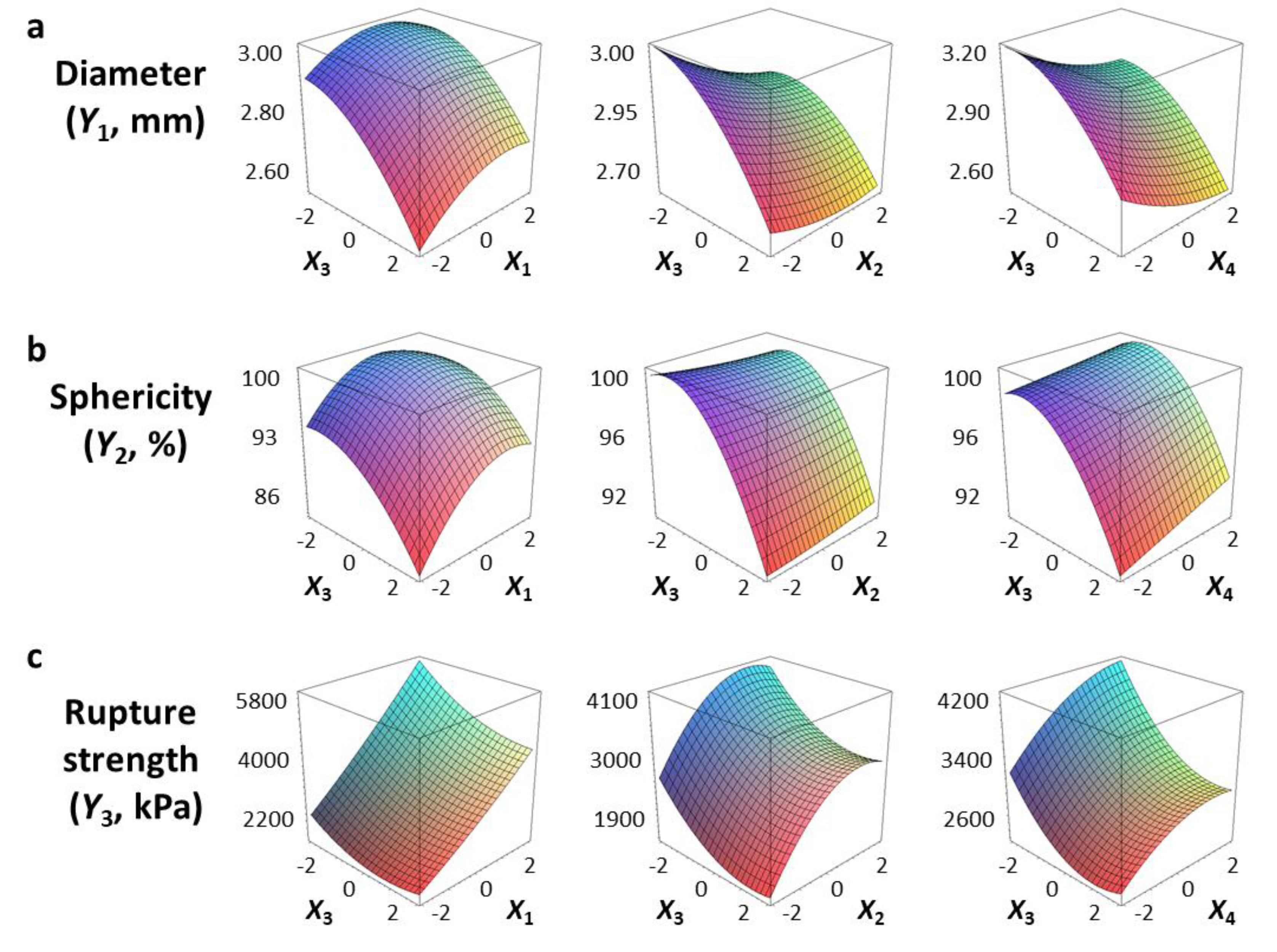
We used a three-dimensional response surface plot to visually display the effects of gelation temperature (X3) and other independent variables [sodium alginate (X1) and calcium lactate (X2) concentration, gelation time (X4)] on the physical properties of the CAG beads.
Figure 2a shows that the diameter (Y1) of the CAG beads increased with increasing sodium alginate concentration (X1) and decreased with increasing gelation temperature (X3). The effect of sodium alginate concentration and gelation temperature on the diameter of CAG beads might be explained by the viscosity of sodium alginate, which increased with increasing concentration or decreasing gelation temperature. As viscosity increased, the size of the sodium alginate droplets on the nozzle tip also increased, thereby increasing the diameter of the CAG beads [25,26]. Moreover, it can be seen from Figure 2a that the diameter (Y1) of the CAG beads decreased when the calcium lactate concentration (X2) or gelation time (X4) increased. These results can be explained by the study of Klokk et al. [26], in which the gel network was contracted by diffusing calcium ions into the sodium alginate droplets in the reactor.
Figure 2b shows that the sphericity (Y2) of the CAG beads increased as gelation temperature (X3) was slightly increased, and then decreased gradually. This is in contrast to the effect of sodium alginate concentration on the sphericity of CAG beads. CAG bead sphericity is closely related to sodium alginate viscosity; the shape of sodium alginate droplets is significantly altered when they hit the calcium lactate solution surface under low viscosity conditions, but sphericity is recovered by increasing the surface tension and gelation above a certain viscosity [9]. Consequently, the sphericity of the CAG beads gradually improved when sodium alginate viscosity increased (increasing sodium alginate concentration or decreasing gelation temperature); however, if the viscosity is too high, the falling sodium alginate droplets develop tails and the CAG beads eventually become tear-shaped [27]. An increase in the gelation time (X4) seemed to increase the sphericity of CAG beads slightly; however, the P-value of the X4 term coefficient for sphericity was greater than 0.05 (Table 3). These results suggest that gelation time does not have a significant effect on the sphericity of CAG beads likewise the calcium lactate concentration (X2). These results indicate that the shape of the falling sodium alginate droplets is an important factor determining the sphericity of CAG beads, and the influence of other factors is not significant after the formation of CAG beads formed through the reaction between alginate and calcium ions. In this study, CAG beads with sphericity greater than 95% were indistinguishable compared to perfect spheres when observed with the naked eye. Thus, preparation conditions must be carefully controlled to produce CAG beads with excellent visual sphericity.

3.3. Rupture Strength

Figure 2c shows that the rupture strength (Y3) of CAG beads increased proportionally with the sodium alginate concentration (X1), calcium lactate concentration (X2), and gelation time (X4). These results are consistent with previous studies that determined CAG gel strength according to the degree of interaction between calcium ions and α-L-guluronic acid, finding that strength was directly proportional to sodium alginate concentration, calcium concentration, and the duration of the interaction between alginate and calcium [28,29,30].
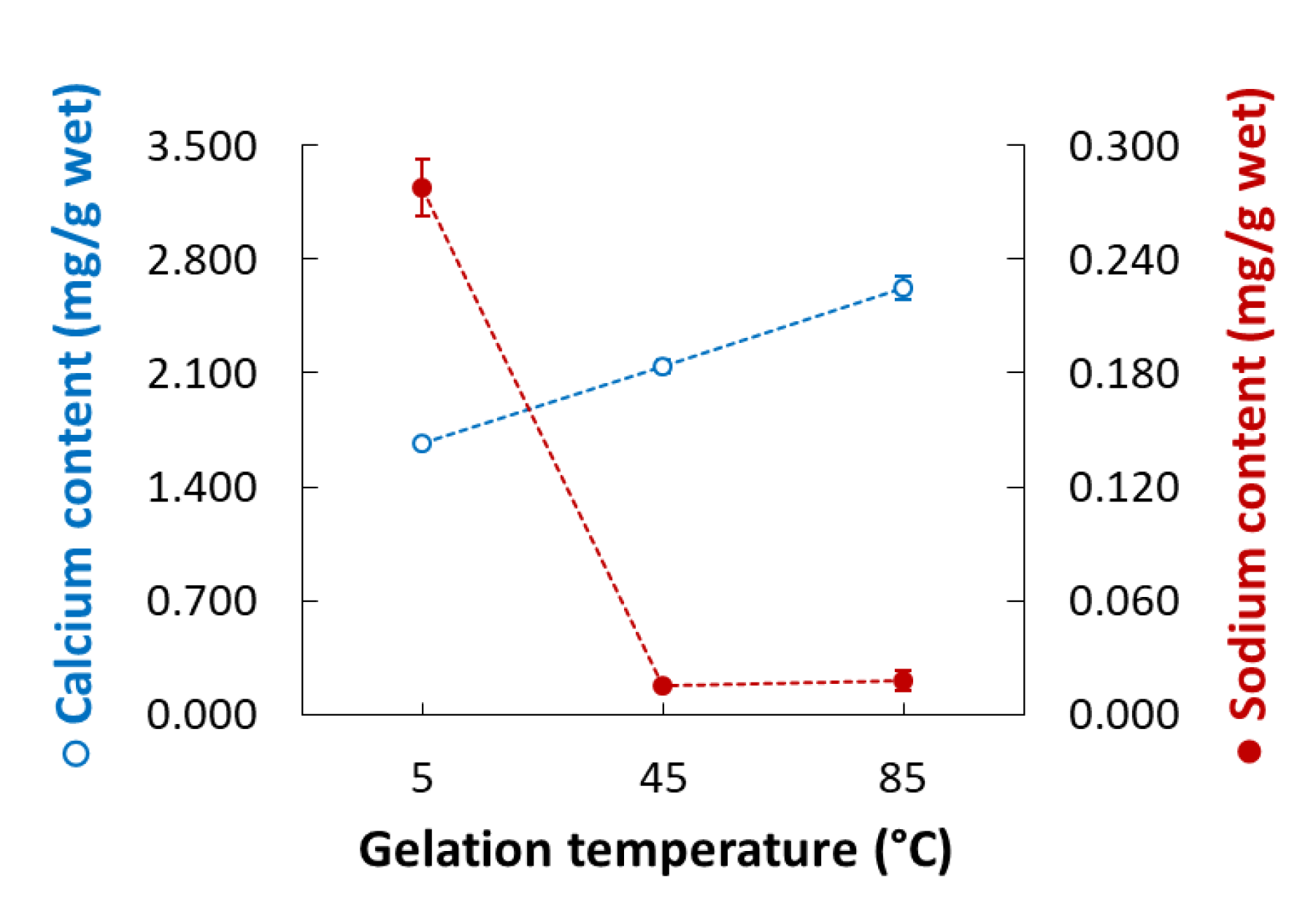
Gelation temperature is an important factor that significantly affects the rupture strength of CAG beads but has not been studied in detail to date. As Figure 2c indicates, the rupture strength (Y3) of the CAG beads increased when the gelation temperature (X3) decreased. Some studies have hypothesized that the increase in gel strength with low gelation temperature might be caused by the formation of a more dense internal structure due to a reduced calcium ion diffusion rate [31,32,33]; however, no experimental results or explanations yet support this theory. Consequently, we measured the calcium and sodium ion content of CAG beads prepared at different temperatures (5, 45, and 85 °C). Other factors were set as follows: sodium alginate concentration, 2.4%; calcium lactate concentration, 2.5%; gelation time, 18 min. As shown in Figure 3, the calcium ion content of the CAG beads decreased from 2.627 to 1.670 mg/g wet weight when the gelation temperature decreased from 85 to 5 °C. This means that the diffusion rate of calcium ions into sodium alginate droplets decreases with decreasing gelation temperature. For the same reasons, the sodium ion content of the CAG beads was highest (0.278 mg/g wet weight) when the gelation temperature was 5 °C.
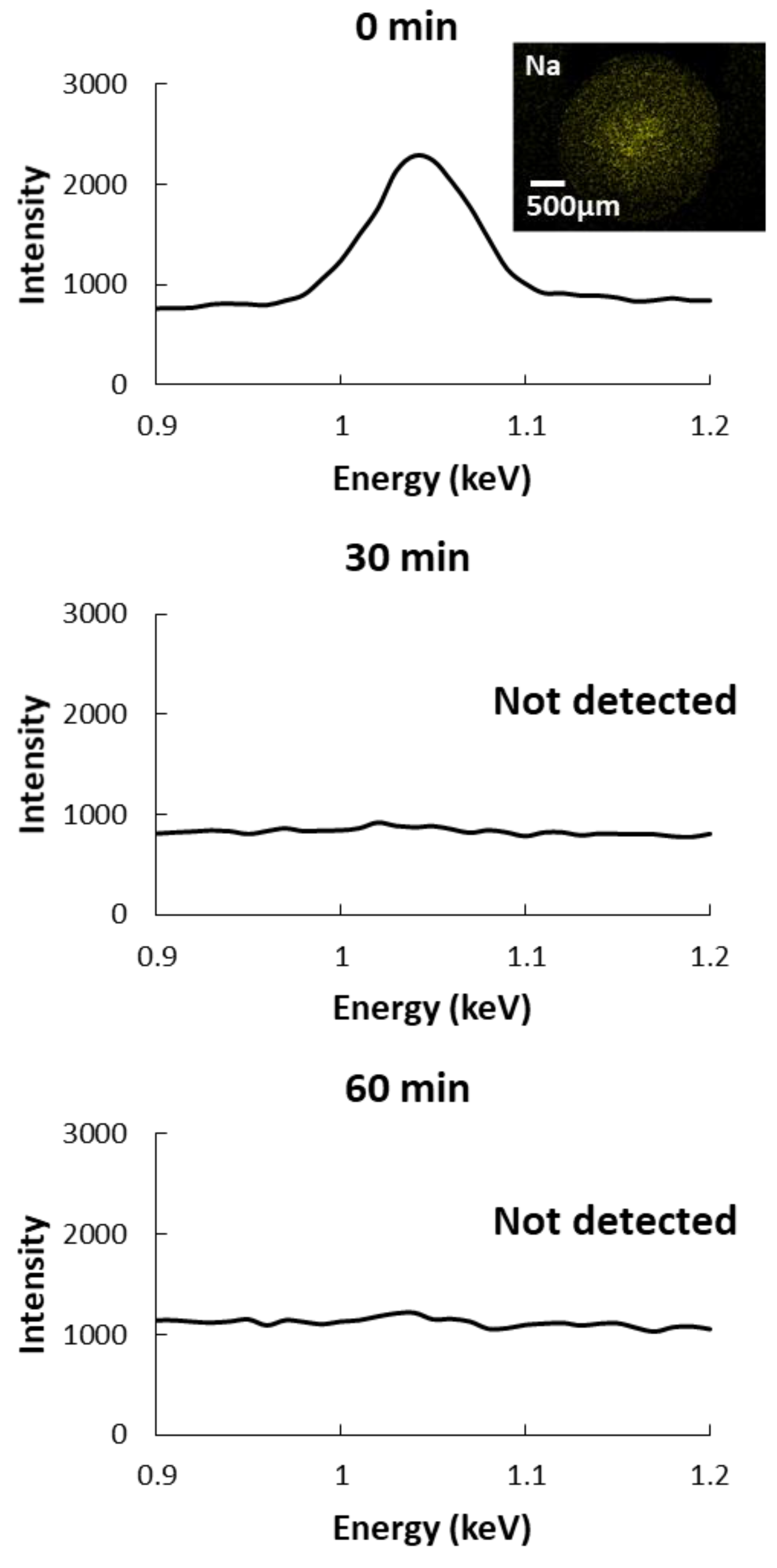
Moreover, the sodium ion content of the CAG beads was highest (0.278 mg/g wet weight) when the gelation temperature was 5 °C. This might cause the CAG beads to swell because of ion-exchange between the residual sodium and calcium ions and can be a problem for their storage stability [34,35]. We assumed that sodium ions might remain in the core of CAG beads, produced at a low gelation temperature because of a reduced diffusion rate out of the beads. Thus, we analyzed the distribution of residual sodium ions in CAG beads prepared at 5 °C and immersed in distilled water for 0, 30, and 60 min. Figure 4 shows that residual sodium ions were detected in the core of the non-immersed CAG beads but not in CAG beads immersed in distilled water for 30 or 60 min. These results indicate that sodium ions remain in the core of the CAG beads because the rate of ion diffusion slows down at low gelation temperatures. Furthermore, immersion allows for sodium ion release, improving the storage stability of CAG beads prepared at low temperatures. The rupture strength of CAG beads generated at 5 °C was decreased upon immersion in distilled water (Table 6). Immersion for 30 min did not significantly alter the rupture strength. Accordingly, to improve the storage stability of CAG beads generated at low temperatures, the immersion time is required to be appropriately set.

3.4. Microstructure

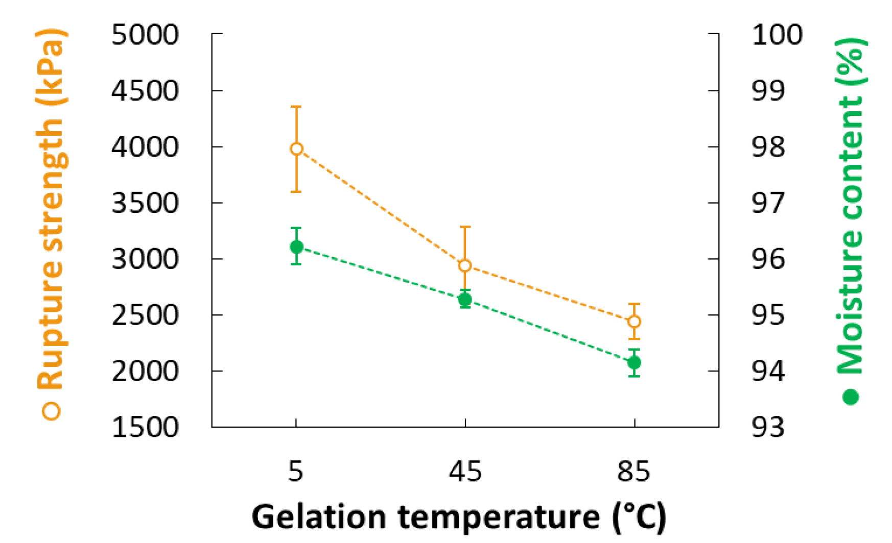
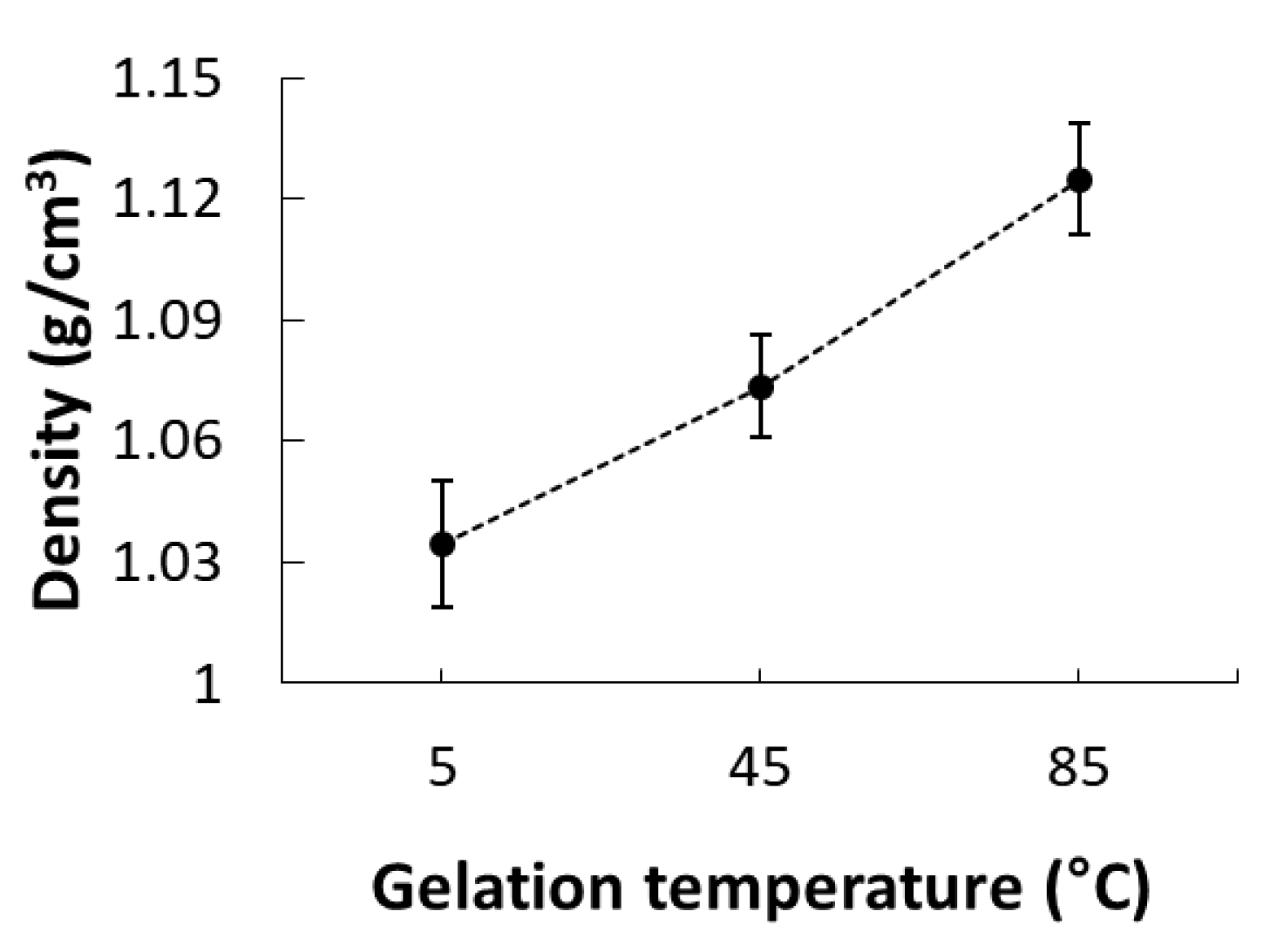
Next, we investigated the effect of gelation temperature on the internal structure of CAG beads. To prevent excessive shrinkage during lyophilization and ensure easy cutting, the beads were lyophilized after being halved while frozen [10,36]. The CAG beads were prepared at a sodium alginate concentration, calcium lactate concentration, and gelation time of 2.4%, 2.5%, and 18 min, respectively. Figure 5 depicts the internal structure of CAG beads prepared at gelation temperatures of 5, 45, and 85 °C. At 30× magnification, the CAG beads prepared at 85 °C had a smooth and homogeneous microstructure, whereas the beads prepared at 5 °C had a rough microstructure containing some big cracks which were likely caused when the CAG beads were freeze-dried due to the relatively high moisture content and rupture strength (Figure 6). At 500× magnification, the CAG beads prepared at 5 °C displayed a microstructure with a well-connected bonding structure and a pattern, unlike those prepared at 45 and 85 °C. This regular microstructure is similar to the SEM image of CAG obtained by Topuz et al. [37]. This difference was better observed at 3000× magnification. Figure 7 shows that the density of CAG beads increased with an increase in the gelation temperature. The calcium ion diffusion rate increased with an increase in the gelation temperature; therefore, the CAG beads generated at a low gelation temperature had a more regular internal structure (not a more dense structure), thus conferring increased rupture strength.

3.5. Optimal Conditions for Maximum Rupture Strength

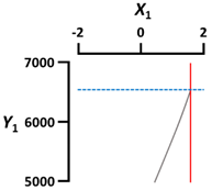
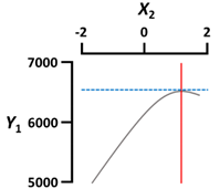
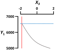
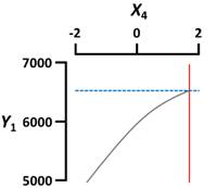
Lastly, we optimized the conditions to prepare CAG beads with maximum rupture strength (Y3). The optimal X1 (sodium alginate concentration), X2 (calcium lactate concentration), X3 (gelation temperature), and X4 (gelation time) conditions for preparing CAG beads with a maximum rupture strength were 3.6%, 4%, 5 °C, and 30 min, respectively (Table 7). Table 8 shows the percentage error verifying the accuracy of the predicted values under the optimal conditions. The predicted Y1 (diameter), Y2 (sphericity), and Y3 (rupture strength) values were 2.85 mm, 94.5%, and 6676 kPa, respectively. We prepared CAG beads under optimal conditions, yielding similar experimental Y1, Y2, and Y3 values of 2.88 ± 0.01 mm, 97.5 ± 0.9%, and 6444 ± 692 kPa, respectively. Consequently, the percentage error values (1.05, 3.17, and 3.48%, respectively) among the experimental and predicted values of Y1, Y2, and Y3 were very small, implying that the developed models were considerably fitted [38].

4. Conclusions

Here, we showed that gelation temperature is an important factor affecting the physical properties of CAG beads. Moreover, our study demonstrated that low gelation temperatures lowered the calcium ion diffusion rate, yielding a more regular microstructure to CAG beads. For this reason, the rupture strength of CAG beads increased as gelation temperature decreased. Furthermore, the CAG beads produced at a low gelation temperature might contain sodium ions; therefore, appropriate immersion is necessary to release these ions and improve the storage stability of CAG beads. Thus, we suggest that gelation temperature should be considered carefully in future research and development using CAGs.

Author Contributions

Working the experiment and writing the manuscript, C.J.; Designing and assisting the experiment, S.K. and C.L.; Designing the experiment and assisting editing the manuscript, S.C.; Supervising this study and revising the manuscript, S.-B.K. All authors have read and agreed to the published version of the manuscript.

Funding

This research received no external funding.

Acknowledgments

We would like to thank Editage (www.editage.co.kr) for English language editing.

Conflicts of Interest

The authors declare no conflict of interest.

References

  1. Vargas, P.O.; Pereira, N.R.; Guimarães, A.O.; Waldman, W.R.; Pereira, V.R. Shrinkage and Deformation during Convective Drying of Calcium Alginate. LWT Food Sci. Technol. 2018, 97, 213–222. [Google Scholar] [CrossRef]
  2. Tavassoli-Kafrani, E.; Shekarchizadeh, H.; Masoudpour-Behabadi, M. Development of Edible Films and Coatings from Alginates and Carrageenans. Carbohydr. Polym. 2016, 137, 360–374. [Google Scholar] [CrossRef] [PubMed]
  3. di Cocco, M.E.; Bianchetti, C.; Chiellini, F. 1H NMR Studies of Alginate Interactions with Amino Acids. Journal of bioactive and compatible polymers. J. Bioact. Compat. Polym. 2003, 18, 283–296. [Google Scholar] [CrossRef]
  4. Grasdalen, H.; Larsen, B.; Smidsrød, O. A pmr study of the composition and sequence of uronate residues in alginates. Carbohydr. Res. 1979, 68, 23–31. [Google Scholar] [CrossRef]
  5. Li, J.; Kim, S.Y.; Chen, X.; Park, H.J. Calcium-alginate beads loaded with gallic acid: Preparation and characterization. LWT Food Sci. Technol. 2016, 68, 667–673. [Google Scholar] [CrossRef]
  6. Vemmer, M.; Patel, A.V. Review of encapsulation methods suitable for microbial biological control agents. Biol. Control 2013, 67, 380–389. [Google Scholar] [CrossRef]
  7. Ha, B.B.; Jo, E.H.; Cho, S.; Kim, S.B. Production Optimization of Flying Fish Roe Analogs Using Calcium Alginate Hydrogel Beads. Fish. Aquat. Sci. 2016, 19, 1–7. [Google Scholar] [CrossRef] [Green Version]
  8. Li, Y.; Hu, M.; Du, Y.; Xiao, H.; McClements, D.J. Control of lipase digestibility of emulsified lipids by encapsulation within calcium alginate beads. Food Hydrocoll. 2011, 25, 122–130. [Google Scholar] [CrossRef]
  9. Lee, B.B.; Ravindra, P.; Chan, E.S. Size and shape of calcium alginate beads produced by extrusion dripping. Chem. Eng. Technol. 2013, 36, 1627–1642. [Google Scholar] [CrossRef]
  10. Voo, W.P.; Ooi, C.W.; Islam, A.; Tey, B.T.; Chan, E.S. Calcium alginate hydrogel beads with high stiffness and extended dissolution behaviour. Eur. Polym. J. 2016, 75, 343–353. [Google Scholar] [CrossRef]
  11. Wu, T.; Li, Y.; Shen, N.; Yuan, C.; Hu, Y. Preparation and characterization of calcium alginate-chitosan complexes loaded with lysozyme. J. Food Eng. 2018, 233, 109–116. [Google Scholar] [CrossRef]
  12. Costa, M.J.; Marques, A.M.; Pastrana, L.M.; Teixeira, J.A.; Sillankorva, S.M.; Cerqueira, M.A. Physicochemical properties of alginate-based films: Effect of ionic crosslinking and mannuronic and guluronic acid ratio. Food Hydrocoll. 2018, 81, 442–448. [Google Scholar] [CrossRef] [Green Version]
  13. Yuasa, M.; Tagawa, Y.; Tominaga, M. The texture and preference of “mentsuyu (Japanese noodle soup base) caviar” prepared from sodium alginate and calcium lactate. Int. J. Gastron. Food Sci. 2019, 18, 100178. [Google Scholar] [CrossRef]
  14. Yamagiwa, K.; Kozawa, T.; Ohkawa, A. Effects of alginate composition and gelling conditions on diffusional and mechanical properties of calcium-alginate gel beads. J. Chem. Eng. Jpn. 1995, 28, 462–467. [Google Scholar] [CrossRef] [Green Version]
  15. Sun, Y.; Liu, J.; Kennedy, J.F. Application of response surface methodology for optimization of polysaccharides production parameters from the roots of Codonopsis pilosula by a central composite design. Carbohydr. Polym. 2010, 80, 949–953. [Google Scholar] [CrossRef]
  16. Kim, S.; Jeong, C.; Cho, S.; Kim, S.B. Effects of Thermal Treatment on the Physical Properties of Edible Calcium Alginate Gel Beads: Response Surface Methodological Approach. Foods 2019, 8, 578. [Google Scholar] [CrossRef] [Green Version]
  17. Izadiyan, P.; Hemmateenejad, B. Multi-response optimization of factors affecting ultrasonic assisted extraction from Iranian basil using central composite design. Food Chem. 2016, 190, 864–870. [Google Scholar] [CrossRef]
  18. Caglar, A.; Sahan, T.; Cogenli, M.S.; Yurtcan, A.B.; Aktas, N.; Kivrak, H. A novel Central Composite Design based response surface methodology optimization study for the synthesis of Pd/CNT direct formic acid fuel cell anode catalyst. Int. J. Hydrog. Energy 2018, 43, 11002–11011. [Google Scholar] [CrossRef]
  19. Cho, S.M.; Gu, Y.S.; Kim, S.B. Extracting Optimization and Physical Properties of Yellowfin Tuna (Thunnus albacares) Skin Gelatin Compared to Mammalian Gelatins. Food Hydrocoll. 2005, 19, 221–229. [Google Scholar] [CrossRef]
  20. Joglekar, A.M.; May, A.T. Product excellence through design of experiments. Cereal Foods World 1987, 32, 857. [Google Scholar]
  21. Su, S.N.; Nie, H.L.; Zhu, L.M.; Chen, T.X. Optimization of adsorption conditions of papain on dye affinity membrane using response surface methodology. Bioresour. Technol. 2009, 100, 2336–2340. [Google Scholar] [CrossRef] [PubMed]
  22. Hashtjin, A.M.; Abbasi, S. Nano-emulsification of orange peel essential oil using sonication and native gums. Food Hydrocoll. 2015, 44, 40–48. [Google Scholar] [CrossRef]
  23. Bezerra, M.A.; Santelli, R.E.; Oliveira, E.P.; Villar, L.S.; Escaleira, L.A. Response Surface Methodology (RSM) as a Tool for Optimization in Analytical Chemistry. Talanta 2008, 76, 965–977. [Google Scholar] [CrossRef]
  24. Hadzir, M.H.; Abbasiliasi, S.; Ariff, A.B.; Yusoff, S.B.; Ng, H.S.; Tan, J.S. Partitioning behavior of recombinant lipase in Escherichia coli by ionic liquid-based aqueous two-phase systems. RSC Adv. 2016, 6, 82571–82580. [Google Scholar] [CrossRef]
  25. Florián-Algarín, V.; Acevedo, A. Rheology and thermotropic gelation of aqueous sodium alginate solutions. J. Pharm. Innov. 2010, 5, 37–44. [Google Scholar] [CrossRef]
  26. Klokk, T.I.; Melvik, J.E. Controlling the size of alginate gel beads by use of a high electrostatic potential. J. Microencapsul. 2002, 19, 415–424. [Google Scholar] [CrossRef]
  27. Li, Z.Q.; Hou, L.D.; Li, Z.; Zheng, W.; Li, L. Study on shape optimization of calcium–alginate beads. Adv. Mat. Res. 2013, 648, 125–130. [Google Scholar] [CrossRef]
  28. Kaklamani, G.; Cheneler, D.; Grover, L.M.; Adams, M.J.; Bowen, J. Mechanical properties of alginate hydrogels manufactured using external gelation. J. Mech. Behav. Biomed. Mater. 2014, 36, 135–142. [Google Scholar] [CrossRef]
  29. Mammarella, E.J.; De Piante Vicín, D.A.; Rubiolo, A.C. Evaluation of stress-strain for characterization of the rheological behavior of alginate and carrageenan gels. Braz. J. Chem. Eng. 2002, 19, 403–409. [Google Scholar] [CrossRef] [Green Version]
  30. Ramdhan, T.; Ching, S.H.; Prakash, S.; Bhandari, B. Time dependent gelling properties of cuboid alginate gels made by external gelation method: Effects of alginate-CaCl2 solution ratios and pH. Food Hydrocoll. 2019, 90, 232–240. [Google Scholar] [CrossRef]
  31. Augst, A.D.; Kong, H.J.; Mooney, D.J. Alginate hydrogels as biomaterials. Macromol. Biosci. 2006, 6, 623–633. [Google Scholar] [CrossRef] [PubMed]
  32. Drury, J.L.; Dennis, R.G.; Mooney, D.J. The tensile properties of alginate hydrogels. Biomaterials 2004, 25, 3187–3199. [Google Scholar] [CrossRef] [PubMed]
  33. Kuo, C.K.; Ma, P.X. Ionically crosslinked alginate hydrogels as scaffolds for tissue engineering: Part 1. Structure, gelation rate and mechanical properties. Biomaterials 2001, 22, 511–521. [Google Scholar] [CrossRef]
  34. Bajpai, S.K.; Sharma, S. Investigation of swelling/degradation behaviour of alginate beads crosslinked with Ca2+ and Ba2+ ions. React. Funct. Polym. 2004, 59, 129–140. [Google Scholar] [CrossRef]
  35. Martinsen, A.; Skjåk-Bræk, G.; Smidsrød, O. Alginate as immobilization material: I. Correlation between chemical and physical properties of alginate gel beads. Biotechnol. Bioeng. 1989, 33, 79–89. [Google Scholar] [CrossRef] [PubMed]
  36. Ayarza, J.; Coello, Y.; Nakamatsu, J. SEM–EDS study of ionically cross-linked alginate and alginic acid bead formation. Int. J. Polym. Anal. Charact. 2017, 22, 1–10. [Google Scholar] [CrossRef]
  37. Topuz, F.; Henke, A.; Richtering, W.; Groll, J. Magnesium ions and alginate do form hydrogels: A rheological study. Soft Matter 2012, 8, 4877–4881. [Google Scholar] [CrossRef]
  38. Wang, X.; Chen, H.; Liu, H.; Li, P.; Yan, Z.; Huang, C.; Zhao, Z.; Gu, Y. Simulation and optimization of continuous laser transmission welding between PET and titanium through FEM, RSM, GA and experiments. Opt. Lasers Eng. 2013, 51, 1245–1254. [Google Scholar] [CrossRef]
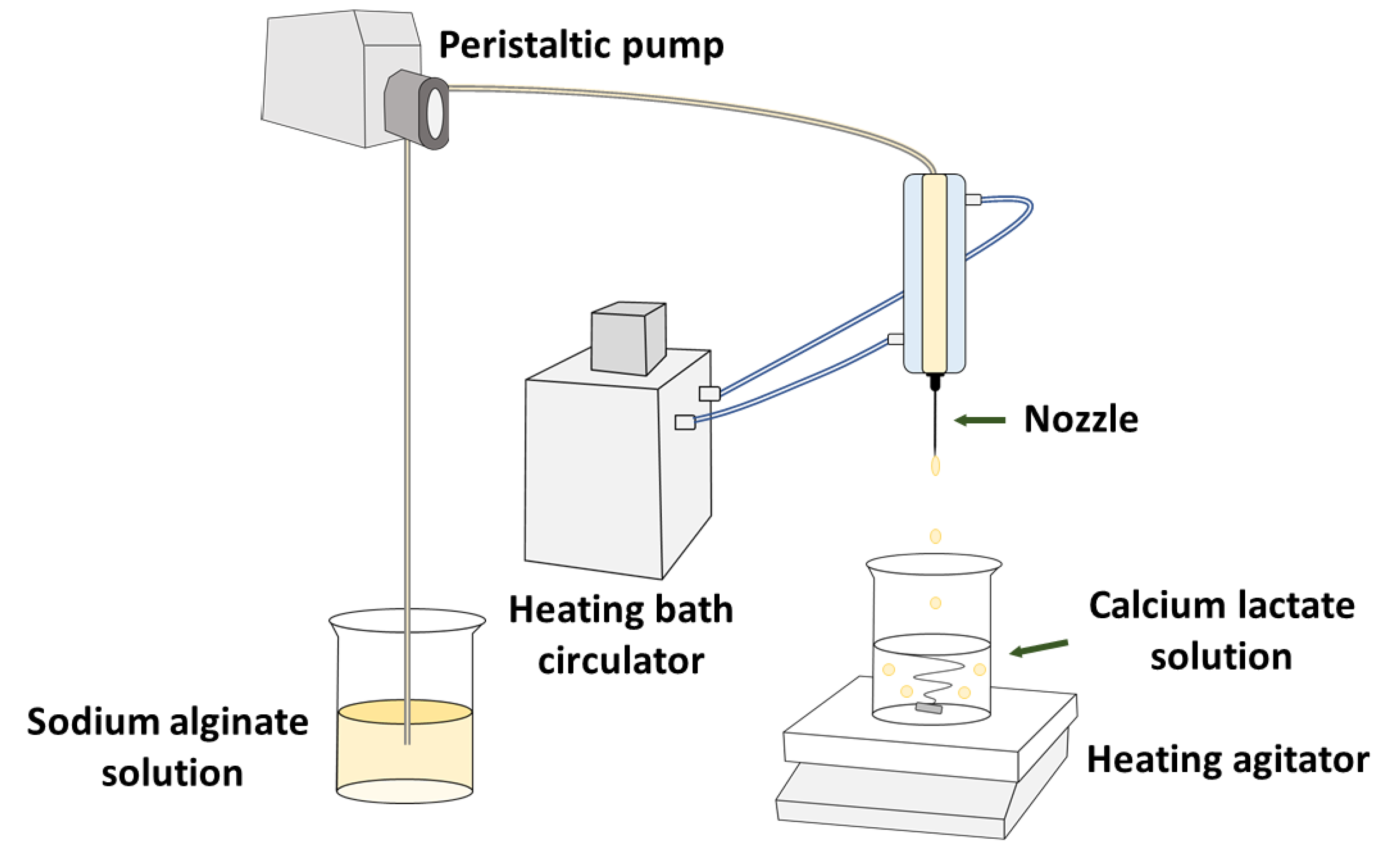
Figure 1. Simple schematic diagram of calcium alginate gel (CAG) bead preparation.
Foods 09 00180 g001
Figure 2. Three-dimensional response surface plots of the physical properties ((a), Diameter; (b), Sphericity; (c), Rupture strength) of CAG beads. X1, sodium alginate concentration (%, w/v); X2, calcium lactate concentration (%, w/v); X3, gelation temperature (°C); X4, gelation time (min).
Foods 09 00180 g002
Figure 3. Calcium (open circles) and sodium ion (filled circles) content of CAG beads prepared at different gelation temperatures.
Foods 09 00180 g003
Figure 4. Energy-dispersive X-ray spectrometer (EDS) spectra and mapping results for sodium ions in CAG beads prepared at 5 °C after immersion in distilled water for different lengths of time.
Foods 09 00180 g004
Figure 5. Visual appearance and low-vacuum scanning electron microscope (LV-SEM) images of CAG beads prepared at different gelation temperatures.
Foods 09 00180 g005
Figure 6. Rupture strength (open circles) and moisture content (filled circles) of CAG beads prepared at different gelation temperatures.
Foods 09 00180 g006
Figure 7. Density (g/cm3) of CAG beads prepared at different gelation temperatures.
Foods 09 00180 g007
Table 1. The range and levels of the independent variables in central composite design (CCD) for monitoring the effects of preparation conditions on the physical properties.
Independent VariablesSymbolRange and Levels
−21012
Sodium alginate concentration (%, w/v)X11.21.82.43.03.6
Calcium lactate concentration (%, w/v)X20.51.52.53.54.5
Gelation temperature (°C)X3525456585
Gelation time (min)X4612182430
Table 2. The CCD matrix and experimental values of the dependent variables for each independent variable.
Run No.Independent VariablesDependent Variables
Coded ValuesUncoded Values
X1X2X3X4X1X2X3X4Y1Y2Y3
Factorial1−1−1−1−11.81.525123.0796.71993
portions21−1−1−13.01.525123.0898.93473
3−11−1−11.83.525123.0096.22274
411−1−13.03.525123.0298.14005
5−1−11−11.81.565122.8292.11901
61−11−13.01.565122.8895.42629
7−111−11.83.565122.8191.62195
8111−13.03.565122.8795.73606
9−1−1−111.81.525242.9397.82420
101−1−113.01.525242.9999.23832
11−11−111.83.525242.9198.32601
1211−113.03.525242.9197.84500
13−1−1111.81.565242.7294.61959
141−1113.01.565242.7795.53575
15−11111.83.565242.7094.22087
1611113.03.565242.7795.43902
Axial17−20001.22.545182.7389.41436
portions1820003.62.545182.9998.54420
190−2002.40.545183.1496.61044
2002002.44.545182.8298.13414
2100−202.42.55183.0498.13976
2200202.42.585182.6290.72440
23000−22.42.54563.0996.72065
2400022.42.545302.8897.83111
Center2500002.42.545182.9798.32788
points2600002.42.545182.9296.62942
2700002.42.545182.8897.53110
X1: Sodium alginate concentration (%, w/v), X2: Calcium lactate concentration (%, w/v), X3: Gelation temperature (°C), X4: Gelation time (min). Y1: Diameter (mm), Y2: Sphericity (%), Y3: Rupture strength (kPa). Each experiment was carried out ten times and the mean value was used.
Table 3. The regression coefficients of the fitted quadratic polynomial models for monitoring the effects of preparation conditions on the physical properties.
ParameterY1Y2Y3
Coefficientp-ValueCoefficientp-ValueCoefficientp-Value
Constant2.923330.00197.46670.0012946.670.001
X10.035420.0111.36250.001752.500.001
X2−0.037920.0070.00420.986338.670.001
X3−0.100420.001−1.80420.001−263.170.003
X4−0.052920.0010.42920.088203.830.013
X1X1−0.019690.141−0.81980.00628.040.713
X2X20.010310.4260.03020.904−146.710.072
X3X3−0.027190.050−0.70730.01498.040.212
X4X40.011560.3730.00520.983−56.960.459
X1X2−0.001880.899−0.06880.812101.250.262
X1X30.009370.5280.28130.340−59.500.502
X1X40.001870.899−0.53130.08587.000.331
X2X30.011880.4270.09380.7464.000.964
X2X40.001870.8990.00630.983−48.750.581
X3X40.000630.9660.10630.714−26.000.767
X1: Sodium alginate concentration (%, w/v), X2: Calcium lactate concentration (%, w/v), X3: Gelation temperature (°C), X4: Gelation time (min). Y1: Diameter (mm), Y2: Sphericity (%), Y3: Rupture strength (kPa).
Table 4. The response surface model equations for monitoring the effects of preparation conditions on the physical properties.
Quadratic Polynomial Model EquationsR2Adj R2Sp-Value
Y1 = 2.92333 + 0.03542X1 − 0.03792X2 − 0.10042X3 − 0.05292X4
− 0.01969X12 + 0.01031X22 − 0.02719X32 + 0.01156X42 − 0.00188X1X2
+ 0.00937X1X3 + 0.00187X1X4 + 0.01188X2X3 + 0.00187X2X4
+ 0.00062X3X4
0.9130.8110.05774100.001
Y2 = 97.4667 + 1.3625X1 + 0.0042X2 − 1.8042X3 + 0.4292X4
− 0.8198X12 + 0.0302X22 − 0.7073X32 + 0.0052X42 − 0.0688X1X2
+ 0.2813X1X3 − 0.5313X1X4 + 0.0938X2X3 + 0.0063X2X4
+ 0.1063X3X4
0.9120.8091.133360.001
Y3 = 2946.67 + 752.50X1 + 338.67X2 − 263.17X3 + 203.83X4
+ 28.04X12 − 146.71X22 + 98.04X32 − 56.96X42 + 101.25X1X2
− 59.50X1X3 + 87.00X1X4 + 4.00X2X3 − 48.75X2X4 − 26.00X3X4
0.9350.860343.7290.001
X1: Sodium alginate concentration (%, w/v), X2: Calcium lactate concentration (%, w/v), X3: Gelation temperature (°C), X4: Gelation time (min). Y1: Diameter (mm), Y2: Sphericity (%), Y3: Rupture strength (kPa).
Table 5. The analysis of variance (ANOVA) of response surface model equations for monitoring the effects of preparation conditions on the physical properties.
Dependent VariablesSourcesDFSSMSf-Valuep-Value
Y1 Diameter (mm)Regression
Linear40.3738170.09345428.030.001
Square40.0404040.0101013.030.061
Interaction60.0038380.0006400.190.973
Residual
Lack of fit100.0359420.0035941.770.415
Pure error20.0040670.002033--
Total260.458067---
Y2 Sphericity (%)Regression
Linear4127.09531.773824.740.001
Square425.6776.41935.000.013
Interaction66.1791.02980.800.587
Residual
Lack of fit1013.9671.39671.930.389
Pure error21.4470.7233--
Total26174.365---
Y3 Rupture strength (kPa)Regression
Linear419,002,1464,750,53740.210.001
Square41,093,213273,3032.310.117
Interaction6390,87065,1450.550.760
Residual
Lack of fit101,365,918136,5925.270.170
Pure error251,87525,937--
Total2621,904,022---
DF (Degrees of freedom), SS (Sum of square), MS (Mean square).
Table 6. Rupture strength of CAG beads generated at 5 °C after immersion in distilled water for different periods.
Immersion Time0 min30 min60 min
Rupture strength3910 ± 150 a3784 ± 119 a3187 ± 114 b
a,b The same letter indicates no significant difference (p < 0.05, Tukey’s range test).
Table 7. The optimal conditions for preparing CAG beads with a maximum rupture strength.
Optimal ConditionsY3 Rupture Strength (kPa)
Target ValueMaximum
X1 Sodium alginate concentration (%, w/v)Coded value2 Foods 09 00180 i001
Actual value3.6
X2 Calcium lactate concentration (%, w/v)Coded value1.5 Foods 09 00180 i002
Actual value4
X3 Gelation temperature (°C)Coded value−2 Foods 09 00180 i003
Actual value4
X4 Gelation time (min)Coded value2 Foods 09 00180 i004
Actual value30
Table 8. Verification of experimental and predicted values under optimal conditions.
Y1 Diameter (mm)Y2 Sphericity (%)Y3 Rupture Strength (kPa)
Predicted values2.8594.56676
Experimental values2.88 ± 0.0197.5 ± 0.96444 ± 692
Error (%)1.053.173.48
Optimal conditions: Sodium alginate concentration = 3.6%; Calcium lactate concentration = 4% min; Gelation temperature = 4 °C; Gelation time = 30 min. Error (%) = (Difference among predicted value and actual value/predicted value) × 100.