Article

PfAgo-Enhanced LAMP Assay for Rapid and Specific Detection of Vibrio alginolyticus and Vibrio vulnificus


College of Food Science and Technology, Nanjing Agricultural University, Nanjing 210095, China
*
Author to whom correspondence should be addressed.
These authors contributed equally to this work.
Foods 2026, 15(14), 2506; https://doi.org/10.3390/foods15142506
Submission received: 11 June 2026 / Revised: 12 July 2026 / Accepted: 13 July 2026 / Published: 15 July 2026
(This article belongs to the Section Food Microbiology)

Abstract

Rapid and accurate detection of Vibrio alginolyticus and Vibrio vulnificus is important for seafood safety and foodborne infection control. In this study, we developed a detection platform that combines loop-mediated isothermal amplification (LAMP) with Pyrococcus furiosus Argonaute (PfAgo)-mediated sequence-specific cleavage. The LAMP rapidly amplified pathogen-specific target sequences under isothermal conditions, while PfAgo provides an additional layer of sequence verification through a programmable guide DNA-directed cleavage step to reduce false-positive signals caused by nonspecific amplification. Under optimized conditions, the assay achieved limits of detection of 3 × 101 CFU·mL−1 for V. alginolyticus and 102 CFU·mL−1 for V. vulnificus, with no positive signals observed among the tested non-target bacteria. In artificially contaminated salmon samples, both V. alginolyticus and V. vulnificus were detected without enrichment after simple boiling-based DNA extraction, with a practical detection limit of 2.9 × 10² CFU·mL−1 in the final homogenate. Compared with culture-based methods and instrument-intensive molecular assays, the proposed LAMP-PfAgo platform exhibits a rapid two-step workflow with an added sequence-verification layer. These results support LAMP coupled with Argonaute-mediated cleavage as a practical strategy for foodborne pathogen monitoring in seafood.

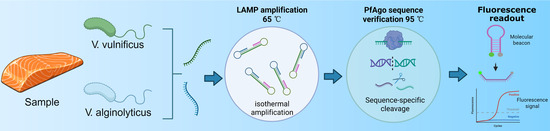
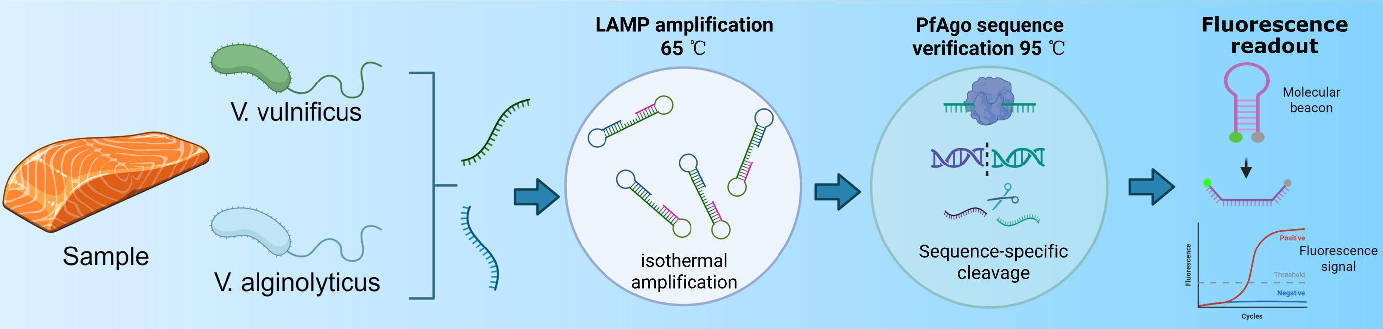
Graphical Abstract

1. Introduction

Vibrio alginolyticus (V. alginolyticus) and Vibrio vulnificus (V. vulnificus) are important halophilic bacteria that are widely distributed in marine and estuarine environments. Both pathogens threaten aquaculture by causing mortality in aquatic organisms and substantial economic losses [1]. V. alginolyticus can cause wound infections in humans, which may progress to severe disease, and has therefore attracted increasing attention [2]. V. vulnificus is recognized as one of the most virulent foodborne pathogens and can cause septicemia and necrotizing wound infections with high mortality rates [3]. Both species also carry virulence-associated genes and show increasing antimicrobial resistance, further complicating seafood safety management [4,5]. Rapid and sensitive detection methods are therefore needed for seafood safety control.
The high genetic relatedness and similar phenotypes of Vibrio species, particularly between V. alginolyticus and V. vulnificus, can lead to misidentification and delays in timely clinical or food-safety interventions [6]. Culture-based methods are reliable but time-consuming, whereas immunological assays require specific antibodies and may be less suitable for early and rapid pathogen detection [7]. Loop-mediated isothermal amplification (LAMP) can detect target genes under constant-temperature conditions, but conventional LAMP results often rely on gel electrophoresis or visual observation of precipitation. These results can be subjective, contamination-prone, or vulnerable to false-positive signals, indicating the need for a more specific detection workflow [8].
Clustered regularly interspaced short palindromic repeats (CRISPR) and CRISPR-associated proteins (Cas) provide highly specific nucleic acid recognition and have been combined with LAMP to improve assay specificity. For example, LAMP-CRISPR-Cas12a has been used for specific detection of Escherichia coli O157:H7 with ultraviolet (UV)-based visual readout, reducing reliance on complex instruments and mitigating false-positive signals [9]. However, CRISPR-Cas assays still face practical constraints. Cas13a-based detection requires an additional transcription step to convert DNA into RNA, increasing assay complexity, cost, and time [10]. Cas12a can directly target double-stranded DNA, but its activity depends on protospacer adjacent motif (PAM) sequences, restricting target-site selection and assay flexibility [11]. In addition, Cas proteins can be sensitive to reaction conditions, which may affect robustness in resource-limited on-site testing [12]. Alternative nucleic acid recognition tools that are compatible with LAMP therefore remain valuable.
Argonaute (Ago) proteins are programmable nucleases that specifically recognize and cleave nucleic acid targets [13]. Compared with CRISPR-Cas systems, Ago proteins do not require PAM sequences, thus offering greater flexibility in target site selection. They use short guide DNAs (gDNAs) that are stable, easy to design, and readily synthesized, which reduce the off-target risks [14]. Pyrococcus furiosus Argonaute (PfAgo) from the hyperthermophilic archaeon Pyrococcus furiosus has been widely explored for nucleic acid detection because of its thermostability and high cleavage efficiency. In PfAgo-based assays, amplified nucleic acid fragments can be recognized by PfAgo-gDNA complexes, leading to probe cleavage and fluorescence signal release [15]. Recent studies have demonstrated that the integration of specific primers, exonuclease I, and PfAgo enables one-step cleavage, facilitating ultrasensitive detection of foodborne pathogens. This assay enabled rapid and sensitive detection of Salmonella Typhi and Staphylococcus aureus in actual food samples, highlighting the potential of PfAgo-based strategies for rapid foodborne pathogen screening [16]. Because LAMP and PfAgo-mediated cleavage can be integrated in a sequential workflow, their combination provides a direct route for target amplification followed by sequence-specific verification [17].
Here, we established a rapid and specific LAMP-PfAgo assay for V. alginolyticus and V. vulnificus. We optimized key parameters of the LAMP and PfAgo cleavage modules, evaluated analytical specificity and sensitivity, and tested practical applicability using artificially contaminated salmon samples. This work addresses the limitations of conventional methods and CRISPR-Cas based detection assays by developing a two-step LAMP-PfAgo detection workflow for V. alginolyticus and V. vulnificus.

2. Materials and Methods

2.1. Bacterial Strains and Genomic DNA Extraction

The standard strain and isolates of V. alginolyticus and V. vulnificus were used as target strains in this study. The biotypes of the tested V. vulnificus strains were not further determined in the present study. Several non-target bacterial species, including other Vibrio species and common foodborne pathogens, were used to evaluate the specificity of the developed assay. All strains used in this study are summarized in Table 1. All strains were maintained in 40% glycerol stocks at −80 °C in our laboratory. Before use, bacterial strains were streaked onto Luria–Bertani (LB) agar plates and incubated at 37 °C for activation. A single colony was subsequently inoculated into LB broth and cultured overnight at 37 °C with shaking. All Vibrio strains were cultured in LB medium supplemented with 3% (w/v) NaCl.
Genomic DNA was extracted as described previously [18]. Briefly, 1 mL of bacterial culture was centrifuged at 12,000× g for 5 min. The pellet was resuspended in 100 μL TE buffer, boiled in a water bath at 100 °C for 10 min, and immediately placed on ice for 10 min. After centrifugation at 12,000× g for 5 min, the supernatant containing genomic DNA was collected and stored at −20 °C until use.

2.2. Design of LAMP Primers, gDNAs, and Probes

Species-specific target genes, N646_RS01740 for V. alginolyticus and AOT11_08030 for V. vulnificus, were selected based on previous genomic screening in our laboratory. Four LAMP primer sets were designed for each target pathogen using PrimerExplorer V5 software (http://primerexplorer.jp/e/index.html, accessed on 7 November 2024) according to previously established LAMP primer design principles [19,20]. Primer sequences are listed in Table 2 and Table 3.
To establish the PfAgo-mediated cleavage system, three groups of 16 nt gDNAs were designed for each pathogen using SnapGene 6.0.2 software. All gDNAs were synthesized with a 5′-phosphate modification to enable PfAgo loading. Corresponding molecular beacon (MB) probes labeled with fluorescent and quenching groups were also designed for fluorescence signal generation. The sequences of all gDNAs and probes are shown in Table 4 and Table 5. The design strategy was based on the programmable cleavage characteristics of Argonaute-mediated nucleic acid detection systems.
All primers, gDNAs, and molecular beacon probes were synthesized by GenScript Biotech Corporation (Nanjing, China). PfAgo (Cat. No. JY0316-L) was purchased from Tiosbio (Beijing, China). Bst 3.0 DNA polymerase, isothermal amplification buffer, and dNTPs were purchased from New England Biolabs (Ipswich, MA, USA).

2.3. LAMP Reaction and Optimization Conditions

The LAMP reaction was performed in a total volume of 25 μL containing outer primers (F3/B3, 0.2 μmol/L each), inner primers (FIP/BIP, 0.8 μmol/L each), loop primers (LF/LB, 0.4 μmol/L each), 1.4 mmol/L dNTPs, 6.0 mmol/L MgSO4, 1× isothermal amplification buffer, 0.32 U/μL Bst 3.0 DNA polymerase, 2 μL genomic DNA template, and nuclease-free water to volume. Amplification was initially conducted at 65 °C for 40 min, consistent with commonly reported LAMP conditions for foodborne pathogen detection [8].
To improve amplification efficiency, key reaction parameters were systematically optimized, including Mg2+ concentration (2.0–10.0 mM), dNTP concentration (1.0–1.8 mM), amplification temperature (60–65 °C), Bst 3.0 DNA polymerase dosage, and inner-to-outer primer ratio (2:1–10:1). During optimization, only one parameter was varied at a time while all other reaction conditions were kept constant. Amplification performance was evaluated according to fluorescence intensity and threshold cycle (Ct) values obtained from real-time fluorescence monitoring.

2.4. Optimization of PfAgo-Mediated Cleavage Reaction Conditions

The PfAgo cleavage reaction was performed in a total volume of 20 μL containing 2 μL PfAgo protein (Tiosbio, JY0316-L), 1 μL gDNA, 0.5 μL Mn2+ solution, 2 μL 10× PfAgo Endonuclease Reaction Buffer (supplied with the commercial PfAgo protein), 1 μL molecular beacon probe, 2 μL LAMP product, and nuclease-free water to final volume. The final concentrations of gDNA, Mn2+, and molecular beacon probe were initially set at 0.5 μM, 1 mM, and 0.5 μM, respectively.
Following LAMP, the PfAgo-assisted nucleic acid detection step was performed as previously described with minor modifications [17]. In total, 2 μL of the LAMP product was transferred to the PfAgo cleavage reaction mixture. The PfAgo-mediated cleavage reaction was then incubated at 95 °C for 30 min, followed by fluorescence measurement. The fluorescence signal was generated through a target-dependent PfAgo cleavage cascade. Briefly, 5′-phosphorylated gDNAs guided PfAgo to cleave the LAMP amplicons at specific sites. The resulting short DNA fragments served as secondary guides to direct PfAgo-mediated cleavage of the molecular beacon probe, separating the fluorophore from the quencher and producing fluorescence signals [21].
The PfAgo cleavage system was further optimized by varying gDNA concentration (0–1.0 μmol/L), PfAgo protein dosage, Mn2+ concentration (0–1.0 mmol/L), and cleavage reaction time (5–30 min). All optimization experiments were performed under otherwise identical conditions, and fluorescence intensity was used as the primary evaluation indicator.

2.5. Specificity and Sensitivity Evaluation

The specificity of the developed LAMP-PfAgo assay was evaluated using genomic DNA extracted from target strains and non-target bacterial species listed in Table 1. Nuclease-free water was used as the negative control. Cross-reactivity within the tested bacterial panel was assessed according to fluorescence signal generation after amplification and PfAgo cleavage.
To determine the limit of detection (LOD), overnight cultures of V. alginolyticus and V. vulnificus were serially diluted 10-fold in sterile saline. Bacterial concentrations were determined by plate counting, and genomic DNA extracted from each dilution was used as the template for the optimized LAMP-PfAgo assay. The lowest bacterial concentration that consistently produced detectable fluorescence signals in three independent experiments was defined as the limit of detection (LOD), according to previously reported molecular diagnostic evaluation criteria [22].

2.6. Preparation of Artificially Contaminated Samples

Artificial contamination experiments were performed to evaluate the practical applicability of the LAMP-PfAgo method for detecting V. alginolyticus and V. vulnificus in food samples. Briefly, 22.5 g of salmon was placed into a stomacher bag containing 225 mL of alkaline peptone water (APW). Target bacterial suspensions were prepared at approximately 100-fold higher concentrations than the intended final contamination levels, with the highest prepared suspension concentration of approximately 108 CFU·mL−1. For each contamination level, 2.5 mL of bacterial suspension was added to the corresponding sample to obtain final bacterial concentrations ranging from approximately 106 to 100 CFU·mL−1 in the homogenates. After homogenization, 1 mL of each homogenate was collected for genomic DNA extraction and tested using the established LAMP-PfAgo method.

2.7. Statistical Analysis

All experiments were performed in triplicate, and the results are presented as mean ± standard deviation (SD). Statistical analysis and graphical visualization were performed using GraphPad Prism 8.0 software (GraphPad Software, Boston, MA, USA). Differences among groups were analyzed by one-way analysis of variance (ANOVA), with p < 0.05 considered statistically significant. Different lowercase letters indicate significant differences among groups.

3. Results

3.1. Optimization of the LAMP System

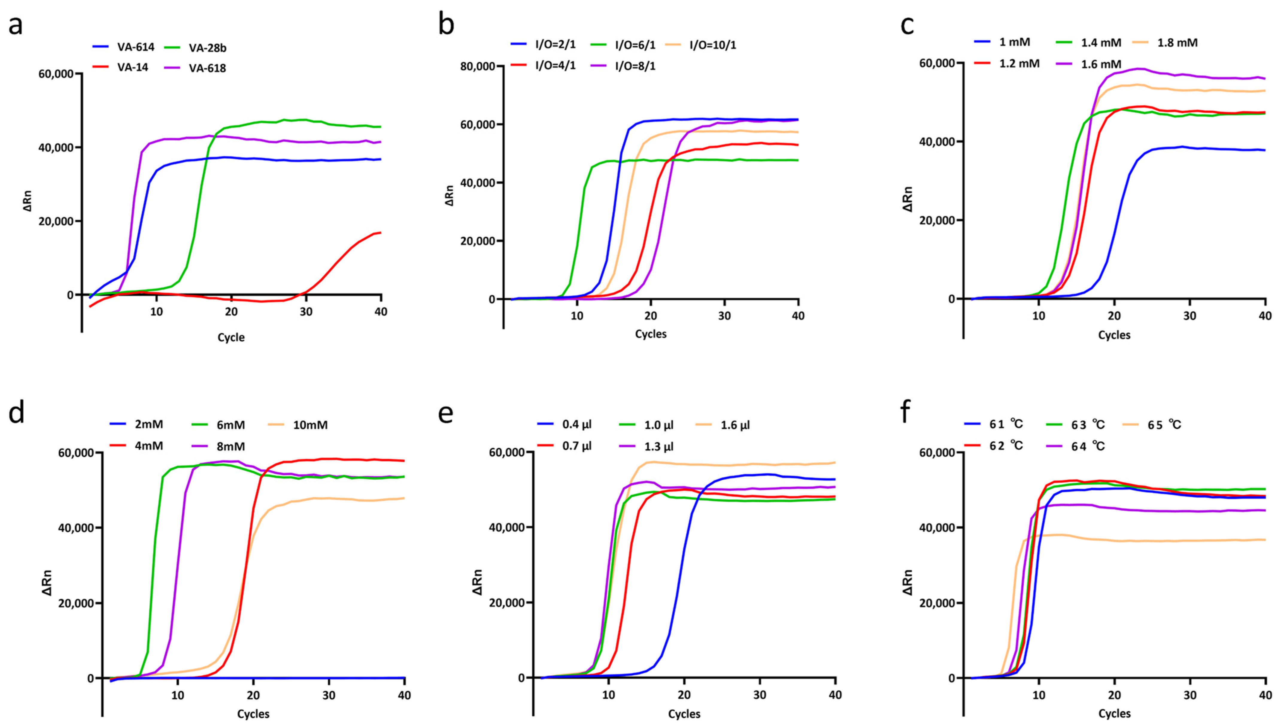
The LAMP conditions were independently optimized for V. alginolyticus and V. vulnificus to establish efficient upstream amplification modules for the subsequent PfAgo-mediated detection assay. For V. alginolyticus, four primer sets were evaluated using genomic DNA as the template. Among them, VA-618 produced an early amplification curve with a stable fluorescence signal and was selected for further optimization (Figure 1a). The inner-to-outer (I/O) primer ratio was then screened, and an I/O ratio of 6:1 yielded rapid amplification onset with a high ΔRn value (Figure 1b). Further optimization showed that 1.4 mM dNTPs, 6 mM Mg2+, and 1.0 μL Bst 3.0 DNA polymerase supported rapid and stable amplification (Figure 1c–e). Although several temperatures generated detectable amplification, 65 °C provided rapid signal development and was selected as the reaction temperature for the V. alginolyticus LAMP assay (Figure 1f).
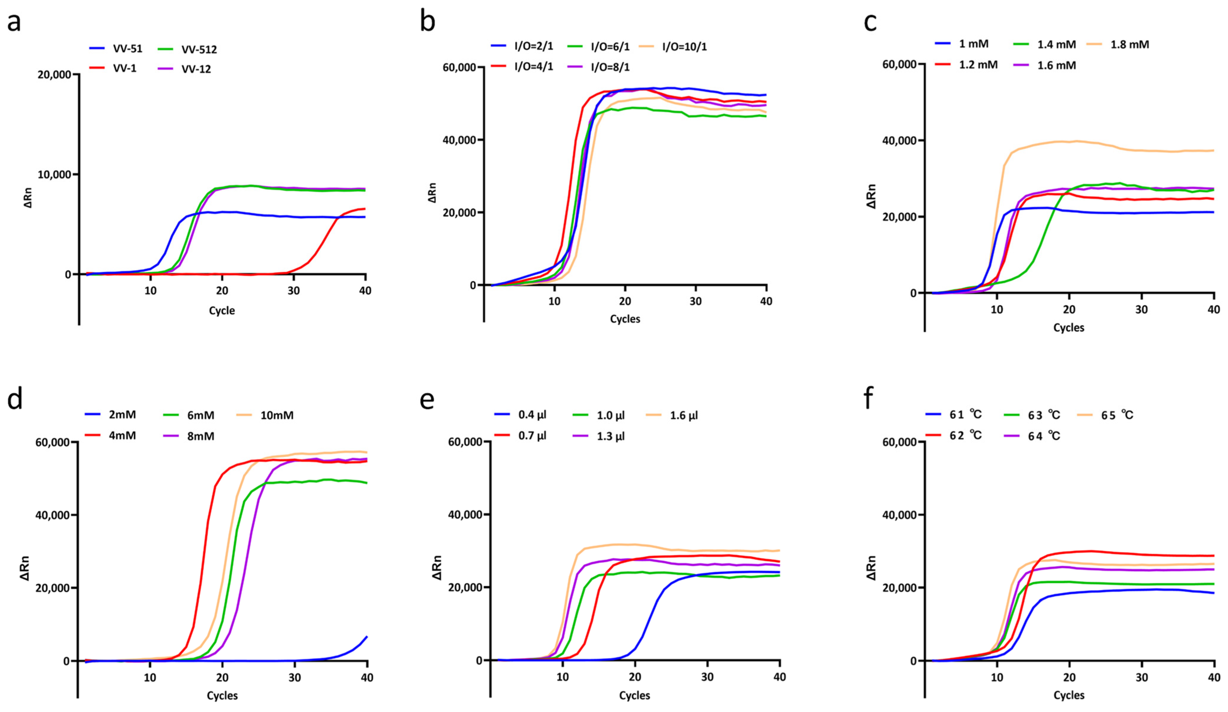
The same optimization strategy was applied to the V. vulnificus LAMP system. Primer set VV-51 showed the earliest amplification among the four candidate primer sets and was selected for subsequent assays (Figure 2a). Screening of primer ratios indicated that an I/O ratio of 4:1 produced rapid amplification and a strong fluorescence response (Figure 2b). For the reaction components, 1.8 mM dNTPs and 4 mM Mg2+ produced favorable amplification profiles (Figure 2c,d). In the Bst 3.0 DNA polymerase screening, the plotted curves indicate that 1.6 μL supported efficient amplification (Figure 2e). For temperature optimization, 65 °C enabled rapid amplification and was selected for the V. vulnificus LAMP reaction (Figure 2f). Together, these results established species-specific optimized LAMP systems for V. alginolyticus and V. vulnificus, providing reliable amplified products for downstream PfAgo cleavage.

3.2. Establishment and Optimization of the LAMP-PfAgo System

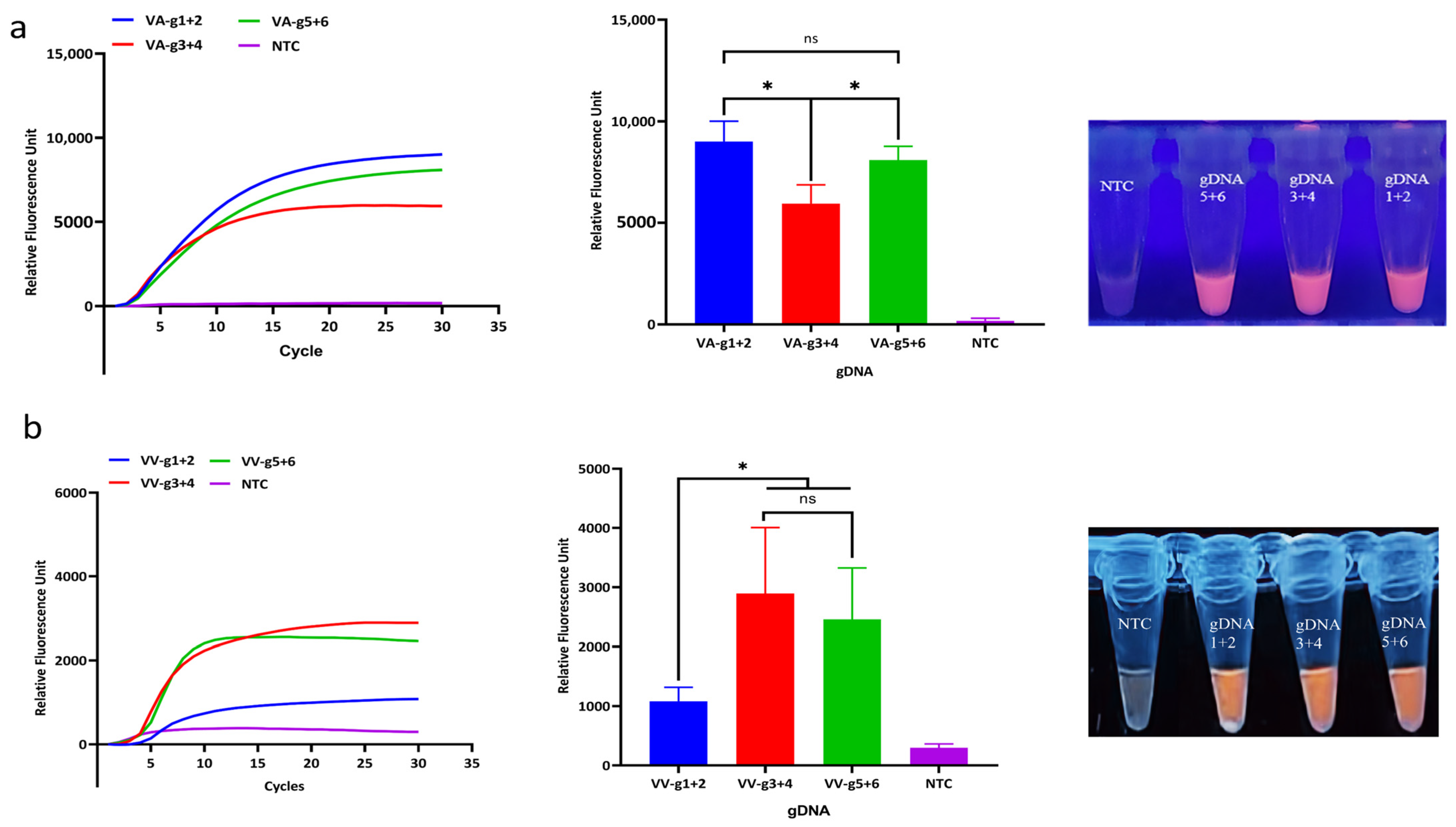
To identify the suitable gDNA combinations for PfAgo-mediated cleavage, three candidate gDNA groups were evaluated for both V. alginolyticus and V. vulnificus. For V. alginolyticus, the tested gDNA groups generated distinguishable fluorescence signals relative to the no-template control (NTC) (p < 0.05) (Figure 3a). For V. vulnificus, the candidate gDNA groups also differed in fluorescence intensity, allowing selection of the group with the strongest signal and clearest separation from the negative control (Figure 3b). These results demonstrated that different gDNA combinations exhibited distinct cleavage efficiencies toward amplified target sequences. Therefore, the VA-gDNA1+2 combination for V. alginolyticus and the VV-gDNA3+4 combination for V. vulnificus were selected for subsequent optimization experiments.
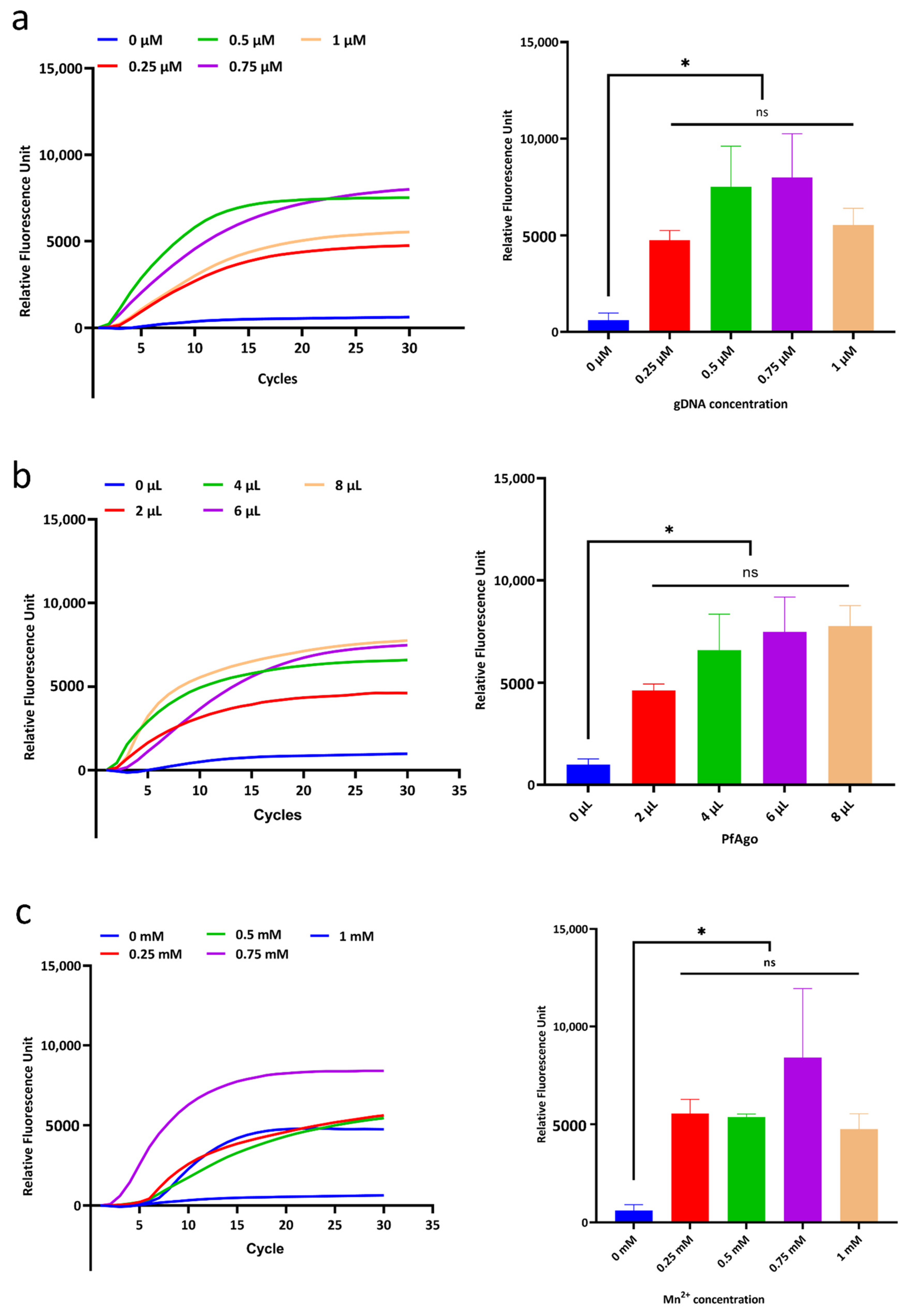
The reaction parameters of the PfAgo cleavage system were further optimized to improve fluorescence signal intensity and detection efficiency for both target pathogens. For the V. alginolyticus detection system, fluorescence intensity increased with increasing gDNA and Mn2+ concentrations and reached maximum values at 0.75 μM gDNA and 0.75 mM Mn2+, respectively (Figure 4a,c). The highest fluorescence signal was obtained with 8 μL PfAgo protein (Figure 4b). Thus, the optimized PfAgo cleavage conditions for V. alginolyticus were 0.75 μM gDNA, 0.75 mM Mn2+, and 8 μL PfAgo protein.
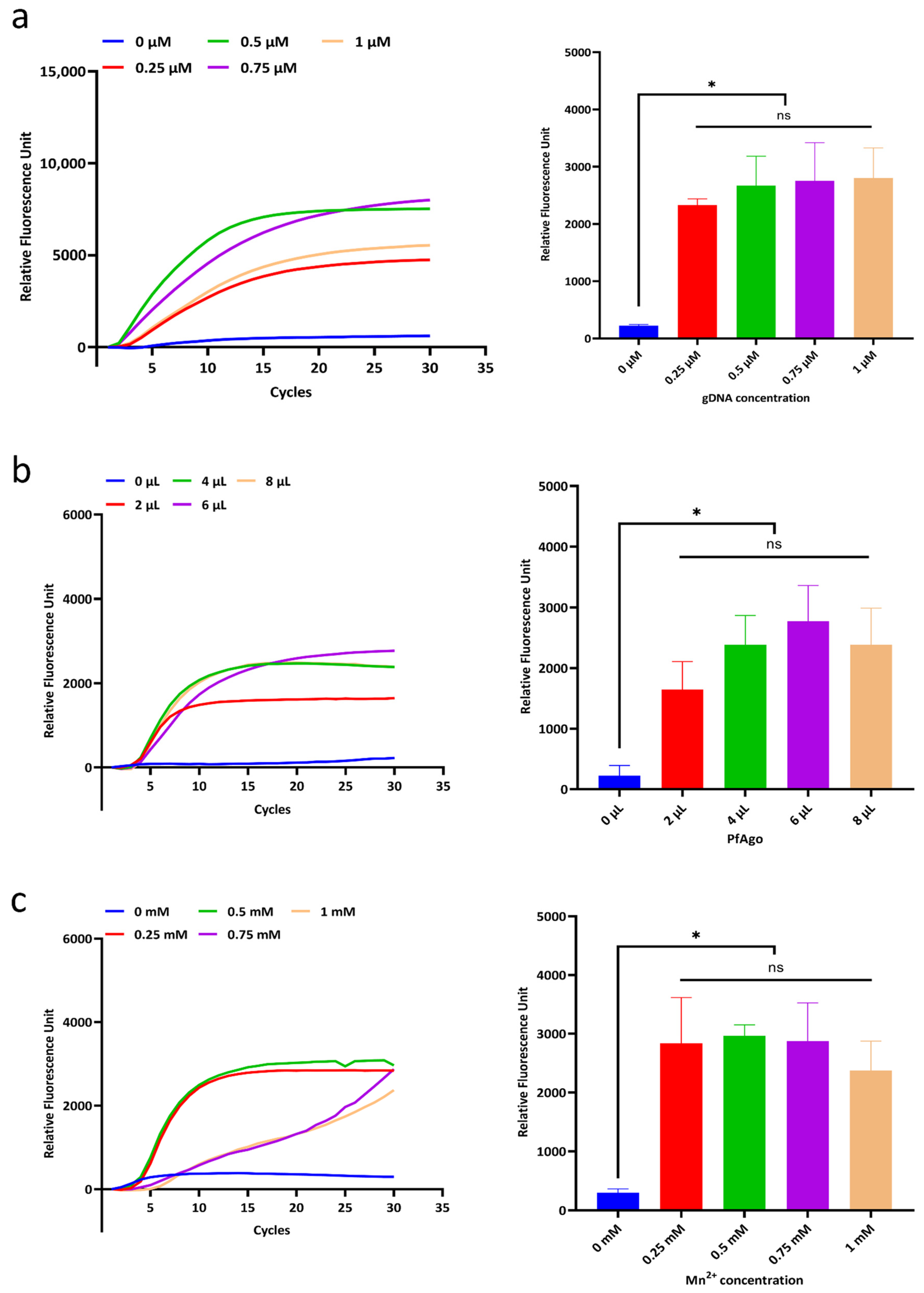
For the V. vulnificus detection system, the optimized conditions were identified as 1.0 μM gDNA, 0.5 mM Mn2+, and 6 μL PfAgo protein, based on the highest fluorescence intensities observed under these conditions (Figure 5a–c).

3.3. Sensitivity and Specificity Evaluation

The specificity and sensitivity of the optimized LAMP-PfAgo systems for V. alginolyticus and V. vulnificus were evaluated using target and non-target bacterial strains.
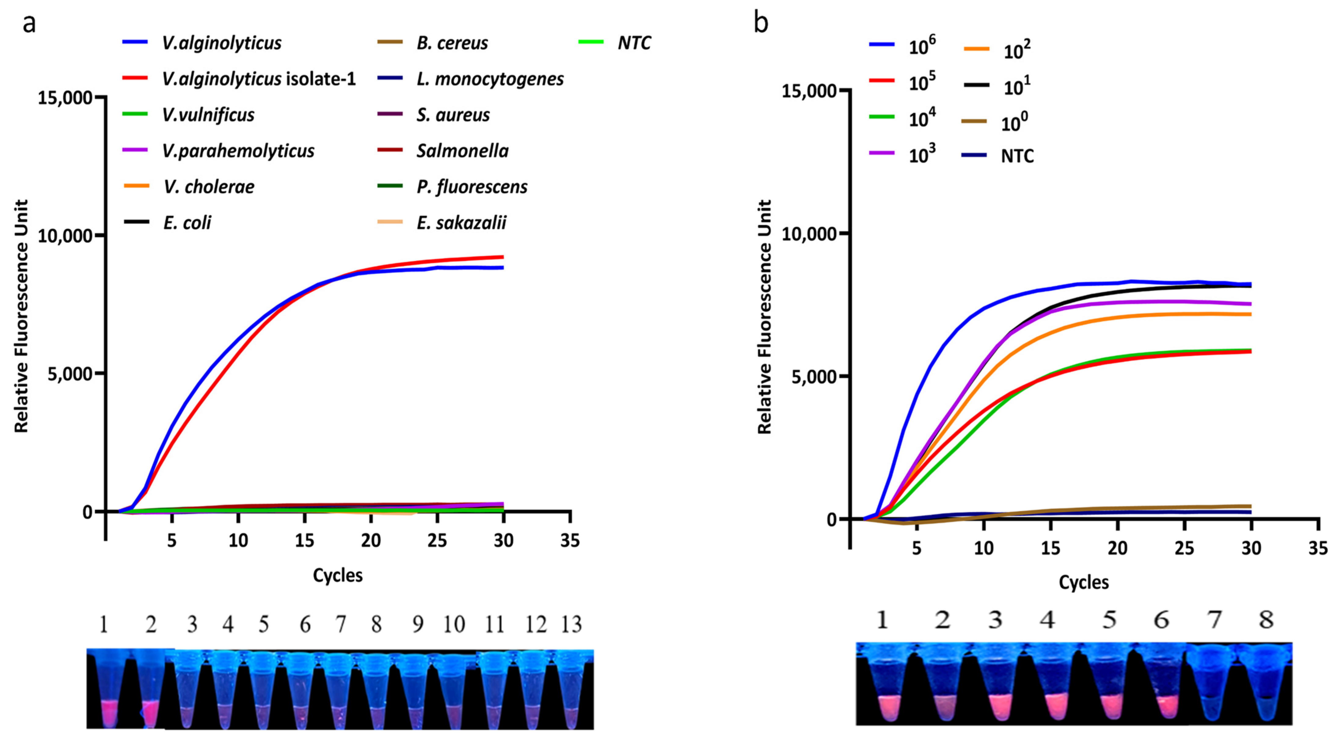
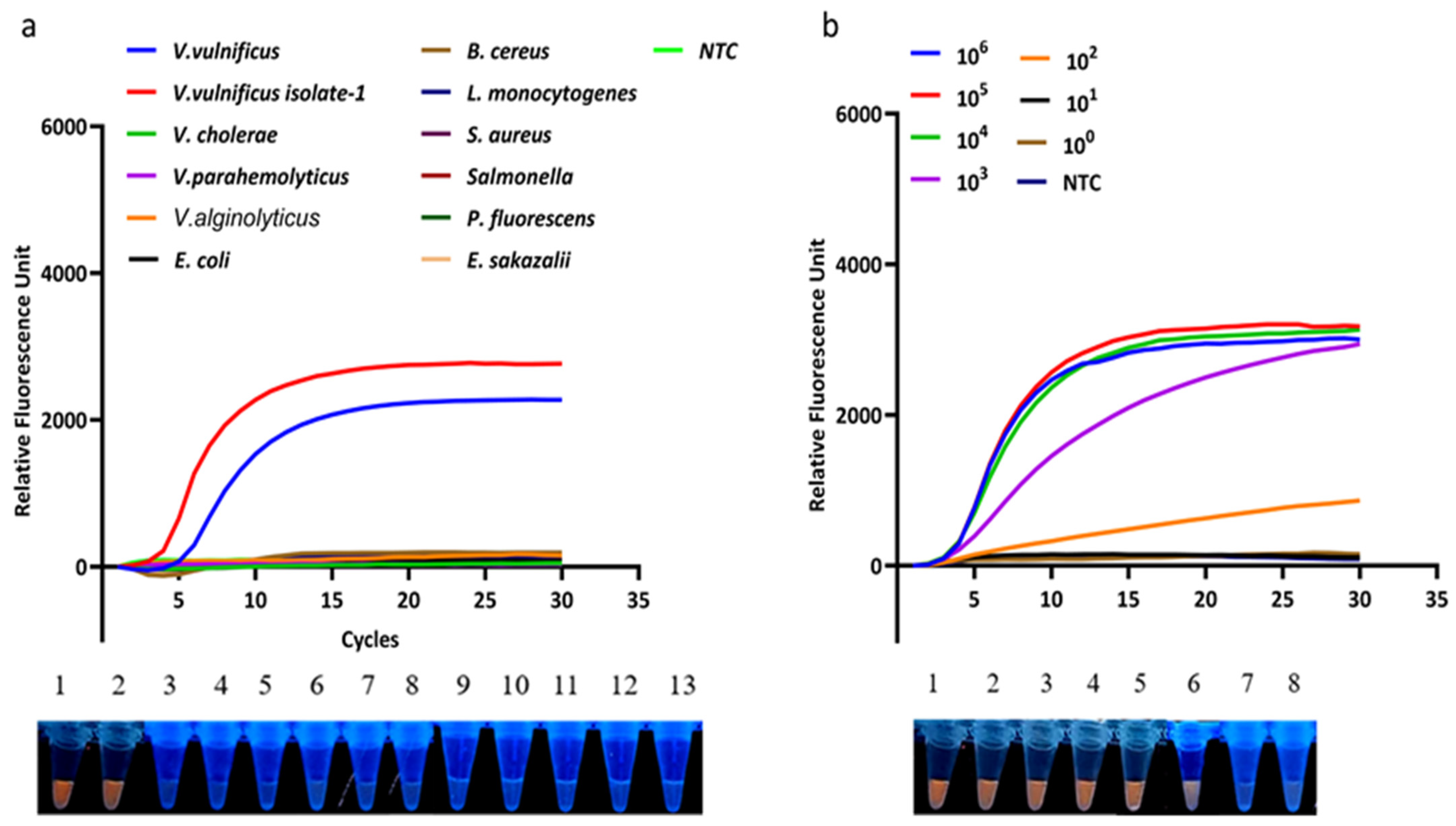
For the V. alginolyticus detection system, strong fluorescence signals were observed only for the standard strain and isolates of V. alginolyticus, whereas non-target bacteria produced weak signals and were judged negative (Figure 6a). These results indicate that the assay specifically detected the tested V. alginolyticus strains within the bacterial panel used in this study. In the sensitivity assay, positive fluorescence signals were detected in bacterial suspensions ranging from 3 × 10⁶ to 3 × 101 CFU·mL−1, whereas the fluorescence intensity at 3 × 100 CFU·mL−1 did not differ significantly from the negative sample (Figure 6b). The LOD of the V. alginolyticus LAMP-PfAgo assay was determined to be 3 × 101 CFU·mL−1.
The V. vulnificus detection system exhibited specific detection of the tested V. vulnificus strains, with no obvious fluorescence signals observed for the non-target bacteria included in this study (Figure 7a). In the sensitivity evaluation, the LOD of the V. vulnificus LAMP-PfAgo assay was 102 CFU·mL−1 (Figure 7b).

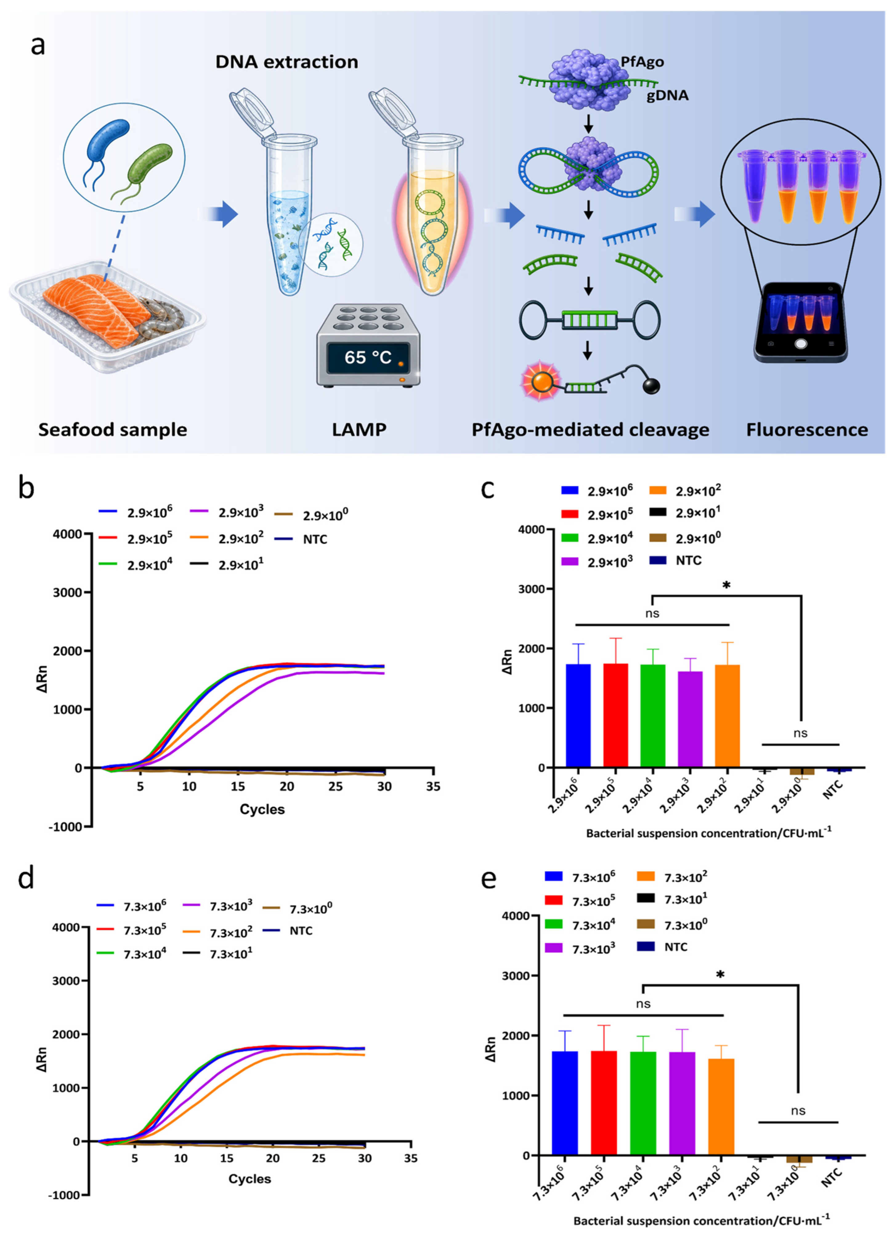
3.4. Artificial Contamination Assay

To assess the applicability of the LAMP-PfAgo assay in a food matrix, salmon samples artificially contaminated with V. alginolyticus or V. vulnificus were analyzed without enrichment culture after boiling-based DNA extraction (Figure 8a). For V. alginolyticus, real-time fluorescence curves increased rapidly for samples prepared with final contamination levels from 2.9 × 10⁶ to 2.9 × 10² CFU·mL−1 in the sample homogenate, whereas lower concentrations remained at background levels (Figure 8b). Endpoint fluorescence analysis further separated the positive groups from the negative controls, with robust ΔRn values retained at the 2.9 × 10² CFU·mL−1 level (Figure 8c). Similar results were obtained for V. vulnificus. Positive fluorescence curves were observed for salmon samples prepared with final contamination levels from 7.3 × 10⁶ to 7.3 × 10² CFU·mL−1 in the sample homogenate, while the 7.3 × 101 and 7.3 × 100 CFU·mL−1 groups remained comparable to the no-template control (Figure 8d). Endpoint fluorescence analysis confirmed that the positive groups were distinguishable from the negative controls down to 7.3 × 10² CFU·mL−1 (Figure 8e). These results indicate that the LAMP-PfAgo assay can detect both V. alginolyticus and V. vulnificus in salmon samples under the tested conditions. For both pathogens, the practical detection limit was approximately 102 CFU·mL−1 in the final homogenate.

4. Discussion

This study establishes a two-step LAMP-PfAgo workflow that couples rapid isothermal amplification with Argonaute-mediated sequence verification for the detection of V. alginolyticus and V. vulnificus. LAMP provides high-yield amplification under constant temperature, whereas PfAgo adds a programmable cleavage step guided by short gDNAs. In this assay, molecular beacon cleavage is mediated by target-derived secondary guide fragments generated after PfAgo cleavage of the LAMP amplicons. This two-layer recognition strategy is well suited to Vibrio detection, where closely related species and nonspecific amplification can complicate interpretation.
V. alginolyticus and V. vulnificus are both important marine and seafood-associated pathogens, but their high relatedness to other Vibrio species can make species-level identification difficult [6]. Culture-based identification remains reliable but is time-consuming, whereas conventional PCR shortens detection time but still requires thermal cycling instrumentation and laboratory infrastructure, with reported detection limits typically around 102–103 CFU mL−1 [23]. Although traditional LAMP assays are sensitive, their reliance on nonspecific signal readouts can lead to false-positive results [8]. The PfAgo-mediated secondary recognition step used here addresses this limitation by adding sequence verification after LAMP, thereby improving confidence in positive signals while preserving the simplicity of a two-step workflow. Consistent with this design, the assay achieved a detection limit of 3 × 101 CFU mL−1 for V. alginolyticus and remained detectable in salmon samples without enrichment, with a practical detection limit of 2.9 × 10² CFU mL−1 in the final sample. For V. vulnificus detection, Tian et al. established a visual LAMP assay targeting the gyrB gene, with a reported analytical sensitivity of 10 fg·μL−1 genomic DNA and a 30 min reaction at 65 °C [24]. Yang et al. developed an RPA-lateral flow strip assay for raw seafood that completed detection within 35 min at 37 °C, with a sensitivity of 2 copies or 10−1 CFU per reaction; in spiked seafood samples, the method detected 1 CFU per 10 g after enrichment [25]. A concise comparison of the present assay with representative nucleic acid detection methods is provided in Table 6. Compared with these assays, the present LAMP-PfAgo method retains the rapid and simple features of isothermal amplification, while introducing PfAgo-mediated sequence verification after amplification. This additional recognition step provides stronger target confirmation and may reduce false-positive signals caused by nonspecific amplification, which is advantageous for species-level identification of closely related Vibrio pathogens.
Compared with CRISPR-Cas-based detection systems, the LAMP-PfAgo assay offers a complementary route with flexible target design. LAMP-CRISPR/Cas12a assays can achieve extremely low detection limits [30], but Cas12a recognition depends on PAM sequences and requires careful reaction optimization [11]. In addition, Cas13a-based assays require an additional transcription step, which increases assay complexity and time [31]. In contrast, PfAgo does not require PAM sequences and uses short, stable gDNAs for target recognition [13]. Previous studies have shown that Ago-based detection systems combined with isothermal amplification can reach detection limits as low as 6.68 CFU·mL−1 [32], supporting the broader feasibility of this strategy.
The specificity results further support the value of the PfAgo verification step. The developed assay distinguished the tested target Vibrio species from the non-target bacteria included in this study. This is important because high genetic similarity among Vibrio species can cause misidentification in some molecular assays [33]. The PfAgo-mediated sequence-specific cleavage therefore helps compensate for the nonspecific amplification risk inherent to LAMP [14]. V. cholerae and V. parahaemolyticus are also major seafood-associated pathogens with greater recognized importance in human disease. In the present study, they were included as non-target Vibrio species for specificity evaluation, whereas V. alginolyticus and V. vulnificus were selected as target organisms to evaluate the LAMP-PfAgo strategy. Although the specificity panel included V. parahaemolyticus and V. cholerae as seafood- and foodborne-associated non-target Vibrio species, other closely related species, such as V. fluvialis, V. mimicus, and V. harveyi, were not included in the present study. In addition, V. vulnificus biotype information was not fully evaluated in the present study. Therefore, further validation using defined biotype 1 and biotype 2 strains is needed before extending the assay claim to all V. vulnificus biotypes. Including these species in future studies would further strengthen the specificity evaluation and support broader validation of the assay using closely related Vibrio species and foodborne or environmental isolates.
The artificial contamination experiment demonstrated that the assay can detect both V. alginolyticus and V. vulnificus in salmon samples with a practical LOD of approximately 102 CFU·mL−1 in the final sample homogenate under the tested conditions. This workflow is faster than conventional culture-based methods, which often require 24–72 h [23], and may be more suitable for rapid screening in food safety monitoring. However, performance in additional seafood matrices and field settings remains to be evaluated.
Overall, the LAMP-PfAgo assay combines LAMP-based isothermal amplification with sequence-specific Argonaute cleavage. Within the tested sample types and bacterial panels, this strategy improved analytical specificity while maintaining practical sensitivity, supporting its further development for on-site foodborne pathogen detection.

5. Conclusions

This study demonstrates a two-step LAMP-PfAgo detection strategy for V. alginolyticus and V. vulnificus that combines rapid LAMP with guide-directed Argonaute cleavage. The assay achieved specific detection of the tested target strains with limits of detection of 3 × 101 CFU·mL−1 for V. alginolyticus and 102 CFU·mL−1 for V. vulnificus. In artificially contaminated salmon samples, the practical detection limits were 2.9 × 10² CFU·mL−1 for V. alginolyticus and 7.3 × 10² CFU·mL−1 for V. vulnificus in the final homogenates. These results support LAMP-PfAgo as a practical route for sequence-verified seafood pathogen screening, while further work is needed to validate broader matrices, closed-tube operation, and multiplex detection.

Author Contributions

Conceptualization, L.H. and X.B.; methodology, L.H.; software, L.H. and C.S.; validation, C.S. and X.B.; formal analysis, C.S. and X.B.; investigation, L.H. and X.B.; resources, Z.L. and X.B.; data curation, L.H.; writing—original draft preparation, C.S. and L.H.; writing—review and editing, X.B. and C.S.; visualization, L.H.; supervision, X.B. and Z.L.; project administration, X.B. and Z.L.; funding acquisition, X.B. All authors have read and agreed to the published version of the manuscript.

Funding

This research was funded by the Agricultural Independent Innovation Program in Jiangsu Province (Grant No. CX (23) 3043).

Institutional Review Board Statement

Not applicable.

Informed Consent Statement

Not applicable.

Data Availability Statement

The original contributions presented in the study are included in the article; further inquiries can be directed to the corresponding author.

Conflicts of Interest

The authors declare no conflicts of interest.

Abbreviations

The following abbreviations are used in this manuscript:
gDNAguide DNA
I/Oinner-to-outer primer ratio
LAMPloop-mediated isothermal amplification
LODlimit of detection
NTCno-template control
PfAgoPyrococcus furiosus Argonaute

References

  1. Jara-Medina, N.R.; Cedeño-Pinargote, A.C.; Beltrán-Noboa, A.; Tejera, E.; Machado, A. Managing Vibrio parahaemolyticus and Vibrio alginolyticus Infections in the Whiteleg Shrimp (Penaeus vannamei): A Systematic Review. Molecules 2025, 30, 3620. [Google Scholar] [CrossRef] [Scilit] [PubMed]
  2. Gao, R.; Sun, K.; Abdalla, A.E.; Tian, Z.; An, H.; Zhang, Z.; Liu, Y.; Zeng, X.; He, X.; Fan, X. Isolation, characterization, and preliminary application of three Vibrio phages in controlling Vibrio alginolyticus. LWT 2024, 191, 115638. [Google Scholar] [CrossRef] [Scilit]
  3. Oliver, J.D. The Biology of Vibrio vulnificus. Microbiol. Spectr. 2015, 3, 10-1128. [Google Scholar] [CrossRef] [Scilit] [PubMed]
  4. Shahimi, S.; Elias, A.; Abd Mutalib, S.; Salami, M.; Fauzi, F.; Mohd Zaini, N.A.; Abd Ghani, M.A.; Azuhairi, A. Antibiotic resistance and determination of resistant genes among cockle (Anadara granosa) isolates of Vibrio alginolyticus. Environ. Sci. Pollut. Res. 2021, 28, 44002–44013. [Google Scholar] [CrossRef] [Scilit] [PubMed]
  5. Heng, S.-P.; Letchumanan, V.; Deng, C.-Y.; Ab Mutalib, N.-S.; Khan, T.M.; Chuah, L.-H.; Chan, K.-G.; Goh, B.-H.; Pusparajah, P.; Lee, L.-H. Vibrio vulnificus: An environmental and clinical burden. Front. Microbiol. 2017, 8, 997. [Google Scholar] [CrossRef] [Scilit] [PubMed]
  6. Dong, Y.; Zhao, P.; Chen, L.; Wu, H.; Si, X.; Shen, X.; Shen, H.; Qiao, Y.; Zhu, S.; Chen, Q.; et al. Fast, simple and highly specific molecular detection of Vibrio alginolyticus pathogenic strains using a visualized isothermal amplification method. BMC Vet. Res. 2020, 16, 76. [Google Scholar] [CrossRef] [Scilit] [PubMed]
  7. Wang, Y.; Hou, Y.; Liu, X.; Lin, N.; Dong, Y.; Liu, F.; Xia, W.; Zhao, Y.; Xing, W.; Chen, J.; et al. Rapid visual nucleic acid detection of Vibrio alginolyticus by recombinase polymerase amplification combined with CRISPR/Cas13a. World J. Microbiol. Biotechnol. 2024, 40, 51. [Google Scholar] [CrossRef] [Scilit] [PubMed]
  8. Wang, Z.; Cui, X.; Hu, A.; Lu, Z.; Meng, F.; Zhou, L.; Bie, X. Establishment of real-time fluorescence and visual LAMP for rapid detection of Escherichia coli O157: H7 and kits construction. Lett. Appl. Microbiol. 2023, 76, ovad122. [Google Scholar] [CrossRef] [Scilit] [PubMed]
  9. Wang, Z.; Chen, H.; Hu, A.; Cui, X.; Shi, C.; Lu, Z.; Meng, F.; Lv, F.; Zhao, H.; Bie, X. Establishment of LAMP-CRISPR/Cas12a for rapid detection of Escherichia coli O157: H7 and one-pot detection. Food Microbiol. 2024, 124, 104622. [Google Scholar] [CrossRef] [Scilit] [PubMed]
  10. Zhu, T.; Jiang, W.; Wu, Y.; Fang, R.; Deng, F.; Yang, D. Advances in CRISPR/Cas13a-based biosensors for non-coding RNA detection. Talanta 2025, 294, 128223. [Google Scholar] [CrossRef] [Scilit] [PubMed]
  11. Dronina, J.; Samukaite-Bubniene, U.; Ramanavicius, A. Towards application of CRISPR-Cas12a in the design of modern viral DNA detection tools. J. Nanobiotechnol. 2022, 20, 41. [Google Scholar] [CrossRef] [Scilit] [PubMed]
  12. Chao, A.; Wang, J.; Xiu, L.; Bold, B.; Ghonaim, A.H.; Chen, J.; Hu, Q.; Yin, K. CRISPR/Cas-Based Biosensing Strategies for Non-Nucleic Acid Contaminants in Food Safety: Status, Challenges, and Perspectives. J. Agric. Food Chem. 2025, 73, 18063–18075. [Google Scholar] [CrossRef] [Scilit] [PubMed]
  13. Li, Y.; Zhao, L.; Wang, J.; Ma, L.; Bai, Y.; Feng, F. Argonaute-based nucleic acid detection technology: Advantages, current status, challenges, and perspectives. ACS Sens. 2024, 9, 5665–5682. [Google Scholar] [CrossRef] [Scilit] [PubMed]
  14. Hong, M.; Wu, G.; Ren, Y.; Wu, S.; Zhu, H.; Chen, Z. Advancements in Pathogen Detection: Argonaute-Based Nucleic Acid Detection Technology. Pathogens 2025, 14, 554. [Google Scholar] [CrossRef] [Scilit] [PubMed]
  15. Qin, Y.; Li, Y.; Hu, Y. Emerging Argonaute-based nucleic acid biosensors. Trends Biotechnol. 2022, 40, 910–914. [Google Scholar] [CrossRef] [Scilit] [PubMed]
  16. Li, Y.; Kou, J.; Han, X.; Qiao, J.; Zhang, W.; Man, S.; Ma, L. Argonaute-triggered visual and rebuilding-free foodborne pathogenic bacteria detection. J. Hazard. Mater. 2023, 454, 131485. [Google Scholar] [CrossRef] [Scilit] [PubMed]
  17. Ye, X.; Zhou, H.; Guo, X.; Liu, D.; Li, Z.; Sun, J.; Huang, J.; Liu, T.; Zhao, P.; Xu, H.; et al. Argonaute-integrated isothermal amplification for rapid, portable, multiplex detection of SARS-CoV-2 and influenza viruses. Biosens. Bioelectron. 2022, 207, 114169. [Google Scholar] [CrossRef] [Scilit] [PubMed]
  18. Hu, Y.-q.; Wang, W.-y.; Fath, T.; Li, F.-x.; Fang, L.-f.; Zhou, Z.-h.; Zhang, D.-f. Rapid and simultaneous detection of viable Vibrio parahaemolyticus, Vibrio alginolyticus, and Vibrio cholerae by PMA-mPCR assay in aquatic products. LWT 2023, 180, 114663. [Google Scholar] [CrossRef] [Scilit]
  19. Nagamine, K.; Hase, T.; Notomi, T. Accelerated reaction by loop-mediated isothermal amplification using loop primers. Mol. Cell. Probes 2002, 16, 223–229. [Google Scholar] [CrossRef] [Scilit] [PubMed]
  20. Notomi, T.; Okayama, H.; Masubuchi, H.; Yonekawa, T.; Watanabe, K.; Amino, N.; Hase, T. Loop-mediated isothermal amplification of DNA. Nucleic Acids Res. 2000, 28, e63. [Google Scholar] [CrossRef] [Scilit] [PubMed]
  21. He, R.; Wang, L.; Wang, F.; Li, W.; Liu, Y.; Li, A.; Wang, Y.; Mao, W.; Zhai, C.; Ma, L. Pyrococcus furiosus Argonaute-mediated nucleic acid detection. Chem. Commun. 2019, 55, 13219–13222. [Google Scholar] [CrossRef] [Scilit] [PubMed]
  22. Broughton, J.P.; Deng, X.; Yu, G.; Fasching, C.L.; Servellita, V.; Singh, J.; Miao, X.; Streithorst, J.A.; Granados, A.; Sotomayor-Gonzalez, A.; et al. CRISPR–Cas12-based detection of SARS-CoV-2. Nat. Biotechnol. 2020, 38, 870–874. [Google Scholar] [CrossRef] [Scilit] [PubMed]
  23. Fu, K.; Li, J.; Wang, Y.; Liu, J.; Yan, H.; Shi, L.; Zhou, L. An innovative method for rapid identification and detection of Vibrio alginolyticus in different infection models. Front. Microbiol. 2016, 7, 651. [Google Scholar] [CrossRef] [Scilit] [PubMed]
  24. Tian, Z.; Yang, L.; Qi, X.; Zheng, Q.; Shang, D.; Cao, J. Visual LAMP method for the detection of Vibrio vulnificus in aquatic products and environmental water. BMC Microbiol. 2022, 22, 256. [Google Scholar] [CrossRef] [Scilit] [PubMed]
  25. Yang, X.; Zhao, P.; Dong, Y.; Chen, S.; Shen, H.; Jiang, G.; Zhu, H.; Dong, J.; Gao, S. An isothermal recombinase polymerase amplification and lateral flow strip combined method for rapid on-site detection of Vibrio vulnificus in raw seafood. Food Microbiol. 2021, 98, 103664. [Google Scholar] [CrossRef] [Scilit] [PubMed]
  26. Xiao, X.; Lin, Z.; Huang, X.; Lu, J.; Zhou, Y.; Zheng, L.; Lou, Y. Rapid and Sensitive Detection of Vibrio vulnificus Using CRISPR/Cas12a Combined With a Recombinase-Aided Amplification Assay. Front. Microbiol. 2021, 12, 767315. [Google Scholar] [CrossRef] [Scilit] [PubMed]
  27. Wang, R.; Qian, C.; Pang, Y.; Li, M.; Yang, Y.; Ma, H.; Zhao, M.; Qian, F.; Yu, H.; Liu, Z.; et al. Duplex On-Site Detection of Vibrio cholerae and Vibrio vulnificus by Recombinase Polymerase Amplification and Three-Segment Lateral Flow Strips. Biosensors 2021, 11, 151. [Google Scholar] [CrossRef] [Scilit] [PubMed]
  28. Fang, W.; Wang, H.; Lu, Y.; Ding, M.; Liu, Y.; Chen, Y.; Wang, Y.; Yang, X. A Novel RAA Combined Test Strip Method Based on Dual Gene Targets for Pathogenic Vibrio vulnificus in Aquatic Products. Foods 2023, 12, 3605. [Google Scholar] [CrossRef] [Scilit] [PubMed]
  29. Zhang, W.; Liu, H.; Zhu, Y.; Wang, Q.; Li, J.; Cao, Y.; Ma, L. Endonuclease V activated Pyrococcus furiosus Argonaute for the detection of food contaminated bacteria. npj Sci. Food 2025, 10, 27. [Google Scholar] [CrossRef] [Scilit] [PubMed]
  30. Chen, J.S.; Ma, E.; Harrington, L.B.; Da Costa, M.; Tian, X.; Palefsky, J.M.; Doudna, J.A. CRISPR-Cas12a target binding unleashes indiscriminate single-stranded DNase activity. Science 2018, 360, 436–439. [Google Scholar] [CrossRef] [Scilit] [PubMed]
  31. Gootenberg, J.S.; Abudayyeh, O.O.; Lee, J.W.; Essletzbichler, P.; Dy, A.J.; Joung, J.; Verdine, V.; Donghia, N.; Daringer, N.M.; Freije, C.A.; et al. Nucleic acid detection with CRISPR-Cas13a/C2c2. Science 2017, 356, 438–442. [Google Scholar] [CrossRef] [Scilit] [PubMed]
  32. Zhou, H.; Yang, J.; Xue, F.; Shen, W.; Cheng, Y.; Liu, X. Argonaute combined with isothermal amplification for simultaneous detection of Vibrio parahaemolyticus and the tetracycline resistance gene tetA in water and food samples. Curr. Res. Food Sci. 2025, 11, 101246. [Google Scholar] [CrossRef] [Scilit] [PubMed]
  33. Wang, W.; Liu, J.; Guo, S.; Liu, L.; Yuan, Q.; Guo, L.; Pan, S. Identification of Vibrio parahaemolyticus and Vibrio Spp. specific outer membrane proteins by reverse vaccinology and surface proteome. Front. Microbiol. 2021, 11, 625315. [Google Scholar] [CrossRef] [Scilit] [PubMed]
Figure 1. Optimization of the LAMP system for V. alginolyticus. Real-time fluorescence amplification curves were used to optimize the LAMP reaction conditions for V. alginolyticus. (a) Screening of four LAMP primer sets. (b) Optimization of the inner-to-outer primer ratio. (c) Optimization of dNTP concentration. (d) Optimization of Mg2+ concentration. (e) Optimization of Bst 3.0 DNA polymerase dosage. (f) Optimization of reaction temperature. Amplification performance was evaluated according to amplification onset and ΔRn values, and the optimized conditions were selected for subsequent PfAgo-mediated detection.
Foods 15 02506 g001
Figure 2. Optimization of the LAMP system for V. vulnificus. Real-time fluorescence amplification curves were used to optimize the LAMP reaction conditions for V. vulnificus. (a) Screening of four LAMP primer sets. (b) Optimization of the inner-to-outer primer ratio. (c) Optimization of dNTP concentration. (d) Optimization of Mg2+ concentration. (e) Optimization of Bst 3.0 DNA polymerase dosage. (f) Optimization of reaction temperature. The optimal reaction parameters were determined based on rapid amplification kinetics and stable fluorescence signals, providing the upstream amplification system for subsequent PfAgo cleavage analysis.
Foods 15 02506 g002
Figure 3. Screening of optimal gDNA combinations for the LAMP-PfAgo cleavage system targeting V. alginolyticus and V. vulnificus. (a) Evaluation of different gDNA combinations for V. alginolyticus based on relative fluorescence intensity, and fluorescence visualization under UV illumination. (b) Evaluation of different gDNA combinations for V. vulnificus based on relative fluorescence intensity. “*” indicates statistically significant differences (p < 0.05), and “ns” indicates no significant difference.
Foods 15 02506 g003
Figure 4. Optimization of the LAMP-PfAgo cleavage system for V. alginolyticus. (a) Optimization of gDNA concentration. (b) Optimization of PfAgo protein dosage. (c) Optimization of Mn2+ concentration. Amplification curves and corresponding relative fluorescence intensities are shown for each condition. “*” indicates statistically significant differences (p < 0.05), and “ns” indicates no significant difference.
Foods 15 02506 g004
Figure 5. Optimization of the PfAgo-mediated cleavage system for V. vulnificus. (a) Optimization of gDNA concentration. (b) Optimization of PfAgo protein dosage. (c) Optimization of Mn2+ concentration. Amplification curves and corresponding relative fluorescence intensities are shown for each condition. “*” indicates statistically significant differences (p < 0.05), and “ns” indicates no significant difference.
Foods 15 02506 g005
Figure 6. Specificity and sensitivity evaluation of the V. alginolyticus LAMP-PfAgo detection system. (a) Specificity analysis using V. alginolyticus strains and non-target bacterial species. (b) Sensitivity evaluation using 10-fold serial dilutions of V. alginolyticus bacterial suspensions.
Foods 15 02506 g006
Figure 7. Specificity and sensitivity evaluation of the V. vulnificus LAMP-PfAgo detection system. (a) Specificity analysis using V. vulnificus strains and non-target bacterial species. (b) Sensitivity evaluation using 10-fold serial dilutions of V. vulnificus bacterial suspensions.
Foods 15 02506 g007
Figure 8. Detection of artificially contaminated salmon samples using the LAMP-PfAgo assay. (a) Schematic overview of the assay workflow, including DNA extraction from seafood samples, LAMP at 65 °C, PfAgo-mediated guide-directed cleavage at 95 °C and fluorescence-based result interpretation. (b) Real-time fluorescence curves for salmon samples spiked with V. alginolyticus. (c) Endpoint fluorescence intensities of the corresponding V. alginolyticus reactions. (d) Real-time fluorescence curves for salmon samples spiked with V. vulnificus. (e) Endpoint fluorescence intensities of the corresponding V. vulnificus reactions. NTC, no-template control. “*” indicates statistically significant differences (p < 0.05), and “ns” indicates no significant difference. Note: The bacterial concentrations shown in panels (be) refer to the final concentrations in the salmon/APW homogenates after addition of bacterial suspensions to salmon samples. The bacterial suspensions used for spiking were approximately 100-fold more concentrated than the final homogenates.
Foods 15 02506 g008
Table 1. Bacterial strains used in this study.
Bacterial Strain No.
V. alginolyticusATCC17749
V. alginolyticusVA Isolates-1
V. vulnificusATCC 27562
V. vulnificusVV Isolates-1
V. choleraeCICC 23794
V. parahaemolyticusCICC 21528
Pseudomonas fluorescensCICC 21620
Bacillus cereusCMCC 63301
Enterobacter sakazakiiCICC 21563
Escherichia coliCICC 21530
Listeria monocytogenesCICC 21662
Staphylococcus aureusCICC 22942
Salmonella enteritidisCICC 21482
Table 2. LAMP primer sequences designed for V. alginolyticus.
Primer NameSequences (5′→3′)
VA-614F3AGTTAATCAAGCCGCCTCAG
B3ACCCGAGCTCACAACCTC
FIPTAGAGCAAAGTAAGCTCGCTCGCAAAACGTTGCGCGTGAGTT
BIPTTAGAAACTGCCCTTCCCGAAGCTCTTAATTGGCTCGCTGTACG
LFTTGCATTGCTTAGTTGGC
LBCGTTTAAGCGGTGAAATAGG
VA-14F3AGTTAATCAAGCCGCCTCAG
B3ACCCGAGCTCACAACCTC
FIPTAGAGCAAAGTAAGCTCGCTCGCAAAACGTTGCGCGTGAGTT
BIPTTAGAAACTGCCCTTCCCGAAGCTCTTAATTGGCTCGCTGTACG
VA-28F3ACTGACTGGCTCGCTCAC
B3GGACGCAACCAACGATGA
FIPCGTCCGGCGATTTCTTCCAAACCAACCCTTCACGCTACCG
BIPGCTATGGTTCATGGTGGACCCTGCCCCGACAGATGTTGAG
LBATCCGGCTTCAACCAATTCTGC
VA-618F3ATCGCAATTGGCCCTGTC
B3TGCAATAGCGTGAAGATGGC
FIPGTGCCGAAGCCGTATCACCACCCGTGTTTGGTGCGTCTA
BIPCCACCCTGGCACCAGTCAACGCGGTAAGCACTCTTTCTCA
LFCGTAGAAAATGCGAGCGGAAAAT
LBCAGAGTGTATTGAGGCTGCAC
Table 3. LAMP primer sequences designed for V. vulnificus.
Primer NameSequences (5′→3′)
VV-51F3ACTGCGAGTGGTTTCCATC
B3GCTCTCTGGTGAAGCAAGAA
FIPCGCCGGATACGTACCAAAGTGATCTACCATCACTTGCTTGGC
BIPAACTTGCTACCGAGACCCGCCCGGCTGAAATCGATCTCAT
LFTGAAGCATGGCCTTTTTGGC
VV-1F3ACTGCGAGTGGTTTCCATC
B3GCTCTCTGGTGAAGCAAGAA
FIPCGCCGGATACGTACCAAAGTGATCTACCATCACTTGCTTGGC
BIPAACTTGCTACCGAGACCCGCCCGGCTGAAATCGATCTCAT
VV-512F3CTGCGAGTGGTTTCCATCTC
B3TTCCTCTGTACTGGCTCTCT
FIPTTAAGCTGCGCTTTTTCGCCGCTTGGCTCACCCGACTCA
BIPCCGCTGAAGCATGGCCTTTTTGGGTGAAGCAAGAATCCCCG
LFGCAAAAGATCACTCGTGATGAGAT
VV-12F3CTGCGAGTGGTTTCCATCTC
B3TTCCTCTGTACTGGCTCTCT
FIPTTAAGCTGCGCTTTTTCGCCGCTTGGCTCACCCGACTCA
BIPCCGCTGAAGCATGGCCTTTTTGGGTGAAGCAAGAATCCCCG
Table 4. gDNA and probe sequence for V. alginolyticus.
Primer Group Sequences (5′→3′)
VA-gDNA1+2VA-gDNA1GGTATGGCCTTTTACA
VA-gDNA2ATCACAGGACAGCCAG
VA-gDNA3+4VA-gDNA3ATGGCCTTTTACAATC
VA-gDNA4ACAGGACAGCCAGCTG
VA-gDNA5+6VA-gDNA5CGGTATGGCCTTTTAC
VA-gDNA6AATCACAGGACAGCCA
MBVA-MB5′ 6-ROX-ATGGCCTTTTACAATCACAGGACAGCCAGCT-3′ BHQ-2
Note: All gDNAs were synthesized with a 5′-phosphate modification.
Table 5. gDNA and probe sequence for V. vulnificus.
Primer Group Sequences (5′→3′)
VV-gDNAs1+2VV-gDNA1AAAGCGCAGCTTAATC
VV-gDNA2ACACGTTACCACTCAA
VV-gDNAs3+4VV-gDNA3AAGCGCAGCTTAATCA
VV-gDNA4CACGTTACCACTCAAT
VV-gDNAs5+6VV-gDNA5AGCGCAGCTTAATCAC
VV-gDNA6ACGTTACCACTCAATA
MBVV-MB5′-6-NED-AAGCGCAGCTTAATCACACGTTACCACTCA-3′BHQ2
Note: All gDNAs were synthesized with a 5′-phosphate modification.
Table 6. Comparison of the present LAMP-PfAgo assay with previously reported nucleic acid detection methods for related foodborne pathogens.
TargetMethodLODAssay TimeEquipment/ReadoutSampleReference
V. alginolyticus
V. vulnificus
LAMP-PfAgo3 × 101 CFU·mL−1 for V. alginolyticus, 102 CFU·mL−1 for V. vulnificus, 2.9 × 10² CFU·mL−1 in the final sample for V. alginolyticus and 7.3 × 10² CFU·mL−1 in the final sample for V. vulnificus40 min LAMP and 30 min PfAgo cleavageConstant-temperature amplification device plus fluorescence readoutBacterial suspensions, spiked salmonThis study
V. vulnificusVisual LAMP10 fg uL−1 genomic DNA30 min at 65 °CWater bath or heating block, visual readoutAquatic products and environmental water[24]
V. vulnificusRPA-LFS2 genome copies or 10−1 CFU per reaction, 1 CFU/10 g in spiked seafood after enrichment35 min at 37 °CLow-temperature incubator plus lateral flow stripRaw seafood[25]
V. vulnificusRAA-CRISPR/Cas12a2 copies per reaction40 minIsothermal amplification plus UV/fluorescence visual readoutSpiked blood, stool and shrimp samples[26]
V. cholerae
V. vulnificus
Duplex RPA-LFS101 gene copies per reaction, 1 CFU/10 g in spiked food30 min at 37 °C and 5 min strip visualizationLow-temperature incubator plus three-segment lateral flow stripSpiked shrimp and clinical samples[27]
V. vulnificusRAA combined with dual-target test strip10 CFU·mL−1 for gyrB and 100 CFU·mL−1 for vvhAWithin 50 min including sample preparationLow-temperature isothermal amplification plus test strip/smartphone grayscale analysisOyster, fish and shrimp[28]
V. alginolyticusRPA-CRISPR/Cas13a-LFD10 copies uL−1<50 minIsothermal amplification plus lateral flow dipstickBacterial isolates and infected mouse blood[7]
E. coli O157:H7
S. aureus,
S. typhimurium
C. sakazakii
Endo V-activated PfAgo detection (VPN)101 CFU·mL−1 for four foodborne pathogensRPA/Endo V step plus 40 min PfAgo cleavageIsothermal amplification plus fluorescence readoutBeef and milk samples[29]
Note: LOD values are reported as described in the cited studies and are not directly interchangeable because template types, sample preparation procedures and matrix validation differed among assays. RPA, recombinase polymerase amplification; RAA, recombinase-aided amplification; LFS/LFD, lateral flow strip/dipstick.
Disclaimer/Publisher’s Note: The statements, opinions and data contained in all publications are solely those of the individual author(s) and contributor(s) and not of MDPI and/or the editor(s). MDPI and/or the editor(s) disclaim responsibility for any injury to people or property resulting from any ideas, methods, instructions or products referred to in the content.