Article

Glycyrrhiza glabra L. Extracts Prevent LPS-Induced Inflammation in RAW264.7 Cells by Targeting Pro-Inflammatory Cytokines, Mediators and the JAK/STAT Signaling Pathway

by
Maria Rosaria Perri
1,†,‡,
Michele Pellegrino
1,†,
Claudia-Crina Toma
2,
Pierfrancesco Prezioso
1,
Vincenzo Tagliaferri
1,
Mariangela Marrelli
1,
Filomena Conforti
1 and
Giancarlo Statti
1,*
1
Department of Pharmacy, Health and Nutritional Sciences, University of Calabria, 87036 Rende, Italy
2
Pharmacognosy Department, Faculty of Pharmacy, Vasile Goldis Western University of Arad, 310045 Arad, Romania
*
Author to whom correspondence should be addressed.
These authors contributed equally to this work.
Current address: National Research Council of Italy, Institute for Agriculture and Forestry System in the Mediterranean (CNR-ISAFoM), 87036 Rende, Italy.
Foods 2025, 14(21), 3746; https://doi.org/10.3390/foods14213746
Submission received: 22 September 2025 / Revised: 17 October 2025 / Accepted: 29 October 2025 / Published: 31 October 2025
(This article belongs to the Section Plant Foods)

Abstract

Glycyrrhiza glabra L. is a species widely spread all over the world, with a long tradition of use in folk medicine. Here, raw and hydrolyzed extracts obtained from roots collected in different geographical areas belonging to the Mediterranean basin were standardized as regards the amount of three main compounds: glycyrrhizin, the most abundant triterpene saponin of licorice, the 18β-glycyrrhetinic acid and the chalcone isoliquiritigenin. Raw and hydrolyzed extracts, as well as their pure single compounds, were investigated for their potential anti-inflammatory properties. The hydrolyzed extracts significantly reduced the production of pro-inflammatory cytokines such as TNF-α, IL-6, NO mediator in LPS-stimulated RAW 264.7 cells. Moreover, they were able to inhibit JAK2 and STAT3 phosphorylated proteins more than pure single standards tested at the same final concentrations, displaying a strength synergism of action. These findings suggest that G. glabra extracts and, more specifically, the hydrolyzed ones could represent interesting sources of potential anti-inflammatory agents able to inhibit the JAK/STAT signaling pathway.

1. Introduction

Plants have always represented a rich source of bioactive compounds with potential nutraceutical and pharmacological properties. Currently, the demand for natural origin products is dramatically increasing, and the power of plant-based products is attributable to their bioactive constituents including triterpenoids, saponins, flavonoids, etc. [1,2,3,4,5].
Plants from the Mediterranean basin constitute a library of valuable plants, among which Glycyrrhiza glabra L. (licorice), belonging to the Fabaceae (Leguminosae) family, represents one of the most ancient and important species. It is an herbaceous perennial plant growing in European countries, India and China [6]. The licorice root apparatus consists of a taproot composed of 3–5 secondary roots, each about 1.25 cm long; root and rhizome barks are green/brown colored. Flowers are narrow, ranging in color from purple to blue, while fruits are small legumes. G. glabra has a long tradition in folk medicine; its recognized pharmacological properties include antitussive, antiulcerogenic, anticancer, antidiabetic, anti-asthmatic, anti-atherogenic, anticoagulant, antispasmodic, anti-microbial, antioxidant, anti-inflammatory and anti-allergic activities [7]. The major problem related to licorice intake is “pseudohyperaldosteronism”, a phenomenon causing increased mineralocorticoid activity because of the 11 beta-hydroxysteroid dehydrogenase (11β-HSD2) inhibitory activity. This effect is due to the presence of glycyrrhetinic acid, the most abundant phytochemical in G. glabra [8]. Despite this, activity can be observed even at a very low serum concentration. The Food and Drug Administration (FDA) has declared licorice as “GRAS” (Generally Recognized As Safe) for food use and it is also included in some over-the-counter drugs. Even if an official Admitted Daily Intake (ADI) does not exist, different organizations all over the world have established some guidelines for safe use of licorice [9]. Currently, the European Medicine Agency (EMA) is discussing a draft concerning Liquiritiae radix (licorice root), with the aim of creating a monograph including information about its qualitative and quantitative composition, therapeutic indications, posology, contraindications, special warnings, precautions for use and interaction and effects on fertility, pregnancy and lactation [10]. The idea behind this work was to partially mimic what usually happens inside the human body by removing glycyrrhizin from the extracts through a hydrolysis process. Indeed, evidence has shown that, both in humans and in rats, a glycyrrhizin peak was not detected in plasma by high performance liquid chromatography (HPLC) after oral administration because Streptococcus and Eubacterium, thanks to their β-glucuronidase activity, are able to hydrolyze glycyrrhizin into its aglycone form [11]. Glycyrrhizin, a triterpene saponin, and its corresponding aglycone, the triterpenoid glycyrrhetinic acid, are the most investigated phytochemicals of G. glabra, known for their antioxidant, anti-inflammatory, antiviral, antitumor and immunoregulatory properties [12]. More than 400 phytochemicals have been isolated from licorice and most of them are flavonoids. The chalcone isoliquiritigenin (2′,4′, 4-trihydroxycalchone), a phytochemical belonging to the flavonoid family, is a naturally occurring pigment in licorice roots and shallot [13] which exerts a wide range of bioactive activities among which are anti-inflammatory properties [14]. In this frame, the aim of this work was to investigate and compare the potential anti-inflammatory activity of both pure single standards and the whole G. glabra extracts.
In fact, both the role of inflammatory process onset and the consequences it drives, have been currently well established: evidence has shown how chronic inflammation plays a key role in cancer development, and epidemiological studies have demonstrated that non-steroidal anti-inflammatory drugs (NSAIDs) can be considered effective chemo-preventive agents [15,16]. In the context of prevention, the inhibition of pro-inflammatory responses promoted by the JAK/STAT signaling pathway modulation is a very promising approach. In fact, the activation of STAT transcriptional factors induces the creation of a pro-tumorigenic microenvironment and promotes cell differentiation and renewal. STAT3 aberrant activation is considered a key player in tumorigenesis initiation and progression processes: it acts as a transcription inducer affecting not only gene expression but also contributing to the formation of the tumor microenvironment [17].

2. Materials and Methods

2.1. Reagents

RAW 264.7 cells were purchased from ATCC, Glasgow, UK (No. TIB-71). Dulbecco’s Modified Eagle’s Medium (DMEM), fetal bovine serum (FBS), L-glutamine, penicillin/streptomycin, phosphate buffered saline (PBS), bovine serum albumin (BSA), protease inhibitors, trypan blue, lipopolysaccharides (LPS) from E. coli, trichloroacetic acid (TCA), sulphorhodamine B (SRB), Tris base, isoliquiritigenin, glycyrrhizin and 18β-glycyrrhetinic acid were obtained from Sigma-Aldrich S.p.a. (Milan, Italy). ELISA kits were from ThermoFisher Scientific (Vienna, Austria). The ECL System was from Bio-Rad (Milan, Italy). Anti-phosphoJak2 (#PA5120138), anti-Jak2 (#PA511267), anti-phosphoSTAT3 (#PA5121259) and anti-STAT3 (#PA5120138) were from ThermoFisher Scientific. Anti-β-actin (AC-15; sc-69879) was from Santa Cruz Biotechnology, Inc., Heidelberg, Germany. Solvents were HPLC-grade and reagent-grade, purchased from VWR International s.r.l. (Milan, Italy).

2.2. Plant Material Recovery, Extraction Procedure and Hydrolysis Process

G. glabra L. investigated extracts were from the Mediterranean area: Morocco, Southern Italy (Rossano (CS) and Montalto Uffugo (CS)). As detailed in Table 1, while one root sample was collected in October (leg. det. G. Statti), others were commercially and locally available. All samples were properly dried at room temperature and shredded before the extraction process by means of a disintegrator, then passed through a 600 µm sieve [18]; pulverized samples were extracted through ultrasonication (MeOH 80%, 20 mins, 25 °C) according to Zhang and colleagues’ method (2013) [19]. Achieved yields were 7.2%, 6.8%, 6.7% and 6.5% for the MON, ROS1, ROS2 and MAR samples, respectively. An aliquot of each obtained extract was dried, while the others were subjected to the hydrolysis process through the addition of HCl (100 °C, 60 min, water bath). Then, the obtained mixture was extracted in ethyl acetate, and the organic solution was mixed with NaCl. The organic phase, dried with Na2SO4, was filtered and dried using a rotary evaporator.

2.3. HPLC Analyses

The three main phytochemicals characterizing the G. glabra species, isoliquiritigenin, glycyrrhizin and 18β-glycyrrhetinic acid, were researched and quantified within both the raw and hydrolyzed extracts. Analyses were carried out in a Kinetex XB-C18 250 × 4.6 mm 5 µm column (Phenomenex Inc., Torrence, CA, USA), with a gradient mobile phase consisting of water 0.1% formic acid (solvent A) and acetonitrile 0.1% formic acid (solvent B), as reported by Zhang and colleagues (2013) [19]. The linear gradient separation applied was as follows: 25–45% B from 0 to 30 min, 45–100% B from 30 to 60 min. The instrument was equipped with an MD2010 Plus PDA detector (Jasco, Hachioji, Tokyo, Japan) working at 255 nm, 370 nm and at maximum absorbance between 210 nm and 410 nm. Data were interpolated with standard calibration curves of isoliquiritigenin, glycyrrhizin and 18β-glycyrrhetinic acid, respectively.

2.4. Cell Culture

RAW 264.7 macrophages were cultured in DMEM implemented with FBS, 1% L-glutamine and 1% penicillin/streptomycin and incubated at 37 °C in 5% CO2 atmosphere [20]. Cells were tested monthly for mycoplasma negativity (MycoAlert Mycoplasma Detection Kit, Lonza, Gampel-Bratsch, Switzerland).

2.5. Cell Viability (SRB) Assay

The sulphorhodamine B (SRB) assay, a widely standardized test evaluating cell protein content, was performed in order to investigate cell viability. To start, 3000 cells/well were seeded into a 96-well/plate and incubated overnight to favor attachment. After 24 h, cells were treated with proper concentrations of samples (in DMSO 0.5%) and, after 30 min, stimulated with LPS from E. coli (1 µg/mL). The next day, the cells were fixed with TCA 10% (1 h, 4 °C), treated with an SRB solution and washed with acetic acid 1% (three times). The absorbance was measured at 540 nm using a microplate reader (Stat fax 3200 Awareness Technology Inc., Palm City, FL, USA) [21].

2.6. Cytokine Measurements

To start, 3 × 10⁵ RAW 264.7 cells/well were seeded into a 24-well/plate and incubated overnight. After 24 h, cells were treated with samples and stimulated with LPS, as reported above. The next day, media was collected, and the presence of pro-inflammatory Tumor Necrosis Factor-α, Interleukin-6 (TNF-α, IL-6) and anti-inflammatory cytokine Interleukin-10 (IL-10) was assessed by means of ELISA kit according to the manufacturer’s instructions (ThermoFisher Scientific, Bender MedSystems GmbH, Vienna, Austria) [22].

2.7. Nitric Oxide (NO) Production Inhibition

In order to evaluate the presence of nitrite, a stable oxidized NO product, 3 × 10⁵ RAW 264.7 cells/well were seeded into a 24-well/plate for 24 h, and then they were treated with samples and stimulated with LPS (1 µg/mL). The next day, the collected medium was mixed with Griess reagent (media: Griess reagent, 1:1). The absorbance was measured at 550 nm with a microplate reader (Stat fax 3200 Awareness Technology Inc., Palm City, FL, USA) [23].

2.8. Western Blot Analysis

Treated and LPS-stimulated RAW 264.7 cells were lysed in a RIPA lysis buffer [20 mM Tris-HCl (pH 7.5), 150 mM NaCl, 1 mM Na2 EDTA, 1 mM EGTA, 1% NP-40, 1% sodium deoxycholate, 2.5 mM sodium pyrophosphate] supplemented with phosphatase and a protease inhibitor cocktail. Equal lysed protein amounts were run on SDS-PAGE 11% gel and electroblotted onto a nitrocellulose membrane. Proteins were revealed through specific polyclonal or monoclonal antibodies (Abs) and recognized by IRDye secondary Abs (LI-COR Corporate, Milan, Italy). Densitometry readings/intensity ratio was assessed by using ImageJ software 1.53k (Rasband, W.S. ImageJ, U.S. National Institutes of Health, Bethesda, MD, USA) [24].

2.9. Statistical Analyses

Data were reported as mean ± S.E.M. of four independent experiments (for SRB assay) and three independent experiments for all the other experiments, each performed in three technical replicates. Phytochemical data were expressed as mean ± S.D. Normality of data and homogeneity of variances were assessed by D’Agostino–Pearson’s K2 test and Levene’s test, respectively, in order to satisfy ANOVA assumption criteria. Statistical differences among groups and treated groups were investigated by Dunnett’s multiple comparison test via one-way ANOVA. Western blot images were analyzed through ImageJ (Rasband, W.S. ImageJ, U.S. National Institutes of Health, Bethesda, MD, USA).

3. Results and Discussion

3.1. HPLC Analyses

The content of the secondary metabolites may vary among different licorice populations, as also underlined by Esmaeili and colleagues, who reported the quantitative variability of glycyrrhizic acid, glabridin, liquiritin and liquiritigenin content in 22 G. glabra populations from Iran [25]. Moreover, the findings from Eghlima and colleagues pointed out that the location where licorice is grown significantly influences its chemical characteristics, influencing the glycyrrhizic acid and liquiritigenin content [26]. Here, through HPLC analyses, raw and hydrolyzed G. glabra extracts were standardized in the content of three main bioactive compounds: 18β-glycyrrhetinic acid, glycyrrhizin and isoliquiritigenin (Table S1). The highest content of 18β-glycyrrhetinic acid and isoliquiritigenin was assessed in the hydrolyzed extracts while, as expected, glycyrrhizin was not detected. The triterpenoid saponin glycyrrhizin, in fact, undergoes the attack of some intestinal bacteria, which transform it in its aglycone form, glycyrrhetinic acid [27]. The highest amount of 18β-glycyrrhetinic acid was detected in MON H hydrolyzed extract (40 ± 0.834 µg/1 mg of extract), consistently with the highest amount of glycyrrhizin detected in the corresponding raw extract (MON 78.04 ± 0.76 µg/1 mg of extract). As reported, isoliquiritigenin was found in all the investigated G. glabra samples, both raw and hydrolyzed, with the highest amount in ROS2 H samples (38 ± 0.036 µg/1 mg of extract, p < 0.05 Bonferroni post hoc test).

3.2. Effects of G. glabra L. Extracts and Standards in RAW264.7 Cell Viability

Cells previously tested for mycoplasma negativity (ratio values < 1), were investigated for viability by means of an SRB assay in the presence of G. glabra extracts. Two different concentrations, 50 and 100 µg/mL [28], of raw and hydrolyzed extracts were tested in presence of LPS (1 µg/mL). As reported in Figure 1, none of the tested concentrations showed cytotoxic effects on the treated cell line.
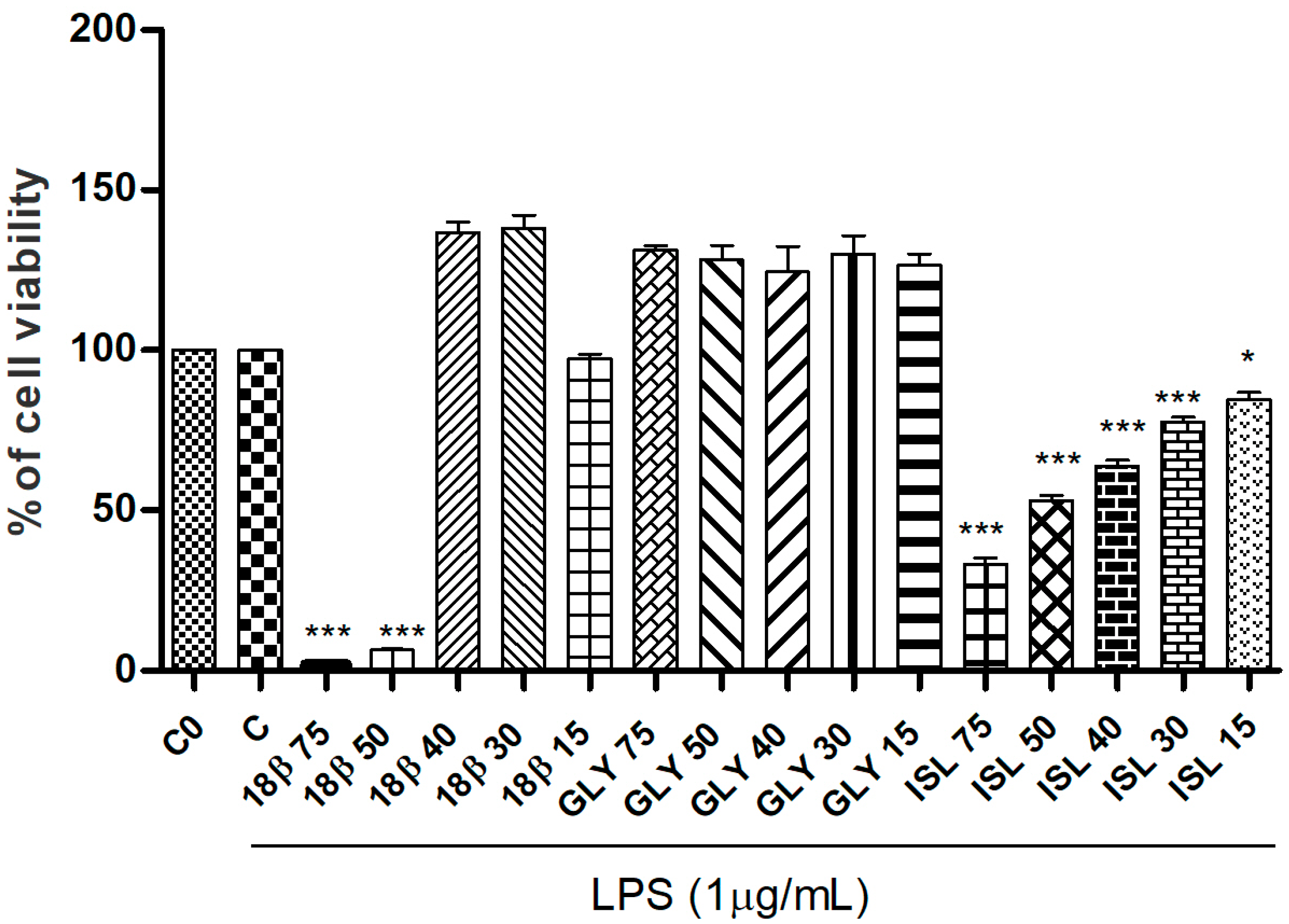
Each investigated licorice standard (18β-glycyrrhetinic acid, glycyrrhizin and isoliquiritigenin) was tested for cell viability at five increasing concentrations (15, 30, 40, 50 and 75 µM) in presence of LPS (1 µg/mL). As shown in Figure 2, glycyrrhizin did not affect cell viability at any tested concentration, while the 18β-glycyrrhetinic acid, at the higher tested doses, 50 and 75 µM, significantly induced a reduction of RAW 264.7 cell viability. As regards isoliquiritigenin, it induced a not negligible cell viability reduction even at the lowest tested dose (15 µM). For this reason, isoliquiritigenin was not selected and carried forward for further biological investigations.
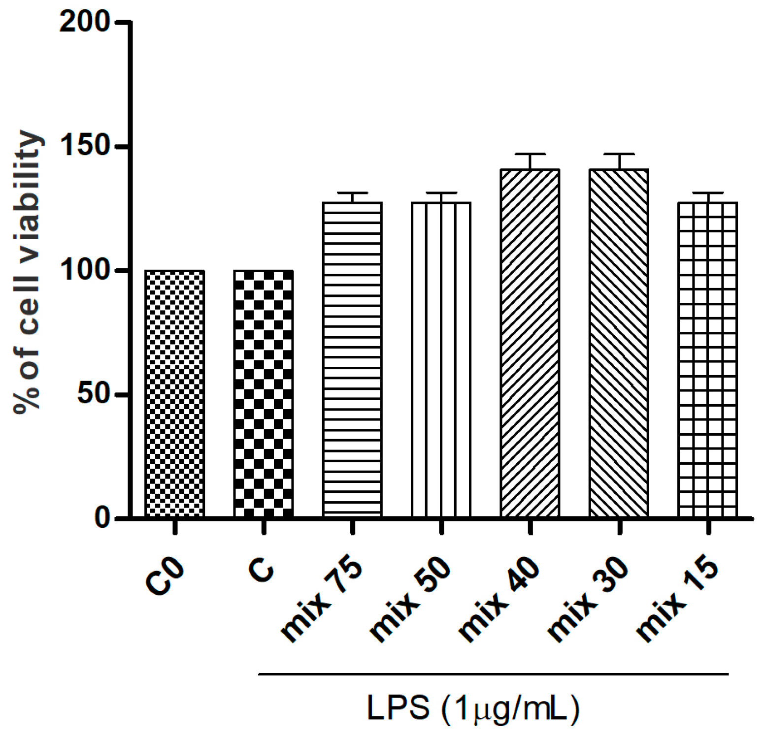
Starting from the results detailed by HPLC quantification of the standards within the extracts, the main licorice compounds, glycyrrhizin and 18β-glycyrrhetinic acid, respectively, were mixed in a combination mimicking their natural ratio in the raw extracts (previously calculated through HPLC analyses). In order to compare the activity of the pure single standards with these formulated mixtures, the final concentrations corresponded to that of the most abundant compound, glycyrrhizin. The formulated mixtures were tested for cell viability with an SRB assay and, as reported, none of the tested samples induced cytotoixc effects on the RAW 264.7 cell line (Figure 3).
In order to compare pure single standards and the mixture at the same final concentration, the highest non-toxic dose in RAW 264.7 cells was selected for each analyzed sample and carried forward for further investigations. Concerning the extracts, they were analyzed at the highest sub-cytotoxic dose of 100 µg/mL.

3.3. Effects of G. glabra L. Extracts and Standards on LPS-Induced Production of Pro-Inflammatory Cytokines (TNF-α, IL-6), the NO Mediator and the Induction of the Anti-Inflammatory Cytokine (IL-10)

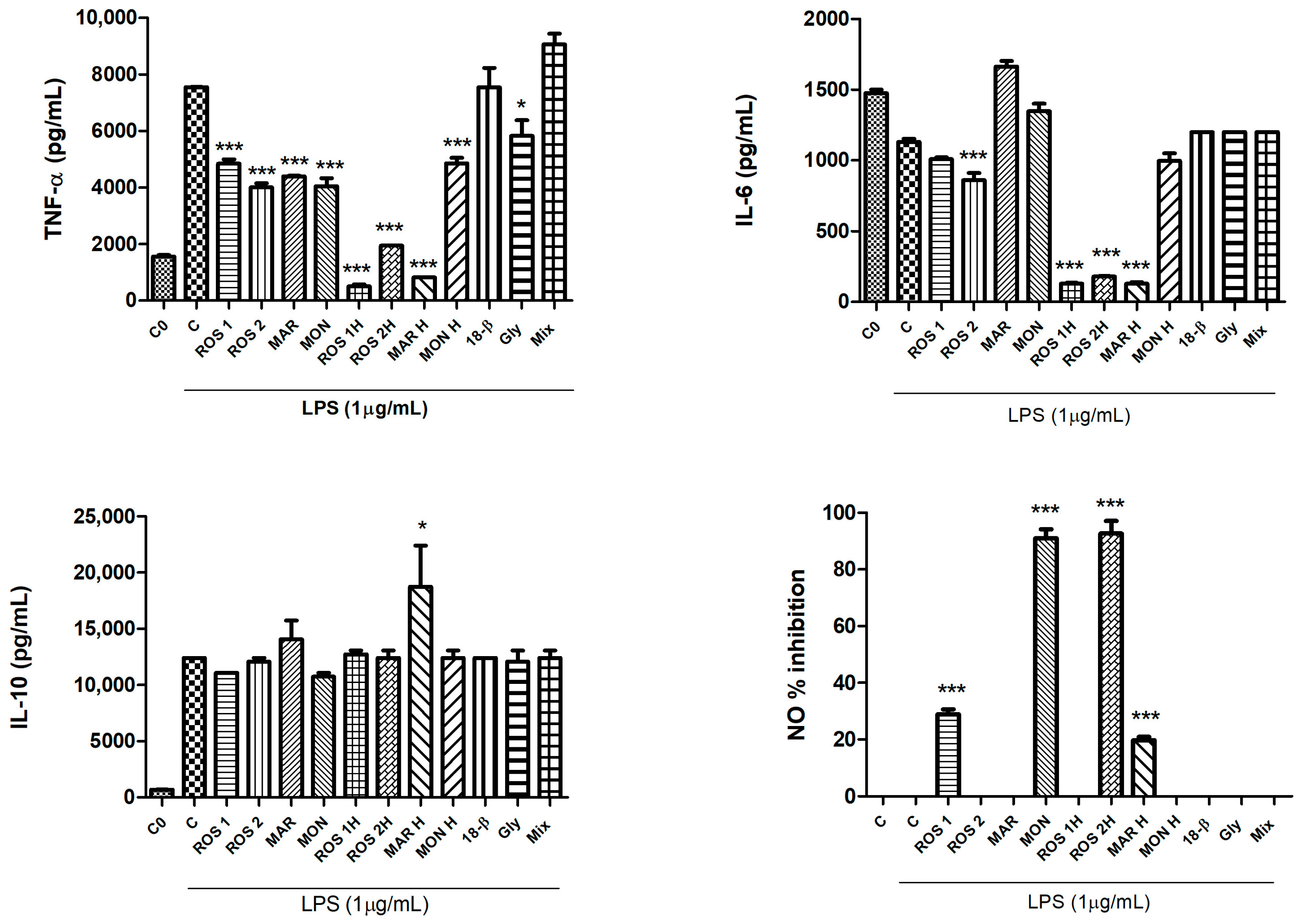
The anti-inflammatory potential of G. glabra raw and hydrolyzed extracts, together with that of glycyrrhizin (40 µM), 18β-glycyrrhetinic acid (40 µM) and their mixture (glycyrrhizin and 18β-glycyrrhetinic acid mixed in the proportion detailed by the HPLC ratio for extract) was investigated. The anti-inflammatory potential of both glycyrrhizin and glycyrrhetinic acid was explained through cytokines inhibition and transcription factors (NF-ĸB and STAT3) downregulation [12]. All the hydrolyzed extracts significantly reduced the production of the pro-inflammatory cytokine TNF-α compared to the control in the presence of 1 µg/mL LPS (C). Also, the corresponding raw extracts, even if to a minor extent, significantly inhibited TNF-α release. Among the standards and the mixture, only glycyrrhizin 40 µM significantly inhibited TNF-α release (p < 0.05 at Dunnett’s multiple comparison test). Following the same trend, ROS 1H, ROS 2H and MAR H hydrolyzed extracts, as well as ROS 2 raw extract significantly reduced the production and release of IL-6 (p < 0.001 at Dunnett’s multiple comparison test). The secretion of the anti-inflammatory cytokine IL-10 was significantly stimulated only by MAR H hydrolyzed extract (p < 0.05 at Dunnett’s multiple comparison test). The inhibition of NO oxidized stable products was assessed by ROS 2H and MON G. glabra extracts, which had the highest inhibition percentage, followed by ROS 1 and MAR H extracts (inhibition percentage lower than 40%) (Figure 4).

3.4. Effects of G. glabra L. Extracts and Standards on LPS-Induced Activation of the JAK/STAT Signaling Pathway

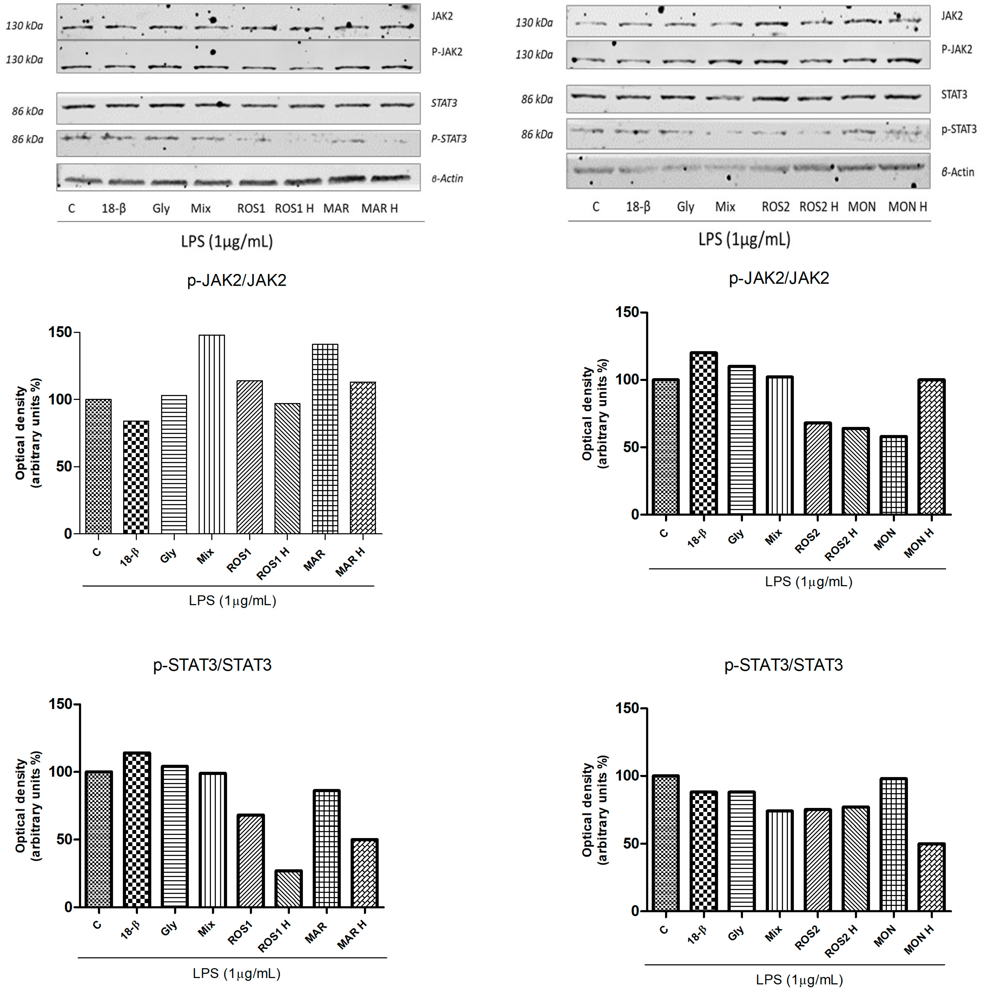
Samples were also tested for their ability to inhibit the phosphorylation of JAK2 and STAT3 proteins through Western blot analyses. 18β-glycyrrhetinic acid (40 µM) and glycyrrhizin (40 µM) as well as ROS 2, ROS 2H and MON inhibited JAK2 phosphorylation, while all raw and hydrolyzed extracts downregulated STAT3 phosphorylated proteins (Figure 5).
As reported by Thiyagarajan and colleagues (2011), G. glabra extracts significantly inhibited the production of IL-1β and IL-6 pro-inflammatory cytokines and NO (with an IC50 value equal to 24 µg/mL) in J774A.1 murine macrophages stimulated with LPS (0.1 µg/mL). On the contrary, glycyrrhizin did not trigger any anti-inflammatory activity versus LPS-induced mediators [29]. It was already demonstrated that both licorice aqueous extract and one its main components, glycyrrhetinic acid, possess an anti-inflammatory activity comparable with that of diclofenac sodium, one of the main drugs actually used for treatment of inflammatory disease. Moreover, it seems that the formulation of diclofenac and licorice aqueous extract improves the anti-inflammatory potential of the commercial drug on its own. Glycyrrhiza ethanolic extracts decreased TNF-α and IL-6 and ameliorated plasma levels of IL-10 cytokine in mice stimulated with LPS [30]. Moreover, the G. glabra extracts not containing glycyrrhizin showed interesting anti-inflammatory potential in mammalian cells by inhibiting Prostaglandin E2 (PGE-2), Thromboxane B2 (TXB-2) and Leukotriene B4 (LTB-4) [31]. Another study conducted by Wang and colleagues (2019), showed that glycyrrhizin exerted both anti-inflammatory and analgesic potential by inhibiting the production of Cyclooxygenase-2 (COX-2) and inducible Nitric Oxide Synthase (iNOS) and by decreasing the release of TNF-α and IL-6 cytokines [32]. Bodet and colleagues (2008) showed that the extract from G. uralensis inhibited IL-1β, IL-6, IL-8 and TNF-α periodontopathogen triggered by LPS in macrophages [33]. Wang and colleagues (2012) demonstrated that both glycyrrhizin and 18β-glycyrrhetinic acid were effective in the inhibition of NO, PGE2 and Reactive Oxygen Species (ROS) and in the downregulation of pro-inflammatory cytokines such as IL-6 and IL-1β in a dose-dependent manner [34]. Zhou and Wink (2019) demonstrated that G. glabra extracts inhibited NO induction and NF-ĸB translocation inside the nucleus in LPS-stimulated RAW264.7 macrophages [35]. Furthermore, other studies demonstrated that G. glabra ethanolic or methanolic extracts inhibited NO and PGE2 production in LPS-activated RAW264.7 cells [36,37]. G. uralensis roots from South Korea showed anti-inflammatory potential by suppressing protein expression of IL-1β, IL-6, COX-2 and iNOS in LPS-stimulated RAW264.7 cells and by downregulating NF-ĸB luciferase activity in HT-29 colon cancer cells [38].
These results assumed a central role in prevention. It is already known that prolonged exposure to pro-inflammatory stimuli and mediators, together with the aberrant activation of signal-transduction pathways can promote a condition of mild but chronic inflammation strictly correlated with a plethora of chronic pathologies including metabolic syndrome-related disorders and cancer [39]. All the discussed works highlighted the great anti-inflammatory potential of G. glabra extracts and the secondary metabolites they produce, which is in accordance with the data shown in this study.
Each plant species requires specific conditions for its growth and development. On the one hand, genetic factors significantly influence the quality and amounts of secondary metabolites produced; on the other hand, the environmental factors such as cultivation areas, habitat, temperature, light, humidity rate and soil characteristics can affect metabolic pathways, gene expression, and enzymatic activities. A study conducted by Eghlima et al. (2025) compared several different G. glabra samples collected in various Uzbekistan regions; data showed that the glycyrrhizic acid content may vary from 3.3% to 6.1% of dry weight, while glabridin amounts range from 0.08% to 0.35% of dry weight [26]. A study by Montoro and colleagues (2011) investigated the glycyrrhizic acid amount extracted from G. glabra root samples from Italy, China, Turkey and Iran: The data showed significant differences in concentrations, namely 51 mg/g, 53 mg/g, 33 mg/g and 32 mg/g of dry weight, respectively [40]. Glycyrrhizin content detected in the Zagros region (Iran) showed amount ranging from 0.03% to 0.23%. A study conducted by Semenescu et al. (2024) investigated the phytochemical profile and the biological activities of G. glabra samples collected in Romania [41]. Among the secondary metabolites identified, glycyrrhizin was detected during HPLC analyses at 13.927 mg/g.

4. Conclusions

Over the past years, G. glabra extracts, as well as their secondary metabolites, were largely studied for their interesting biological properties. In this paper, the anti-inflammatory potential activities of both the extracts and their main standards (glycyrrhizin, 18β-glycyrrhetinic acid and isoliquiritigenin) were investigated and compared with each other. Here, the best efficacy in inhibiting the production of pro-inflammatory cytokines (TNF-α, IL-6) and NO mediator and the best downregulation of the JAK/STAT signaling pathway was attributable to G. glabra extracts if compared to the standards. Among the extracts, the best activity was exerted by the hydrolyzed extracts, if compared to the raw ones; this process mainly involves glycyrrhizin, a triterpene saponin considered to be the most abundant compound in licorice that hydrolyzes in 18β-glycyrrhetinic acid. In particular, MAR H, by affecting all the investigated pro-inflammatory stimuli (TNF-α, IL-6), NO mediator and phosphorylation of JAK2 and STAT3 proteins, and being the only sample inducing a significant release of the IL-10 anti-inflammatory cytokine, can be considered the most effective extract. In conclusion, in this article, the potential anti-inflammatory activity of G. glabra extracts and of their main standards were investigated, suggesting they could represent potential valuable candidates for the treatment of inflammatory diseases acting through the inhibition of the JAK/STAT signaling pathway.

Supplementary Materials

The following supporting information can be downloaded at https://www.mdpi.com/article/10.3390/foods14213746/s1, Table S1: 18β-glycyrrhetinic acid, Isoliquiritigenin and Glycyrrhizin HPLC determination and quantification in G. glabra samples collected in different geographical areas.

Author Contributions

Conceptualization, G.S.; methodology, M.R.P. and G.S.; validation, F.C. and G.S.; formal analysis, M.R.P. and F.C.; investigation, M.R.P., M.P., C.-C.T., P.P., V.T., F.C. and M.M.; resources, G.S.; data curation, M.R.P., M.P., C.-C.T., M.M. and F.C.; writing—original draft preparation, M.R.P., M.P. and C.-C.T.; writing—review and editing, M.M., F.C. and G.S.; visualization, M.R.P., C.-C.T. and M.M.; supervision, G.S. All authors have read and agreed to the published version of the manuscript.

Funding

This research received no external funding. M.R. Perri was supported by POR Calabria 2014/2020.

Institutional Review Board Statement

Not applicable.

Informed Consent Statement

Not applicable.

Data Availability Statement

The data presented in this study are available in the article.

Conflicts of Interest

The authors declare no conflicts of interest.

Abbreviations

The following abbreviations are used in this manuscript:
EMAEuropean Medicine Agency
NSAIDsNon-steroidal anti-inflammatory drugs
TNF-αTumor Necrosis Factor-α
IL-6Interleukin-6
NONitric Oxide
IL-10Interleukin-10
LPSLipopolysaccharide
JAK2Janus Kinase
STAT-3Signal Transducer and Activator of Transcription 3
11β-HSD211 beta-hydroxysteroid dehydrogenase
FDAFood and Drug Administration
GRASGenerally Recognized As Safe
ADIAdmitted Daily Intake
HPLCHigh Performance Liquid Chromatography
DMEMDulbecco’s Modified Eagle’s Medium
FBSFetal Bovin Serum
PBSPhosphate Buffered Saline
BSABovine Serum Albumin
TCATrichloroacetic Acid
SRBSulphorhodamine B
ISLIsoliquiritigenin
GlyGlycyrrhizin
18-β18-β glycyrrhetinic acid
PGE-2Prostaglandin E2
TXB-2Thromboxane B2
LTB-4Leukotriene B4
COX-2Cyclooxygenase-2
iNOSInducible Nitrix Oxide Synthase
ROSReactive Oxygen Species

References

  1. Narayanankutty, A.; Famurewa, A.C.; Oprea, E. Natural bioactive compounds and human health. Molecules 2024, 29, 3372. [Google Scholar] [CrossRef]
  2. Walia, A.; Gupta, A.K.; Sharma, V. Role of bioactive compounds in human health. Acta Sci. Med. Sci. 2019, 3, 25–33. [Google Scholar]
  3. Samtiya, M.; Aluko, R.E.; Dhewa, T.; Moreno-Rojas, J.M. Potential health benefits of plant food-derived bioactive components: An overview. Foods 2021, 10, 839. [Google Scholar] [CrossRef]
  4. Najmi, A.; Javed, S.A.; Al Bratty, M.; Alhazmi, H.A. Modern approaches in the discovery and development of plant-based natural products and their analogues as potential therapeutic agents. Molecules 2022, 27, 349. [Google Scholar] [CrossRef]
  5. Hossain, M.S.; Wazed, M.A.; Asha, S.; Amin, M.R.; Shimul, I.M. Dietary phytochemicals in health and disease: Mechanisms, clinical evidence, and applications—A comprehensive review. Food Sci. Nutr. 2025, 13, e70101. [Google Scholar] [CrossRef]
  6. Sharma, V.; Katiyar, A.; Agrawal, R.C. Glycyrrhiza glabra: Chemistry and Pharmacological Activity. In Sweeteners; Springer: Cham, Switzerland, 2018; pp. 87–100. [Google Scholar]
  7. Hasan, M.K.; Ara, I.; Mondal, M.S.A.; Kabir, Y. Phytochemistry, pharmacological activity, and potential health benefits of Glycyrrhiza glabra. Heliyon 2021, 7, e07240. [Google Scholar] [CrossRef] [PubMed]
  8. El-Saber Batiha, G.; Magdy Beshbishy, A.; El-Mleeh, A.; M. Abdel-Daim, M.; Prasad Devkota, H. Traditional uses, bioactive chemical constituents, and pharmacological and toxicological activities of Glycyrrhiza glabra L. (Fabaceae). Biomolecules 2020, 10, 352. [Google Scholar] [CrossRef]
  9. Sidhu, P.; Shankargouda, S.; Rath, A.; Ramamurthy, P.H.; Fernandes, B.; Singh, A.K. Therapeutic benefits of liquorice in dentistry. J. Ayurveda Integr. Med. 2020, 11, 82–88. [Google Scholar] [CrossRef]
  10. European Medicines Agency. Draft European Union Herbal Monograph on Glycyrrhiza glabra L.; Gycyrrhiza inflata Bat.; Glycyrrhiza uralensis Fisch., Radix—Revision 1. Available online: https://www.ema.europa.eu/en/medicines/herbal/liquiritiae-radix (accessed on 12 October 2025).
  11. Isbrucker, R.A.; Burdock, G.A. Risk and safety assessment on the consumption of Licorice root (Glycyrrhiza sp.), its extract and powder as a food ingredient, with emphasis on the pharmacology and toxicology of glycyrrhizin. Regul. Toxicol. Pharmacol. 2006, 46, 167–192. [Google Scholar] [CrossRef]
  12. Richard, S.A. Exploring the pivotal immunomodulatory and anti-inflammatory potentials of glycyrrhizic and glycyrrhetinic acids. Mediat. Inflamm. 2021, 1, 6699560. [Google Scholar] [CrossRef] [PubMed]
  13. Guo, J.; Shang, E.; Zhao, J.; Fan, X.; Duan, J.; Qian, D.; Tao, W.; Tang, Y. Data mining and frequency analysis for licorice as a “Two-Face” herb in Chinese Formulae based on Chinese Formulae Database. Phytomedicine 2014, 21, 1281–1286. [Google Scholar] [CrossRef]
  14. Peng, F.; Du, Q.; Peng, C.; Wang, N.; Tang, H.; Xie, X.; Shen, J.; Chen, J. A review: The pharmacology of isoliquiritigenin. Phytother. Res. 2015, 29, 969–977. [Google Scholar] [CrossRef]
  15. Crusz, S.M.; Balkwill, F.R. Inflammation and cancer: Advances and new agents. Nat. Rev. Clin. Oncol. 2015, 12, 584–596. [Google Scholar] [CrossRef]
  16. Gurpinar, E.; Grizzle, W.E.; Piazza, G.A. COX-independent mechanisms of cancer chemoprevention by anti-inflammatory drugs. Front. Oncol. 2013, 3, 181. [Google Scholar] [CrossRef]
  17. Groner, B.; von Manstein, V. Jak Stat signaling and cancer: Opportunities, benefits and side effects of targeted inhibition. Mol. Cell. Endocrinol. 2017, 451, 1–14. [Google Scholar] [CrossRef] [PubMed]
  18. Jang, S.; Lee, A.Y.; Lee, A.R.; Choi, G.; Kim, H.K. Optimization of ultrasound-assisted extraction of glycyrrhizic acid from licorice using response surface methodology. Integr. Med. Res. 2017, 6, 388–394. [Google Scholar] [CrossRef]
  19. Zhang, Y.; Cao, J.; Wang, Y.; Xiao, S. Simultaneous determination of glycyrrhizin and 15 flavonoids in licorice and blood by high performance liquid chromatography with ultraviolet detector. Int. Sch. Res. Not. 2013, 2013, 786151. [Google Scholar] [CrossRef]
  20. Conforti, F.; Perri, M.R.; Guerrini, A.; Sacchetti, G.; Statti, G. Lavandula austroapennina and Lavandula angustifolia essential oils and bioactive components: In vitro anti-denaturation effect of lavender from the Pollino massif (Southern Italy). Plant Biosyst. 2023, 157, 339–345. [Google Scholar] [CrossRef]
  21. Mazzei, R.; Piacentini, E.; Nardi, M.; Poerio, T.; Bazzarelli, F.; Procopio, A.; Di Gioia, M.L.; Rizza, P.; Ceraldi, L.; Morelli, C.; et al. Production of plant-derived oleuropein aglycone by a combined membrane process and evaluation of its breast anticancer properties. Front. Bioeng. Biotechnol. 2020, 8, 908. [Google Scholar] [CrossRef]
  22. Perri, M.R.; Pellegrino, M.; Marrelli, M.; Aquaro, S.; Cavaliere, F.; Grande, F.; Occhiuzzi, M.A.; Lupia, C.; Toma, C.C.; Conforti, F.; et al. Identification of Pinosylvin in Pinus nigra subsp. laricio: A Naturally Occurring Stilbenoid Suppressing LPS-Induced Expression of Pro-Inflammatory Cytokines and Mediators and Inhibiting the JAK/STAT Signaling Pathway. Pharmaceuticals 2023, 16, 718. [Google Scholar] [CrossRef] [PubMed]
  23. Musolino, V.; Marrelli, M.; Perri, M.R.; Palermo, M.; Gliozzi, M.; Mollace, V.; Conforti, F. Centranthus ruber (L.) DC. and Tropaeolum majus L.: Phytochemical Profile, In Vitro Anti-Denaturation Effects and Lipase Inhibitory Activity of Two Ornamental Plants Traditionally Used as Herbal Remedies. Molecules 2023, 28, 32. [Google Scholar] [CrossRef]
  24. Perri, M.R.; Pellegrino, M.; Aquaro, S.; Cavaliere, F.; Lupia, C.; Uzunov, D.; Marrelli, M.; Conforti, F.; Statti, G. Cachrys spp. from Southern Italy: Phytochemical Characterization and JAK/STAT Signaling Pathway Inhibition. Plants 2022, 11, 2913. [Google Scholar] [CrossRef]
  25. Esmaeili, H.; Karami, A.; Hadian, J.; Saharkhiz, M.J.; Ebrahimi, S.N. Variation in the phytochemical contents and antioxidant activity of Glycyrrhiza glabra populations collected in Iran. Ind. Crops Prod. 2019, 137, 248–259. [Google Scholar] [CrossRef]
  26. Eghlima, G.; Tafreshi, Y.M.; Aghamir, F.; Ahadi, H.; Hatami, M. Regional environmental impacts on growth traits and phytochemical profiles of Glycyrrhiza glabra L. for enhanced medicinal and industrial use. BMC Plant Biol. 2025, 25, 116. [Google Scholar] [CrossRef] [PubMed]
  27. Graebin, C.S. The Pharmacological Activities of Glycyrrhizinic Acid (“Glycyrrhizin”) and Glycyrrhetinic Acid. In Sweeteners; Springer: Cham, Switzerland, 2018; pp. 245–261. [Google Scholar]
  28. Li, C.; Eom, T.; Jeong, Y. Glycyrrhiza glabra (L.) extract inhibits LPS-induced inflammation in RAW macrophages. J. Nutr. Sci. Vitaminol. 2015, 61, 375–381. [Google Scholar] [CrossRef]
  29. Thiyagarajan, P.; Chandrasekaran, C.V.; Deepak, H.B.; Agarwal, A. Modulation of lipopolysaccharide-induced pro-inflammatory mediators by an extract of Glycyrrhiza glabra and its phytoconstituents. Inflammopharmacology 2011, 19, 235–241. [Google Scholar] [CrossRef] [PubMed]
  30. Kim, J.K.; Oh, S.M.; Kwon, H.S.; Oh, Y.S.; Lim, S.S.; Shin, H.K. Anti-inflammatory effect of roasted licorice extracts on lipopolysaccharide-induced inflammatory responses in murine macrophages. Biochem. Biophys. Res. Commun. 2006, 345, 1215–1223. [Google Scholar] [CrossRef]
  31. Bisht, D.; Rashid, M.; Arya, R.K.K.; Kumar, D.; Chaudhary, S.K.; Rana, V.S.; Sethiya, N.K. Revisiting liquorice (Glycyrrhiza glabra L.) as anti-inflammatory, antivirals and immunomodulators: Potential pharmacological applications with mechanistic insight. Phytomed. Plus 2022, 2, 100206. [Google Scholar] [CrossRef]
  32. Wang, H.L.; Li, Y.X.; Niu, Y.T.; Zheng, J.; Wu, J.; Shi, G.J.; Ma, L.; Niu, Y.; Sun, T.; Yu, J.Q. Observing anti-inflammatory and anti-nociceptive activities of glycyrrhizin through regulating COX-2 and pro-inflammatory cytokines expressions in mice. Inflammation 2015, 38, 2269–2278. [Google Scholar] [CrossRef]
  33. Bodet, C.; La, V.D.; Gafner, S.; Bergeron, C.; Grenier, D. A licorice extract reduces lipopolysaccharide-induced proinflammatory cytokine secretion by macrophages and whole blood. J. Periodontol. 2008, 79, 1752–1761. [Google Scholar] [CrossRef]
  34. Wang, C.Y.; Kao, T.C.; Lo, W.H.; Yen, G.C. Glycyrrhizic acid and 18β-glycyrrhetinic acid modulate lipopolysaccharide-induced inflammatory response by suppression of NF-κB through PI3K p110δ and p110γ inhibitions. J. Agric. Food Chem. 2011, 59, 7726–7733. [Google Scholar] [CrossRef]
  35. Zhou, J.X.; Wink, M. Evidence for anti-inflammatory activity of isoliquiritigenin, 18β glycyrrhetinic acid, ursolic acid, and the traditional Chinese medicine plants Glycyrrhiza glabra and Eriobotrya japonica, at the molecular level. Medicines 2019, 6, 55. [Google Scholar] [CrossRef] [PubMed]
  36. Han, M.H.; Lee, M.H.; Hong, S.H.; Choi, Y.H.; Moon, J.S.; Song, M.K.; Kim, M.J.; Shin, S.J.; Hwang, H.J. Comparison of anti-inflammatory activities among ethanol extracts of Sophora flavescens, Glycyrrhiza uralensis and Dictamnus dasycarpus, and their mixtures in RAW 246.7 murine macrophages. J. Life Sci. 2014, 24, 329–335. [Google Scholar] [CrossRef]
  37. Yoon, T.S.; Cheon, M.S.; Kim, S.J.; Lee, A.; Moon, B.C.; Chun, J.M.; Choo, B.K.; Kim, H.K. Evaluation of solvent extraction on the anti-inflammatory efficacy of Glycyrrhiza uralensis. Korean J. Med. Crop Sci. 2010, 18, 28–33. [Google Scholar]
  38. Wu, T.Y.; Khor, T.O.; Saw, C.L.L.; Loh, S.C.; Chen, A.I.; Lim, S.S.; Park, J.H.Y.; Kong, A.N.T. Anti-inflammatory/Anti-oxidative stress activities and differential regulation of Nrf2-mediated genes by non-polar fractions of tea Chrysanthemum zawadskii and licorice Glycyrrhiza uralensis. AAPS J. 2011, 13, 1–13. [Google Scholar] [CrossRef] [PubMed]
  39. Nunes, C.D.R.; Barreto Arantes, M.; de Menezes, F.P.S.; da Leandro, C.L.; de Souza, P.M.; de Pereira, M.L.; Vieira, I.J.C.; de Barros, O.D. Plants as sources of anti-inflammatory agents. Molecules 2020, 25, 3726. [Google Scholar] [CrossRef] [PubMed]
  40. Montoro, P.; Maldini, M.; Russo, M.; Postorino, S.; Piacente, S.; Pizza, C. Metabolic profiling of roots of liquorice (Glycyrrhiza glabra) from different geographical areas by ESI/MS/MS and determination of major metabolites by LC-ESI/MS and LC-ESI/MS/MS. J. Pharm. Biomed. Anal. 2011, 54, 535–544. [Google Scholar] [CrossRef]
  41. Semenescu, I.; Avram, S.; Similie, D.; Minda, D.; Diaconeasa, Z.; Muntean, D.; Lazar, A.E.; Gurgus, D.; Danciu, C. Phytochemical, Antioxidant, Antimicrobial and Safety Profile of Glycyrrhiza glabra L. Extract Obtained from Romania. Plants 2024, 13, 3265. [Google Scholar] [CrossRef]
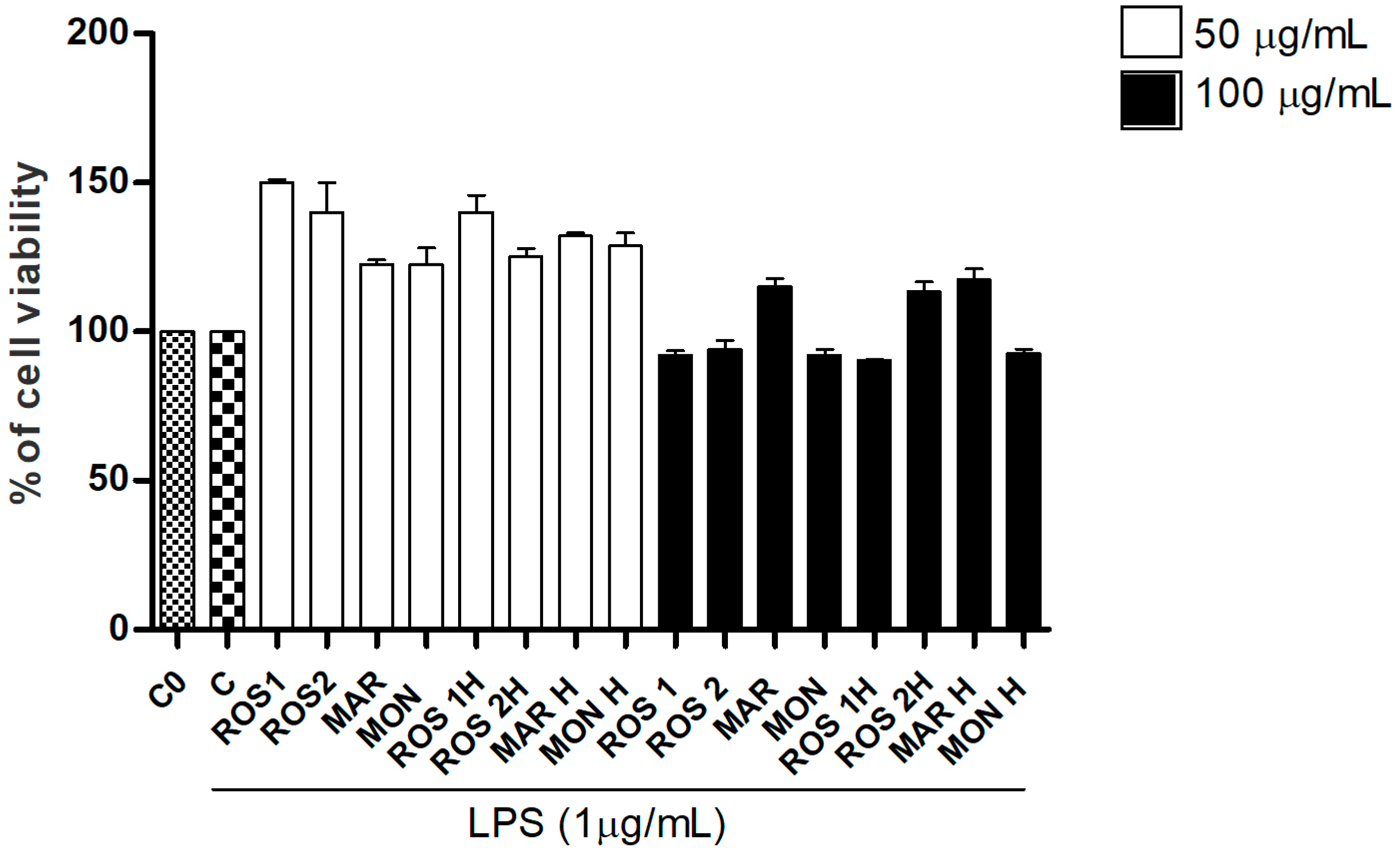
Figure 1. Cell viability of LPS-stimulated RAW 264.7 cells treated with G. glabra extracts as evaluated by SRB assay. Abbreviation codes are reported in Table 1. C0: control (untreated cells); C: control (untreated cells) with LPS. Data are expressed as mean ± S.E.M. from four independent experiments (n = 4), each one performed in three technical replicates.
Foods 14 03746 g001
Figure 2. Cell viability of LPS-stimulated RAW 264.7 cells treated with 18β-glycyrrhetinic acid (18β), glycyrrhizin (Gly) and isoliquiritigenin (ISL) (75, 50, 40, 30, 15 µM) evaluated at SRB assay. C0: control (untreated cells); C: control (untreated cells) with LPS. Data are expressed as mean ± S.E.M. from four independent experiments (n = 4), each one performed in three technical replicates. Mean values of samples showing significant difference compared to C were denoted with *** p < 0.001, * p < 0.05 (Dunnett’s multiple comparison test).
Foods 14 03746 g002
Figure 3. Effects of 18β-glycyrrhetinic acid and glycyrrhizin combinations (Mix) at different concentrations on LPS-stimulated RAW 264.7 cells viability. Combination proportions derive from HPLC detailed ratio (ROS 1 proportion). C0: control (untreated cells); C: control (untreated cells) with LPS. Data are expressed as mean ± S.E.M. from four independent experiments (n = 4), each one performed in three technical replicates.
Foods 14 03746 g003
Figure 4. G. glabra L. raw and hydrolyzed extracts (100 µg/mL), single standards of 18β-glycyrrhetinic acid (18β) 40 µM and glycyrrhizin (Gly) 40 µM and a mixture of them (Mix) (ROS1 proportion) and their potential inhibitory activity on the release of pro-inflammatory cytokines (TNF-α and IL-6), induction of the anti-inflammatory cytokine IL-10 release and production of nitric oxide (NO) mediator inhibition in LPS-stimulated RAW264.7 cells. C0: control (untreated cells); C: control (untreated cells) with LPS. Significant differences versus control in presence of LPS (C): * p < 0.05, *** p < 0.001 (Dunnett’s multiple comparison test).
Foods 14 03746 g004
Figure 5. G. glabra L. raw and hydrolyzed extracts (100 µg/mL), single standards of 18β-glycyrrhetinic acid 40 µM (18β) and glycyrrhizin 40 µM (Gly) and a mixture of them (Mix) (ROS 1 proportion) and their potential ability to downregulate p-JAK2 and p-STAT3 phosphorylated proteins. Cells were pretreated with samples and stimulated with LPS (1 µg/mL) after 30 min. After 2 h cells were lysed and equal amounts (70 µg) of total cellular extracts were analyzed. β-actin, as a protein not subjected to modulation by treatments, was used as loading control. Histograms refer to the densitometric analyses of the Western blot shown and were obtained by normalizing phosphorylated forms on relative total proteins, previously normalized versus β-actin.
Foods 14 03746 g005
Table 1. G. glabra geographical area origin, starting material and abbreviation codes.
G. glabra Sample OriginMaterialRaw/Hydrolyzed
Extracts
Codes
Coordinates
Montalto Uffugo (CS), ItalyCollectedMON/MON H39°24′ N; 16°09′ E
Rossano (CS), ItalyCommercially availableROS1/ROS1 H-
Rossano (CS), ItalyCommercially availableROS2/ROS2 H-
MoroccoCommercially availableMAR/MAR H-
Disclaimer/Publisher’s Note: The statements, opinions and data contained in all publications are solely those of the individual author(s) and contributor(s) and not of MDPI and/or the editor(s). MDPI and/or the editor(s) disclaim responsibility for any injury to people or property resulting from any ideas, methods, instructions or products referred to in the content.