Article

Study on the Effect of Radish Sprouts on Short-Chain Fatty Acids and Gut Microbial Diversity in Healthy Individuals

1
College of Food and Biological Engineering, Xuzhou Institute of Technology, Xuzhou 221018, China
2
College of Food Science and Nutritional Engineering, China Agricultural University, Beijing 100083, China
*
Author to whom correspondence should be addressed.
Foods 2025, 14(2), 170; https://doi.org/10.3390/foods14020170
Submission received: 2 December 2024 / Revised: 18 December 2024 / Accepted: 4 January 2025 / Published: 8 January 2025

Abstract

:
This study aimed to assess the impact of radish sprouts on the gut microbiota of healthy individuals. Radish sprout additives, subjected to short-term storage and steam treatment, were used to intervene in an in vitro culture of human gut microbiota. The influence of radish sprouts on the gut microbiota was evaluated by monitoring short-chain fatty acid (SCFA) content and proportion in the fermentation broth, and microbial diversity was assessed using 16S rDNA amplicon sequencing. The results indicated that the gut microbiota produced a substantial amount of SCFA within 48 h of fermentation, with a right-skewed distribution across all groups. The addition of both digestates enhanced Firmicutes diversity, while Bacteroidetes and Proteobacteria diversity remained stable between the control and fresh sprout groups. The 30 s steam treatment group showed an increase in Bacteroidetes and a decrease in Proteobacteria diversity. The abundance of Bacilli, Bacillaceae, and Bacillus was significantly higher in both the fresh and steam-treated groups compared to the control. Both fresh and steam-treated radish sprout digestates enriched gut microbiota diversity, with steam treatment showing superior effects. These findings suggest that radish sprout consumption may positively influence gut microbiota, with steam treatment potentially enhancing these benefits.

Graphical Abstract

1. Introduction

The human microbiome, with the majority residing in the gut, is composed of an immense number of symbiotic microorganisms, particularly in the colon where counts reach 1011 to 1012 cfu/mL [1,2]. This gut microbiota, boasting potent physiological functions, is considered an essential microecosystem for human health [3]. Nevertheless, the equilibrium of the gut microbiota is highly susceptible to modulation by diverse factors. Disruptions in this balance have been implicated in a spectrum of diseases, with connections now established between gut microbiota dysbiosis and conditions affecting both the gastrointestinal tract and the brain [4].
Diet has been established as a significant determinant of gut microbiota composition and metabolic activities [5]. Research, both in vitro and in vivo, demonstrates that dietary polyphenols from fruits and vegetables can selectively suppress harmful gut bacteria while fostering the growth of beneficial species such as Bifidobacterium [6,7]. This balance is further maintained by reducing the prevalence of detrimental microbes like Bacteroides, Enterococcus, and Staphylococcus [8,9]. Concurrently, the gut microbiota engages in the metabolism of dietary components, influencing their health functionalities [10]. Extensive research underscores the profound impact of the interplay between gut microbiota and dietary constituents on human physiology [11]. Deciphering the intricate relationships and mechanisms among dietary components, gut microbiota, and physiological health is not only of paramount importance but also a focal point of contemporary scientific inquiry.
Radish sprouts (Raphanus sativus L.), germinated from radish seeds, are enriched with carbohydrates, an array of vitamins, dietary fiber, and amylase [12]. These nutrients promote the absorption of nutrients, decrease the consumption of additional foods, and stimulate intestinal motility, assisting in digestion and easing constipation, which can aid in weight management [13]. The sprouts also contain bioactive compounds, including phenolics, which exhibit antioxidant activities and are hypothesized to influence the gut microbiota’s structure and function [14]. Phenolic compounds in radish sprouts, such as phenolic acids and flavonoids, are known for their antioxidant and anti-inflammatory properties. Specific phenolics like sinapic acid, ferulic acid, and kaempferol glycosides have been identified in radish sprouts [15]. These compounds are suggested to contribute to the prebiotic effect on the gut microbiota by selectively promoting the growth of beneficial bacteria, such as Bifidobacterium and Lactobacillus species, while inhibiting the growth of potentially harmful bacteria [7]. Beyond their prebiotic potential, these sprouts are low in calories and high in nutrients, making them a valuable component of a balanced diet. They are particularly rich in vitamin C, which supports the immune system, and vitamin K, which is essential for blood clotting and bone health [13].
Short-chain fatty acids (SCFAs) are predominantly generated as metabolic byproducts following the fermentation of undigested dietary substances, such as fiber from fruits and vegetables, by microorganisms within the large intestine [16]. Specific gut microbiota, including beneficial species within the Bifidobacterium genus, contribute to SCFA production by fermenting carbohydrates and proteins present in the intestinal chyme [17]. Research indicates that carotenoids present in radish sprouts are intricately linked to the gut microbiota [18]. These carotenoids, including β-carotene, lutein, lycopene, and astaxanthin, have been shown to modulate the gut microbiota’s composition by enhancing the growth of beneficial bacteria and suppressing the proliferation of harmful species [19]. Notably, these carotenoids significantly augment the overall production of SCFA and enrich the population of beneficial bacteria like Roseburia and Parasutterella, while concurrently curbing the growth of detrimental bacteria such as Collinsella [20,21]. The polyphenolic content in radish sprouts is also linked to the enhancement of short-chain fatty acid (SCFA) production, which plays a crucial role in maintaining gut health and overall well-being [16].
In recent years, the development of in vitro gut microbiota fermentation technology has greatly facilitated the study of gut microbiota, allowing for a thorough investigation into the effects of additives on the microbiota and its metabolites [22]. Therefore, based on the bioactive substances present in radish sprouts, fecal samples are collected from individuals who meet the requirements according to health and dietary surveys. The impact of stored and steamed radish sprout digestates on human gut microbiota is studied through in vitro culture, with the changes in the content and proportion of SCFA in the fermentation broth reflecting the influence of the functional substances in radish sprouts on the gut microbiota. Concurrently, high-throughput sequencing methods (16S RNA sequencing) are employed to investigate the diversity of the gut microbiota in the fermentation broth, thereby exploring the effects of radish sprout digestates on the gut microbiota. The novelty of our study lies in its comprehensive approach to understanding the impact of radish sprouts on the gut microbiota. While previous studies have explored the effects of various sprouts on gut health, our research delves specifically into the role of radish sprouts in modulating microbial diversity and function. By employing in vitro fermentation models and 16S rDNA sequencing, we provide a deeper insight into the mechanisms by which radish sprouts may exert their prebiotic effects.

2. Materials and Methods

2.1. Radish Sprout Cultivation

Plump, mold-free Shengfeng white radish seeds (from Beijing Jingyan Shengfeng Seed Research Institute) were selected. The radish seeds were thoroughly cleaned and then soaked in distilled water for 5 h. After soaking, the seeds were rinsed 2–3 times. The soaked seeds were evenly spread on a 30 × 20 cm seedling tray lined with four layers of gauze, with approximately 200 seeds per tray. The seeds were germinated in the dark at 25 °C for 3 days, with deionized water sprayed in the morning and evening. After three days, light was reintroduced with an intensity of 100 mmol/m2/s, under a 16 h/8 h (day/night) photoperiod, at a temperature of 25 °C and a relative humidity of 70% to 80%. After 7 days, the radish sprouts were ready for harvest [15].

2.2. Acquisition of Additives for Radish Sprout Cultivation

Uniformly cultivated radish sprouts were subjected to short-term storage (at 25 °C for 12 h (S1) and at 4 °C for 12 h (S2)) and short-term steaming (in boiling water for 30 s (P1) and 300 s (P2)). These treated sprouts, along with fresh radish sprouts (fresh sprouts, FS), were ground into powder with liquid nitrogen and stored at −80 °C for subsequent in vitro simulated gastrointestinal digestion [15,23]. Simulated saliva was prepared by dissolving 2.38 g of Na2HPO4, 0.19 g of KH2PO4, and 8 g of NaCl in 1 L of deionized water, with the pH adjusted to 6.75, and the addition of 12.5 mg of α-amylase (E.C. 3.2.1.1, A3176-500KU, 16 U/mg solid, Sigma, Kawasaki-shi, Japan) to achieve an enzyme activity of 200 U/mL. Simulated gastric fluid was prepared by adding pepsin A (EC3.4.23.1, P7012-250 MG, 3641 U/mg protein, 87% protein content, Sigma) (from porcine gastric mucosa) to 0.03 mol/L NaCl to achieve an enzyme activity of 300 U/mL, with the pH adjusted to 1.2. Simulated intestinal fluid was prepared by dissolving 0.05 g of pancreatin (equivalent to 4 times USP, P1750-25G, Sigma) and 0.3 g of bile extract (B8631-100g, Sigma) in 35 mL of 0.1 mol/L NaHCO3. The final solution contained 120 mmol/L NaCl and 5 mmol/L KCl. For the in vitro simulated gastrointestinal digestion process, 5 g of each sample powder was homogenized in 5 mL of simulated saliva and incubated at 37 °C for 10 min. Then, the samples were adjusted to pH 1.2 with HCl (5 mmol/L), suspended in 15 mL of simulated gastric fluid, and incubated at 37 °C for 120 min. Subsequently, the samples were adjusted to pH 6 with 0.1 mol/L NaHCO3, suspended in 15 mL of simulated intestinal fluid, adjusted to pH 7 with 1 mol/L NaOH, and then 5 mL of the final solution was added, with in vitro digestion for 120 min. The final concentration of each sample was 0.1 g/mL [24,25].

2.3. Assessment of Health and Dietary Practices in a Study Population

A survey questionnaire was meticulously designed to evaluate the health and dietary practices of the study population, and informed consent was secured from all participants. Participants were excluded based on three primary criteria: the intake of antibiotics or probiotic medications within the three-month period preceding the study, doubts concerning the participants’ willingness or capacity to adhere to the experimental protocol, and the existence of significant medical conditions, including but not limited to kidney diseases requiring dialysis. A cohort of 12 participants was selected, exhibiting similar demographic profiles and dietary behaviors, such as age, gender, smoking status, alcohol consumption, and dietary habits [26].

2.4. Collection and Pre-Treatment of Fecal Samples

Researchers provided fecal sampling tubes, gloves, and sample information strips to the subjects. Fecal samples were collected by eligible subjects at home using the provided fecal sampling tubes. The requirements for the samples are as follows [27]: fresh, not mixed with urine, free from disinfectants and sewage, and ideally not in contact with any impurities. After sample collection, the sample information strip (including name, gender, sampling location, and sampling time (accurate to minutes)) should be filled out accurately and transferred to the laboratory’s cold storage within 3 h, and the preliminary treatment of the fecal samples should be completed within 8 h. The remaining samples are stored at −80 °C. This study has been approved by the Human Research Ethics Committee of China Agricultural University (CAUHR-2019007).
Fresh feces of 0.8 g were weighed and added to 8 mL of sterilized PBS solution (pH 7.2–7.4) (mass concentration of 0.1 g/mL), mixed evenly by shaking, and then centrifuged at 3000× g at room temperature for 10 min. The supernatant after sedimentation was taken for subsequent experiments [28].

2.5. Media for In Vitro Cultivation of Intestinal Microbiota

The medium consists of (per 100 mL) 0.1 g of casein peptone, 0.25 g of yeast extract, 0.4 g of NaHCO3, 0.1 g of cysteine, 0.045 g of K2HPO4, 0.045 g of KH2PO4, 0.09 g of NaCl, 0.009 g of MgSO4·7H2O, 0.009 g of CaCl2, 0.1 mg of gentian violet, 1 mg of hemin, 1 mg of biotin, 1 mg of cobalamin, 3 mg of p-aminobenzoic acid, 5 mg of folic acid, and 15 mg of pyridoxine, with a pH of 6.5 [29]. The medium is sterilized by autoclaving and then distributed into 10 mL glass medical sealable vials under anaerobic conditions, 5 mL per vial, for storage at room temperature.

2.6. In Vitro Cultivation of Intestinal Microbiota with Radish Sprout Digesta

The experimental setup was divided into six groups, with each group containing 12 bottles, and the culture medium was appropriately marked. Alongside the FS, S1, S2, P1, and P2 groups, a control group (CK), which received no intervention substances, was also included. The operations were carried out as follows: To each bottle within the groups, 500 μL of fecal supernatant from 12 distinct subjects was introduced. Subsequently, 100 μL of the intervention substances were added, with the CK group receiving 100 μL of sterilized PBS, and the remaining groups receiving 100 μL of the respective radish sprout digesta. The cultures were incubated at 37 °C for a duration of 48 h to facilitate ex vivo fermentation. Upon completion of the fermentation period, the fermented broth was transferred to 2 mL centrifuge tubes for further processing, while the surplus samples were cryopreserved at −80 °C [30,31].

2.7. Detection of Short-Chain Fatty Acids

External standard compounds included acetic acid, propionic acid, butyric acid, isobutyric acid, pentanoic acid, and isovaleric acid, with crotonic acid serving as the internal standard. Standards, fermentation broths (CK, FS, S1, S2, P1, and P2), and fecal suspensions (FeS), each 500 μL in volume, were aliquoted into 1.5 mL centrifuge tubes. To each, 100 μL of a crotonic acid and metaphosphoric acid mixture was added, with a sample-to-internal standard volume ratio of 5:1. The mixtures were vortexed to achieve uniformity and then placed in a −20 °C freezer overnight. After thawing, the samples were centrifuged at 14,000 rpm for 5 min. The supernatant was collected in its entirety using a 1 mL syringe, filtered through a hydrophilic filter membrane, and transferred to fresh centrifuge tubes. A 100 μL aliquot of the filtrate was then pipetted into a sample vial equipped with a liner, ensuring that any air bubbles at the bottom of the liner were expelled prior to analysis. The gas chromatographic analysis was conducted using a GC9720 (Fuli) instrument, equipped with an HP-FFAP column (30 m × 0.25 mm × 0.25 μm, Agilent, Santa Clara, CA, USA). The injector temperature was maintained at 250 °C; the column temperature program initiated at 75 °C, with a ramp rate of 20 °C/min, and a final temperature of 220 °C for a duration of 2 min; the FID detector temperature was set at 250 °C; the gas flow rates were configured to 40 mL/min for hydrogen, 400 mL/min for air, and 20 mL/min for tail-blow nitrogen; the injection mode was split, with a split ratio of 5:1; and the injection volume was 1 μL [31,32].

2.8. Detection of Intestinal Microbiota Diversity

Following the outcomes of the initial experiments, fermentation broths from the CK, FS, and P1 groups were selected for further analysis. The notation F (Female) and M (Male) signifies the gender of the subjects, with the numerals indicating their respective identification numbers. For instance, CK.F.1 denotes the fermentation broth sample associated with Female subject number 1 in the control group that received no intervention substances.
The DNA extraction from fecal samples obtained post in vitro fermentation was conducted utilizing the OMEGA Stool DNA Kit (D4015). Agarose gel electrophoresis was employed to assess the purity and concentration of the extracted DNA. Aliquots of the sample DNA were placed into centrifuge tubes and diluted with sterile water to a concentration of 1 ng/μL. Subsequently, the diluted genomic DNA served as a template for PCR amplification, which was carried out using barcoded specific primers (515F and 806R) corresponding to the V3–V4 region, Phusion® High-Fidelity PCR Master Mix with GC Buffer from New England Biolabs, and high-fidelity enzymes to ensure amplification efficiency and accuracy. The PCR products were electrophoresed on a 2% agarose gel to verify the integrity and were then mixed in equal amounts. The PCR products were purified using a 1×TAE buffered 2% agarose gel, and the target bands were excised and recovered. The Thermo Scientific GeneJET Gel Recovery Kit (Thermo Fisher Scientific, Waltham, MA, USA) was utilized for the purification of the excised products. Library construction was performed using the Thermofisher Ion Plus Fragment Library Kit 48 rxns (Thermo Fisher Scientific, Waltham, MA, USA), and following quantification with Qubit and library validation, the constructed libraries were sequenced on the Thermofisher Ion S5TMXL platform (Thermo Fisher Scientific, Waltham, MA, USA). Data obtained were subsequently processed for analysis.
The 16S rDNA amplicon sequencing approach was employed, targeting the hypervariable V3–V4 regions, to amplify and sequence the DNA extracts from the in vitro fermentation broths [33]. Universal primers designed for the conserved regions facilitated PCR amplification. Following amplification, the variable regions were sequenced and analyzed for microbial identification. Characteristic of the amplified region, the IonS5™ XL sequencing platform was utilized, employing single-end sequencing to construct small fragment libraries. After sequencing, the reads were trimmed and filtered (Raw data were extracted from the IonS5™ XL platform in fastq file format. Sample data were delineated based on barcode sequences, and chimeras are eliminated to obtain clean reads. Quality control metrics, including raw reads, clean reads, total base count, average length, Q20 percentage, GC content, and effective percentage, were assessed). Operational Taxonomic Units (OTUs) were clustered, and species annotation and abundance analysis were conducted to reveal the composition of species within the samples. Further complexity analysis of the samples was performed, including alpha diversity (α-diversity analysis) for intra-sample diversity assessment and beta diversity (β-diversity analysis) for inter-sample comparison to uncover differences among the samples.

2.9. Data Processing

Data statistics and single-factor analysis of variance (ANOVA) were conducted using Excel 2010 and SPSS 20.0 software, followed by Duncan’s multiple range test for significance analysis (p < 0.05). Graphs were created using Origin 9.1 software. Results are presented as mean ± standard deviation. All experiments were repeated three times.

3. Results and Discussion

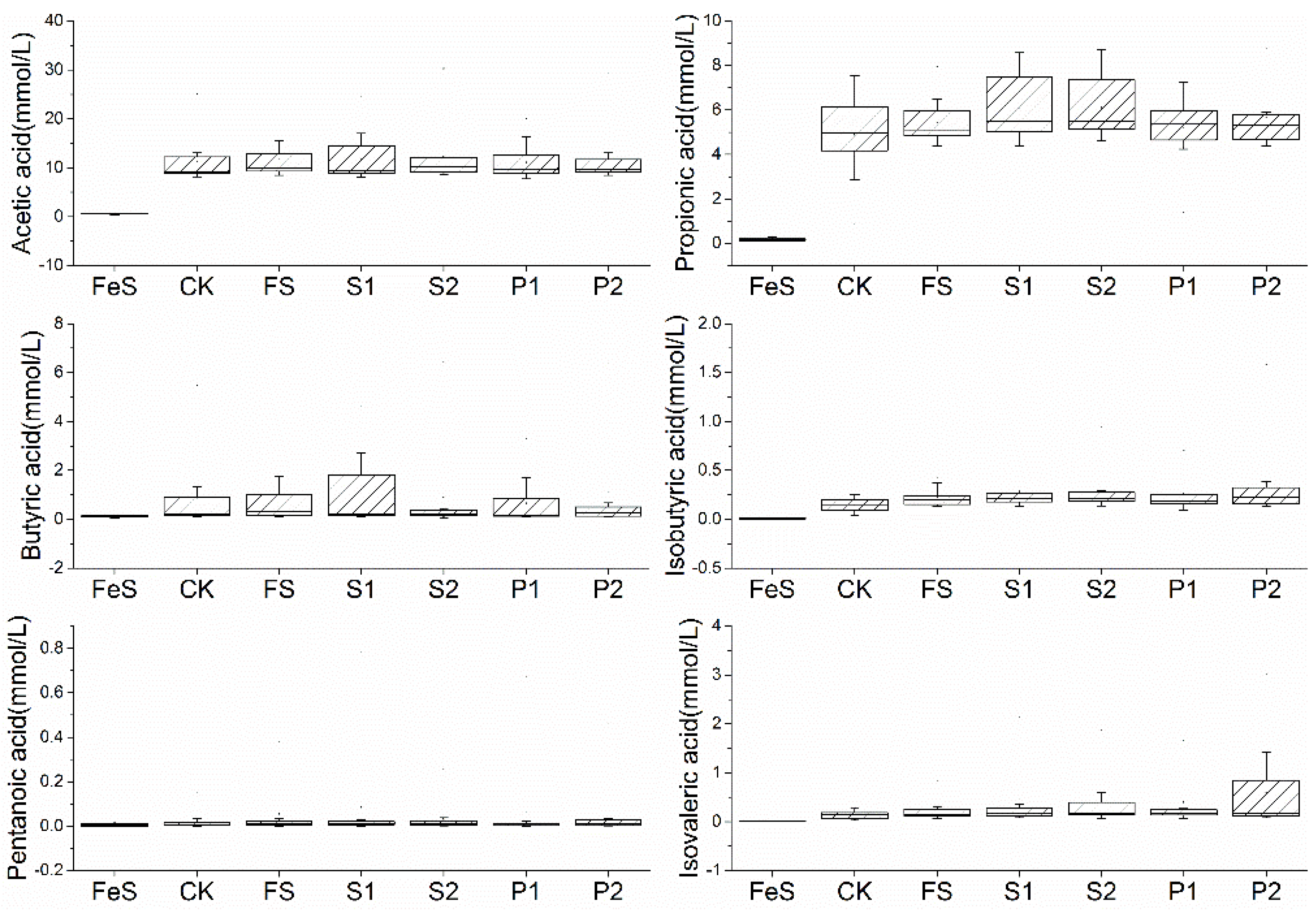
3.1. The Effect of Radish Sprout Digesta Supplementation on Short-Chain Fatty Acids

Short-chain fatty acids (SCFAs), primarily consisting of acetic acid, propionic acid, and butyric acid, maintain the intestinal barrier, promote gut health, regulate blood sugar and body weight, enhance cardiac and skeletal health, possess anti-inflammatory and immune-modulating effects, and may even contribute to cancer prevention [34,35,36]. SCFAs can also improve neurological health, exerting positive effects on mood and cognitive functions [37]. The levels of acetic, propionic, and isobutyric acids in all experimental groups were elevated after 48 h of fermentation compared to the FeS group, with the exception of valeric acid (Figure 1). Post in vitro fermentation, a greater dispersion was observed in other SCFAs, and a notable right skewness was apparent, with the exception of propionic acid. The distribution of acetic acid did not yield any directional outcomes except for an increased dispersion in the S1 group. The incorporation of foreign materials in the FS, S1, S2, P1, and P2 groups resulted in higher propionic acid levels overall when compared to the control group, with more significant effects noted in the S1, S2, P1, and P2 groups, and a reduction in dispersion observed in the FS, P1, and P2 groups. The distribution of butyric acid revealed greater dispersion in the S1 group relative to CK, while S2 and P2 exhibited the opposite trend. The addition of digesta to the fermentation fluids increased isobutyric acid content, with P1 showing a less pronounced increase compared to P2, and except for increased dispersion in the P2 group, the other groups remained relatively consistent. The fermentation fluid in the P1 group demonstrated reduced dispersion of valeric acid, and the P2 group had an increased valeric acid content relative to CK. The distribution of isovaleric acid saw an increase in the S1, S2, P1, and P2 groups, with S2 and P2 displaying greater dispersion. The elevated levels of acetic, propionic, and isobutyric acids post-fermentation suggest a shift in microbial metabolism, which could be attributed to the fermentative capabilities of the introduced digestates [38,39]. Collectively, the supplementation of various digesta positively influenced the elevation in certain SCFAs in the fermentation fluid, particularly after the radish sprout digesta had been subjected to storage and steaming processes.

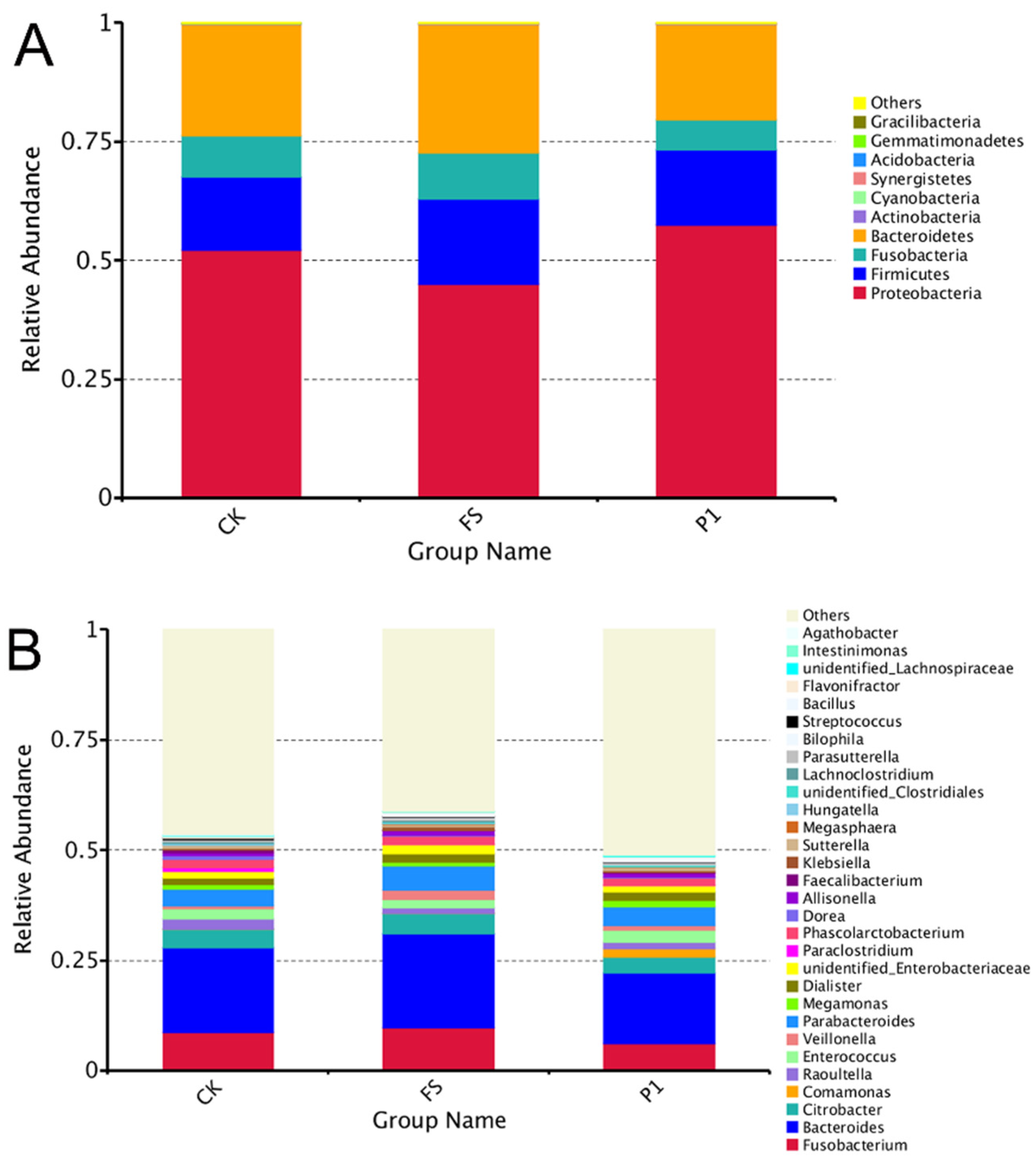
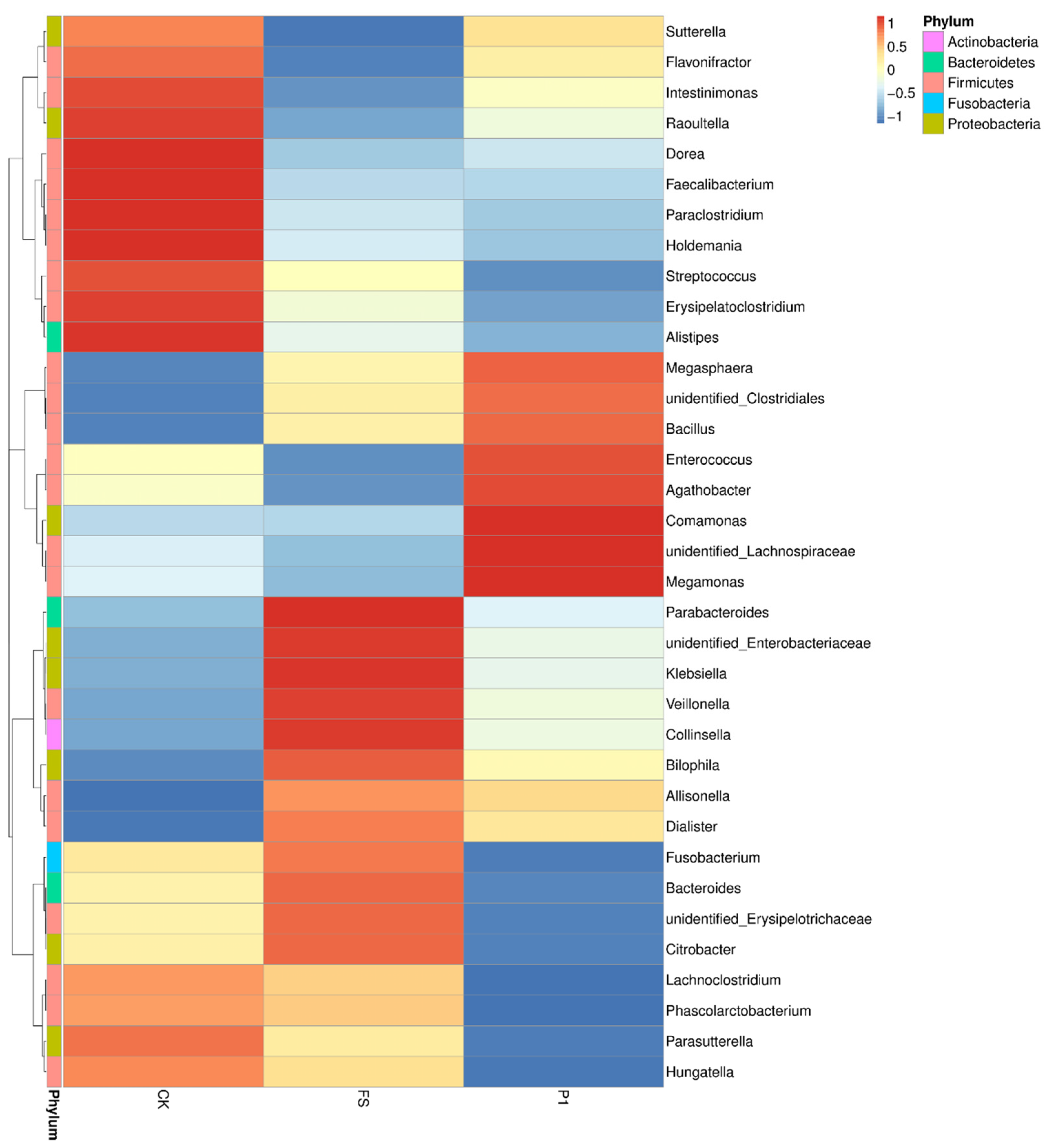
3.2. The Effect of Radish Sprout Digesta Supplementation on Gut Microbiota Diversity—A Comparative Analysis of Microbial Diversity Based on OUT

In light of initial research outcomes, the aim was to delve deeper into the effects of radish sprout digesta on intestinal microbiota. Consequently, ex vivo fermentation broths from the CK, the FS, and the P1 were chosen for further investigation. A 16S bioinformatics analysis was performed on these selected samples. The effective data from all samples were clustered into Operational Taxonomic Units (OTUs) at a 97% similarity level, after which the representative sequences of these OTUs underwent species annotation in preparation for subsequent analyses. As depicted in the bar chart illustrating the top 10 phyla by relative abundance (Figure 2), based on species annotation results, Proteobacteria emerges as the predominant phylum across all experimental groups, with Escherichia and Desulfovibrio being its principal constituents [40]. The supplementation of fresh radish sprout digesta (FS) and steamed radish sprout digesta for 30 s (P1) has been observed to modulate the relative abundance of Proteobacteria, with a decrease in the FS group and an increase in the P1 group. Bacteroidetes, which includes Bacteroides and Prevotella as key genera [41], ranks second in abundance and exhibits an inverse response to Proteobacteria, with an increase in the FS group and a decrease in the P1 group, mirroring the pattern observed for Fusobacteria, the fourth-ranked phylum. Firmicutes, the third-most abundant phylum, encompasses genera such as Clostridium, Lactobacillus, Ruminococcus, Eubacterium, Faecalibacterium, and Roseburia [42], and its relative abundance is notably higher in the FS group compared to the P1 group. Actinobacteria, also present in the fermentation liquids, is characterized primarily by the genus Bifidobacterium, with several species recognized for their probiotic properties [43].
Figure 2B illustrates a cumulative representation of the top 30 genera based on their relative abundance, categorized according to species annotation outcomes. Excluding the “other” category, Bacteroides (from the Bacteroidetes phylum) emerges as the predominant genus, with its abundance notably elevated in the fermentation group supplemented with fresh radish sprout digestate (FS) and diminished in the group with steamed radish sprout digestate for 30 s (P1). It has been documented that the prevalence of Bacteroides in individuals with type 2 diabetes significantly surpasses that in healthy populations [44]. Fusobacterium (belonging to the Fusobacteria phylum) follows suit, with its abundance influenced by the additives in a manner congruent with Bacteroides, showing an increase in the FS group and a decrease in the P1 group. Evidence suggests that Fusobacterium is more abundant in colorectal cancer patients compared to healthy individuals [45]. Citrobacter (Proteobacteria phylum) and Parabacteroides (Bacteroidetes phylum) exhibit similar proportions, while Phascolarctobacterium (Firmicutes phylum) and Dialister (Firmicutes phylum) are detected, albeit in lower quantities. Citrobacter, a common non-pathogenic gut bacterium that can become pathogenic under compromised immunity, bears a striking resemblance to the significant pathogen Salmonella within the gut [46]. Parabacteroides, integral to the microbiota of healthy individuals, is known for its production of beneficial metabolites such as acetic acid and succinic acid [47]. Research has indicated that individuals predisposed to weight loss harbor higher levels of Phascolarctobacterium, whereas those resistant to weight loss exhibit increased Dialister levels [48]. Collectively, it appears that the consumption of steamed radish sprouts for 30 s, as opposed to fresh radish sprouts, leads to a reduction in the abundance of certain bacterial genera detrimental to health, including Bacteroides and Fusobacterium. The increased diversity of Firmicutes and the reduction in potentially harmful genera such as Bacteroides and Fusobacterium upon steaming radish sprouts for 30 s underscore the importance of food processing methods in shaping the gut microbiota [8]. These findings have implications for the development of dietary strategies aimed at enhancing gut health and potentially mitigating the risk of associated diseases.
Species annotation and abundance data at the genus level across all samples (grouped) were analyzed using the maximum value sorting method to identify the top 35 genera (with group abundance defined as the average abundance across all samples within the group). Clustering at the species level was performed based on the abundance information in each sample, and heatmaps were generated using the pheatmap package in R 3.6.0 software. These visualizations facilitate the identification of species that are either abundant or scarce in specific samples. Figure 3 depicts the clustering heatmaps at the genus level (grouped), with values representing Z-scores derived from the standardized relative abundance of each species. The Z-score for a sample in a given taxon is calculated by subtracting the average relative abundance across all samples from the sample’s relative abundance in that taxon, and then dividing it by the standard deviation of all samples in that taxon. This approach revealed 35 of the most abundant bacterial genera, marked with different colors across the three groups. Notably, significant clustering differences were observed among the CK, FS, and P1 groups. Genera such as Sutterella (Proteobacteria), Flavonifractor (Firmicutes), Intestinimonas (Firmicutes), Raoultella (Proteobacteria), Dorea (Firmicutes), Faecalibacterium (Firmicutes), Parabacteroides (Bacteroidetes), Holdemania, Streptococcus (Firmicutes), Erysipelatoclostridium, and Alistipes were found to be significantly more abundant in the control group compared to the FS and P1 groups. Sutterella has been identified in the gut of children with autism and is associated with digestive disorders [49]; a high abundance of Flavonifractor correlates with a low quality of life in large populations [50]; Intestinimonas is significantly reduced in obesity [51]; Raoultella shows a positive correlation with inflammatory bowel disease [52]; Dorea, a major gas-producing bacterium in the human gut, is prevalent in children with irritable bowel syndrome [53]; Faecalibacterium is ubiquitous in the gut of healthy individuals [54]; Parabacteroides has been positively correlated with anti-epileptic effects in gut microbiota studies [55]; Holdemania is significantly less abundant in the gut of kidney stone patients compared to healthy individuals [56]; Streptococcus exhibits a 30% higher variation in diarrhea populations than in asymptomatic populations [57]; Erysipelatoclostridium is significantly increased in kidney stone patients [58]; Alistipes is a dominant genus in the gut of patients with depression [59]. These findings suggest that consuming fresh radish sprouts and steamed radish sprouts for 30 s may help reduce the abundance of detrimental gut bacteria. Lachnoclostridium, Phascolarctobacterium, Parasutterella, and Hungatella exhibit relatively higher abundance in the CK group. Parasutterella is significantly reduced in the gut of kidney stone patients [59,60]; Hungatella is more abundant in the gut of Parkinson’s disease patients compared to healthy individuals [61]. Megasphaera (Firmicutes), Bacillus (Firmicutes), Enterococcus (Firmicutes), Agathobacter, Comamonas (Proteobacteria), Megamonas (Firmicutes), and two unidentified genera (unidentified_Clostridiales and unidentified_Lachnospiraceae) show the highest abundance in the P1 group; the remaining genera are most abundant in the FS group. Megasphaera is less abundant in the gut of patients with rheumatoid arthritis [62]; Bacillus is a normal, beneficial bacterial genus in the human gut [63]; Enterococcus is a normal gut flora, often residing in the gut and female urinary reproductive tract, and is an opportunistic pathogen [64]; Megamonas may be very abundant in the gut of Asian colorectal cancer patients [65], indicating that consuming steamed radish sprouts for 30 s is beneficial for maintaining normal gut flora.

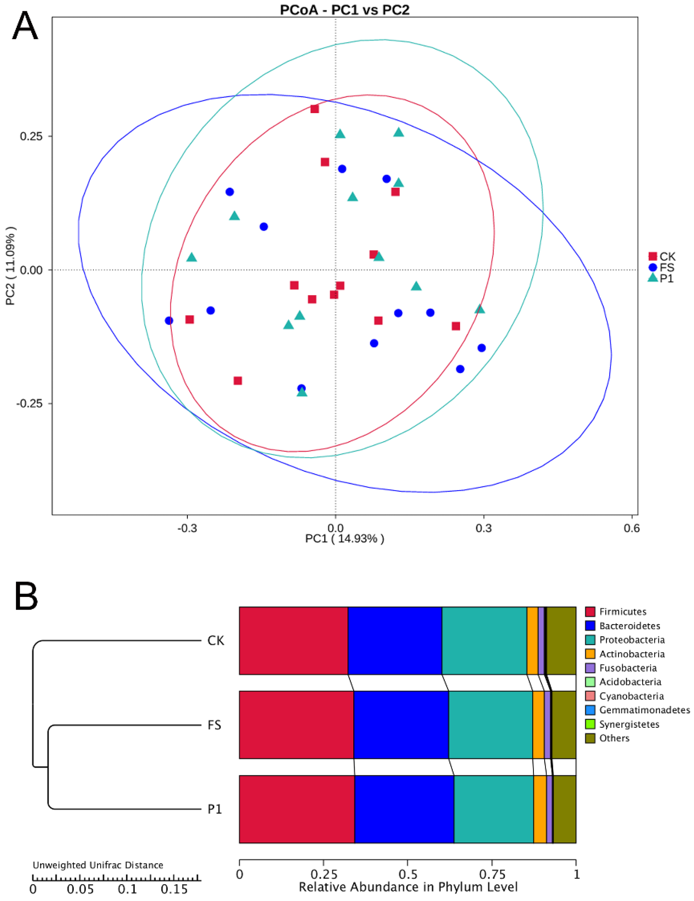
3.3. The Impact of Radish Sprout Digesta Supplementation on Gut Microbiota Diversity—β-Diversity Analysis

Figure 4A illustrates the Principal Co-ordinates Analysis (PCoA) outcomes premised on Unweighted Unifrac distances. This analytical technique elucidates the principal constituents and structures within multidimensional datasets through the arrangement of eigenvalues and eigenvectors. The depiction delineates two principal components, which are responsible for explicating 14.93% and 11.09% of the dataset’s variance, respectively. Group P1 encompasses Group CK entirely, signifying an augmentation in microbial diversity consequent to the introduction of exogenous substances. Conversely, Group FS, while partially overlapping with Group CK, exhibits a considerably expanded area, denoting a pronounced enhancement in microbial diversity when compared to Group CK. The divergence in microbial diversity between Groups FS and P1 is ascribed to the dissimilarities in the composition of the additives. Collectively, the integration of both fresh and steam-processed radish sprout digestates enriches gut microbial diversity, with the steam-processed variant demonstrating a more pronounced impact.
Additionally, a UPGMA (Unweighted Pair Group Method with Arithmetic Mean) clustering analysis was conducted utilizing the Unweighted Unifrac distance matrix, and the clustering outcomes were amalgamated with the relative abundance of taxa at the phylum level for each sample, as depicted in Figure 4B. The underlying principle of UPGMA involves initially clustering the two samples with the shortest distance to form a new node, with the branch point positioned midway between the two samples. Subsequently, the average distance between this new “sample” and the remaining samples is determined, and the two samples with the smallest average distance are clustered together. This iterative process continues until all samples are integrated into a single cluster, culminating in a comprehensive dendrogram. The findings reveal that Firmicutes, Bacteroidetes, and Proteobacteria constitute over 80% of the microbial communities, aligning with prior research [66]. The dominance of Firmicutes, Bacteroidetes, and Proteobacteria in our study is consistent with their established roles in the gut ecosystem, and the observed changes in their proportions following the introduction of radish sprout digestates highlight the potential for dietary manipulation to modulate microbial balance [67]. The introduction of both digestates resulted in an increased diversity within Firmicutes, whereas the diversity of Bacteroidetes and Proteobacteria exhibited minimal variation between the CK and FS groups. In contrast to the CK group, the diversity of Bacteroidetes and Proteobacteria in the P1 group experienced a notable increase and decrease, respectively. Actinobacteria and Fusobacteria displayed negligible differences across the three groups.
To discern differential species across taxonomic hierarchies (Order, Family, Genus, Species) between groups, t-tests were employed to identify species with statistically significant differences (p < 0.05), as depicted in Figure 5. The FS and P1 groups exhibited a significantly higher abundance of Bacillales (Order), Bacillaceae (Family), and Bacillus (Genus) compared to the control group at their respective taxonomic levels. These taxa are classified under the Bacilli class of Firmicutes and are Gram-positive bacteria. Constituting a vital part of the gut microbiota, Bacillus species, including Bacillus subtilis (encompassing Bacillus natto), Bacillus cereus, and Bacillus licheniformis, are extensively utilized as probiotics in human applications [68,69]. Through their adhesive properties to the intestinal lining and antagonistic effects on pathogenic bacteria, Bacillus species contribute to the maintenance of gut microbiota equilibrium and intestinal health [70,71]. The incorporation of both fresh and steamed radish sprout digestates appears to be advantageous for gut microbiota balance and intestinal well-being. Moreover, at the species level, the FS group demonstrated a significantly higher abundance of Clostridium-beijerinckii compared to the control group. Clostridium-beijerinckii, a member of the Clostridium genus within Firmicutes, is recognized as a key butyrate-producing bacterium in the gut, aligning with our prior findings on short-chain fatty acids [72,73]. The findings suggest that the supplementation of fresh and steamed radish sprout digestates can enhance the abundance of Bacillus, which is instrumental in elevating the ratio of beneficial bacteria. Additionally, the enrichment of Clostridium-beijerinckii due to the addition of fresh radish sprout digestate implies a positive impact on butyrate synthesis.

4. Conclusions

In vitro fermentation of radish sprout digestates led to a significant increase in SCFAs such as acetic, propionic, and isobutyric acids within 48 h, with a right-skewed distribution. Valeric acid showed increased variability. The FeS group had lower propionic and isovaleric acids but higher butyric and valeric acids. Phyla Firmicutes, Bacteroidetes, Proteobacteria, Fusobacteria, and Actinobacteria constituted over 80% of the microbiota, with notable genera including Bacteroides and Fusobacterium. The introduction of radish sprout digestates increased Firmicutes diversity, with steamed sprouts (P1 group) enhancing diversity more effectively and reducing potentially harmful genera like Bacteroides and Fusobacterium, suggesting improved gut health benefits over fresh sprouts. t-test analysis showed higher abundances of Bacillales, Bacillaceae, and Bacillus in treated groups compared to the control. Future research should explore the long-term effects of such dietary interventions on gut microbiota composition and function, as well as the mechanisms underlying the observed changes in SCFA production. Additionally, the translational potential of these findings to human health and disease prevention warrants further investigation.

Author Contributions

Conceptualization, R.L. and Y.Z.; methodology, R.L. and Y.Z.; validation, R.L., X.C., C.S. and Y.Z.; investigation, R.L.; resources, Y.Z.; data curation, C.S.; writing—original draft preparation, R.L.; writing—review and editing, R.L., X.C., C.S. and Y.Z.; visualization, C.S. and Y.Z.; supervision, X.C.; project administration, X.C. and C.S.; funding acquisition, Y.Z. All authors have read and agreed to the published version of the manuscript.

Funding

This research was funded by National Natural Science Foundation of China, grant number 31101263 and Xuzhou Key Research and Development Plan Project, grant number KC23312.

Institutional Review Board Statement

This study has been approved by the Hu-man Research Ethics Committee of China Agricultural University (CAUHR-2019007, 16 January 2019).

Informed Consent Statement

Informed consent was obtained from all subjects involved in the study.

Data Availability Statement

The original contributions presented in this study are included in the article. Further inquiries can be directed to the corresponding author.

Acknowledgments

The authors wish to thank Novogene Co., Ltd. (China, Beijing) for the assistance in sample analysis.

Conflicts of Interest

The authors declare no conflicts of interest.

References

  1. Hitch, T.C.A.; Afrizal, A.; Riedel, T.; Kioukis, A.; Haller, D.; Lagkouvardos, I.; Overmann, J.; Clavel, T. Recent Advances in Culture-Based Gut Microbiome Research. Int. J. Med. Microbiol. 2021, 311, 151485. [Google Scholar] [CrossRef] [PubMed]
  2. Noecker, C.; Turnbaugh, P.J. Emerging Tools and Best Practices for Studying Gut Microbial Community Metabolism. Nat. Metab. 2024, 6, 1225–1236. [Google Scholar] [CrossRef] [PubMed]
  3. Fan, Y.; Pedersen, O. Gut Microbiota in Human Metabolic Health and Disease. Nat. Rev. Microbiol. 2021, 19, 55–71. [Google Scholar] [CrossRef] [PubMed]
  4. Ashique, S.; Mohanto, S.; Ahmed, M.G.; Mishra, N.; Garg, A.; Chellappan, D.K.; Omara, T.; Iqbal, S.; Kahwa, I. Gut-Brain Axis: A Cutting-Edge Approach to Target Neurological Disorders and Potential Synbiotic Application. Heliyon 2024, 10, e34092. [Google Scholar] [CrossRef] [PubMed]
  5. Mithul Aravind, S.; Wichienchot, S.; Tsao, R.; Ramakrishnan, S.; Chakkaravarthi, S. Role of Dietary Polyphenols On Gut Microbiota, their Metabolites and Health Benefits. Food Res. Int. 2021, 142, 110189. [Google Scholar] [CrossRef]
  6. Ross, F.C.; Patangia, D.; Grimaud, G.; Lavelle, A.; Dempsey, E.M.; Ross, R.P.; Stanton, C. The Interplay Between Diet and the Gut Microbiome: Implications for Health and Disease. Nat. Rev. Microbiol. 2024, 22, 671–686. [Google Scholar] [CrossRef]
  7. Wang, X.; Qi, Y.; Zheng, H. Dietary Polyphenol, Gut Microbiota, and Health Benefits. Antioxidants 2022, 11, 1212. [Google Scholar] [CrossRef]
  8. Kumar, S.A.; Cabral, C.; Kumar, R.; Ganguly, R.; Kumar, R.H.; Gupta, A.; Rosaria, L.M.; Carbone, C.; Reis, F.; Pandey, A.K. Beneficial Effects of Dietary Polyphenols On Gut Microbiota and Strategies to Improve Delivery Efficiency. Nutrients 2019, 11, 2216. [Google Scholar] [CrossRef]
  9. Plamada, D.; Vodnar, D.C. Polyphenols-Gut Microbiota Interrelationship: A Transition to a New Generation of Prebiotics. Nutrients 2021, 14, 137. [Google Scholar] [CrossRef]
  10. Zhang, P. Influence of Foods and Nutrition On the Gut Microbiome and Implications for Intestinal Health. Int. J. Mol. Sci. 2022, 23, 9588. [Google Scholar] [CrossRef]
  11. Perler, B.K.; Friedman, E.S.; Wu, G.D. The Role of the Gut Microbiota in the Relationship Between Diet and Human Health. Annu. Rev. Physiol. 2023, 85, 449–468. [Google Scholar] [CrossRef] [PubMed]
  12. Attri, P.; Ishikawa, K.; Okumura, T.; Koga, K.; Shiratani, M.; Mildaziene, V. Impact of Seed Color and Storage Time On the Radish Seed Germination and Sprout Growth in Plasma Agriculture. Sci. Rep. 2021, 11, 2539. [Google Scholar] [CrossRef] [PubMed]
  13. Manivannan, A.; Kim, J.H.; Kim, D.S.; Lee, E.S.; Lee, H.E. Deciphering the Nutraceutical Potential of Raphanus sativus—A Comprehensive Overview. Nutrients 2019, 11, 402. [Google Scholar] [CrossRef] [PubMed]
  14. Vidal, J.D.; Beres, C.; Brito, F.O.; Zago, L.; Miyahira, R.F. Unlocking the Functional Potential of Sprouts: A Scientific Exploration On Simulated Gastrointestinal Digestion and Colonic Fermentation. J. Funct. Foods 2024, 117, 106235. [Google Scholar] [CrossRef]
  15. Li, R.; Hao, R.; Zhu, Y. Steam Radish Sprout (Raphanus sativus L.): Active Substances, Antioxidant Activities and Non-Targeted Metabolomics Analysis. Int. J. Food Sci. Technol. 2019, 54, 3138–3148. [Google Scholar] [CrossRef]
  16. Fusco, W.; Lorenzo, M.B.; Cintoni, M.; Porcari, S.; Rinninella, E.; Kaitsas, F.; Lener, E.; Mele, M.C.; Gasbarrini, A.; Collado, M.C.; et al. Short-Chain Fatty-Acid-Producing Bacteria: Key Components of the Human Gut Microbiota. Nutrients 2023, 15, 2211. [Google Scholar] [CrossRef]
  17. Mann, E.R.; Lam, Y.K.; Uhlig, H.H. Short-Chain Fatty Acids: Linking Diet, the Microbiome and Immunity. Nat. Rev. Immunol. 2024, 24, 577–595. [Google Scholar] [CrossRef]
  18. Valdes, A.M.; Louca, P.; Visconti, A.; Asnicar, F.; Bermingham, K.; Nogal, A.; Wong, K.; Michelotti, G.A.; Wolf, J.; Segata, N.; et al. Vitamin a Carotenoids, but Not Retinoids, Mediate the Impact of a Healthy Diet On Gut Microbial Diversity. BMC Med. 2024, 22, 321. [Google Scholar] [CrossRef]
  19. Li, X.; Meng, L.; Shen, L.; Ji, H. Regulation of Gut Microbiota by Vitamin C, Vitamin E and Β-Carotene. Food Res. Int. 2023, 169, 112749. [Google Scholar] [CrossRef]
  20. Bernabeu, M.; Gharibzahedi, S.; Ganaie, A.A.; Macha, M.A.; Dar, B.N.; Castagnini, J.M.; Garcia-Bonillo, C.; Meléndez-Martínez, A.J.; Altintas, Z.; Barba, F.J. The Potential Modulation of Gut Microbiota and Oxidative Stress by Dietary Carotenoid Pigments. Crit. Rev. Food. Sci. Nutr. 2023, 64, 12555–12573. [Google Scholar] [CrossRef]
  21. Li, Z.; Dai, Z.; Shi, E.; Wan, P.; Chen, G.; Zhang, Z.; Xu, Y.; Gao, R.; Zeng, X.; Li, D. Study On the Interaction Between Β-Carotene and Gut Microflora Using an in Vitro Fermentation Model. Food Sci. Human Wellness 2023, 12, 1369–1378. [Google Scholar] [CrossRef]
  22. Pérez-Burillo, S.; Molino, S.; Navajas-Porras, B.; Valverde-Moya, Á.J.; Hinojosa-Nogueira, D.; López-Maldonado, A.; Pastoriza, S.; Rufián-Henares, J.Á. An in Vitro Batch Fermentation Protocol for Studying the Contribution of Food to Gut Microbiota Composition and Functionality. Nat. Protoc. 2021, 16, 3186–3209. [Google Scholar] [CrossRef]
  23. Li, R.; Zhu, Y. The Primary Active Components, Antioxidant Properties, and Differential Metabolite Profiles of Radish Sprouts (Raphanus sativus L.) Upon Domestic Storage: Analysis of Nutritional Quality. J. Sci. Food. Agric. 2018, 98, 5853–5860. [Google Scholar] [CrossRef]
  24. Świeca, M.; Sęczyk, A.; Gawlik-Dziki, U.; Dziki, D. Bread Enriched with Quinoa Leaves–the Influence of Protein–Phenolics Interactions On the Nutritional and Antioxidant Quality. Food Chem. 2014, 162, 54–62. [Google Scholar] [CrossRef]
  25. Minekus, M.; Alminger, M.; Alvito, P.; Ballance, S.; Bohn, T.; Bourlieu, C.; Carrière, F.; Boutrou, R.; Corredig, M.; Dupont, D.; et al. A Standardised Static in Vitro Digestion Method Suitable for Food—An International Consensus. Food Funct. 2014, 5, 1113–1124. [Google Scholar] [CrossRef]
  26. Cena, H.; Calder, P.C. Defining a Healthy Diet: Evidence for the Role of Contemporary Dietary Patterns in Health and Disease. Nutrients 2020, 12, 334. [Google Scholar] [CrossRef]
  27. Hosseinkhani, F.; Dubbelman, A.; Karu, N.; Harms, A.C.; Hankemeier, T. Towards Standards for Human Fecal Sample Preparation in Targeted and Untargeted LC-HRMS Studies. Metabolites 2021, 11, 364. [Google Scholar] [CrossRef]
  28. Wang, Z.; Zolnik, C.P.; Qiu, Y.; Usyk, M.; Wang, T.; Strickler, H.D.; Isasi, C.R.; Kaplan, R.C.; Kurland, I.J.; Qi, Q.; et al. Comparison of Fecal Collection Methods for Microbiome and Metabolomics Studies. Front. Cell. Infect. Microbiol. 2018, 8, 301. [Google Scholar] [CrossRef]
  29. Duncan, S.H.; Louis, P.; Thomson, J.M.; Flint, H.J. The Role of Ph in Determining the Species Composition of the Human Colonic Microbiota. Environ. Microbiol. 2010, 11, 2112–2122. [Google Scholar] [CrossRef]
  30. Mannaa, M.; Han, G.; Seo, Y.S.; Park, I. Evolution of Food Fermentation Processes and the Use of Multi-Omics in Deciphering the Roles of the Microbiota. Foods 2021, 10, 2861. [Google Scholar] [CrossRef]
  31. O’ Donnell, M.; Rea, M.; Shanahan, F.; Ross, R. The Use of a Mini-Bioreactor Fermentation System as a Reproducible, High-Throughput Ex Vivo Batch Model of the Distal Colon. Front. Microbiol. 2018, 9, 1844. [Google Scholar] [CrossRef]
  32. Wu, Q.; Pi, X.; Liu, W.; Chen, H.; Yin, Y.; Yu, H.D.; Wang, X.; Zhu, L. Fermentation Properties of Isomaltooligosaccharides are Affected by Human Fecal Enterotypes. Anaerobe 2017, 48, 206–214. [Google Scholar] [CrossRef] [PubMed]
  33. Louca, S.; Polz, M.F.; Mazel, F.; Albright, M.B.N.; Huber, J.A.; Connor, M.I.O.; Ackermann, M.; Hahn, A.S.; Srivastava, D.S.; Crowe, S.A.; et al. Function and Functional Redundancy in Microbial Systems. Nat. Ecol. Evol. 2018, 2, 936–943. [Google Scholar] [CrossRef]
  34. Li, J.; Zhao, J.; Tian, C.; Dong, L.; Kang, Z.; Wang, J.; Zhao, S.; Li, M.; Tong, X. Mechanisms of Regulation of Glycolipid Metabolism by Natural Compounds in Plants: Effects On Short-Chain Fatty Acids. Nutr. Metab. 2024, 21, 49. [Google Scholar] [CrossRef]
  35. Xiong, R.G.; Zhou, D.D.; Wu, S.X.; Huang, S.Y.; Saimaiti, A.; Yang, Z.J.; Shang, A.; Zhao, C.N.; Gan, R.Y.; Li, H.B. Health Benefits and Side Effects of Short-Chain Fatty Acids. Foods 2022, 11, 2863. [Google Scholar] [CrossRef]
  36. Kalyanaraman, B.; Cheng, G.; Hardy, M. The Role of Short-Chain Fatty Acids in Cancer Prevention and Cancer Treatment. Arch. Biochem. Biophys. 2024, 761, 110172. [Google Scholar] [CrossRef]
  37. Mirzaei, R.; Bouzari, B.; Hosseini-Fard, S.R.; Mazaheri, M.; Ahmadyousefi, Y.; Abdi, M.; Jalalifar, S.; Karimitabar, Z.; Teimoori, A.; Keyvani, H.; et al. Role of Microbiota-Derived Short-Chain Fatty Acids in Nervous System Disorders. Biomed. Pharmacother. 2021, 139, 111661. [Google Scholar] [CrossRef]
  38. Roberfroid, M.; Gibson, G.R.; Hoyles, L.; McCartney, A.L.; Rastall, R.; Rowland, I.; Wolvers, D.; Watzl, B.; Szajewska, H.; Stahl, B.; et al. Prebiotic Effects: Metabolic and Health Benefits. Br. J. Nutr. 2010, 104 (Suppl. 2), S1–S63. [Google Scholar] [CrossRef]
  39. Selma, M.V.; Espín, J.C.; Tomás-Barberán, F.A. Interaction Between Phenolics and Gut Microbiota: Role in Human Health. J. Agric. Food. Chem. 2009, 57, 6485–6501. [Google Scholar] [CrossRef]
  40. Sharma, V.; Vashishtha, A.; Jos, A.L.M.; Khosla, A.; Basu, N.; Yadav, R.; Bhatt, A.; Gulani, A.; Singh, P.; Lakhera, S.; et al. Phylogenomics of the Phylum Proteobacteria: Resolving the Complex Relationships. Curr. Microbiol. 2022, 79, 224. [Google Scholar] [CrossRef]
  41. Pan, X.; Raaijmakers, J.M.; Carrión, V.J. Importance of Bacteroidetes in Host–Microbe Interactions and Ecosystem Functioning. Trends Microbiol. 2023, 31, 959–971. [Google Scholar] [CrossRef] [PubMed]
  42. Singh, V.; Lee, G.; Son, H.; Koh, H.; Kim, E.S.; Unno, T.; Shin, J.H. Butyrate Producers, “the Sentinel of Gut”: Their Intestinal Significance with and Beyond Butyrate, and Prospective Use as Microbial Therapeutics. Front. Microbiol. 2022, 13, 1103836. [Google Scholar] [CrossRef] [PubMed]
  43. Meile, L.; Ludwig, W.; Rueger, U.; Gut, C.; Kaufmann, P.; Dasen, G.; Wenger, S.; Teuber, M. Bifidobacterium Lactis Sp. Nov., A Moderately Oxygen Tolerant Species Isolated From Fermented Milk. Syst. Appl. Microbiol. 1997, 20, 57–64. [Google Scholar] [CrossRef]
  44. Wang, J.; Li, W.; Wang, C.; Wang, L.; He, T.; Hu, H.; Song, J.; Cui, C.; Qiao, J.; Qing, L.; et al. Enterotype Bacteroides is Associated with a High Risk in Patients with Diabetes: A Pilot Study. J. Diabetes Res. 2020, 2020, 6047145. [Google Scholar] [CrossRef]
  45. Ou, S.; Wang, H.; Tao, Y.; Luo, K.; Ye, J.; Ran, S.; Guan, Z.; Wang, Y.; Hu, H.; Huang, R. Fusobacterium Nucleatum and Colorectal Cancer: From Phenomenon to Mechanism. Front. Cell. Infect. Microbiol. 2022, 12, 1020583. [Google Scholar] [CrossRef]
  46. Pilar, A.V.C.; Petronella, N.; Dussault, F.M.; Verster, A.J.; Bekal, S.; Levesque, R.C.; Goodridge, L.; Tamber, S. Similar Yet Different: Phylogenomic Analysis to Delineate Salmonella and Citrobacter Species Boundaries. BMC Genom. 2020, 21, 377. [Google Scholar] [CrossRef]
  47. Ezeji, J.C.; Sarikonda, D.K.; Hopperton, A.; Erkkila, H.L.; Cohen, D.E.; Martinez, S.P.; Cominelli, F.; Kuwahara, T.; Dichosa, A.; Good, C.E.; et al. Parabacteroides Distasonis: Intriguing Aerotolerant Gut Anaerobe with Emerging Antimicrobial Resistance and Pathogenic and Probiotic Roles in Human Health. Gut Microbes 2021, 13, 1922241. [Google Scholar] [CrossRef]
  48. Muñiz, P.D.; Jensen, M.D.; Van Dyke, C.T.; Murray, J.A.; Woods, J.A.; Chen, J.; Kashyap, P.C.; Nehra, V. Gut Microbial Carbohydrate Metabolism Hinders Weight Loss in Overweight Adults Undergoing Lifestyle Intervention with a Volumetric Diet. Mayo. Clin. Proc. 2018, 93, 1104–1110. [Google Scholar] [CrossRef]
  49. Wang, L.; Christophersen, C.T.; Sorich, M.J.; Gerber, J.P.; Angley, M.T.; Conlon, M.A. Increased Abundance of Sutterella Spp. and Ruminococcus torques in Feces of Children with Autism Spectrum Disorder. Mol. Autism. 2013, 4, 42. [Google Scholar] [CrossRef]
  50. Liu, R.T.; Rowan-Nash, A.D.; Sheehan, A.E.; Walsh, R.F.L.; Sanzari, C.M.; Korry, B.J.; Belenky, P. Reductions in Anti-Inflammatory Gut Bacteria are Associated with Depression in a Sample of Young Adults. Brain Behav. Immun. 2020, 88, 308–324. [Google Scholar] [CrossRef]
  51. Lin, H.; An, Y.; Tang, H.; Wang, Y. Alterations of Bile Acids and Gut Microbiota in Obesity Induced by High Fat Diet in Rat Model. J. Agric. Food. Chem. 2019, 67, 3624–3632. [Google Scholar] [CrossRef] [PubMed]
  52. Zhang, Y.; Si, X.; Yang, L.; Wang, H.; Sun, Y.; Liu, N. Association Between Intestinal Microbiota and Inflammatory Bowel Disease. Anim. Model. Exp. Med. 2022, 5, 311–322. [Google Scholar] [CrossRef] [PubMed]
  53. Yang, J.; Wang, P.; Liu, T.; Lin, L.; Li, L.; Kou, G.; Zhou, R.; Li, P.; Li, Y. Involvement of Mucosal Flora and Enterochromaffin Cells of the Caecum and Descending Colon in Diarrhoea-Predominant Irritable Bowel Syndrome. BMC Microbiol. 2021, 21, 316. [Google Scholar] [CrossRef] [PubMed]
  54. Miquel, S.; Martín, R.; Rossi, O.; Bermúdez-Humarán, L.G.; Chatel, J.M.; Sokol, H.; Thomas, M.; Wells, J.M.; Langella, P. Faecalibacterium Prausnitzii and Human Intestinal Health. Curr. Opin. Microbiol. 2013, 16, 255–261. [Google Scholar] [CrossRef]
  55. Olson, C.A.; Vuong, H.E.; Yano, J.M.; Liang, Q.Y.; Nusbaum, D.J.; Hsiao, E.Y. The Gut Microbiota Mediates the Anti-Seizure Effects of the Ketogenic Diet. Cell 2018, 173, 1728–1741. [Google Scholar] [CrossRef]
  56. Yuan, T.; Xia, Y.; Li, B.; Yu, W.; Rao, T.; Ye, Z.; Yan, X.; Song, B.; Li, L.; Lin, F.; et al. Gut Microbiota in Patients with Kidney Stones: A Systematic Review and Meta-Analysis. BMC Microbiol. 2023, 23, 143. [Google Scholar] [CrossRef]
  57. Le, S.H.; Nguyen Ngoc Minh, C.; de Sessions, P.F.; Jie, S.; Tran Thi Hong, C.; Thwaites, G.E.; Baker, S.; Pham, D.T.; Chung The, H. The Impact of Antibiotics On the Gut Microbiota of Children Recovering From Watery Diarrhoea. Npj Antimicrob. Resist. 2024, 2, 12. [Google Scholar] [CrossRef]
  58. Kim, H.; Kim, J.H.; Chang, Y.; Yang, D.; Joo, K.J.; Cho, Y.; Park, H.J.; Kim, H.; Ryu, S. Gut Microbiota and the Prevalence and Incidence of Renal Stones. Sci. Rep. 2022, 12, 3732. [Google Scholar] [CrossRef]
  59. Caso, J.R.; MacDowell, K.S.; González-Pinto, A.; García, S.; de Diego-Adeliño, J.; Carceller-Sindreu, M.; Sarramea, F.; Caballero-Villarraso, J.; Gracia-García, P.; De la Cámara, C.; et al. Gut Microbiota, Innate Immune Pathways, and Inflammatory Control Mechanisms in Patients with Major Depressive Disorder. Transl. Psychiatry 2021, 11, 645. [Google Scholar] [CrossRef]
  60. Cao, C.; Li, F.; Ding, Q.; Jin, X.; Tu, W.; Zhu, H.; Sun, M.; Zhu, J.; Yang, D.; Fan, B. Potassium Sodium Hydrogen Citrate Intervention on Gut Microbiota and Clinical Features in Uric Acid Stone Patients. Appl. Microbiol. Biotechnol. 2024, 108, 51. [Google Scholar] [CrossRef]
  61. Zhao, Z.; Chen, J.; Zhao, D.; Chen, B.; Wang, Q.; Li, Y.; Chen, J.; Bai, C.; Guo, X.; Hu, N.; et al. Microbial Biomarker Discovery in Parkinson’S Disease through a Network-Based Approach. NPJ Park. Dis. 2024, 10, 203. [Google Scholar] [CrossRef] [PubMed]
  62. Wang, Y.; Wei, J.; Zhang, W.; Doherty, M.; Zhang, Y.; Xie, H.; Li, W.; Wang, N.; Lei, G.; Zeng, C. Gut Dysbiosis in Rheumatic Diseases: A Systematic Review and Meta-Analysis of 92 Observational Studies. EBioMedicine 2022, 80, 104055. [Google Scholar] [CrossRef] [PubMed]
  63. Elshaghabee, F.; Rokana, N.; Gulhane, R.D.; Sharma, C.; Panwar, H. Bacillus as Potential Probiotics: Status, Concerns, and Future Perspectives. Front. Microbiol. 2017, 8, 1490. [Google Scholar] [CrossRef] [PubMed]
  64. Krawczyk, B.; Wityk, P.; Gałęcka, M.; Michalik, M. The Many Faces of Enterococcus Spp.-Commensal, Probiotic and Opportunistic Pathogen. Microorganisms 2021, 9, 1900. [Google Scholar] [CrossRef]
  65. Berbert, L.; Santos, A.; Magro, D.O.; Guadagnini, D.; Assalin, H.B.; Lourenço, L.H.; Martinez, C.; Saad, M.; Coy, C. Metagenomics Analysis Reveals Universal Signatures of the Intestinal Microbiota in Colorectal Cancer, Regardless of Regional Differences. Braz. J. Med. Biol. Res. 2022, 55, e11832. [Google Scholar] [CrossRef]
  66. Blaut, M. Gut Microbiota and Energy Balance: Role in Obesity. Proc. Nutr. Soc. 2015, 74, 227–234. [Google Scholar] [CrossRef]
  67. David, L.A.; Maurice, C.F.; Carmody, R.N.; Gootenberg, D.B.; Button, J.E.; Wolfe, B.E.; Ling, A.V.; Devlin, A.S.; Varma, Y.; Fischbach, M.A.; et al. Diet Rapidly and Reproducibly Alters the Human Gut Microbiome. Nature 2014, 505, 559–563. [Google Scholar] [CrossRef]
  68. Lee, N.K.; Kim, W.S.; Paik, H.D. Bacillus Strains as Human Probiotics: Characterization, Safety, Microbiome, and Probiotic Carrier. Food Sci. Biotechnol. 2019, 28, 1297–1305. [Google Scholar] [CrossRef]
  69. Golnari, M.; Bahrami, N.; Milanian, Z.; Rabbani Khorasgani, M.; Asadollahi, M.A.; Shafiei, R.; Fatemi, S.S. Isolation and Characterization of Novel Bacillus Strains with Superior Probiotic Potential: Comparative Analysis and Safety Evaluation. Sci. Rep. 2024, 14, 1457. [Google Scholar] [CrossRef]
  70. Zhu, J.; Chen, Y.; Imre, K.; Arslan-Acaroz, D.; Istanbullugil, F.R.; Fang, Y.; Ros, G.; Zhu, K.; Acaroz, U. Mechanisms of Probiotic Bacillus Against Enteric Bacterial Infections. One Health Adv. 2023, 1, 21. [Google Scholar] [CrossRef]
  71. Amoah, K.; Cai, J.; Huang, Y.; Wang, B.; Shija, V.M.; Wang, Z.; Jin, X.; Cai, S.; Lu, Y.; Jian, J. Identification and Characterization of Four Bacillus Species From the Intestine of Hybrid Grouper (Epinephelus fuscoguttatus♀ × E. lanceolatus♂), their Antagonistic Role On Common Pathogenic Bacteria, and Effects On Intestinal Health. Fish Shellfish Immunol. 2024, 152, 109795. [Google Scholar] [CrossRef] [PubMed]
  72. Zou, W.; Ye, G.; Liu, C.; Zhang, K.; Li, H.; Yang, J. Comparative Genome Analysis of Clostridium Beijerinckii Strains Isolated From Pit Mud of Chinese Strong Flavor Baijiu Ecosystem. G3-Genes Genomes Genet. 2021, 11, jkab317. [Google Scholar] [CrossRef] [PubMed]
  73. Yao, X.; Zhang, Q.; Fan, Y.; Xu, X.; Liu, Z. Butanol–Isopropanol Fermentation with Oxygen-Tolerant Clostridium Beijerinckii XH29. AMB Express 2022, 12, 57. [Google Scholar] [CrossRef] [PubMed]
Figure 1. Effects of radish sprouts additives on short-chain fatty acids during the in vitro fermentation of intestinal bacteria in healthy individuals. FeS: fecal suspension. CK: control fermentation broth without intervention. FS: fermentation broth with fresh radish digest. S1: fermentation broth with radish sprout digest for 12 h storage at 25 ± 1 °C. S2 fermentation broth with radish sprout digest for 12 h storage at 4 ± 1 °C. P1: fermentation broth with radish sprout digest steamed 30 s. P2: fermentation broth with radish sprout digest steamed 300 s.
Foods 14 00170 g001
Figure 2. Species relative abundance of effects of radish sprouts additives on intestinal microflora in the in vitro fermentation broth. (A): At the phylum level; (B): at the genus level. CK: control fermentation broth without intervention; FS: fermentation broth with fresh radish digest; P1: fermentation broth with radish sprout digest steamed 30 s. Others: the sum of the relative abundances of all other species at the phylum (A)/genus (B) levels.
Foods 14 00170 g002
Figure 3. Cluster thermogram of radish sprouts additives on intestinal flora in the in vitro fermentation broth at the genus level. The longitudinal direction is the sample information, and the horizontal direction is the species annotation information. The cluster tree on the left side of the figure is the species clustering tree. CK: control fermentation broth without intervention; FS: fermentation broth with fresh radish digest; P1: fermentation broth with radish sprout digest steamed 30 s.
Foods 14 00170 g003
Figure 4. PCoA analysis (A) and UPGMA cluster tree (B) based on Unweighted Unifrac distance for the effect of radish sprouts additives on intestinal flora in fermentation broth. (A) The abscissa represents a principal component, the ordinate represents another principal component, and the percentage represents the contribution of the principal component to the sample difference; each point in the graph represents one sample, and the samples of the same group are represented by the same color. CK: control fermentation broth without intervention; FS: fermentation broth with fresh radish digest; P1: fermentation broth with radish sprout digest steamed 30 s. (B) On the left is the UPGMA clustering tree structure, and on the right is the relative abundance distribution of the species at the door level. CK: control fermentation broth without intervention; FS: fermentation broth with fresh radish digest; P1: fermentation broth with radish sprout digest steamed 30 s.
Foods 14 00170 g004
Figure 5. Species with significant differences between groups by t-test (from top to bottom: order, family, genus, and species) of the effect of radish sprouts additives on intestinal flora in the in vitro fermentation broth. CK: control fermentation broth without intervention; FS: fermentation broth with fresh radish digest; P1: fermentation broth with radish sprout digest steamed 30 s.
Foods 14 00170 g005
Disclaimer/Publisher’s Note: The statements, opinions and data contained in all publications are solely those of the individual author(s) and contributor(s) and not of MDPI and/or the editor(s). MDPI and/or the editor(s) disclaim responsibility for any injury to people or property resulting from any ideas, methods, instructions or products referred to in the content.

