Article

Solid Dispersion of Hesperidin Alleviates Acetic Acid-Induced Colitis Through Modulating the Gut Microbiota Dysbiosis in Rats

College of Veterinary Medicine, Hebei Agricultural University, Baoding 071001, China
*
Authors to whom correspondence should be addressed.
Foods 2025, 14(18), 3252; https://doi.org/10.3390/foods14183252
Submission received: 3 August 2025 / Revised: 28 August 2025 / Accepted: 17 September 2025 / Published: 19 September 2025
(This article belongs to the Section Food Nutrition)

Abstract

Hesperidin (HD) is predominantly found in citrus fruits, and has been shown to possess various biological properties, such as anti-inflammatory, antioxidant, and anti-carcinogenic. However, its application is limited by poor solubility. In this study, a new solid dispersion (SD) of hesperidin was prepared by ball milling using PVPK30 as the carrier, and the in vitro and in vivo studies and the therapeutic effects about colitis in rats were evaluated. In vitro analysis revealed that the solid dispersion showed a better release effect. The cumulative release of HD-SD reached 48.24% at 120 min, which was 5.9 times that of pure HD. In vivo studies demonstrated that Cmax and AUC024 were significantly higher in HD-SD compared with pure HD (p < 0.01), which were 2.67 and 1.50 times that of HD, respectively. Furthermore, treatment with HD-SD significantly alleviates symptoms and histological features in acetic acid (AA)-induced colitis in rats. Furthermore, HD-SD treatment significantly ameliorated the disease severity of acetic acid (AA)-induced colitis in rats, as evidenced by improved clinical signs, attenuated histological damage, and decreased levels of inflammatory factors (TNF-α, IL-6, and IL-1β). Moreover, the structure and relative abundance of the gut microbiota were modulated. Specifically, the relative abundance of Erysipelotrichaceae was decreased and the relative abundance of Bacteroidota, Lachnospiraceae, and [Eubacterium]_coprostanoligenes_group were increased. These results suggest that HD-SD could serve as a gut-health-promoting functional ingredient, potentially contributing to the dietary management of colitis through microbiota modulation.

1. Introduction

Inflammatory bowel disease (IBD) is a chronic inflammatory disorder in the gastrointestinal system which includes Crohn’s disease (CD) and ulcerative colitis (UC) [1]. The rates of incidence and prevalence of UC in Western Europe and North America are greater than those found in Asia. However, in recent years, there has been a rising trend in the incidence of UC in Asian populations [2]. The most common symptoms of UC include abdominal pain, diarrhea, mucus and blood in the stool, and weight loss. While the exact etiology of UC remains unclear, growing evidence supports the notion that poor dietary patterns and gut microbial dysbiosis significantly increase the risk of developing UC [3,4]. Increased secretion of inflammatory factors in the intestinal mucosa and dysbiosis in the gut microbiota play significant roles in the pathogenesis of UC [5]. Recent studies have indicated that flavonoids exhibit promising effects in preventing and ameliorating UC [6]. Additionally, natural active compounds are advantageous due to their low toxicity, minimal side effects, and affordability, making them potential candidates for further exploration as preventive or therapeutic agents for chronic UC.
Fortunately, plant-derived functional components from dietary sources have emerged as promising candidates for the prevention and management of colonic disorders. Researchers in food science and nutrition are actively investigating how bioactive compounds sourced from food can modulate intestinal inflammation and maintain microbial homeostasis, providing innovative strategies for the intervention of UC [7,8]. Hesperidin (HD) is a natural flavonoid compound found in the Rutaceae family, primarily extracted from citrus fruits such as oranges, sweet oranges, and lemons, and it is the most abundant component in sweet orange juice, with a concentration of 28.6 mg/100 mL [9,10]. Hesperidin can be used as a dietary supplement to treat obesity, atherosclerosis, and promote cardiovascular health [11,12]. In addition, HD possesses a range of pharmacological effects, including antibacterial [13], antioxidant [14], anti-inflammatory [15], the modulation of gut microbiota [16], and anticancer [17]. Earlier studies have shown that supplementation with HD can upregulate the Nrf2 antioxidant pathway in mice and reduce the levels of inflammatory cytokines, thereby improving the symptoms of colitis [18]. Previous studies have indicated that 200 mg/kg hesperidin in rats regulated the gut microbiota by increasing the total bacterial count, particularly increasing the proportion of Lactobacillus and Bifidobacterium [19]. Despite numerous studies that demonstrated that HD had broad application prospects as a new additive and therapeutic agent in the pharmaceutical and food industries, its poor water solubility (only 4.95 μg/mL) led to low bioavailability, which limited its applications in clinical medicine and functional food field [20,21]. In recent years, various formulations of HD, such as nanoparticles [22], nanosuspensions [23], and inclusion complexes [24] have been utilized to address its poor aqueous solubility and low oral bioavailability.
Solid dispersions facilitate the transformation of the active pharmaceutical ingredient (API) from a crystalline state to a molecular, microcrystalline, or amorphous state, which enhances the solubility and bioavailability of the API [25]. The SD technology can enhance the solubility of poorly plant-derived functional components and improve oral bioavailability, which has been widely applied in the functional food field [26]. Common methods for preparing SD include solvent evaporation, spray drying, hot-melt extrusion, and ball milling. This study employed the environmentally friendly and cost-effective ball milling method with PVPK30 as the carrier to prepare the hesperidin solid dispersion (HD-SD). The physicochemical properties, solubility, and pharmacokinetic behaviors were evaluated. Furthermore, this study evaluated the therapeutic effects of HD-SD on acetic acid-induced colitis in rats.

2. Methods and Materials

2.1. Materials

HD (purity ≥ 95%) was provided by Shanghai yuanye Bio-Technology Co., Ltd. (Shanghai, China). HD standard (purity ≥ 98%) was obtained from Beijing Vokai Biotechnology Co., Ltd. (Beijing, China). PVPK30 was purchased from Aladdin Biochemical Technology Co., Ltd. (Shanghai, China). Chromatographic grade methanol was purchased from Thermo Fisher Scientific (Shanghai, China). Carboxymethylcellulose sodium (CMC-Na) was purchased from Beijing Solarbio Science & Technology Co., Ltd. (Beijing, China). Acetic acid (AA) was purchased from Fuyu Chemical Co., Ltd. (Tianjin, China).

2.2. Preparation of HD Solid Dispersion by Ball Milling Method

HD and PVPK30 were mixed in different weight ratios (1:9, 2:8, 3:7, 4:6, 5:5) in 50 mL centrifuge tubes using a vortex mixer (VORTEX-5, Kylin-Bell, Haimen, China) for 3 min to obtain the physical mixture (named HD-PM). Appropriate amounts of the physical mixture were transferred into 2 mL centrifuge tubes containing one zirconium oxide ball (3 mm diameter). Then, the physical mixture was milled at 1200 rpm using a micro ball mill GT300 (Grinder, Beijing, China) at room temperature (22–25 °C). The milling time was 30 min. After the completion of the milling treatment, the obtained systems were named HD-SD.

2.3. The Characterization of HD Solid Dispersions

2.3.1. Powder X-Ray Diffraction (PXRD)

PXRD of HD, PVPK30, and different weight ratios of HD-SD and HD-PM were obtained at room temperature using a D8 Advance PXRD (TD-3700, Dandong Tongda Science & Technology Co., Ltd., Liaoning, China). Measurements were conducted using Cu Kα radiation (λ = 1.54178 Å) with the X-ray tube operating at 45 kV and 40 mA. A scanning range of 3° to 40° (2θ) was applied with a step size of 0.015° and a dwell time of 0.1 s per step.

2.3.2. Differential Scanning Calorimetry (DSC)

HD, PVPK 30, HD-SD (1:9), and HD-PM (1:9) were set in standard aluminum pans for DSC analysis using a Mettler Toledo instrument (Zurich, Switzerland). The samples were heated from 25 to 300 °C at a scanning rate of 10 °C/min under a nitrogen flow rate of 50 mL/min.

2.3.3. Fourier Transform Infrared Spectroscopy (FTIR)

FT-IR spectra of HD, PVPK30, HD-PM (1:9), and HD-SD (1:9) samples were recorded using a Bruker ALPHA spectrometer (Bruker GmbH, Ettlingen, Germany). Each sample was mixed with KBr powder in a 1:100 ratio. The resulting powder was then pressed into pellets for scanning, with a spectral range of 4000 to 400 cm−1 and a resolution of 0.1 cm−1.

2.4. Study on Dissolution In Vitro

The dissolution study of HD, HD-PM (1:9), and HD-SD (1:9) (containing 36 mg HD) was studied by the paddle method with a dissolution apparatus (RC-8DS, Tianjin Tuopu Instrument Co., Ltd., Tianjin, China) [27]. The studies were carried out at 37 ± 0.5 °C with a stirring speed of 100 rpm in 900 mL of dissolution medium (pH 6.8). The powders were sieved through 100-mesh sieves (150–180 μm). At time intervals of 5, 10, 15, 30, 45, 60, 90, and 120 min, 1 mL of sample was collected, and equivalent volumes of fresh dissolution medium were used for replacement. Each sample was then filtered with a 0.22 μm syringe-driven filter. The samples were examined using UV/vis spectrophotometry (UV-6850, Jenway, Chicago, IL, USA) at 284 nm to avoid interference from PVPK30. The calculation of the cumulative release rate was based on the formula described by Gao et al. [28].

2.5. Stability Analysis

An appropriate amount of HD-SD (1:9) was placed into a humidity chamber (Shanghai Yiheng Scientific Instruments Co., Ltd., Shanghai, China) with 40 ± 0.5 °C and 75 ± 5% RH for 180 d. Then, PXRD analysis was carried out at 0, 30, 90, 120, 150, and 180 days to assess any potential crystallization of solid dispersions.

2.6. In Vivo Pharmacokinetic Assay

All experiments were approved by the Ethics Committee of Experimental Animal Care of Hebei Agricultural University (No. 2021058). Twelve male Sprague–Dawley (SD) rats (220 ± 20 g) were purchased from SPF Biotechnology Co., Ltd. (Beijing, China). After 1 week of adaptation, the rats were randomly divided into the HD group and the HD-SD (1:9) group (n = 6 per group). The rats of the two groups received an oral administration of 100 mg/kg HD (or equivalent to the amount of HD) suspended in a 0.5% CMC-Na aqueous solution, respectively. Blood samples (approximately 500 μL) were withdrawn from the orbital sinus at 0.083, 0.167, 0.25, 0.5, 0.75, 1, 1.5, 2, 4, 6, 8, 10, 12, and 24 h after oral administration and placed in heparinized Eppendorf tubes. Blood samples were centrifuged at 6000× g for 10 min and the plasma was separated and stored at −80 °C until analysis.
Precisely, 100 μL of plasma sample was taken and mixed with 300 μL of methanol, vortexed for 3 min, then centrifuged at 12,000× g for 10 min. The supernatant was taken out, dried by nitrogen gas, and the residue was reconstituted in 150 µL methanol, and then injected for HPLC (Waters 1525 series, Waters Corporation, MA, USA) analysis. An HPLC system equipped with a PDA detector and a C18 column (250 × 4.6 mm, 5 μm) was used, with the column temperature maintained at 37 °C. The mobile phase was a mixture of methanol and water (50:50, v/v), the flow rate was 1 mL/min, and the detection wavelength was 284 nm.

2.7. AA-Induced Acute Colitis Rats

2.7.1. Animals and Experimental Design

Thirty-five male Sprague–Dawley (SD) rats (220 ± 20 g) were purchased from SPF Biotechnology Co., Ltd. (Beijing, China). After 1 week of adaptation, all rats were randomly divided into 5 groups (n = 7 per group): CON group, AA group, HD-SD group, HD-PM group, and HD group (the mass ratio of HD-PVPK30 was 1:9 for HD-SD and HD-PM).
Rats in the CON group and the AA group were administered normal saline by intragastric gavage for consecutive 14 days, while rats in the HD-SD group, the HD-PM group, and the HD group were administered HD-SD, HD-PM, and HD (containing 40 mg/kg of HD, suspended in 0.5% CMC-Na solution) in the same way. On the 8th day, diluted AA (2 mL, 3% v/v) was inserted into the rat (all the rats except the CON group) anus for 30 s using a PVC cannula with a diameter of 2 mm and slowly injected into the rat anus at a depth of 8 cm [29]. To prevent AA leakage, the animals remained in a head-down position for 2 min. All the rats were treated with the specified drugs 1 h after inducing colitis with AA.

2.7.2. Assessment of the DAI (Disease Activity Index) and Sample Collection

From the 8th day, the body weight, stool consistency, and the presence of blood in the stool of each rat were recorded every day. The DAI score was calculated based on the method of the previous report [30]. On day 15, the rats were anesthetized (by inhalation of 2% isoflurane) and sacrificed by cervical dislocation after the blood was collected. The colon of each rat was removed and measured in length. The colon segment was fixed with 4% paraformaldehyde solution. The fixed colon tissue was embedded in paraffin, cut into 5 μm slices, and stained with hematoxylin and eosin (H&E).

2.7.3. ELISA

The levels of interleukin (IL) such as IL-1β, IL-10, IL-6, and tumor necrosis factor-α (TNF-α) were determined from the serum by enzyme-linked immunosorbent assay (ELISA) kits according to the manufacturer’s instructions (Shanghai Jianglai Biological Technology Co., Ltd., Shanghai, China).

2.7.4. Gut Microbiota Analysis

The intestinal contents from the cecum were collected aseptically and the samples were stored at −80 °C for later examination. Total genome DNA from the samples was extracted using the cetyltrimethylammonium bromide/sodium dodecyl sulfate (CTAB/SDS) method. DNA concentration and purity were monitored on 2% agarose gels. The V4 hypervariable regions of the bacterial 16S rRNA gene were amplified using the polymerase chain reaction (PCR) system (Bio-Rad Laboratories) with primers 515F (5′-GTGCCAGCMGCCGCGGTAA-3′) and 806R (5′-GGACTACHVGGGTWTCTAAT′). The PCR products were purified with the Gene JET Gel Extraction Kit (Thermo Scientific, Waltham, MA, USA). The NEB Next® Ultra™ DNA Library Prep Kit for Illumina (NEB, Ipswich, MA, USA) was used to prepare sequencing libraries. The library’s quality was tested using the Qubit@ 2.0 Fluorometer (Thermo Scientific, Waltham, MA, USA) and the Agilent Bioanalyzer 2100 system. Quantified libraries were pooled and sequenced on Illumina platforms based on effective library concentration and data volume requirements. The gut microbiota’s α and β diversity were determined using QIIME (Version QIIME2-202202) software. The species annotation was performed with the QIIME2 program. The ade4 and ggplot2 packages in R (Version 4.0.3) were used to display the principal coordinate analysis (PCoA). Non-metric multi-dimensional scaling (NMDS) analysis was carried out using R software, namely the ade4 and ggplot2 packages.

2.8. Statistical Analysis

Pharmacokinetic parameters in vivo were analyzed using DAS 2.0 and SPSS 23.0 statistical software. One-way ANOVA was performed using SPSS 23.0, followed by Duncan’s multiple. p < 0.05 was considered statistically significant and p < 0.01 was considered to be extremely significant.

3. Results

3.1. The Characterization of HD-SD

3.1.1. PXRD Analysis

The PXRD results of pure HD, PVPK30, SD, and PM are shown in Figure 1C. The characteristic diffraction peaks of pure HD were 12.03°, 15.42°, 19.46°, 21.09°, 22.34°, and 24.67°, which indicated its crystal form. Furthermore, characteristic diffraction peaks of HD were still present in the HD-SD (2:8), HD-SD (3:7), HD-SD (4:6), HD-SD (5:5), and HD-PM (1:9), while they were absent in the HD-SD (1:9). It was demonstrated that HD maintains the amorphous state in HD-SD (1:9), that is, HD-SD was successfully prepared at this ratio.

3.1.2. DSC Analysis

The DSC thermograms of pure HD, PVPK30, HD-SD (1:9), and HD-PM (1:9) are presented in Figure 1D. A characteristic endothermic peak observed at 260 °C for HD corresponds to its melting point, confirming its crystalline nature [22]. The DSC thermogram of HD-SD (1:9) and HD-PM (1:9) displayed the absence of the endothermic peak of HD.

3.1.3. FT-IR Spectrum Analysis

The FTIR spectra of the HD, PVPK30, SD (1:9), and PM (1:9) are shown in Figure 1E. In the spectrum of crystalline HD, two peaks are present at 3476 cm−1 and 3421 cm−1, which was caused by the stretching vibration of -OH. The stretching vibrations of C-H and C = O are 2929 cm−1 and 1648 cm−1, respectively. PVPK30 showed two distinct peaks at 2955 cm−1 and 1666 cm−1, which corresponded to -OH and C = O stretching. In the spectrum of HD-SD (1:9), the -OH peak of the HD became wider; the -OH characteristic peak of PVPK30 changed from 2955 cm−1 to 2923 cm−1, and the C = O characteristic peak of HD changed from 1648.3 cm−1 to 1662 cm−1.

3.2. Powder Dissolution

Powder dissolution profiles for HD, HD-SD, and HD-PM in phosphate-buffered saline (PBS, pH 6.8) are shown in Figure 1F. It can be found that the cumulative release of HD and HD-PM within 120 min was 7.06% and 8.18%, respectively. The cumulative release of HD-SD was 47.69% within 10 min and was about 48.24% at 120 min, which was 5.9 times that of HD.

3.3. Stability of the Samples

As shown in Figure 1G, there were no HD characteristic diffraction peaks in the PXRD curves during the research. HD-SD (1:9) was stored at 40 °C and 75% RH for 120 days. The results indicated that the HD-SD (1:9) remained in an amorphous state and had good physical stability.

3.4. Pharmacokinetic Characteristics

The results of pharmacokinetic studies in vivo are shown in Table 1. Compared with HD, the Cmax and AUC0–24 in HD-SD (1:9) were significantly higher than that of HD (p < 0.01), which were 2.67 and 1.50 times that of HD, respectively.

3.5. HD-SD Enhanced the Amelioration of Ameliorated AA-Induced Acute Colitis

3.5.1. Assessment of DAI and Colonic Damage

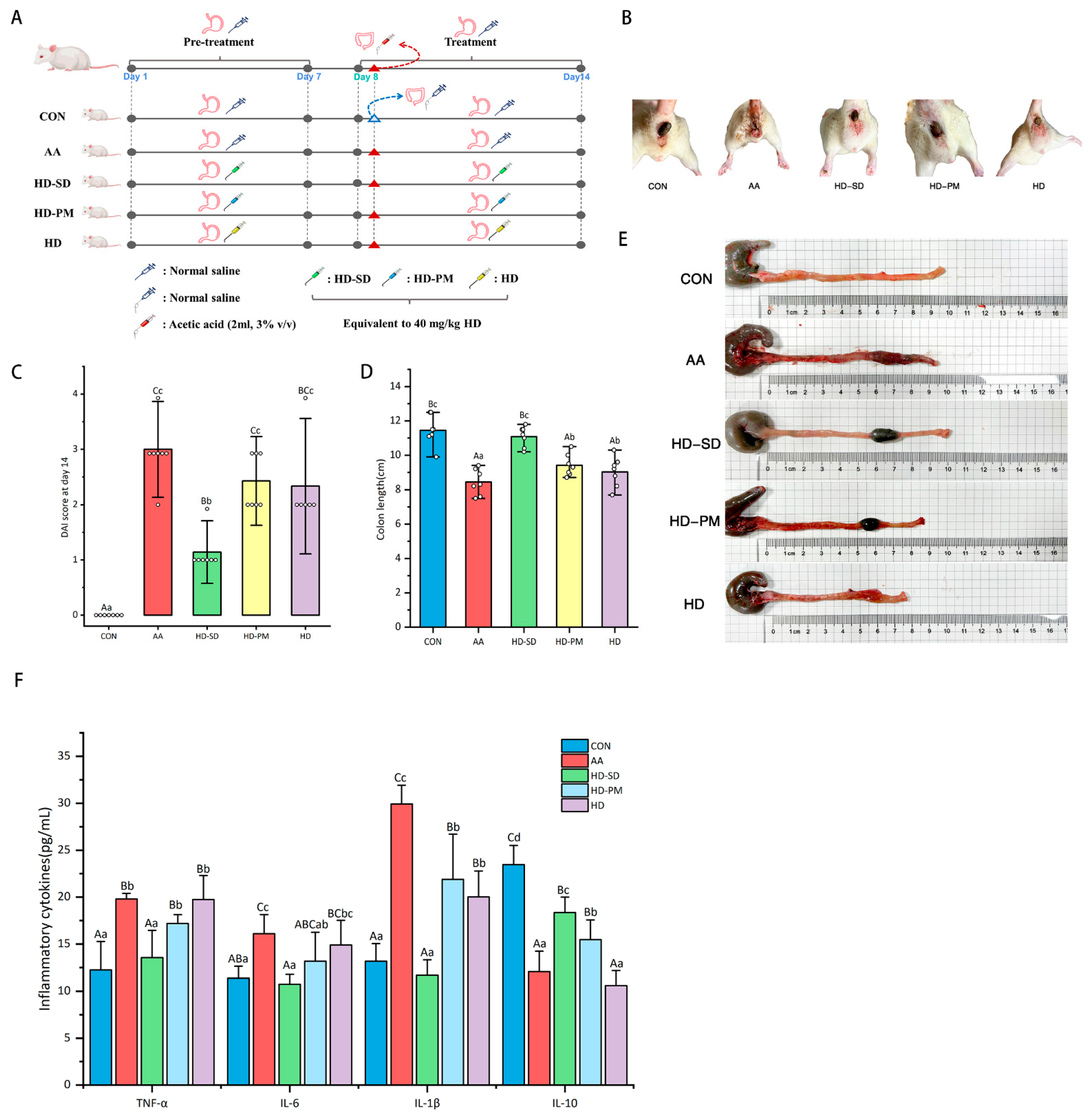
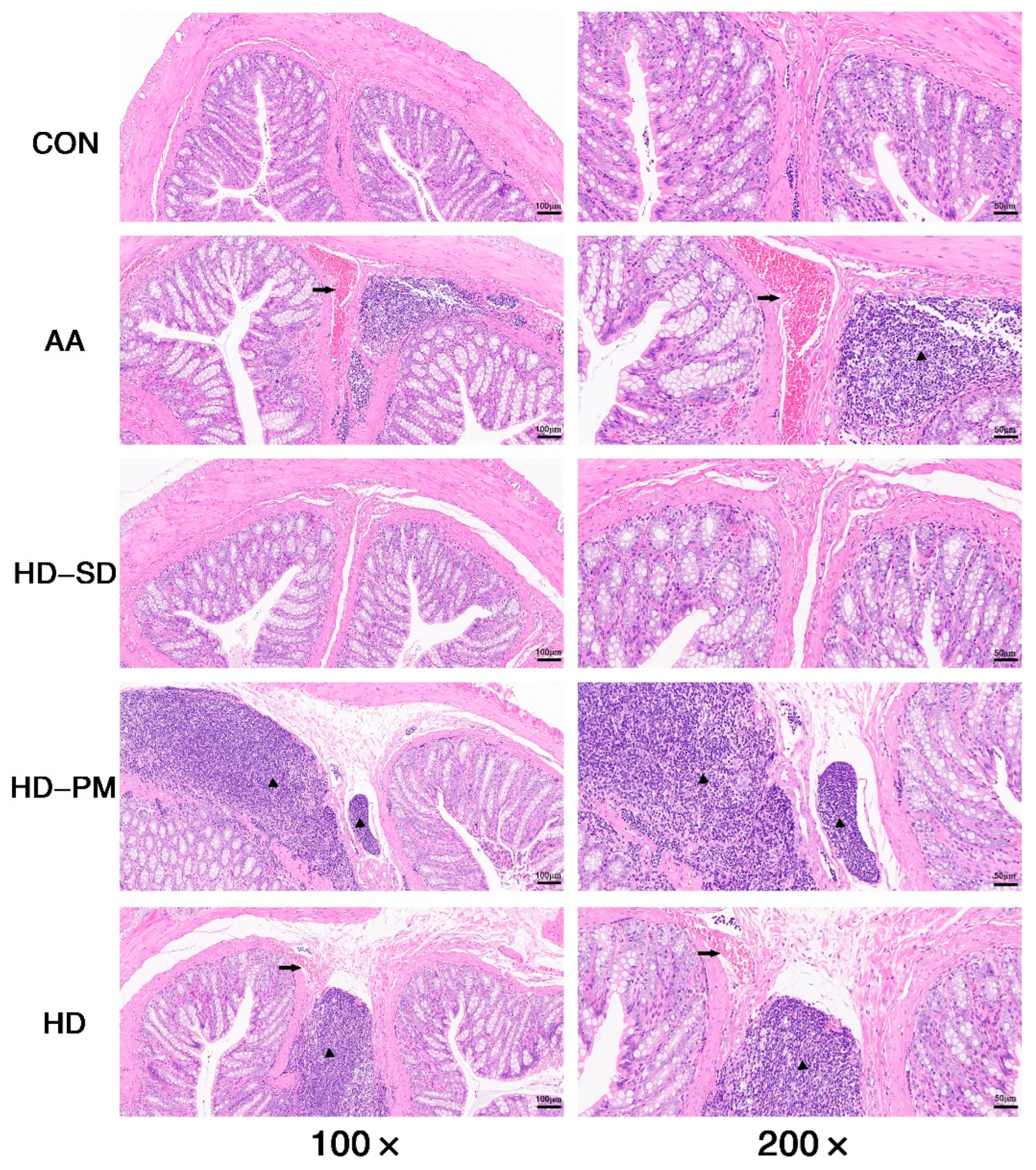
The animal experimental plan is illustrated in Figure 2A. The fecal status of the rats is shown in Figure 2B. The results showed that the AA group had diarrhea which significantly improved after HD treatment. The severity of the AA-induced colitis was evaluated using the DAI score. At the conclusion of the experiment, the highest DAI scores were observed in the AA group, while the HD-SD group exhibited a significant decrease in these scores (p < 0.01). The rats of the HD and HD-PM group exhibited lower DAI scores, and there was no significant difference between these two groups (Figure 2C). The literature reported that AA causes severe inflammation and shortening of the colon [31,32]. As depicted in Figure 2D,E, treatment with HD prevented colon shortening, and the effect of HD-SD was better than PM and HD (p < 0.01). Additionally, the colon in the AA group displayed pronounced dark red swelling and the HD-SD group reversed these effects (Figure 2E). In addition, Figure 3 displays the pathological characteristics of the colon slice as assessed by H&E staining; the colon of the AA group exhibited various inflammatory features such as evident bleeding, extensive infiltration of inflammatory cells, and goblet cell depletion. In contrast, HD-SD, HD-PM, and HD supplementation alleviated AA-induced histopathological alterations of the colon.

3.5.2. Examination of Anti-Inflammatory Effects

The effects of HD-SD on TNF-α, IL-6, IL-1β, and IL-10 levels in the serum after AA administration were evaluated. Compared with the CON group, the levels of TNF-α, IL-6, and IL-1β in the AA group were significantly elevated, while the level of IL-10 was significantly reduced (Figure 2F) (p < 0.01). In contrast, compared to the AA group, the treatment with HD-SD significantly reduced AA-induced TNF-α, IL-6, and IL-1β production, and significantly enhanced the secretion of the anti-inflammatory cytokine IL-10 (Figure 2F) (p < 0.01). Through careful observation, we found that the levels of TNF-α and IL-1β in the HD-SD group were significantly lower than HD-PM and HD groups (p < 0.01). Compared to the HD group, the HD-SD group exhibited significantly lower/higher levels of IL-6/IL-10 (p < 0.01). Although a change of 27.88%/73.28% was observed, this difference was not statistically significant. All these results indicated that HD with the ability of inhibiting pro-inflammatory cytokines and the effects of HD-SD was better than the PM and HD.

3.5.3. HD-SD Administration Modulated Gut Dysbiosis in Colitic Rat

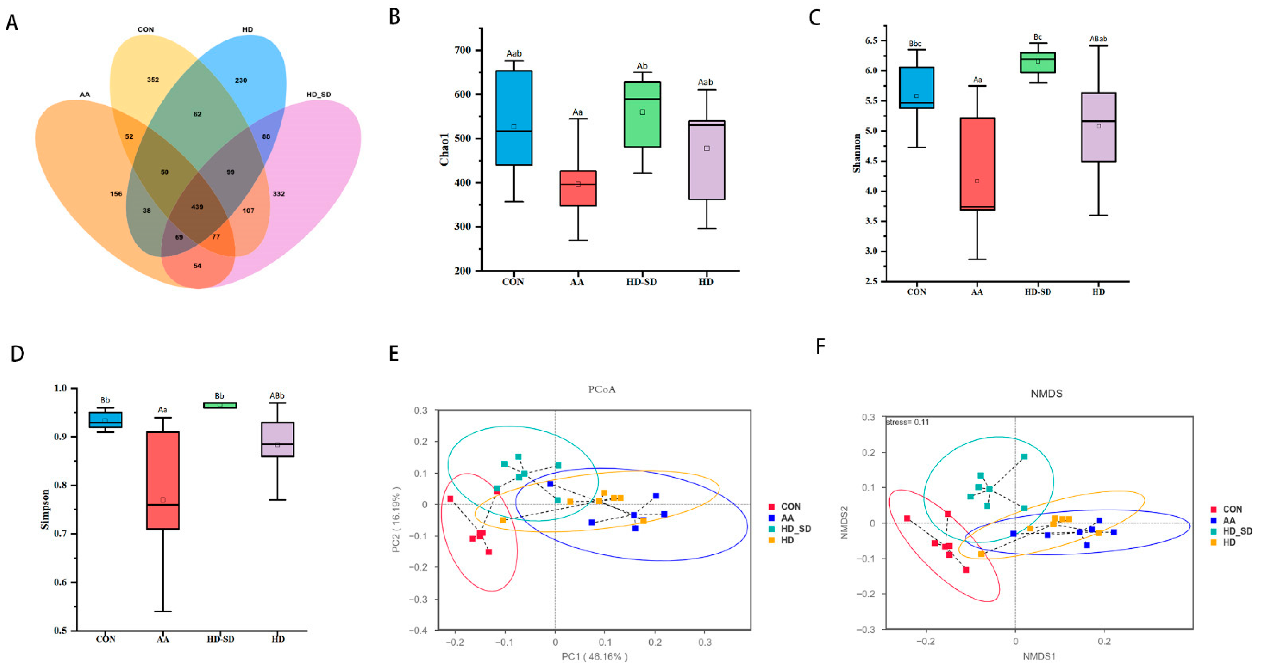
To investigate the effects of HD-SD on the gut microbiota composition of rats with acetic acid (AA)-induced colitis, 16S rDNA gene amplicons in the intestinal contents of the rats were sequenced. As shown in Figure 4A, the Venn diagram indicated that 2203 operational taxonomic units (OTUs) in all were identified across the four groups of fecal samples. Unique OTUs of the CON, AA, HD-SD, and HD groups were 352, 156, 332, and 230, respectively, indicating greater OUT diversity in the HD-SD group.
Compared to the CON group, the Shannon and Simpson indices were significantly decreased (p < 0.01 or p < 0.05) in the AA group (Figure 4B–D), showing that AA decreased the gut microbiota’s richness and diversity. There was no statistically significant difference in the Chao1, Shannon, and Simpson indices between the HD-SD and CON groups, suggesting that HD-SD treatment restored the microbiota that resemble that of the CON group. PCoA and NMDS plots were generated to assess the similarities and differences in microbial communities among groups (Figure 4E,F). The results revealed a clear separation in bacterial community composition between the CON and AA groups. Interestingly, the bacterial community of the HD-SD group clustered more closely to the CON group than the AA group, suggesting that HD-SD alleviated the gut microbiota dysbiosis in rats with colitis.
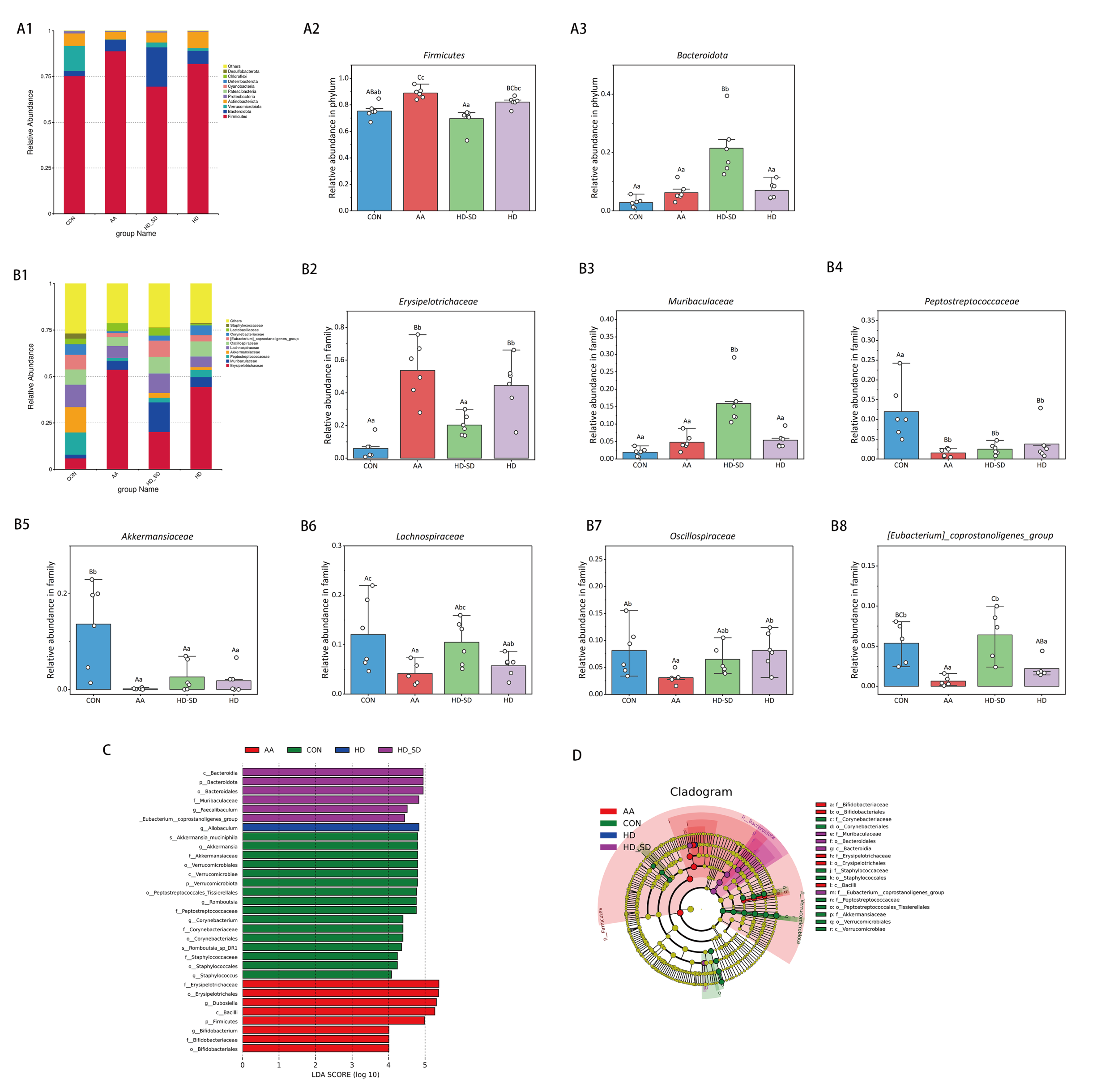
The composition of the gut microbiota at the phylum level in intestinal content samples is presented in Figure 5(A1). The relative abundance of Firmicutes and Verrucomicrobiota was obviously declined in the AA group (p < 0.01), whereas HD-SD supplementation prominently reversed the relative abundance of Firmicutes caused by AA treatment (p < 0.01) (Figure 5(A2)). Moreover, the relative abundance of Bacteroidota was significantly boosted in the HD-SD group (p < 0.01) (Figure 5(A3)).
The relative abundance of gut microbiota at the family level in intestinal contents was illustrated in Figure 5(B1). At the family level, the relative abundance of Erysipelotrichaceae was significantly elevated in the AA group (p < 0.01); these results were consistent with the previous literature [2] (Figure 5(B2)). The administration HD-SD significantly reversed the increase in Erysipelo-trichaceae. Moreover, the relative abundances of Akkermansiaceae and Lachnospiraceae were significantly reduced in the AA group (p < 0.01 or p < 0.05), whereas HD-SD supplementation prominently reversed the relative abundance of Lachnospiraceae caused by AA treatment (p < 0.01) (Figure 5(B5,B6)). AA treatment decreased the relative abundance of Oscillospiraceae and [Eubacterium]_coprostanoligenes_group compared with that of normal saline treatment (p < 0.05 or p < 0.01), whereas HD-SD supplementation prominently reversed the relative abundance of [Eubacterium]_coprostanoligenes_group caused by AA treatment (p < 0.01).
We then used LEfSe analysis to identify statistically significant taxa in each experimental group. Figure 5C,D display the outcomes of the LEfSe analysis. Notably, the levels of Erysipelotrichaceae and Erysipelotrichales were broadened in the AA group. Importantly, Erysipelotrichaceae was found to have a positive correlation with ulcerative colitis (UC) disease [33]. The HD-SD group has nine significantly enriched marker species, including Bacteroidia, Bacteroidota, Bacteroidales, muribaculaceae, and oscillospirates. These bacterial genera are recognized for their capacity to break down complex polysaccharides via fermentation, resulting in the production of short-chain fatty acids [34].

4. Discussion

Currently, UC has become a globally prevalent gastrointestinal disorder closely associated with modern dietary patterns, significantly compromising life quality. While current therapeutic approaches often yield suboptimal outcomes, bioactive compounds derived from functional foods emerge as promising candidates for UC prevention and dietary management. Solid dispersion is a kind of solid form that can satisfactorily improve the solubility and the dissolution rates [21]. There are probably two reasons for this: first, the bioactive compounds were uniformly dispersed in the carriers in an amorphous form; second, no energy was required for the bioactive compounds to break the crystal lattice [28]. Polyvinylpyrrolidone K30 (PVPK30) is a commonly used carrier which is a hydrophilic polymer composed of N-vinylpyrrolidone monomers. The molecular weight of PVP ranges from approximately 2.5 to 3000 kDa, with “K” values ranging from 12 to 120, depending on the length of the polymer chain [35]. PVP K30, a hydrophilic and biocompatible polymer, demonstrates low toxicity and dissolves readily in a range of polar solvents. It is widely utilized in diverse drug delivery applications such as oral, topical, and ocular formulations [36]. Additionally, PVP K30 is widely used in the cosmetics, food, and biomedical fields [21]. The novel curcumin analog C086 solid dispersion (C086-SD) was prepared using PVP K30 as a carrier, which had a cumulative dissolution of 100% within 20 min and an oral bioavailability of 28 times that of C086 [37].
The crystalline state of a drug is crucial for the formation of amorphous solid dispersions. PXRD can detect the crystallinity of drugs [38]. It has been recognized that no crystalline diffraction peaks of HD were observed in the HD-SD 1:9 system, indicating the effective production of an amorphous solid dispersion. DSC can analyze the thermal properties of drugs such as melting point, boiling point, and crystallinity [39]. From the DSC analysis, a significant endothermic peak was observed for HD at 260 °C which was consistent with the literature [22]. A broad endotherm ranging from roughly 50 °C to 100 °C was observed in the thermogram of the PVP, showing water loss due to the extremely hygroscopic properties of PVP polymers [40]. No endothermic peak of HD was observed in the PM, possibly due to a higher proportion of polymer. And with the temperature increased, PVPK30 melted before HD, obscuring HD’s melting peak [28,41]. The absence of an endothermic peak of HD in HD-SD indicates the amorphous state.
The low solubility of HD leads to low oral bioavailability [42]. The dissolution curve showed that the cumulative release of HD-SD (1:9) was higher than that of HD and HD-PM (1:9), indicating that the amorphous state of HD could improve the dissolution performance of HD. The enhancement of release of HD-SD in vivo and in vitro is facilitated by the presence of the hydrophilic PVP carrier, which improves the wetting properties of the amorphous drug. PVPK30 promotes fast release in addition to improving the solubility of poorly soluble medications [43]. Importantly, polymer adsorption onto the surface of crystalline medicines may be encouraged by interactions between the drug and carrier, inhibit crystal growth and disperse them into an amorphous or metastable microcrystalline state within the water-soluble carrier, and thereby enhance the solubility [44]. Additionally, mechanical milling induces changes in particle morphology and distribution, promotes favorable surface effects, size effects, and quantum effects, thereby enhancing drug activity and adsorption properties, and consequently improving the dissolution rate and bioavailability [38]. Furthermore, pharmacokinetic results indicate that the Cmax and AUC0–24 of HD-SD are 2.67 and 1.50 times higher than HD, respectively. This improvement was attributed to the close relationship between drug solubility and bioavailability [45].
The model of colitis in rats was established using the rectal administration of acetic acid, which was reported in the previous literature [46,47]. DAI is a comprehensive assessment method used to measure disease activity, particularly in IBD. The DAI score combines measurements of weight loss, stool consistency, and bleeding, calculating these indicators to assess the severity of the disease. This method plays a crucial role in the diagnosis and monitoring of inflammatory diseases such as IBD [48]. Our results indicated that the AA group had the highest DAI score, while the HD-SD group had the lowest DAI score. Additionally, a significant characteristic of UC is the marked shortening of the colon, which is consistent with our experimental results [49]. As shown in the figure, the AA-treated colons were notably shortened and exhibited congestion, whereas the HD-SD intervention significantly improved these conditions. Histopathological examination is a critical tool in pathology research, allowing for the observation of inflammatory infiltration, ulcer formation, and mucosal hemorrhage in colitis [50]. Histopathological results revealed significant hemorrhage and inflammatory infiltration in the AA group, while the HD-SD intervention ameliorated these symptoms. Furthermore, this model induces the excessive production of pro-inflammatory cytokines such as TNF-α, IL-1β, and IL-6, and is characterized by congestion, ulceration, and a reduction in goblet cells, leading to persistent inflammation [29]. This is consistent with our experimental findings. Overall, every treatment in this study improved colitis; however, the HD-SD intervention showed superior efficacy compared to the other treatment groups. This may be closely related to the water solubility of hesperidin. As evidenced by research, optimized hesperidin magnesium improved the water solubility of pure hesperidin, resulting in better therapeutic effects on UC compared to pure hesperidin [51].
Imbalance in the gut microbiota is closely associated with impaired intestinal barrier function and inflammatory responses. The balanced state of gut microbial communities is crucial for maintaining the stability of the intestinal ecosystem [52]. To investigate the effects of HD-SD on the gut microbiota in rats with colitis, this study analyzed the alpha and beta diversity, as well as the composition of gut communities, based on 16S rDNA high-throughput sequencing. Research indicates that UC patients’ gut microbiota diversity is significantly reduced than that of healthy individuals [34]. The α-diversity and β-diversity analysis indicated that the diversity and compositional structure of intestinal flora in the CON group were remarkably different from those in the AA group, and the CON group showed more similarity with the HD-SD groups. These results showed that HD-SD could recover the variety and composition of the gut microbial community in AA-treated rats. Furthermore, at the phylum level, the relative abundances of Firmicutes were significantly decreased in the AA group, and the relative abundances of Bacteroidota were significantly increased in the HD-SD group. The two most abundant phyla in the intestines of both humans and rats are Bacteroidetes and Firmicutes, relying mainly on carbohydrate-active enzymes for the utilization of dietary polysaccharides [53]. They can be major degraders of complex carbohydrates. At the family level, the relative abundances of Akkermansiaceae and Lacnnospiraceae were significantly reduced in the AA group, which was in line with what was found in earlier research. Lachnospiraceae is consistently decreased in patients with IBD, as has been demonstrated. It has been reported that Lachnospiraceae exhibits strong capability in polysaccharide degradation and production of SCFAs, potentially contributing to the breakdown of ASPP [54]. Lachnospiraceae is a major generator of short-chain fatty acids in the gut, which is essential for intestinal health [55]. Akkermansiaceae, a microbe that plays a positive role in maintaining intestinal barrier homeostasis, can increase the levels of mucin, thereby restoring intestinal barrier function and reducing colonic inflammation [2,6]. Compared to the AA group, HD-SD intervention enriched Lachnospiraceae and Akkermansiaceae. Additionally, an increase in Erysipelotrichaceae is frequently observed in animals with UC [2], in agreement with the results obtained herein. In addition, HD-SD supplementation significantly reversed the decrease in the abundance of [Eubacterium]_coprostanoligenes_group caused by AA treatment. One study found that [Eubacterium]_coprostanoligenes_group can strengthen the integrity of the intestinal mucus barrier by promoting mucin secretion by goblet cells, which helps to resist microbial invasion and subsequently reduce inflammation [56].
The LEfSe results indicated that the marker species included Erysipelotrichaceae and Erysipelotrichales which were significantly enriched in the AA group. Feng et al. showed that the polysaccharides can ameliorate the Erysipelatoclostridium elevation caused by DSS [57], which is consistent with the results of this study. In addition, AA treatment notably decreased the relative abundance of Oscillospiraceae. Oscillospiraceae produces SCFAs, such as butyrate, which play an important role in gut health. Butyrate is not only an energy source, but also has anti-inflammatory properties. However, HD-SD supplementation considerably alleviated the aforementioned alterations generated by the AA treatment. Bacteroidetes, recognized as beneficial gut microbes, play a critical role in preventing intestinal diseases such as inflammation and maintaining gut health [34]. The HD-SD group showed significant enrichment of Bacteroidota, suggested that HD-SD intervention improves the gut microbiota structure. Collectively, the integration of food-derived HD with PVPK30 presents a promising treatment strategy for UC management, exemplifying the translational potential of food-grade delivery systems in preventive health applications.

5. Conclusions

HD-SD were successfully synthesized using PVPK30 as the carrier in this study. Compared with pure HD, HD-SD showed a higher cumulative dissolution rate and higher bioavailability. In addition, HD-SD has advantages in effectively alleviating bloody diarrhea, colonic tissue damage, and colonic inflammation in AA-induced colitis rats. HD-SD was able to correct the dysregulated gut microbiota structure through increasing the relative abundance of beneficial bacteria and decreasing that of harmful bacteria. Therefore, the results suggested that HD-SD has potential as a functional food or as a novel therapeutic strategy for UC.

Author Contributions

Conceptualization, Q.W. (Qiru Wang), X.H. and X.Z.; Data curation, Q.W. (Qiru Wang), X.H. and X.Z.; Funding acquisition, X.H. and X.Z.; Investigation, Q.W. (Qiru Wang), D.L., Q.W. (Qi Wu) and Y.S.; Methodology, Q.W. (Qiru Wang), X.H. and X.Z.; Supervision, X.H. and X.Z.; Writing—original draft, Q.W. (Qiru Wang); Writing–review and editing, Q.W. (Qiru Wang), Y.S., N.M., X.H. and X.Z. All authors have read and agreed to the published version of the manuscript.

Funding

This work was supported by the National Natural Science Foundation of China, (Grant Nos. 32172898, 32473073).

Institutional Review Board Statement

All animal experiments were conducted in accordance with protocols approved by the Animal Ethics Committee of Hebei Agricultural University (Approval No. 2021058). Approval 1 March 2021.

Informed Consent Statement

Not applicable.

Data Availability Statement

The original contributions presented in the study are included in the article. Further inquiries can be directed to the corresponding authors.

Conflicts of Interest

The authors declare that they have no known competing financial interests or personal relationships.

Abbreviations

HD, Hesperidin; PVPK30, polyvinylpyrrolidone K30; SD, solid dispersion; IBD, inflammatory bowel disease; CD, Crohn’s disease; UC, ulcerative colitis; AA, acetic acid; API, active pharmaceutical ingredient; HD-SD, hesperidin solid dispersion; PXRD, Powder X-ray Diffraction; DSC, Differential Scanning Calorimetry; FTIR, Fourier Transform Infrared Spectroscopy; DAI, disease activity index; H&E, hematoxylin and eosin; PCoA, principal coordinate analysis; NMDS, non-metric multi-dimensional scaling.

References

  1. Hong, C.J.; Chen, S.Y.; Hsu, Y.H.; Yen, G.C. Protective effect of fermented okara on the regulation of inflammation, the gut microbiota, and SCFAs production in rats with TNBS-induced colitis. Food Res. Int. 2022, 157, 111390. [Google Scholar] [CrossRef] [PubMed]
  2. Cheng, H.; Zhang, D.; Wu, J.; Liu, J.; Tan, Y.; Feng, W.; Peng, C. Atractylodes macrocephala Koidz. volatile oil relieves acute ulcerative colitis via regulating gut microbiota and gut microbiota metabolism. Front. Immunol. 2023, 14, 1127785. [Google Scholar] [CrossRef] [PubMed]
  3. Zhu, M.Z.; Xu, H.M.; Liang, Y.J.; Xu, J.; Yue, N.N.; Zhang, Y.; Tian, C.M.; Yao, J.; Wang, L.S.; Nie, Y.Q.; et al. Edible exosome-like nanoparticles from Portulaca oleracea L. mitigate DSS-induced colitis via facilitating double-positive CD4+CD8+T cells expansion. J. Nanobiotechnol. 2023, 21, 309. [Google Scholar] [CrossRef]
  4. Szymon, S.; Anna, S.; Andrzej, M.; Marcin, Ż.; Judyta, C. Zein as an Effective Carrier for Hesperidin Delivery Systems with Improved Prebiotic Potential. Molecules 2023, 28, 5209. [Google Scholar] [CrossRef]
  5. Franzosa, E.A.; Sirota-Madi, A.; Avila-Pacheco, J.; Fornelos, N.; Haiser, H.J.; Reinker, S.; Vatanen, T.; Hall, A.B.; Mallick, H.; McIver, L.J.; et al. Gut microbiome structure and metabolic activity in inflammatory bowel disease. Nat. Microbiol. 2019, 4, 293–305. [Google Scholar] [CrossRef]
  6. Wang, H.; Huang, X.; Xia, S.; Chen, C.; Chen, X.; Zhang, Y.; Farag, M.A.; Xiao, J.; Nie, S. Celery soluble dietary fiber antagonize flavonoids ameliorative effect on dextran-sodium-sulfate-induced colitis in mice. J. Adv. Res. 2023, 52, 73–88. [Google Scholar] [CrossRef]
  7. Wan, L.; Qian, C.; Yang, C.; Peng, S.; Dong, G.; Cheng, P.; Zong, G.; Han, H.; Shao, M.; Gong, G.; et al. Ginseng polysaccharides ameliorate ulcerative colitis via regulating gut microbiota and tryptophan metabolism. Int. J. Biol. Macromol. 2024, 265, 130822. [Google Scholar] [CrossRef]
  8. Sarah, M.N.; Peddha, M.S.; Kempaiah, B.B.; Singh, N.P. Efficacy of a functional food ingredient from Ensete superbum Roxb. Cheesman peel in reducing the severity of ulcerative colitis in a murine model. Food Funct. 2022, 13, 3732–3745. [Google Scholar] [CrossRef]
  9. Khalilabad, S.N.; Mirzaei, A.; Askari, V.R.; Mirzaei, A.; Khademi, R.; Rahimi, V.B. How hesperidin and Hesperetin, as promising food Supplements, combat cardiovascular Diseases: A systematic review from bench to bed. J. Funct. Foods 2024, 120, 106358. [Google Scholar] [CrossRef]
  10. Gattuso, G.; Barreca, D.; Gargiulli, C.; Leuzzi, U.; Caristi, C. Flavonoid Composition of Citrus Juices. Molecules 2007, 12, 1641. [Google Scholar] [CrossRef]
  11. Bhattacharyya, S.; Pal, S.; Mohamed, R.; Singh, P.; Chattopadhyay, S.; China, S.P.; Porwal, K.; Sanyal, S.; Gayen, J.R.; Chattopadhyay, N. A nutraceutical composition containing diosmin and hesperidin has osteogenic and anti-resorptive effects and expands the anabolic window of teriparatide. Biomed. Pharmacother. 2019, 118, 109207. [Google Scholar] [CrossRef]
  12. Assini, J.M.; Mulvihill, E.E.; Huff, M.W. Citrus flavonoids and lipid metabolism. Curr. Opin. Lipidol. 2013, 24, 34–40. [Google Scholar] [CrossRef]
  13. SungSook, C.; SunHyung, L.; KyungAe, L. A Comparative Study of Hesperetin, Hesperidin and Hesperidin Glucoside: Antioxidant, Anti-Inflammatory, and Antibacterial Activities In Vitro. Antioxidants 2022, 11, 1618. [Google Scholar] [CrossRef]
  14. Zhang, P.; Zhang, S.; Hu, H.; Hu, T.; Shi, K.; Xu, Y.; Xu, G.; Hu, H.; Pan, S. Synthesis and evaluation of novel hesperidin selenium- enriched derivatives as potential anti-inflammatory and antioxidant agents. Food Biosci. 2024, 58, 103651. [Google Scholar] [CrossRef]
  15. He, D.; Gao, X.; Wen, J.; Zhang, Y.; Yang, S.; Sun, X.; Cui, M.; Li, Z.; Fu, S.; Liu, J.; et al. Orally administered neohesperidin attenuates MPTP-induced neurodegeneration by inhibiting inflammatory responses and regulating intestinal flora in mice. Food Funct. 2024, 15, 1460–1475. [Google Scholar] [CrossRef]
  16. Li, X.; Yao, Y.; Wang, Y.; Hua, L.; Wu, M.; Chen, F.; Deng, Z.-Y.; Luo, T. Effect of Hesperidin Supplementation on Liver Metabolomics and Gut Microbiota in a High-Fat Diet-Induced NAFLD Mice Model. J. Agric. Food Chem. 2022, 70, 11224–11235. [Google Scholar] [CrossRef] [PubMed]
  17. Radha, G.; Raghunandhakumar, S.; Balakumar, S. Dual therapeutic 5-fluorouracil and hesperidin loaded chitosan nanocarrier system: Understanding its synergism on anti-cancer activity. J. Drug Deliv. Sci. Technol. 2023, 80, 104184. [Google Scholar] [CrossRef]
  18. Guo, K.; Ren, J.; Gu, G.; Wang, G.; Gong, W.; Wu, X.; Ren, H.; Hong, Z.; Li, J. Hesperidin Protects Against Intestinal Inflammation by Restoring Intestinal Barrier Function and Up-Regulating Treg Cells. Mol. Nutr. Food Res. 2020, 64, e1970058. [Google Scholar] [CrossRef]
  19. Estruel-Amades, S.; Massot-Cladera, M.; Pérez-Cano, F.J.; Franch, À.; Castell, M.; Camps-Bossacoma, M. Hesperidin Effects on Gut Microbiota and Gut-Associated Lymphoid Tissue in Healthy Rats. Nutrients 2019, 11, 324. [Google Scholar] [CrossRef]
  20. Zhang, X.; Liu, Z.; Huang, Y.; Niu, Y.; Zhang, L.; Xiong, T.; Zhang, Y.; Zhang, R.; Zhang, H. Preparation and in vitro evaluation of hesperidin nanoparticles by antisolvent recrystallization in a double homogenate system. Food Chem. X 2023, 18, 100639. [Google Scholar] [CrossRef]
  21. Natalia, R.; Kamil, W.; Ewa, T.; Judyta, C. Amorphous Solid Dispersion of Hesperidin with Polymer Excipients for Enhanced Apparent Solubility as a More Effective Approach to the Treatment of Civilization Diseases. Int. J. Mol. Sci. 2022, 23, 15198. [Google Scholar] [CrossRef]
  22. Rajendra, J.; Osman, E.G.; Sulekha, K.; Deependra, S.; Manju, S.; Kumar, S.R.; Jiyauddin, K. Hesperidin-Loaded Lipid Polymer Hybrid Nanoparticles for Topical Delivery of Bioactive Drugs. Pharmaceuticals 2022, 15, 211. [Google Scholar] [CrossRef]
  23. Long, J.; Song, J.; Zhang, X.; Deng, M.; Xie, L.; Zhang, L.; Li, X. Tea saponins as natural stabilizers for the production of Hesperidin nanosuspensions. Int. J. Pharm. 2020, 583, 119406. [Google Scholar] [CrossRef] [PubMed]
  24. Kamil, W.; Natalia, R.; Ewa, T.; Marcin, Ż.; Anita, P.; Wojciech, P.; Judyta, C. Amorphous Inclusion Complexes: Molecular Interactions of Hesperidin and Hesperetin with HP-Β-CD and Their Biological Effects. Int. J. Mol. Sci. 2022, 23, 4000. [Google Scholar]
  25. Li, Y.; Xu, J.; Guan, Q.; Zhang, H.; Ding, Z.; Wang, Q.; Wang, Z.; Han, J.; Liu, M.; Zhao, Y. Impact of hypromellose acetate succinate and Soluplus® on the performance of β-carotene solid dispersions with the aid of sorbitan monolaurate: In vitro-in vivo comparative assessment. Int. J. Biol. Macromol. 2023, 253, 126639. [Google Scholar] [CrossRef] [PubMed]
  26. Qiu, C.; Zhang, Y.; Fan, Y.; Li, S.; Gao, J.; He, X.; Zhao, X. Solid Dispersions of Genistein via Solvent Rotary Evaporation for Improving Solubility, Bioavailability, and Amelioration Effect in HFD-Induced Obesity Mice. Pharmaceutics 2024, 16, 306. [Google Scholar] [CrossRef]
  27. Han, J.; Tong, M.; Li, S.; Yu, X.; Hu, Z.; Zhang, Q.; Xu, R.; Wang, J. Surfactant-free amorphous solid dispersion with high dissolution for bioavailability enhancement of hydrophobic drugs: A case of quercetin. Drug Dev. Ind. Pharm. 2021, 47, 153–162. [Google Scholar] [CrossRef]
  28. Gao, J.; Fan, Y.; Lu, C.; Zhao, X.; He, X. The baicalein amorphous solid dispersion to enhance the dissolution and bioavailability and effects on growth performance, meat quality, antioxidant capacity and intestinal flora in Taihang chickens. Poult. Sci. 2024, 103, 103768. [Google Scholar] [CrossRef]
  29. Mahdy, R.N.E.; Nader, M.A.; Helal, M.G.; Abu-Risha, S.E.; Abdelmageed, M.E. Eicosapentaenoic acid mitigates ulcerative colitis-induced by acetic acid through modulation of NF-κB and TGF-β/EGFR signaling pathways. Life Sci. 2023, 327, 121820. [Google Scholar] [CrossRef]
  30. Tinnirello, V.; Zizzo, M.G.; Conigliaro, A.; Tabone, M.; Ganji, N.R.; Cicio, A.; Bressa, C.; Larrosa, M.; Rappa, F.; Vergilio, G.; et al. Industrial-produced lemon nanovesicles ameliorate experimental colitis-associated damages in rats via the activation of anti-inflammatory and antioxidant responses and microbiota modification. Biomed. Pharmacother. 2024, 174, 116514. [Google Scholar] [CrossRef]
  31. Belém, M.O.; de Andrade, G.M.M.; Carlos, T.M.; Guazelli, C.F.S.; Fattori, V.; Toginho Filho, D.O.; Dias, I.F.L.; Verri, W.A., Jr.; Araújo, E.J.A. Light-emitting diodes at 940nm attenuate colitis-induced inflammatory process in mice. J. Photochem. Photobiol. B Biol. 2016, 162, 367–373. [Google Scholar] [CrossRef]
  32. Guazelli, C.F.; Fattori, V.; Colombo, B.B.; Georgetti, S.R.; Vicentini, F.T.; Casagrande, R.; Baracat, M.M.; Verri, W.A., Jr. Quercetin-loaded microcapsules ameliorate experimental colitis in mice by anti-inflammatory and antioxidant mechanisms. J. Nat. Prod. 2013, 76, 200–208. [Google Scholar] [CrossRef] [PubMed]
  33. Labbé, A.; Ganopolsky, J.G.; Martoni, C.J.; Prakash, S.; Jones, M.L. Bacterial bile metabolising gene abundance in Crohn’s, ulcerative colitis and type 2 diabetes metagenomes. PLoS ONE 2014, 9, e115175. [Google Scholar] [CrossRef]
  34. Ouyang, Q.; Li, X.; Liang, Y.; Liu, R. Sea Buckthorn Polysaccharide Ameliorates Colitis. Nutrients 2024, 16, 1280. [Google Scholar] [CrossRef]
  35. Zhang, J.; Guo, M.; Luo, M.; Cai, T. Advances in the development of amorphous solid dispersions: The role of polymeric carriers. Asian J. Pharm. Sci. 2023, 18, 100834. [Google Scholar] [CrossRef]
  36. Franco, P.; De Marco, I. The Use of Poly(N-vinyl pyrrolidone) in the Delivery of Drugs: A Review. Polymers 2020, 12, 1114. [Google Scholar] [CrossRef]
  37. Deng, Y.; Chen, C.; Xiao, Z.; Huang, X.; Xu, J. Enhanced anti-hepatoma effect of a novel curcumin analog C086 via solid dispersion technology. Drug Deliv. 2020, 27, 927–937. [Google Scholar] [CrossRef] [PubMed]
  38. Li, Z.-R.; Jia, R.-B.; Cai, X.; Luo, D.; Chen, C.; Zhao, M. Characterizations of food-derived ellagic acid-Undaria pinnatifida polysaccharides solid dispersion and its benefits on solubility, dispersity and biotransformation of ellagic acid. Food Chem. X 2023, 413, 135530. [Google Scholar] [CrossRef]
  39. Shahraki, O.; Shayganpour, M.; Hashemzaei, M.; Daneshmand, S. Solid lipid nanoparticles (SLNs), the potential novel vehicle for enhanced in vivo efficacy of hesperidin as an anti-inflammatory agent. Bioorg. Chem. 2023, 131, 106333. [Google Scholar] [CrossRef] [PubMed]
  40. Zhang, Y.; Liu, F.; Cao, Y.; Xu, H.; Xie, Y.; Xiao, X.; Addo, K.A.; Peng, X.-F. Preparation and characterization of a solid dispersion of Hexahydrocolupulone and its application in the preservation of fresh apple juice. Food Chem. X 2023, 424, 136367. [Google Scholar] [CrossRef]
  41. Nandi, U.; Ajiboye, A.L.; Patel, P.; Douroumis, D.; Trivedi, V. Preparation of solid dispersions of simvastatin and soluplus using a single-step organic solvent-free supercritical fluid process for the drug solubility and dissolution rate enhancement. Pharmaceuticals 2021, 14, 846. [Google Scholar] [CrossRef] [PubMed]
  42. Suthar, T.; Patel, P.; Singh, P.; Datusalia, A.K.; Yadav, A.K.; Jain, K. Hesperidin microemulsion: Formulation optimization, characterization, and in vitro evaluation. J. Drug Deliv. Sci. Technol. 2023, 80, 130–149. [Google Scholar] [CrossRef]
  43. Van Duong, T.; Van den Mooter, G. The role of the carrier in the formulation of pharmaceutical solid dispersions. Part II: Amorphous carriers. Expert Opin. Drug Deliv. 2016, 13, 1681–1694. [Google Scholar] [CrossRef]
  44. Shi, Q.; Chen, H.; Wang, Y.; Wang, R.; Xu, J.; Zhang, C. Amorphous Solid Dispersions: Role of the Polymer and Its Importance in Physical Stability and In Vitro Performance. Pharmaceutics 2022, 14, 1747. [Google Scholar] [CrossRef]
  45. Liu, Y.; Liang, Y.; Yuhong, J.; Xin, P.; Han, J.L.; Du, Y.; Yu, X.; Zhu, R.; Zhang, M.; Chen, W.; et al. Advances in Nanotechnology for Enhancing the Solubility and Bioavailability of Poorly Soluble Drugs. Drug Des. Dev. Ther. 2024, 18, 1469–1495. [Google Scholar] [CrossRef]
  46. Mahdy, R.N.E.; Nader, M.A.; Helal, M.G.; Risha, S.E.A.; Abdelmageed, M.E. Tiron ameliorates acetic acid-induced colitis in rats: Role of TGF-β/EGFR/PI3K/NF-κB signaling pathway. Int. Immunopharmacol. 2024, 128, 111587. [Google Scholar] [CrossRef] [PubMed]
  47. Niraj, P.; Kumar, B.L. Topotecan alleviates acetic acid-induced ulcerative colitis in rats via attenuation of the RORγT transcription factor. Life Sci. 2023, 328, 121915. [Google Scholar]
  48. Ge, H.; Cai, Z.; Chai, J.; Liu, J.; Liu, B.; Yu, Y.; Liu, J.; Zhang, T. Egg white peptides ameliorate dextran sulfate sodium-induced acute colitis symptoms by inhibiting the production of pro-inflammatory cytokines and modulation of gut microbiota composition. Food Chem. 2021, 360, 129981. [Google Scholar] [CrossRef]
  49. Jiang, P.; Zhang, Y.; Li, X.; Chen, J. Geniposidic acid attenuates DSS-induced colitis through inhibiting inflammation and regulating gut microbiota. Phytother. Res. PTR 2023, 37, 3453–3466. [Google Scholar] [CrossRef]
  50. Liu, J.; Cai, J.; Fan, P.; Dong, X.; Zhang, N.; Tai, J.; Cao, Y. Salidroside alleviates dextran sulfate sodium-induced colitis in mice by modulating the gut microbiota. Food Funct. 2023, 14, 7506–7519. [Google Scholar] [CrossRef]
  51. Zhang, L.; Miao, C.; Wang, Z.; Guan, X.; Ma, Y.; Song, J.; Shen, S.; Song, H.; Li, M.; Liu, C. Preparation and characterisation of baicalin magnesium and its protective effect in ulcerative colitis via gut microbiota-bile acid axis modulation. Phytomedicine 2024, 126, 155416. [Google Scholar] [CrossRef]
  52. Sun, B.; Wang, Y.; Bai, J.; Li, X.; Ma, L.; Man, S. Litchi Procyanidins Ameliorate DSS-Induced Colitis through Gut Microbiota-Dependent Regulation of Treg/Th17 Balance. J. Agric. Food Chem. 2024, 72, 24823–24832. [Google Scholar] [CrossRef]
  53. El Kaoutari, A.; Armougom, F.; Gordon, J.I.; Raoult, D.; Henrissat, B. The abundance and variety of carbohydrate-active enzymes in the human gut microbiota. Nat. Rev. Microbiol. 2013, 11, 497–504. [Google Scholar] [CrossRef]
  54. Suna, J.; Chen, H.; Kan, J.; Gou, Y.; Liu, J.; Zhang, X.; Wu, X.; Tang, S.; Sun, R.; Qian, C.; et al. Anti-inflammatory properties and gut microbiota modulation of an alkali-soluble polysaccharide from purple sweet potato in DSS-induced colitis mice. Int. J. Biol. Macromol. 2020, 153, 708–722. [Google Scholar] [CrossRef] [PubMed]
  55. Yang, Y.H.; Chen, C.; Zheng, Y.; Wu, Z.J.; Zhou, M.Q.; Liu, X.Y.; Miyashita, K.; Duan, D.L.; Du, L. Fucoxanthin Alleviates Dextran Sulfate Sodium-Induced Colitis and Gut Microbiota Dysbiosis in Mice. J. Agric. Food Chem. 2024, 72, 4142–4154. [Google Scholar] [CrossRef] [PubMed]
  56. Bai, D.; Zhao, J.; Wang, R.; Du, J.; Zhou, C.; Gu, C.; Wang, Y.; Zhang, L.; Zhao, Y.; Lu, N. Eubacterium coprostanoligenes alleviates chemotherapy-induced intestinal mucositis by enhancing intestinal mucus barrier. Acta Pharm. Sin. B 2024, 14, 1677–1692. [Google Scholar] [CrossRef]
  57. Feng, W.; Liu, J.; Tan, Y.; Ao, H.; Wang, J.; Peng, C. Polysaccharides from Atractylodes macrocephala Koidz. Ameliorate ulcerative colitis via extensive modification of gut microbiota and host metabolism. Food Res. Int. 2020, 138, 109777. [Google Scholar] [CrossRef] [PubMed]
Figure 1. PXRD, DSC, FT-IR, stability and in vivo/in vitro release analysis of HD-SD. (A) Chemical structure of HD. (B) Chemical structure of PVPK30. (C) PXRD patterns of different samples. (D) DSC of different samples. (E) FT-IR spectra of different samples. (F) Dissolution diagram in vitro. (G) The PXRD patterns of HD-SD (1:9) at different times. (H) Blood concentration–time curves of HD and HD-SD (n = 6).
Foods 14 03252 g001
Figure 2. HD-SD attenuated symptoms of AA-induced colitis in rats. (A) Diagram of flow for the design of the animal experiment. (B) Diarrhea situation of rats. (C) Disease activity index (DAI) score of rats on day 14. (D) Colon length of the rats. (E) Representative images of the colon from rats. (F) Changes in the levels of inflammatory cytokines in the serum. Data are presented as mean ± standard deviation (SD) (n = 6–7 rats per group). Different lowercase letters indicate significant differences (p < 0.05), and different capital letters indicate extremely significant differences (p < 0.01). The same letters indicate no significant difference (p > 0.05).
Foods 14 03252 g002
Figure 3. Representative H&E staining of colon tissue sections. The black arrow shows inflammation, and the black triangle shows bleeding.
Foods 14 03252 g003
Figure 4. HD-SD improved gut microbiota diversity in AA-treated rats. (A) Venn diagram of OTUs. (B) Chao1 index. (C) Shannon index. (D) Simpson index. (E) PCoA plot. (F) NMDS plot. Data are presented as mean ± SD (n = 6 rats per group). Different lowercase letters indicate significant differences (p < 0.05), and different capital letters indicate extremely significant differences (p < 0.01). The same letters indicate no significant difference (p > 0.05).
Foods 14 03252 g004
Figure 5. Effect of HD-SD on the gut microbiota compositions and relative abundances. (A1) The top 10 microbiota taxa found at the phylum level. (A2) Firmicutes. (A3) Bacteroidota. (B1) The top 10 microbiota taxa found at the family level. (B2) Erysipelotrichaceae. (B3) Muribaculaceae. (B4) Peptostreptococcaceae. (B5) Akkermansiaceae. (B6) Lachnospiraceae. (B7) Oscillospiraceae. (B8) [Eubacterium]_coprostanoligenes_group. (C) The distribution histogram based on LDA (LDA > 4.0). (D) Cladogram of LEfSe. Data are presented as mean ± SD (n = 5–6 rats per group). The same letters indicate no significant difference (p > 0.05), while different lowercase letters indicate significant differences (p < 0.05). Different capital letters indicate extremely significant differences (p < 0.01).
Foods 14 03252 g005
Table 1. Pharmacokinetic parameters of HD and HD-SD (1:9) (n = 6).
ItemsGroup
HDHD-SD
Tmax(0–24) (h)1.15 ± 0.340.96 ± 0.10
Cmax(0–24) (μg/mL)0.61 ± 0.141.63 ± 0.17 **
t1/2(0–24) (h)5.35 ±1.694.30 ± 1.03
AUC0–24 (μg·h/mL)4.05 ± 0.556.06 ±0.81 **
Results are presented as “mean ± standard deviation.” Compared with HD, ** p < 0.01 indicates a significant difference.
Disclaimer/Publisher’s Note: The statements, opinions and data contained in all publications are solely those of the individual author(s) and contributor(s) and not of MDPI and/or the editor(s). MDPI and/or the editor(s) disclaim responsibility for any injury to people or property resulting from any ideas, methods, instructions or products referred to in the content.