Article

The Natural Fermentation of Greek Tsounati Olives: Microbiome Analysis

by
Marina Georgalaki
1,*,†,
Ilario Ferrocino
2,
Davide Buzzanca
2,
Rania Anastasiou
1,†,
Georgia Zoumpopoulou
1,†,
Despoina Giabasakou
1,
Danai Ziova
1,
Alexandra Kokkali
1,
George Paraskevakos
3 and
Effie Tsakalidou
1
1
Laboratory of Dairy Research, Department of Food Science and Human Nutrition, Agricultural University of Athens, Iera Odos 75, 11855 Athens, Greece
2
Department of Agricultural, Forest, and Food Science, University of Turin, Largo Paolo Braccini 2, Grugliasco, 10095 Torino, Italy
3
International Probiotics Association, Los Angeles, CA 90035, USA
*
Author to whom correspondence should be addressed.
These authors contributed equally to this work.
Foods 2025, 14(15), 2568; https://doi.org/10.3390/foods14152568
Submission received: 20 June 2025 / Revised: 14 July 2025 / Accepted: 18 July 2025 / Published: 22 July 2025
(This article belongs to the Section Food Microbiology)

Abstract

The comprehensive analysis of microbial communities reveals the unique microbial identity of different olive varieties, paving the way for new strategies in their development and commercial exploitation. In this context, the present study aimed to explore the microbial diversity and functional characteristics of Tsounati variety olives from the Monemvasia region of Peloponnese, Greece, that were naturally fermented for three months. The bacterial and fungal microbiota of both olives and brines were fingerprinted throughout the fermentation through classical microbiological analysis combined with molecular techniques. Among the 148 isolated bacteria, 85 were lactic acid bacteria (LAB), and 63 belonged to the Enterobacteriaceae family, while the 178 fungal isolates comprised 136 yeasts and 42 non-yeast or yeast-like fungi. Metataxonomic analysis confirmed the dominance of the bacterial genera Lactiplantibacillus, Leuconostoc, along with the Enterobacteriaceae family, and it revealed the presence of Coleofasciculaceae cyanobacteria mostly in olives. The dominant fungal genera were yeasts, namely Saccharomyces, Nakazawaea, and Cyberlindnera. Using the Folin–Ciocalteu assay, the average total polyphenol content of Tsounati fermented olive samples was 761.80 ± 128.87 mg gallic acid equivalents kg−1 after 90 days of fermentation. The concentrations of the triterpenic, maslinic, and oleanolic acids, as determined by HPLC, remained stable throughout fermentation, with average values of 4764 and 1807 mg kg−1, respectively. Finally, sensory analysis revealed the rich aromatic character of Tsounati variety, highlighting its potential to be used for Greek-style table olive production.

1. Introduction

In the kingdom of Plantae, an outstanding family called Oleaceae, also known as the olive and lilac family, comprises 28 genera, one of which is extinct, and an estimated number of 700 species [1]. Notable members include ash trees, jasmine, and several popular ornamental plants, such as privet, forsythia, fringe trees, and lilac [2]. The genus Olea includes 12 species, with the most important one being, by far, the type species of the genus, namely olive (Olea europaea L.) [3]. This species is the only one that produces edible fruits, and it originates from the Middle East, where it was cultivated by 5000 BC, later spread to all countries bordering the Mediterranean Sea, and eventually reached America, Africa, Australia, New Zealand, and China [4,5]. To delve into the past, ancient DNA, trapped in the matrices of ceramic transport jars from Mediterranean shipwrecks, was sequenced, revealing the presence of olive fruits among other plants [6], while next-generation sequencing (NGS) technologies and bioinformatics have been applied to study the singular evolution of the Olea genome structure [7].
The number of olive varieties reported by The World Catalogue of Olive Varieties features 139 varieties from 23 olive-growing countries [8]. However, the cataloguing of the existing olive cultivars in the world has been a permanent challenge. To conserve the genetic heritage of the olive tree evolved over centuries through farmers’ selection, the International Olive Council (IOC) network of germplasm banks conserves cultivated varieties from around the world and currently holds more than 1700 accessions [9]. Nevertheless, different parameters determine their use (production of olive oil, table olives, or both), and only a few of them can be considered commercial [10], while highlighting the probable existence of unknown olive cultivars different from those characterized so far [11].
In Greece, according to the register of cultivated fruit tree varieties [12], a total of 64, namely 33 Greek and 31 foreign ones, are officially destined for table olive production. The total number of Greek cultivars exceeds 40 [9,13]. However, only nine Greek olive varieties, namely Adramitini, Amigdalolia, Halkidikis, Kalamon (or Kalamata), Konservolia (or Conservolea), Koroneiki, Mastoidis, Megaritiki, and Valanolia, are included in The World Catalogue of Olive Varieties, which has been compiled under the guidance of IOC [8,14].
Although the most significant Greek olive varieties are Kalamon, Konservolia, and Halkidikis, there are others with outstanding organoleptic characteristics. The Mastoidis variety (Olea europaea var. mamilaris), which is officially reported as Tsounati in The National Catalogue of Olive Varieties but is also called “Athinaiki” or “Athinolia”, is one of the most important varieties for the economy of the Peloponnese region. It is cultivated in South Laconia as well as in Argolida and Western Crete [15,16,17]. The fruit weight is 137–141 g per 100 olive fruits, while its oil concentration of 507.8–517.3 g kg−1 dry mass makes it ideal for olive oil production [18].
According to the IOC [19] for the crop year 2022–2023, Greece ranked second in table olives exports with 188,861 tons, valued at approximately EUR 739.5 million, trailing behind Spain with 267,142 tons valued at EUR 1005.2 million. These data highlight the significance of table olives in the Greek economy, establishing Greece as a key player in global olive exports. Moreover, table olives are an undoubtable nutritional treasure of high energy content (180–250 kcal/100 g), unsaturated fatty acids, proteins containing essential amino acids, and functional phytochemicals, such as phenolic compounds and triterpenic acids [20,21]. Multiple health benefits have been associated with their consumption with respect to cardiovascular health, protection from oxidative damage, and anti-inflammatory activity [21]. Moreover, multiple biological effects of triterpenic acids both in vitro and in vivo have been reported [22,23].
In Greece, the most used practice for table olive production is the Greek-style natural fermentation of mainly black olives immersed in brine without any debittering treatment [10]. Due to brine’s high salt concentration (6–10%), the spontaneous fermentation process is mainly driven by yeasts and also by lactic acid bacteria (LAB), as well as Gram-negative bacteria that dominate mostly the first phase of the fermentation.
The complex and amazing biosystem of Greek-style table olives has been studied not only by culture-dependent approaches but by metataxonomics as well. There are numerous references concerning the microbiome of natural green olives, belonging to diverse varieties, such as Nocellare etnea [24,25], Bella di Cerignola [26], Aloreña de Málaga [27,28], Halkidikis [29], Picual [30], and Manzanilla [31]. Fewer studies on fermented natural black olives, belonging to Konservolia [29], Nyons [32], and Kalamata [30,33] varieties, have been also reported. Additionally, the bacterial diversity of commercial table olive biofilms belonging to various green and black olive varieties has been explored by metataxonomic and compositional data analysis [34].
Tsounati is a Greek olive variety that, so far, is exclusively used for the production of high-quality olive oil. The objective of the present study was to address the challenge of determining, for the first time, whether Tsounati-variety olives can also be utilized for the production of high-quality table olives. The fermentation process was monitored through both physicochemical and microbiological analyses, while the final product was evaluated from a sensory perspective.

2. Materials and Methods

2.1. Olives Fermentation

Olives of the Tsounati variety were collected from the Monemvasia region of Peloponnese, Greece, in November of the 2022–2023 harvest period (at the green stage of maturation). Drupes were transferred to the laboratory and directly washed with tap water and carved with a knife (split) by scoring in three places lengthwise without reaching the core, to facilitate the removal of their bitter taste. Three technical replicates of Greek-style natural fermentation (Fermentations A, B, and C) were performed. Olives (3 kg per fermentation) were placed in three different plastic containers of 5.0 L volume, and these were filled with brine (2 L, 6% w/v NaCl). Fermentation took place for 90 days at room temperature (~25 °C). Olive and brine samples were collected at various time points (0, 4, 7, 15, 30, 60, and 90 days) and stored at −80 °C until further analyses. Classical microbiological analysis was performed on the day of sampling. The sample coding of olives and brines is presented in Table S1.

2.2. Physicochemical Analysis of Olives and Brines

2.2.1. pH and Salt Content

The pH of brine samples was measured using a pH meter (827 pH lab, Metrohm, Herisau, Switzerland) throughout the fermentation period. Salt concentration (NaCl, % w/v) of the brine samples was measured every seven days using a Beaumé hydrometer (Alla, Chemillé en Anjou, France) and expressed as Beaumé degrees. During the fermentation process, brine salt concentration was adjusted by the periodic addition of salt to 6% (w/v), which represents the minimum NaCl content of naturally fermented olives [35].

2.2.2. HPLC Analysis of Sugars, Organic Acids, Alcohols, and Triterpenic Acids

In brine samples, sugars, organic acids, and ethanol were determined by HPLC analysis (GBC Scientific Equipment Pty Ltd., Dandenong, Victoria, Australia) on an HPX-87H Aminex column (BioRad, Hercules, CA, USA) according to Angelopoulou et al. [36]. Triterpenic acids were determined in olive and brine samples by HPLC analysis using the same equipment on a Spherisorb ODS-2 column (Waters Inc., Mildford, MA, USA) as described by Alexandraki et al. [20]. The extraction of triterpenic acids from the flesh of olive samples as well as from brine samples was performed as described by Romero et al. [37], with slight modifications in the case of olive samples [20]. Pure maslinic and oleanolic acids (Sigma, St. Louis, MO, USA) in a concentration range of 0–3000 mg L−1 were used as standards for standard curve plots and quantification. Each sample was measured in duplicate, and the results were expressed as mM mL−1 brine.

2.2.3. Total Phenolic Content

The Folin–Ciocalteu assay [38] was performed to measure TPC in both olive and brine samples.
Regarding brine samples, the method of Marsilio et al. [39] was followed using 0.1 mL of brine sample. More specifically, 1.0 mL of Folin–Ciocalteu reagent (Carlo Erba Reagents, Val de Reuil Cedex, France) was added to each brine sample. After vortexing, samples remained still for 5 min, and 3.0 mL of 20% w/v Na2CO3 solution (Carlo Erba Reagents) was added. The volume of the samples was adjusted to 10 mL with deionized water, and samples were left undisturbed for 30 min.
The same procedure was performed for the olive samples, which were previously prepared as described by Romero et al. [37], with slight modifications [20]. Samples were finally centrifuged at 3500× g for 2 min, and supernatants were transferred to new tubes. Blanks were prepared by mixing 1.0 mL of Folin–Ciocalteu reagent, 3.0 mL of 20% w/v Na2CO3 solution, and 6.0 mL of deionized water.
Brine and olive samples were measured photometrically (Lambda 20 UV/Vis Spectrophotometer, PerkinElmer, Norwalk, CT, USA) at 725 nm. TPC was determined using a calibration curve created with gallic acid solutions (Sigma-Aldrich, St. Louis, USA) of known concentrations ranging from 0 to 3 mg mL−1. Each sample was measured in duplicate, and the results were expressed as gallic acid equivalents (GAE mg kg−1 olives or mg mL−1 brine).

2.3. Microbiological Analysis of Olives and Brines

Olive samples (10 g of olive flesh) were homogenized for 1 min using 90 mL of sterilized Ringer’s solution (Biokar Diagnostics, Beauvais, France) in a stomacher (Lab-Blender 400, Seward Ltd., London, UK). Homogenized olive and brine samples were serially diluted in the aforementioned medium. The following groups of microorganisms were enumerated throughout the fermentation at days 0, 4, 7, 15, 30, 60, and 90: (1) total mesophilic bacteria on Plate Count agar (PCA, Biokar Diagnostics) at 30 °C for 3 days; (2) mesophilic lactobacilli on De Man–Rogosa–Sharpe agar (MRS, Biokar Diagnostics) with a pH of 6.4 containing cycloheximide (50 μg mL−1) at 30 °C for 3 days; (3) Enterobacteriaceae on Violet Red Bile Glucose agar (VRBG, Biokar Diagnostics) at 37 °C for 24 h, under microaerophilic conditions (double agar layer); and (4) fungi on Yeast Glucose Chloramphenicol agar (YGC, Condalab, Madrid, Spain) at 25 °C for 5 d. Μicrobial counts of olives (log CFU g−1) and brines (log CFU mL−1) were expressed as mean ± standard deviation (SD) values (n = 2).
Different colonies were collected from MRS and YGC agar plates (used for enumerating mesophilic lactobacilli and fungi, respectively) based on their morphology, i.e., shape, size, and color. They were purified by repetitive streaking and finally stored for further study at −80 °C in MRS broth (bacteria) or liquid medium with 0.5% w/v yeast extract and 2.0% w/v glucose (fungi), containing 20% v/v glycerol.

2.4. DNA Extraction and Rep-PCR Fingerprinting of Bacterial and Fungal Isolates

Total bacterial DNA was extracted from 2 mL of fresh pure MRS cultures of all isolates at the exponential phase, following a protocol previously reported [40]. DNA extraction from fungal pure cultures grown overnight in medium containing 5% w/v yeast extract and 20% w/v glucose was performed according to the protocol of Kopsahelis et al. [41]. However, cell pellet was washed twice with dd H2O and incubated at 65 °C for 10 min before lysis, aiming at the partial inactivation of deoxyribonucleases (DNases). Ice-cold 70% v/v ethanol was used to wash the DNA pellet, which was finally resuspended in 30–50 μL of Tris-EDTA (TE) buffer (10 mM Tris-HCl, 1 mM EDTA, pH 8.0). DNA concentration was measured using a NanoDrop One Microvolume UV spectrophotometer (ThermoFisher Scientific, Madison, WI, USA).
Repetitive extragenic palindromic elements-PCR (rep-PCR) was performed for fingerprinting of the isolates using a SimpliAmp™ Thermal Cycler (ThermoFisher Scientific, Waltham, MA, USA). The protocol of Georgalaki et al. [40] was employed for bacteria. The protocol of Silva-Filho et al. [42], slightly modified, was employed for fungi. More specifically, PCR reaction volume (25 µL) contained 50 ng of DNA, 0.3 mM (GTG) of 5 primer (5′-GTG GTG GTG GTG GTG-3′; VBC Biotech, Vienna, Austria) and 12.5 µL of OneTaq-Quick Load 2 × Master Mix (New England Biolabs, Ipswich, MA, USA). Amplification was performed as follows: initial denaturation at 94 °C for 5 min, 35 cycles with denaturation at 94 °C for 15 s, primer annealing at 55 °C for 45 s and primer extension at 72 °C for 90 s, followed by a final extension at 72 °C for 15 min. Separation of bacterial and fungal rep-PCR products was electrophoretically performed, and the BioNumerics version 6.0 software (Applied Maths, Ghent, Belgium) was used for rep-PCR fingerprint clustering [40].

2.5. Identification of Bacterial and Fungal Isolates by 16S and ITS rDNA Sequencing

The selection of representative bacterial and fungal isolates was based on the clustering of the rep-PCR analysis. Identification at the species level of MRS and YGC isolates was performed by 16S rDNA sequencing [43] and ITS rDNA sequencing [44], respectively. The primers used were 16SF (5′-GGA GAG TTA GAT CTT GGC TCA G-3′)/16SR (5′-AGA AAG GAG GTG ATC CAG CC-3′) and ITS1 (5′-TCC GTA GGT GAA CCT TGC GG-3′)/ITS4 (5′-TCC TCC GCT TAT TGA TAT GC-3′), respectively. NucleoSpin® Gel and PCR Clean-up (Macherey-Nagel, Duren, Germany) were further used for DNA purification after electrophoresis. Identification at the species level was achieved using the Basic Local Alignment Tool available at the NCBI (National Center for Biotechnology Information (https://blast.ncbi.nlm.nih.gov/Blast.cgi? (accessed on 8 April 2024)).

2.6. Total DNA Extraction and Metataxonomic Analysis

Olive pulp samples were prepared according to Kazou et al. [33]. The DNeasy® PowerSoil® Pro Kit (Qiagen, Valencia, CA, USA) was used for total genomic DNA extraction and purification from olive pulp samples (1 g of olive pulp pellet obtained from 12 g of olive pulp). The DNeasy® PowerFood® Microbial Kit (Qiagen) was used for total genomic DNA extraction and purification from brine samples (15 mL) according to the manufacturer’s instructions. A NanoDropTM One Microvolume UV Spectrophotometer (ThermoFisher Scientific) was used for DNA content measurement before sequencing. Evaluation of the bacterial and fungal microbiota was performed by amplicon sequencing (bTEFAP®) performed at Molecular Research DNA (MR DNA, Shallowater, TS, USA) on the Illumina MiSeq. Evaluation of the bacterial diversity was performed using the primers 27F (5′-AGR GTT TGA TCM TGG CTC AG-3′)/519R (5′-GTN TTA CNG CGG CKG CTG-3′), targeting the V1–V3 hypervariable region of the 16S rRNA gene. Evaluation of the fungal diversity was performed using the primers ITS1F (5′-CTT GGT CAT TTA GAG GAA GTA A-3′)/ITS2R (5′-GCT GCG TTC TTC ATC GAT GC-3′), targeting the internal transcribed spacer (ITS) DNA region, namely ITS1-ITS2 [33]. PCR and purification of amplicon products were performed according to Papademas et al. [45].

2.7. Bioinformatic and Statistical Analysis

Raw reads were imported in QIIME2 software (vr. 2022.2.0, https://docs.qiime2.org/ (accessed on 10 February 2025)) [46] for denoising by the script qiime dada2 denoise-paired [47]. The amplicon-sequence variants (ASVs) obtained were then used for taxonomic assignment against the greengenes2 [48] or UNITE database for bacteria and fungi, respectively, and rarefied at the lowest number of sequence/sample (43,589 for bacteria and 10,194 for fungi). Alpha and beta diversity calculations were performed by the diversity script of QIIME2. Statistical analysis about taxa was performed with Microeco vr. 1.4.0 [1] in RStudio vr. 2023.09.1 (R vr. 4.4.2; 2024-10-31 ucrt). Homogeneity tests were performed in R (vr. 4.4.2; 2024-10-31 ucrt) using Shapiro–Wilk’s W and Modified Levene’s tests (Brown–Forsythe test). The Kruskal–Wallis test followed by Dunn’s post hoc test and ANOVA followed by Tukey’s post hoc test were used for non-parametric and parametric data, respectively.
The amplicon sequencing raw sequence reads are available at the NCBI (https://www.ncbi.nlm.nih.gov/bioproject/ (accessed on 10 February 2025)) bioproject number PRJNA1222373.

2.8. Sensory Evaluation of Olives

The method of sensory analysis of table olives established by the IOC [49] was used for the sensory evaluation of olive samples. A taste panel consisting of 10 trained people evaluated the samples, while olives were well mixed in the containers prior to their presentation in the tasting glass plates to ensure sample representativeness. An evaluation sheet was used to fulfill the scores, and the intensity ranged from 1 (low level) to 11 (high level). The panel members determined the intensities of the attributes listed in the profile sheet, namely smell, color, abnormal fermentation, salty, bitter, acid, hardness, fibrousness, and crunchiness. Two commercial, naturally fermented green olive samples of Konservolia variety were included in the sensory evaluation.

2.9. Statistical Analysis of Experimental Values

Data were expressed as mean ± SD values per sample (n = 6; three fermentations A, B, and C, and two technical repetitions for each fermentation). Two-sample comparisons were performed with Student’s t-test. Multiple-sample comparisons were performed with one-way analysis of variance (ANOVA) and post hoc Tukey’s. Differences were considered statistically significant for p < 0.05. All statistical procedures were carried out using Statgraphics® CenturionTM XVII software (StatPoint Technologies Inc., Warrenton, VA, USA).

3. Results and Discussion

3.1. Physicochemical Analysis of Olives and Brines

3.1.1. pH and Salt Content of Brines

Regarding brine samples, the initial pH value was 7.30 ± 0.00. It decreased to 4.91 ± 0.11 and 4.65 ± 0.60 at days 4 and 7, respectively, and remained thereafter stable at the value of approximately 4.73 ± 0.30 until the end of the fermentation period. The pH value of 4.73 is higher than the pH values previously reported (≤4.50) for Greek-style olives at the end of fermentation [29,32]. Moreover, it does not comply with the physicochemical characteristics for the safety of the final product that report a maximum pH value of 4.30 [35]. Actually, multiple parameters determine the dynamics of fermentation, namely indigenous olive microbiota itself, variety, degree of ripeness, region, and farming practices [50]. Additionally, the high presence of phenolic compounds in the brine incommodes LAB growth, which is related to the pH decrease in directly brined olives [51]. It is reported that Manzanilla fermented olives, affected by butyric, sulfidic, or putrid spoilage, had pH values > 4.60, 7.70, and 4.80, respectively, while samples following unspoiled fermentation had pH values ≤ 4.3 [52]. The potential risks associated with spoilage and food safety are considerable due to the high pH value, which may permit the growth of bacteria, such as Enterobacteriaceae. However, the 6% w/v NaCl concentration in the brine inhibits the development of abnormal fermentations, as it results in a water activity (aw) of 0.964, a value approaching the tolerance limit for Enterobacteriaceae (0.93) [53]. Additionally, the high concentration of phenolic compounds in the brine further suppresses the growth of Enterobacteriaceae, due to their antimicrobial properties [54].
Sodium intake has been associated with various health outcomes, including blood pressure regulation, cardiovascular disease risk, and bone health. Consequently, numerous studies have investigated the effects of sodium consumption [55]. In addition, the European Food Safety Authority (EFSA) Panel on Nutrition, Novel Foods, and Food Allergens (NDA) established dietary reference values (DRVs) for sodium, as requested by the European Commission, recommending a total daily salt intake of less than 5.0 g, equivalent to a sodium intake of less than 2.0 g per day. As reported by IOC, in 2019, in the EU, the annual per capita consumption of table olives ranged between 0.3 and 4.4 kg (corresponding to 0.8 and 12.1 g olives per capita per day), depending on the country (https://www.internationaloliveoil.org/wp-content/uploads/2021/02/NEWSLETTER_IOC-160_EN.pdf (accessed on 4 June 2025)). Cyprus was the leading consuming country (4.4 kg per capita annually), followed by Spain, the world’s top producer (4.1 kg), Luxembourg (2.7 kg), Bulgaria (1.8 kg), and Malta (1.7 kg). Further down the ranking, Greece had an annual consumption of 1.5 kg per capita. In the present study, the salt concentration in the olives was not directly determined; however, considering that the NaCl concentration in the brine throughout the fermentation was adjusted to 6% w/v, it can be hypothesized that the salt content in the olives did not exceed this value and therefore remains within the recommended limits for NaCl intake set by EFSA [55] and the World Health Organization (WHO 2023, https://www.who.int/news-room/fact-sheets/detail/salt-reduction (accessed on 4 June 2025)).

3.1.2. HPLC Analysis of Sugars, Organic Acids, Alcohols, and Triterpenic Acids

In the present study, sugars, organic acids, and ethanol were determined by HPLC in brine samples. These compounds are either naturally present in the olive fruits or are formed during fermentation in brine. Additionally, sugar diffusion takes place from olives to brine, and an equilibrium takes place after approximately seven days of fermentation, as previously reported [56]. HPLC analysis revealed the presence of glucose, fructose, acetic acid, tartaric acid, glycerol, 2,3-butanediol, and ethanol (Table S2).
Glucose was detected on day 4 of fermentation, as expected because of its diffusion from the olives to the brine. It was then catabolized by bacteria and fungi and was not further detected. Its average concentration of 53.42 ± 10.23 mM was similar to that reported (45 mM) by Anagnostopoulos et al. [57] for green naturally fermented Cypriot-variety olives. Fructose was detected for the first time on day 7 of fermentation. It increased until day 15 (p < 0.05) and then decreased or remained stable, depending on the fermentation, until the end of fermentation. The maximal average fructose concentration of 54.33 ± 3.80 mM on day 15 was higher than the concentration reported by Anagnostopoulos et al. [57], who determined 12 mM after 20 days of fermentation of green Cypriot naturally fermented olives. The total depletion of fructose, reported by Anagnostopoulos et al. [57], was not observed, probably due to the low presence of fructophilic fungal species. The fermentation of both sugars is attributed to microbial catabolism via the glycolysis pathway [58], more specifically the activity of homofermentative, as well as heterofermentative or facultatively heterofermentative, LAB, e.g., Lactococcus spp., Leuconostoc spp., and Lactiplantibacillus spp., respectively. Additionally, fungi that grow in fermented olives are also involved in sugar fermentation [59].
Acetic acid was detected on day 4 and increased until the end of fermentation (p < 0.05), as expected due to the action of heterofermentative LAB and fungi, mostly yeasts, which can oxidize acetaldehyde and ethanol produced during the alcoholic fermentation [60]. Its maximal average concentration of 16.00 mM ± 1.23 on day 90 was similar to the concentration reported by Ruiz-Barba et al. [31], who measured 20 mM after 90 days of fermentation of green Manzanilla Spanish-type olives, but lower than 220 mM, the concentration reported by Anagnostopoulos et al. [57] in naturally fermented green olives after 100 days of fermentation. However, similar concentrations were measured in fermentations performed with starter cultures [57]. Tartaric acid, which is a chemical compound of fresh olives, was detected in the brines from day 7 to the end of fermentation (p > 0.05). Its average concentration (22.21 ± 2.69 mM on day 90) was higher than previously reported, e.g., 8.4 mM [56] and 8 mM [57], after 70 days of fermentation. Moreover, only traces of lactic acid were detected, probably due to its conversion to acetic acid by LAB [61] and/or to its assimilation by some fungal species [62].
Ethanol was first detected on day 4, increased until day 60 (p < 0.05), and was further stable (maximal average concentration 56.21 ± 2.68 mM on day 60). Its increase can be attributed to the fungal, mostly yeast, metabolism, the bacterial heterofermentative fermentation, as well as to the catabolism of citric acid by LAB and fungi [63]. Similar or higher concentrations have been previously reported, e.g., 40 mM after 90 days of fermentation of green Manzanilla Spanish-type olives [31] and 250 mM after 90 d of fermentation of Cypriot olives [57]. Glycerol was first detected on day 4 and increased until day 90 (p < 0.05). Its presence is credited to fungal metabolism, as it acts as an osmoregulatory factor and a reserve of NADH, which is the necessary co-enzyme for “anaerobic” glycolysis [64]. The maximal average concentration of 57.82 ± 4.74 mM detected on day 90 was lower than previously reported, e.g., 200 mM in green naturally fermented Cypriot olives after 100 days of fermentation [57]. Lower concentrations could be attributed to the olive variety, as well as the low salt concentration of the brine, which influences the growth of LAB [65,66,67]. Finally, 2,3 butanodiol was detected from day 4 to the end of fermentation (p < 0.05); however, its average concentration on day 90 (1.74 ± 0.18 mM) was lower than previously detected, e.g., 15 mM on day 12 of the fermentation of green Manzanilla olives of the Spanish type, probably due to the different variety and the lower salt concentration of the brine [68]. The presence of 2,3-butanediol is attributed to the catabolism of citric acid by LAB, as well as to the metabolism of members of the Enterobacteriaceae family [68].
The concentration of triterpenic acids was measured in olives before and throughout fermentation (Table S2). In untreated olives, the average concentrations of maslinic and oleanolic acids were 5041.73 ± 398.13 and 1672.18 ± 174.60 mg kg−1, respectively. The concentrations of both acids were consistently preserved throughout fermentation, despite high standard deviations observed for both acids’ concentrations, probably due to variations during the extraction procedure. The average value of maslinic acid was 4764.53 mg kg−1 and values ranged between 4854.35 (day 0) and 4663.46 mg kg−1 (day 90), while the average value of oleanolic acid was 1807.60 mg kg−1 and values ranged between 1689.84 (day 0) and 1966.13 mg kg−1 (day 90). Dissimilar values determined among different time points were not statistically significant (p > 0.05), supporting the fact that the concentrations of both triterpenic acids were not affected by the fermentation process [20].
The above-mentioned values are similar to or higher than values reported in the literature for table olives at the end of fermentation. Values reported for maslinic acid range from 1260 (Kalamon variety) to 2500 mg kg−1 (Arbequina variety), and values reported for oleanolic acid range from 366 (Cellina di Nardò variety) to 900 mg kg−1 (Empeltre variety) [20,69,70]. These variations may be attributed to multiple factors, which may influence the composition of table olives, e.g., olive variety, geographical location, agronomical and technological practices, fruit ripeness, and crop season [71].
It has already been reported that the natural processing of Greek table olive production (Kalamata and Conservolea) did not decrease the triterpenic acid content in the final product [20]. Additionally, in the present study, maslinic acid content was higher than that of oleanolic acid, which is in agreement with the data previously reported [20,37,69,72]. Finally, as formerly stated [69], no triterpenic acids were detected in the brine samples, as they are not dissolved in brine when the pH value is lower than 7.0 [37].
Triterpenic acids offer multiple health benefits, exhibiting cardioprotective, neuroprotective, anti-tumor, anti-diabetic, anti-oxidant, anti-hypertensive, anti-hyperlipidemic, anti-viral, and anti-inflammatory actions [22,23,73,74]. The detection of high concentrations of both maslinic and oleanolic acids throughout the fermentation process highlights the potential of the Tsounati variety for table olive production and positions it as a natural dietary source of these compounds.

3.1.3. Total Phenol Content of Fermented Olive and Brine Samples

Concerning olive samples, TPC at the beginning of fermentation was rather low (average value 1079.11 ± 58.08 mg GAE kg−1) compared to that of other olive varieties previously reported (Table S2). Anagnostopoulos et al. [57] reported approximately 5500 mg GAE kg−1 at the beginning of fermentation for the green Cypriot cracked olives. It is established that the chemical composition of olives, including TPC, is determined by genetic factors of the variety, as well as by other factors, i.e., climate, size and maturity stage of the fruit, harvest time, and adopted cultivation procedures [75,76,77]. It is worth mentioning the importance of irrigation, not only in the reduction of the pigments, alpha-tocopherols, and oleic acid, but also in the total phenol compound content [78]. García et al. [79] reported that the content in phenolic compounds of black processed olives was higher in fruits from non-irrigated than irrigated trees, particularly those of hydroxytyrosol, tyrosol and luteolin 7-glucoside. Olives used in the present study were derived from daily irrigated olive trees. Thus, we assume that irrigation rather than variety may be the reason for the low TPC of untreated olive samples in the beginning of fermentation.
The average TPC value of Tsounati fermented olive samples was 761.80 ± 128.87 mg GAE kg−1 after 90 days of fermentation (Table S2). Higher TPC values ranging from 1000 to 2410 mg GAE kg−1 have been previously reported for both green and black naturally fermented olive samples at the end of fermentation (green cracked Cypriot olives) [57] (industrially produced black Kalamata olives [80]; Kalamata black olives [81]).
It is established that TPC tends to decrease during natural fermentation, not only because of the occurring diffusion to the brine, but also because of the hydrolysis of olives’ indigenous phenolic compounds, such as oleuropein, driven by endogenous or microbial enzymes. Concerning bacteria, the activity of L. plantarum strains, which can cause the degradation of phenolic acids to their corresponding vinyl derivatives by phenolic acid decarboxylase activity has been reported [76,82]. Additionally, yeasts dominating the microbiome of fermented olives may exhibit high enzymatic activity in the degradation of phenolic compounds and therefore play an important role in the olive debittering process [57,83]. However, the hydrolytic degradation of oleuropein is significantly slower in naturally fermented olives than in olives treated with a sodium hydroxide solution [84]. The detection of a significant loss of TPC during fermentation (p < 0.05) has been previously reported for naturally fermented green olives, e.g., Carolea variety [85], as well as black olives, e.g., Kalamata variety [81]. Moreover, as TPC is influenced by the condition of the olives in the beginning of fermentation, a low content of TPC would be expected at the end of fermentation because of the drupe-splitting procedure, which was performed. The cutting into the olive skin lengthwise of the fruit facilitates brine entering but also leads to a higher lixiviation of phenolic compounds to the brine [77]. However, in the present study, a low decrease (29.4%) of TPC was observed during the fermentation period. The fact that TPC did not suffer a high loss during fermentation could be attributed to lower microbial activity affecting phenolic compounds, although further experiments should be performed to verify this hypothesis. Nevertheless, although the final phenolic content of olives is affected by a plethora of factors, it is sufficiently large enough to protect the oil in the fruit against oxidation during storage [75].
The TPC of all brine samples increased gradually during the fermentation, as also reported by Anagnostopoulos et al. [57]. The TPC average value was 0.85 ± 0.12 mg GAE mL−1 brine at day 4 of fermentation and reached the highest average value 2.63 ± 0.08 mg GAE mL−1 brine on day 60 (Table S2). Slightly lower values were measured on day 90. The increase of TPC in brines was expected as phenolic compounds, including oleuropein, are diffused from olives to brines during fermentation [25,79]. The decrease of TPC observed in brines after 60 days of fermentation may be attributed to the hydrolysis of phenolic compounds, such as oleuropein, driven by endogenous olive enzymes and microbial enzymes, such as esterase and β-glucosidase, which can convert complex phenolic compounds into simpler forms, e.g., hydroxytyrosol and elenolic acid [75,84]. The values observed for the Tsounati brine samples at the end of the fermentation are similar to values previously reported for the brine samples of green fermented olives, ranging from 0.55 to 3.7 mg mL−1 (Moroccan olives [76]; Nocellara Etnea variety [25]; cracked Cypriot olives [57]). Similar or even higher values (2.27 to 6.29 mg GAE mL−1 brine) have been reported for naturally fermented black olives (Kalamata olives [81]).
It is worth noting that, in large-scale fermentations, differences in pH reduction and phenolic content may be observed compared to laboratory-scale ones. These variations are attributed to greater fluctuations in salt concentration, temperature, and microbial distribution within larger tanks [53]. More specifically, polyphenol oxidation can be accelerated at low salt concentrations, since the solubility of oxygen species in saline solutions increases with decreasing salinity and rising temperature. Therefore, careful selection and use of microbial starters, along with the optimization of processing parameters, such as fermentation temperature, are crucial for achieving the desired fermentation outcomes on an industrial scale.

3.2. Microbiological Analysis of Olives and Brines

Results of the classical microbiological analysis of olive and brine samples are summarized in Table S2.
The microbiological analysis of untreated Tsounati olives, before immersion in tap water, resulted in the following microbial populations: total mesophilic bacteria 7.76 ± 0.06, presumptive mesophilic LAB 7.09 ± 0.34, fungi 6.51 ± 0.25, and enterobacteria 7.41 ± 0.14 log CFU g−1. These counts indicate the high initial microbial load of olives before fermentation. In fermented olive samples (Table S2), all microbial groups exhibited stable growth up to day 15 of fermentation (p > 0.05). Until day 30, total mesophilic bacterial counts and presumptive mesophilic LAB decreased less than 2 logs, fungal counts showed a decrease of less than 1 log, while, for enterobacteria, a 4 log reduction was determined (p < 0.05). Afterwards, no significant differences among fermentation time points were observed for all microbial groups (p > 0.05), which plateaued until day 90, except for enterobacteria that were eliminated between day 30 and 60 of fermentation.
Regarding brine samples (Table S2), all microbial populations, except that of enterobacteria, increased gradually from the beginning (day 0) until day 7 of fermentation (p < 0.05) reaching maxima counts of 7.79 ± 0.17 (total mesophilic bacteria), 7.75 ± 0.17 (presumptive mesophilic LAB), and 6.61 ± 0.25 (fungi) log CFU mL−1. Their growth was then gradually decreased, reaching 5.78 ± 0.13, 5.68 ± 0.40, and 6.09 ± 0.22 CFU mL−1, respectively, at day 30 (p < 0.05) and further increased, reaching counts of 6.27 ± 0.27, 6.16 ± 0.19, and 6.22 ± 0.16 log CFU mL−1, respectively, at the end of fermentation. The growth of enterobacteria increased during the first four days of fermentation (p < 0.05) and then decreased by less than 1 log until day 15 (p < 0.05). However, it dropped drastically after 30 days of fermentation (1.35 ± 0.34 log CFU mL−1) and was eliminated by day 60.
The results obtained for microbial counts suggest no significant differences among the growth patterns of the bacterial and fungal populations throughout the fermentation of Tsounati olives. They also indicate their parallel growth until day 90 and an equilibrium between olives and brines, taking place from day 7 until the end of the fermentation (day 90). The growth of fungi was expected, as they can not only grow on residual sugars, reaching population levels above 6 log CFU mL−1 and causing brine clouding [86], but they can also tolerate acidic environments with pH values around 3.5 [87]. Additionally, they can utilize lactic and acetic acids that are produced during fermentation as carbon and energy sources [88], while tolerance to phenolic compounds depends on their chemical nature, as well as the fungal species and strain [89]. The growth of mesophilic LAB and fungi influenced the population of enterobacteria and resulted in their final disappearance on day 60, as it was expected due to pH decrease. Indeed, there are multiple references concerning Gram-negative bacteria, such as Enterobacteriaceae, found at the beginning of the olive fermentation and decreased after a few weeks of fermentation due to the acidification of the environment by LAB [90,91,92]. On the other hand, there are references reporting that the Enterobacteriaceae population was even lower than the detection limit of the method (<1 log CFU g−1) from the beginning of olive fermentation [29].

3.3. Rep-PCR Fingerprinting and Identification of Bacterial Isolates

A total of 148 bacterial isolates (MRS agar plates, 30 °C) were collected from olives (109) and brines (39) (Table 1 and Table S3).
Based on the rep-PCR analysis, bacterial isolates were clustered in 48 groups (Figure S1A). The 16S rDNA sequencing was performed for selected representative isolates of all rep-PCR groups. However, no rep-PCR products were obtained for 11 isolates, namely, 93a, 141, 142, 155, 160, 171, 195, 226, 233, 241, and 249; therefore, since they could not be clustered, they were also identified by 16S rDNA sequencing.
Most of the isolates belonged to LAB species (85; 57%), while numerous belonged to the Enterobacteriaceae family (63; 43%). Most of the LAB isolates (64) were identified as Lactiplantibacillus spp. (basionym Lactobacillus spp.), belonging to the plantarum, paraplantarum, pentosus, or argentoratensis species. The highest number of Lactiplantibacillus spp. isolates were isolated from olives (59), while only five were isolated from brines. Other olive LAB isolates were identified as Leuconostoc mesenteroides/pseudomesenteroides (19), Lactococcus lactis (1), and Enterococcus spp. (1). As expected, due to phylogenetic similarities, 16S rRNA gene sequencing did not clearly discriminate L. plantarum from L. paraplantarum, L. pentosus, or L. argentoratensis [93]; E. faecium from E. faecalis or other enterococcal species [94]; or L. mesenteroides from L. pseudomesenteroides [95], but further discrimination was out of the scope of the present study.
As presumed, 75% of the dominant LAB isolates were lactobacilli, isolated from day 4 to day 90, while only 22% of them were Leuconostoc spp. isolated from olives, either untreated or up to day 30 of fermentation. Several research articles and reviews in the last four decades describe the prevalence of LAB in the microbial ecosystem of Greek-style olives, and their parallel impact with fungi on sensorial properties and shelf life [96], as well as their human health-related traits [97,98,99]. L. plantarum and L. pentosus have been previously reported as the most frequently isolated LAB species from fermented olives and/or brines [24,25,97,100,101]. De Angelis et al. [26] also reported that L. plantarum and L. pentosus dominated the total and metabolically active microbiota of the control (without starter cultures) olives and brines at the end of the fermentation. Moreover, Heperkan [102] reported in a review that the most relevant LAB species to be used as starter cultures, in single or mixed combinations, for naturally fermented (Greek-style) as well as Spanish-style olives are L. plantarum and L. pentosus. The fact that L. plantarum and L. pentosus are the dominant LAB species of naturally fermented green and black olives has also been verified by multiple studies using amplicon-based sequencing analysis, as mentioned above [24,25,26,27,28,29,30,31,32,33]. They are considered to play a significant role in shaping the olives’ sensory profile, not only by exhibiting β-glucosidase and esterase activity and therefore being involved in the debittering of olives, but also because of their facultative heterofermentative metabolism [99]. Heterofermentative LABs, such as Leuconostoc spp., are also commonly detected in fermented olives [101,103,104]. On the other hand, salt-resistant enterococcal species, although not predominant during olive fermentation, are members of LAB other than lactobacilli and Leuconostoc spp., present at lower concentrations along with streptococci and pediococci [105]. Although the role of enterococci during the fermentation of table olives is not clear, the isolation of Enterococcus spp. from olive brines has been reported by several authors, as reviewed by De Castro et al. [106], and several strains have been used as starters along with other LABs and/or yeasts [106].
Concerning the Enterobacteriaceae isolates, most of them were identified as Mangrovibacter spp. (totally 39, 6 from olives and 33 from brines), 22 (all from olives) as Enterobacter/Raoultella/Klebsiella spp., one (from olives) as Kluyvera intermedia, and one (from brine) as Pseudocitrobacter spp. Isolates from this family were found in olives up to day 30 and in brines up to day 15, which is consistent with the respective microbial counts being zeroed between day 15 (brine) and day 30 (olives). Although the fermented olive microbiome is dominated by LABs and fungi, members of Enterobacteriaceae have been detected at the beginning of the process, possibly generating off-odors and -flavors [25,26,104,106]. This group is eliminated during fermentation and is not detected at the end of the process [32,107]. The genus Mangrovibater was described in 2010 [108], and thereafter, multiple reports suggest its abundance and wide distribution across diverse habitats [109], including high-salt sludges and geothermal lakes, root systems, foliage and fruits of plants, where it can exist in an endophytic form, and in several organisms throughout the animal kingdom, as a symbiotic bacterium [109]. The high abundance and wide distribution of Mangrovibacter across diverse habitats indicates its multi-functional role in various ecosystems. In plants, like other endophytic microorganisms, it is possible that it contributes to the growth, by enhancing nutrient absorption, bolstering stress tolerance, and fortifying resistance against diseases; therefore, it may contribute to the enhancement of crop yields [110]. The presence of Mangrovibacter has been recently reported in directly brined green table olives [111], as well as in olive mill wastewater sludge [112]. Moreover, Mennane et al. [113] also reported the occurrence of Klebsiella spp. and Enterobacter sakazakii in Moroccan fermented olives. Several authors demonstrated that the olive fermentation environment supports the growth/survival of pathogen microorganisms, such as Enterobacter cloacae, an opportunistic pathogen for humans, recovered in spontaneously fermented table olives [114]. Interestingly, it is reported that there was a parallel (positive) presence of the genera Enterobacter, Raoultella, Klebsiella, Mangrovibacter, Escherichia, Shigella, Clostridum sensu stricto, and Atlantibacter in spoiled fermented olives, although these genera were associated with low levels of salt, i.e., 1.2–1.4% w/v [52].

3.4. Rep-PCR Fingerprinting and Identification of Fungal Isolates

A total of 178 fungal colonies were isolated (YGC agar plates, 25 °C) from olives (127) and brines (51) (Table 2 and Table S4). Based on the rep-PCR analysis, fungal isolates were clustered in 45 groups (Figure S1B). Representative isolates from each group were identified by ITS rDNA sequencing. However, there was no rep-PCR product for five isolates, namely F128, F180, F194, F200, and F256; therefore, since they could not be clustered, they were also identified by ITS rDNA sequencing.
Among the 178 isolated fungi, 136 isolates were yeasts collected from olives (96) and brines (40). Most of the yeasts belonged to the genera Candida (35.3%) and Nakazawaea (31.6%), which were isolated from untreated olives as well as from both fermented olives (65 isolates) and brines (26 isolates) at all time points of fermentation. Concerning the species, the Candida isolates were identified as C. boidinii (24), C. diddensiae (22), C. adriatica (1), and C. neodendra (1); all Nakazawaea isolates (43) were identified as N. molendinolei (basionym: Candida molendinolei). Saccharomyces cerevisiae/boulardii/paradoxus (21 isolates) and Pichia kluyveri/fermentans (16 isolates) were also isolated. S. cerevisiae, S. boulardii, and S. paradoxus are reported as sibling species with close phylogenetic relatedness; therefore, the sequencing of the ITS region is not sufficient for their identification, and complementary methods, such as restriction digests of the ITS PCR products, are required [115,116]. However, concerning S. cerevisiae, the most emblematic and industrially relevant yeast species, it has been recently proposed that the strains in olives originate from a hybridization between S. cerevisiae and S. paradoxus [117]. It is worth noting that S. cerevisiae has a long list of taxonomical synonyms, including S. boulardii and S. diastaticus. [118]. Furthermore, for P. kluyveri and P. fermentans, distinguishing methods, such as the PCR-RFLP analysis of the 5.8 ITS rRNA region and sequence information for the D1/D2 domains of the 26S rRNA gene, are necessary [119]. However, further discrimination was beyond the scope of the present study. The rest of the eight isolates were identified as Barnettozyma californica (6), Rhodotorula glutinis/graminis (1), and Cyberlindnera fabianii (1). Candida, Nakazawaea, and Saccharomyces were the only genera isolated at the end of fermentation. This fact seems to be in accordance with studies reporting that a broader range of yeast species can be detected in the beginning rather than the end of the fermentation [120,121].
The isolation of Candida spp., Saccharomyces spp., Pichia spp., and Rhodotorula spp. was expected because, staring in 1965, early studies of olive fermentations have mentioned the presence of most of them, as reported by Arroyo-López et al. [122]. Arroyo-López et al. [123] included the detection of the isolated genera/species of the present study in their review of multiple studies.
During the last few years, the role of yeasts has been re-evaluated and appreciated for both green-treated and black naturally fermented olives [124]. Yeasts can play an important role during olive fermentation, i.e., they exhibit desirable biochemical activities (lipase, esterase, β-glucosidase, catalase, production of killer factors, etc.), offering essential technological benefits to the final product [59]. The production of volatile compounds, originating from the yeasts’ metabolism, is the most important yeast property contributing to the final organoleptic character of fermented olives [96,125,126]. Several yeast strains isolated from Portuguese brined olives exhibited the absence of pectinolytic activity, a positive catalase response, high osmotolerance, the ability to take up oleuropein and lactic acid, and the capacity to produce B-complex vitamins [62]. There are many references concerning the biotechnological potential of the yeast species isolated during the present study, as mentioned below. A strain of N. molendinolei was included in a study evaluating yeast starters for the production of industrial-scale Taggiasca black table olives [127]. C. boidinii is widespread in nature and has been isolated from diverse substrates related to human activity, including olive manufacturing [128]. It is a xylose-consuming and methylotrophic yeast and, therefore, a species presenting clear biotechnological potential. When involved in olive processing, it exhibits different multifunctional features, such as lipase activity, biofilm formation on fruit epidermis, and co-aggregation with LAB species, such as L. pentosus, as reviewed by Camiolo et al. [128]. The concomitant importance of S. cerevisiae lies in its unique biological characteristics, e.g., fermentation capacity and production of alcohol and CO2, which contribute to its various biotechnological applications [129]. S. cerevisiae exhibiting glucosidase activity may be involved in the hydrolysis of oleuropein present in olive oil and may therefore contribute to the fading of the bitter taste of newly produced olive oil during storage [130]. P. kluyveri isolated from fermented olives improved the volatilome of Hojiblanca naturally fermented green olives [31]. P. fermentans is another “ester aromatic yeast” that produces polysaccharides [131]. C. fabianii is a species widely distributed in the environment and has been used in biotechnological procedures, as reviewed by Colautti et al. [132]. However, to our knowledge, it has not been isolated from olives before. Finally, Randazzo et al. [25] highlighted the interaction between LAB and yeasts as mentioned above, since some yeast species seem not only to contribute to the aroma compound production but also stimulate the growth of LAB strains [123,133,134,135,136].
However, yeasts have been blamed for spoiling olives, as they exhibit pectolytic and xylanolytic activities and may produce off-flavors and -odors, brine-clouding, gas-pocket formation, and package bulging [124]. Rhodotorula species have been detected in table olive fermentation and were reported to cause the softening of fruits exhibiting protease, xylanase, and pectinase activity [137]. C. fabianii has been recently described as a contaminant of food and fermentation products [132], and it has also been reported as an opportunistic pathogen isolated from clinical specimens [138]. Moreover, yeasts may be involved in the production of undesired acidity in olive oil due to the hydrolysis of triacylglycerols [139,140,141]. Nevertheless, olive oil’s chemical composition is important, and the improvement of its flavor after a period of storage is assigned to enzyme activities of not only plants but also microbes, including fungal origin, during its extraction or sedimentation phase [130,142]. For example, yeasts including C. adriatica and C. diddensiae, originating from the fruit, can damage the quality of the olive oil and therefore are considered harmful; however, the water and polyphenol content of olive oil can modulate their lipolytic activity [141].
Concerning yeast-like or non-yeast fungi, 42 were isolated (Table 2 and Table S4) from olives (31 isolates) and brines (11 isolates). Taking into consideration the new nomenclature code adopting the “one fungus, one name” principle [143], most of the isolates (31) were identified as Geotrichum candidum (synonym: Galactomyces candidus)/Geotrichum australiense (basionym: Dipodascus australiensis)/Geotrichum galactomycetum (basionym: Galactomyces geotrichum) [144]. Species of Geotrichum were isolated from olives until the end and from brines until day 30 of fermentation. Additionally, four isolates (isolated from untreated olives and from olives at day 0 and 4) were identified as Aureobasidium pullulans, two (olives, day 15) as Filobasidium magnum (basionym: Cryptococcus laurentii var. magnus), one (brine, day 15) as Cladosporium halotolerans, one (olives, day 15) as Cladosporium tenellum/herbarum/ramnotenellum/iridis, one (olives, day 0 of fermentation) as Lasiodiplodia macrospora, one (brine, day 4) as Fusarium solani/hoffmannii/ambrosium/ensiforme, and one (brine, day 15) as Aspergillus amstelodami/cristatus/chevalieri/montevidensis.
Geotrichum candidum is a ubiquitous filamentous yeast-like fungus mostly known for its use as a starter in the dairy and brewing industry, but it is also important for agrifood- and bio-industries, comprising strains producing antimicrobial compounds and being capable of bioremediation [145]. It has been isolated from table olives [122] and from alpeorujo, a residue of olive oil production [146]. Additionally, it has been isolated from olive mill wastewater and was used for its treatment, i.e., to control decolorization and biodegradation [147]. The cactophilic D. australiensis (current name: G. australiense) was one of the species that dominated the fungal microbiome of olive mill wastewater sludge, while Galactomyces sp. seemed to be strongly involved in phenol biodegradation and to influence the composting process [148]. The detection of A. pullulans has been reported in green Konservolia [120], black Kalamata [121], and green and black Taggiasca [126] naturally fermented olive varieties. This ubiquitous and widespread black yeast-like species is particularly known for its biotechnological potential, as it produces pullulan (poly-α-1,6-maltotriose), a biodegradable extracellular polysaccharide [149]. F. magnum has been isolated from rhizosphere soil [150] and, recently, from fermented natural black olives [32]. Cladosporium spp. has been isolated from Portuguese olive varieties [151], black Greek-style fermented Konservolia olives [29], Spanish naturally fermented olives [152], and it has been detected in Italian Taggiasca olives by metataxonomic analysis [126]. To our knowledge C. halotolerans, C. tenellum, C. herbarum, C. ramnotenellum, and C. iridis have not been isolated from olives before. Different species of the soil-borne Fusarium genus, i.e., F. oxysporum, F. solani, F. graminearum, and F. eupionnates, have been isolated from multiple olive parts, either healthy or exhibiting different pathogenic symptoms [153,154,155]. The genus Lasiodiplodia is an important plant pathogen with multiple hosts and may cause fruit and root rot, die-back of branches, as well as stem canker [156]; however, to our knowledge, L. macrospora has not been isolated from olives before. Finally, Aspergillus spp. has been isolated from the fruits of Portuguese olive varieties [151], while this genus was one of the most abundant fungal genera in Cypriot spontaneously fermented olives, as revealed through metabarcoding sequencing [30].

3.5. Metataxonomic Analysis

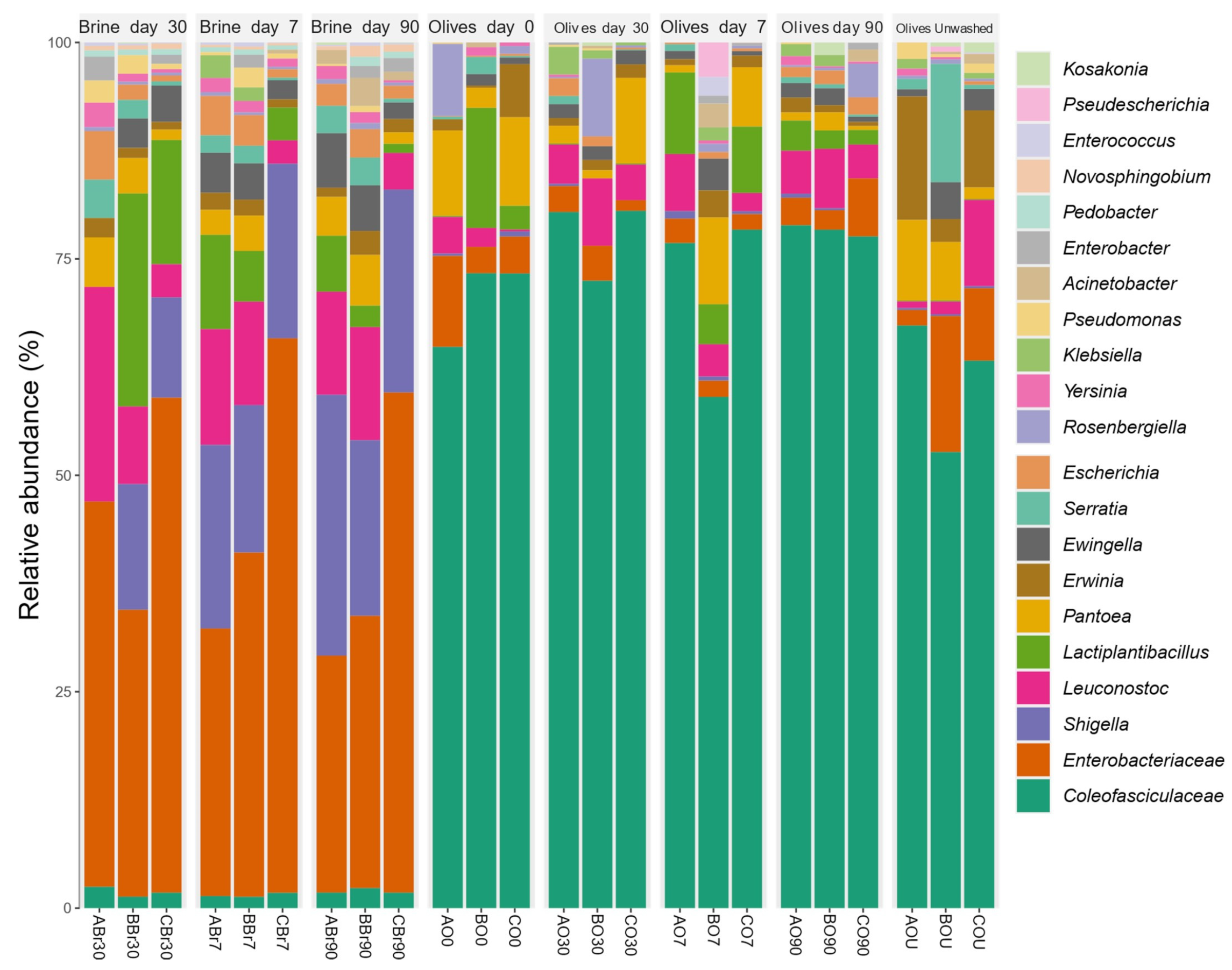
3.5.1. Bacterial Microbiota Composition

A total of 19 bacterial genera were detected in the samples (Table S5). Brine samples presented higher α-diversity (ANOVA, p-value < 0.05) when compared with olive ones (Figure S2). The α-diversity showed lower Simpson’s and inverted Simpson’s indexes of olives fermented after 30 and 90 days when compared to untreated olives (ANOVA, p-value < 0.05) (Figure S2).
Brines showed the highest abundance of Enterobacteriaceae, Shigella, Lactiplantibacillus, and Leuconostoc (Figure 1 and Figure S3). Coleofasciculaceae was the most abundant ASV in olives (>60%), Lactiplantibacillus was more abundant in olives after day 7 (7.2%), while Leuconostoc was the most abundant after days 30 and 90 (5.2 and 5.5%, respectively) (Figure 1 and Figure S3). Untreated olives showed that the second taxon in terms of abundance was Enterobacteriaceae (8.6%) (Figure 1 and Figure S3). Coleofasciculaceae and Novosphingobium spp. were more abundant in olives at day 30 (LDA = 5.81) and at day 90 (LDA = 5.18), respectively (LEfSe, p-value < 0.01).
The PCoA based on Bray–Curtis’s distance matrix of fermented olives showed differences in terms of bacterial taxa when compared to brines (Figure 2).
Several studies using metagenomics analysis have reported the detection of the above-mentioned bacterial genera, such as Lactiplantibacillus in Nyons variety [32], Manzanilla variety [111], Cypriot, Picual, and Kalamata varieties [30]; Leuconostoc in Konservolia variety [29], Kalamata variety [33], Nyons variety [32], Cypriot, Picual, and Kalamata varieties [30]; Shigella in Manzanilla spoiled Spanish-style fermented olives [52]; and Novosphingobium in untreated Aloreña de Málaga olives [28] and Kalamata variety [33], as well as the detection of the families Lactobacillaceae in Konservolia variety [29] and Kalamata variety [33], and Enterobacteriaceae in Aloreña de Málaga olives [28], Nocellara Etnea variety [25], Konservolia variety [29], Kalamata variety [33], Nyons variety [32], and Hojiblanca variety [31]. However, to our knowledge, the Coleofasciculaceae family, which corresponds to terrestrial cyanobacteria playing a significant role as pioneers of biological soil crusts [157], has not been detected in olives using metataxonomic analysis, although it is possible that its presence has not been reported because of its very low abundance. The presence of Coleofasciculaceae in olives could be attributed to contamination from the soil; however, it is possible that this family is a member of the olive microbiome, as its presence is also verified in leaves by metataxonomic analysis during the present study. Moreover, cyanobacteria species have already been isolated from plants’ phyllosphere, e.g., Acanthaceae and Poaceae [158]. Today, cyanobacteria play an important role in global oxygen, carbon, and nitrogen cycles [159]; thus, it is particularly intriguing to investigate their presence in olive fruits.
The results of the metataxonomic analysis are in accordance with the results of the classical microbiological analysis, as a high proportion of the isolated bacterial strains belonged to the Enterobacteriaceae family (63 isolates) and the Lactiplantibacillus and Leuconostoc genera (64 and 19 isolates, respectively). Enterobacteriaceae were eliminated due to pH decrease and the presence of phenolic compounds during the fermentation, as revealed by the classical microbiological analysis. They were, however, detected by metataxonomic analysis until the end of the process in both olive and brine samples, as this analysis enables the detection of both live and dead microbial cells. Moreover, many of the families and genera corresponding to the bacterial ASVs detected were not isolated from either olives or brines, e.g., Coleofasciculaceae and Novosphingobium, while all isolated genera were detected by the metataxonomic analysis. This was expected, as metataxonomics, using evolved sequencing technologies, can detect a broader range of microbial genera, including those not easily cultured using the growth media used in this work, e.g., members of the Coleofasciculaceae family [160]. Moreover, opportunities to guide the isolation and cultivation of microbes of interest are provided by the growing abundance of metagenomic sequence information from a wide range of environments [161].

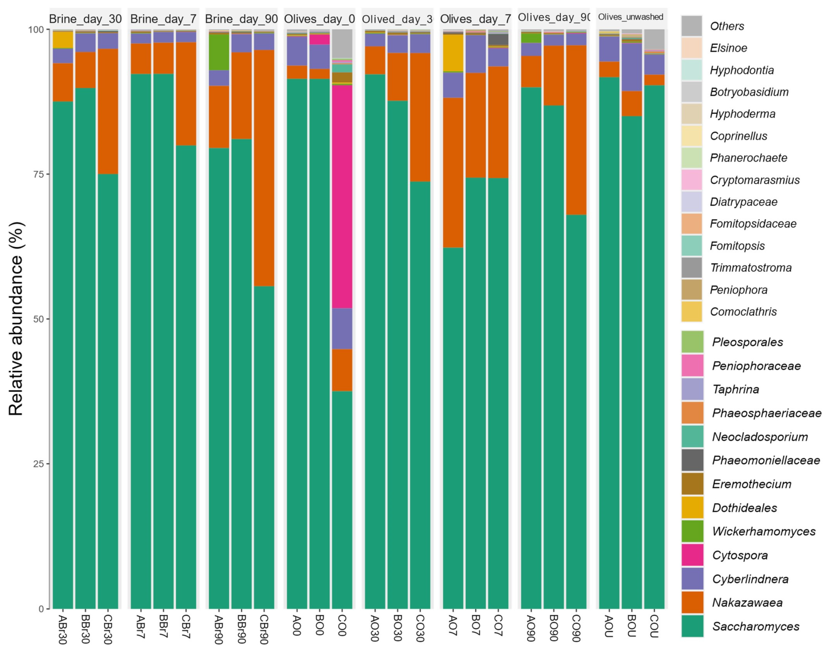
3.5.2. Mycobiota Composition

A total of twenty-six (26) fungal taxa at the lowest taxonomic resolution were detected (Table S6), whereas those detected but not specifically identified were categorized collectively as ‘Others’ in Figure 3. Mycobiota (Figure 3) of olives and brines did not demonstrate differences in terms of α-diversity (Figure S4). Saccharomyces was the most abundant ASV in olives and brines (>70%), followed by Nakazawaea (>9%) in both fermented olives and brines (Figure 3). The second-most abundant taxa of untreated olives and olives at day zero were Cyberlindnera (5.3%) and Cytospora (13.4%), respectively (Figure 3). PCoA did not show separation between sample groups (Figure S5). Cyberlindnera was associated with olives at day 0 (LEfSe p-value < 0.05, LDA = 4.44).
Several studies using metataxonomic analysis have also reported detection of the above-mentioned fungal genera Saccharomyces [32] and Nakazawaea [111]. Additionally, Saccharomyces was one of the dominant genera in fermented olives, such as Aloreña de Málaga variety [27], Kalamata variety, [33], Manzanilla variety [111], Picual and Kalamata varieties [30], and Hojiblanca and Manzanilla varieties [31]; however, a low occurrence (<1%) of Saccharomyces was observed in the yeast consortium in fermented Greek table olives of Halkidikis and Konservolia varieties [29]. Nevertheless, to our knowledge, the presence of the genera Cyberlindnera and Cytospora in olives has not been so far reported, possibly due to its low abundance as reported above.
The results of the metataxonomic analysis are in accordance with the results of the classical microbiological analysis, as a high proportion of the isolated fungal strains belonged to the Saccharomyces and Nakazawaea genera (21 and 43 isolates, respectively), and one isolate was identified as Cyberlindnera sp. However, most of the genera and families corresponding to the fungal ASVs detected were not isolated either from olives or brines, e.g., the genus Cytospora, which was detected in olives at the beginning of fermentation. This filamentous fungal genus commonly inhabits woody plants, includes several important canker-causing species, and has been reported to affect olive trees [162]. On the other hand, all genera isolated from olive and brine samples were detected by the metataxonomic analysis, with the exception of the genus Lasiodiplodia (one isolate). This result was not surprising either, as one of the challenges of the sequencing process for metataxonomic analysis may miss low-abundance microorganisms [160]. The isolation of microorganisms remains critical to directly study them and confirm their metabolic and physiological functions, while cultivated isolates could be useful in a range of applications, such as new probiotics, biocontrol agents, and agents for industrial processes [161]. The high abundance of a fungal species in a single sample, i.e., Cytospora in sample CO0 (41.3% abundance), indicates that the sampling process influences the results of metataxonomic analysis [163].

3.6. Sensory Evaluation of Fermented Tsounati Table Olives

The sensory profile (original scores) of Tsounati olives after 90 days of fermentation is presented in a spider graph (Figure 4).
The evaluation scores of the panel members were similar concerning both gustatory (salty, bitter, acid) and kinesthetic (hardness, fibrousness, crunchiness) sensations (Table S7). Average score values were 5.6 ± 0.1 (salty), 5.8 ± 0.4 (bitter), 4.6 ± 0.2 (acid), 6.3 ± 0.1 (hardness), 6.4 ± 0.2 (fibrousness), and 7.0 ± 0.1 (crunchiness). On the contrary, the commercial samples included in the analysis were perceived to be saltier and more acidic, however, less bitter, indicating higher salt concentration in the brine, longer period of fermentation, and possible addition of acid, respectively. Concerning smell and color, all samples had similar score values (7.04 ± 0.4, 6.06 ± 0.3 respectively). Abnormal fermentation was not indicated for any of the samples. The panelists not only did not perceive spoilage odors, such as those associated with zapatera spoilage, butyric, sulfidic, or putrid fermentation [52], but they also highlighted the mild and fruitful taste of Tsounati fermented olives.

3.7. Overall Discussion

The exploration of microbial diversity in fermented foods has been significantly advanced by the application of high-throughput sequencing (HTS) combined with omics approaches. However, culture-dependent analysis remains essential for estimating viable counts of various microbial groups and for isolating microorganisms for potential future applications. This necessity arises from limitations of culture-independent methods, such as the inclusion of DNA from dead or metabolically inactive cells, the insufficient detection of microbial populations present at low abundance, and variability in DNA quality extracted from microbial communities, all of which can affect sequencing success [164].
The microbiome of olives is closely linked to specific characteristics, including structural, chemical, genetic, and physiological traits. The uniqueness and appeal of traditionally fermented table olives stem from the complex and dynamic fermentation process, driven by a consortium of interacting microorganisms that collectively shape the final product. Furthermore, the abundance, prevalence, and succession of microbial populations during fermentation are influenced by various internal and external factors that affect the process [165].
Abiotic factors, such as weather conditions, including temperature, rainfall, and humidity, along with cultivation practices (traditional or novel), fruit maturation stage, microbial endophytic or epiphytic status, soil characteristics, salinity, and pollutant levels can all influence olive trees and negatively impact both yield and quality, as well as the microbiome of olive fruits [165,166,167].
Biotic stresses, such as olive tree diseases caused by arthropods, bacteria, fungi, nematodes, and viruses, interact with and influence microbial survival and dominance [166]. Additionally, the composition of the endophytic microbial community may vary according to plant age, altitude, and geographic location [167]. Recently, a study investigating the microbial terroir of fresh olive samples from 38 Greek olive varieties, including Tsounati, collected across geographically diverse regions of Greece, was conducted employing metataxonomics [168]. This study revealed a notable association between geographical origin, tree age, altitude, and microbial composition, underscoring their potential role in shaping the fermentation process.
The impact of olive cultivar genotype on the bacterial communities present in olive fruits and leaves has been studied, suggesting that genotype may have a stronger influence than soil and climate conditions, as reported in the reviews by Cardoni and Mercado-Blanco [166] and Melloni and Cardoso [167]. The microbial community of Tsounati fermented olives analyzed in this study, using both culture-dependent and -independent methods, shared several bacterial and fungal taxa commonly found in other olive varieties, including Lactobacillus plantarum, L. pentosus, Leuconostoc spp., and Enterobacteriaceae, and fungi such as Candida, Saccharomyces, Pichia, Rhodotorula, and Geotrichum candidum. However, several taxa not previously detected in table olives, such as Coleofasciculaceae, Cyberlindnera, Cytospora, and Candida fabianii, were identified, possibly due to the factors mentioned above, although the possibility remains that these taxa were present but undetected in earlier studies.
The microbial consortium plays a crucial role during olive fermentation, exhibiting both desirable and undesirable biochemical activities while providing essential technological benefits to the final product, as previously mentioned. However, this consortium is itself directly influenced by the numerous factors discussed above. This multifactorial interaction creates a complex ecosystem within table olives. The phenolic compounds and triterpenic acids present in Tsounati table olives may not only confer human health benefits but also affect the microbial consortium, which in turn directly shapes the distinctive sensory characteristics of Tsounati olives.

4. Conclusions

In the present study, naturally fermented olives of the Greek Tsounati variety from Monemvasia area were produced. The high abundance of Lactiplantibacillus and Leuconostoc, as well as Saccharomyces and Nakazawaea until the end of the fermentation, determined by classical microbiological and metataxonomics analyses, showed that both olives and brines were dominated by LAB and fungi known for their desirable biochemical activities and beneficial impact on the organoleptic character of the fermented olives. However, the detection of Enterobacteriaceae, as well as Cyberlindnera and Cytospora, suggests the occurrence of undesirable bacteria and fungi, which may have spoilage potential or even pathogenic traits. Moreover, members of the aforementioned LAB and fungal microbiota were isolated and identified, confirming the existence of these microorganisms until the end of fermentation and pointing out the disappearance of Enterobacteriaceae after 30 days. The presence of the filamentous fungal species Geotrichum candidum in olives until the end of fermentation reinforces the ambiguous role of fungi. TPC was lower compared to that of other olive varieties previously reported, possibly due to irrigation, but it remained largely unaffected during fermentation. Additionally, two of the most abundant and important bioactive compounds of olive fruits, the triterpenic acids maslinic and oleanolic, were monitored and quantified by HPLC. Their consistent presence showed that natural fermentation did not influence their concentration and suggested multiple beneficial health effects for consumers. Finally, sensory evaluation revealed the mild and fruitful taste of Tsounati fermented olives and strengthened their potential for table olive production. The results show the importance of the Tsounati variety as a promising Greek olive variety for future applications.

Supplementary Materials

The following supporting information can be downloaded at: https://www.mdpi.com/article/10.3390/foods14152568/s1, Figure S1: Fingerprinting of microorganisms isolated from Tsounati fermented olives and brines by rep-PCR analysed with the software BioNumerics v. 6.0. S1A: rep-PCR Clusters C1-C48 of bacterial isolates, S1B: rep-PCR Clusters C1-C45 of fungal isolates. Asterisks denote isolates that were selected for identification by 16S rDNA and ITS rDNA sequencing; Figure S2: Bacterial α-diversities indexes related to olives and brines based on the metataxonomic analysis. The Tukey’s post hoc test results are shown in the boxplots (ANOVA, p-value < 0.05); Figure S3: Pie chart of fermented olives/brines bacterial taxa relative abundances based on the metataxonomic analysis; Figure S4: Fungal α-diversities indexes related to olives and brines based on the metataxonomic analysis. The Tukey’s post hoc test results are shown in the boxplots (ANOVA p-value > 0.05); Figure S5: PCoA analysis about fungal β-diversity (Bray–Curtis’s distance) fermented olives and brines based on the metataxonomic analysis; Table S1: Coding of Tsounati variety Olive and Brine samples; Table S2: Concentration of sugars, organic acids, alcohols, triterpenic acids, Total Phenol Concentration (TPC), and microbial counts in olive and/or brine samples. Values represent the mean ± SD (n = 6; three fermentations A, B, and C of Tsounati variety table olives and two separate technical repetitions for each fermentation). Different lowercase letters indicate statistically significant differences among samples within the same column (p < 0.05). PCA: total microbial counts; MRS: presumptive mesophilic LAB; YGC: fungi; VRBGA: enterobacteria; Table S3: Identification, sample origin and rep-PCR clusters of bacterial isolates. (1) rep-PCR dendrogram is shown in Figure S1A. (2) Isolates in bold letters were selected and identified by 16S rDNA sequencing. (3) C: rep-PCR Cluster; Table S4: Identification, sample origin and rep-PCR clusters of fungal isolates, (1) rep-PCR dendrogram is shown in Figure S1B, (2) Isolates in bold letters were selected and identified by ITS rDNA sequencing, (3) C: rep-PCR Cluster; Table S5: Bacterial taxa abundances ASVs; Table S6: Mycobiota taxa abundances ASVs; Table S7: Sensory profiles (evaluation scores) of Tsounati variety olives after fermentation of 90 days. 1, 2: Commercial samples of green Konservolia variety fermented table olives. Values represent the mean ± SD of ten panel members, and three fermentations (A, B, and C) in the case of Tsounati variety table olives.

Author Contributions

Conceptualization, M.G., R.A., G.Z., G.P. and E.T.; methodology, M.G., I.F., D.B., R.A., G.Z. and E.T.; formal analysis, I.F. and D.B.; investigation, M.G., I.F., D.B., R.A., G.Z., D.G., D.Z. and A.K.; data curation, I.F. and D.B.; writing—original draft preparation, M.G., I.F., D.B., R.A., G.Z. and E.T.; writing—review and editing, M.G., I.F., D.B., R.A., G.Z. and E.T.; supervision, E.T.; funding acquisition, G.P. and E.T. All authors have read and agreed to the published version of the manuscript.

Funding

This work (project code 06.0148) was funded by the company Zoëfarma Inc., Headquarters in #320-3484 Boul. Des Sources, Dollard-des-Ormeaux, Québec, H9B 1Z9 Canada, that is lawfully represented by George Paraskevakos acting in his capacity as President.

Institutional Review Board Statement

Not applicable.

Informed Consent Statement

Not applicable.

Data Availability Statement

The original contributions presented in the study are included in the article/Supplementary Material, further inquiries can be directed to the corresponding author.

Acknowledgments

The authors would like to thank Koen Venema, Microbiome expert at Wageningen Food and Biobased Research, part of Wageningen University and Research, for critically reading the manuscript and for his valuable suggestions. The authors are also obliged to all farmers who kindly provided the olive samples considered in this study.

Conflicts of Interest

The authors declare no conflicts of interest. The funders had no role in the design of the study; in the collection, analyses, or interpretation of data; in the writing of the manuscript; or in the decision to publish the results.

Abbreviations

The following abbreviations are used in this manuscript:
NGSNext-Generation Sequencing
IOCInternational Olive Council
NOGBNational Olive Germplasm Bank of Greece
WOGBCWorld Olive Germplasm Bank of IFAPA, Córdoba
PDOProtected Designation of Origin
PGIProtected Geographical Indication
LABLactic Acid Bacteria
TPCTotal Polyphenol Content
HPLCHigh-Performance Liquid Chromatography
GAEGallic Acid Equivalents
PCAPlate Count agar
MRSDe Man–Rogosa–Sharpe
VRBGViolet Red Bile Glucose agar
YGCYeast Glucose Chloramphenicol agar
CFUColony Forming Units
SDStandard Deviation
TETris-EDTA
PCRPolymerase Chain Reaction
RepRepetitive Extragenic Palindromic Elements
NCBINational Center for Biotechnology Information
NADHNicotinamide Adenine Dinucleotide Hydrogen
WHOWorld Health Organization
ASVAmplicon Sequence Variant
LDALinear Discriminant Analysis
PCoAPrincipal Component Analysis

References

  1. Green, P.S.  Oleaceae. In The Families and Genera of Vascular Plants; Kubitzki, K., Kadereit, J.W., Eds.; Springer: Berlin,/Heidelberg, Germany, 2004; Volume 7, pp. 296–306. ISBN 978-3-540-40593-1. [Google Scholar]
  2. Huxley, A.; Griffiths, M.; Levy, M. The New Royal Horticultural Society Dictionary of Gardening; The Macmillan Press, Ltd.: London, UK, 1992; ISBN 978-0-333-47494-5. [Google Scholar]
  3. Green, P.S. A revision of Olea L. (Oleaceae). Kew Bull. 2002, 57, 91–140. [Google Scholar] [CrossRef]
  4. Cimato, A.; Attilio, C. Worldwide diffusion and relevance of olive culture. In Olive Diseases and Disorders; Schena, L., Agosteo, G.E., Cacciola, S.O., Eds.; Transworld Research Network: Trivandrum, India, 2011; Chapter 1; pp. 1–21. [Google Scholar]
  5. Barazani, O.; Dag, A.; Dunseth, Z. The history of olive cultivation in the southern Levant. Front. Plant Sci. 2023, 14, 1131557. [Google Scholar] [CrossRef]
  6. Foley, B.P.; Hansson, M.C.; Kourkoumelis, D.P.; Theodoulou, T.A. Aspects of ancient Greek trade re-evaluated with amphora DNA evidence. J. Archaeol. Sci. 2012, 39, 389–398. [Google Scholar] [CrossRef]
  7. Mascagni, F.; Barghini, E.; Ceccarelli, M.; Baldoni, L.; Trapero, C.; Díez, C.M.; Natali, L.; Cavallini, A.; Giordani, T. The singular evolution of Olea genome structure. Front. Plant Sci. 2022, 13, 869048. [Google Scholar] [CrossRef] [PubMed]
  8. Barranco, D.; Cimato, A.; Fiorino, P.; Rallo, L.; Touzani, A.; Castañeda, C.; Serafín, F.; Trujillo, I. World Catalogue of Olive Varieties. International Olive Oil Council, Madrid. 2000. Available online: https://www.internationaloliveoil.org/product/world-catalogue-of-olive-varieties/ (accessed on 22 March 2025).
  9. International Olive Council (IOC). The IOC Network of Germplasm Banks and the True Healthy Olive Cultivars Project. In Book of the IOC Network of Germplasm Banks, International Seminar, Córdoba, Argentina, 21–24 October 2019; IOC: Córdoba, Argentina, 2019; Available online: https://www.internationaloliveoil.org/the-iocs-book-of-germplasm-banks/ (accessed on 5 February 2025).
  10. Conte, P.; Fadda, C.; Del Caro, A.; Urgeghe, P.P.; Piga, A. Table olives: An overview on effects of processing on nutritional and sensory quality. Foods 2020, 9, 514. [Google Scholar] [CrossRef]
  11. Gómez-Gálvez, F.J.; Ninot, A.; Rodríguez, J.C.; Compañ, S.P.; Andreva, J.U.; Rubio, J.A.G.; Aragón, I.P.; Viñuales-Andreu, J.; Casanova-Gascón, J.; Ŝatović, Z.; et al. New insights in the Spanish gene pool of olive (Olea europaea L.) preserved ex situ and in situ based on high-throughput molecular markers. Front. Plant Sci. 2024, 14, 1267601. [Google Scholar] [CrossRef]
  12. FEK 4460. 2016. Available online: http://www.minagric.gr/images/stories/docs/agrotis/Pollaplasiastiko_Yliko/nomothesia_pollaplasiastiko_yliko/fek4460_301216.pdf (accessed on 25 September 2024).
  13. Hagidimitriou, M.; Katsiotis, A.; Menexes, G.; Pontikis, C.; Loukas, M. Genetic diversity of major Greek olive cultivars using molecular (AFLPs and RAPDs) markers and morphological traits. J. Am. Soc. Hortic. Sci. 2005, 130, 211–217. [Google Scholar] [CrossRef]
  14. Agriopoulou, S.; Tarapoulouzi, M.; Bedine Boat, M.A.; Rébufa, C.; Dupuy, N.; Theocharis, C.R.; Varzakas, T.; Roussos, S.; Artaud, J. Authentication and chemometric discrimination of six Greek PDO table olive varieties through morphological characteristics of their stones. Foods 2021, 10, 1829. [Google Scholar] [CrossRef]
  15. Stefanoudaki, E.; Kotsifaki, F.; Koutsaftakis, A. Classification of virgin olive oils of the two major cretan cultivars based on their fatty acid composition. J. Am. Oil Chem. Soc. 1999, 76, 623–626. [Google Scholar] [CrossRef]
  16. Andreou, V.; Psarianos, M.; Dimopoulos, G.; Tsimogiannis, D.; Taoukis, P. Effect of pulsed electric fields and high pressure on improved recovery of high-added-value compounds from olive pomace. J. Food Sci. 2020, 85, 1500–1512. [Google Scholar] [CrossRef]
  17. Skiada, V.; Tsarouhas, P.; Varzakas, T. Comparison and discrimination of two major monocultivar extra virgin olive oils in the Southern region of Peloponnese, according to specific compositional/traceability markers. Foods 2020, 9, 155. [Google Scholar] [CrossRef]
  18. Mousa, Y.M.; Gerasopoulos, D.; Metzidakis, I.; Kiritsakis, A. Effect of altitude on fruit and oil quality characteristics of “Mastoides” olives. J. Sci. Food Agric. 1996, 71, 345–350. [Google Scholar] [CrossRef]
  19. International Olive Council (IOC). (2024/2025 Crop Year). Export Figures of Table Olives in the European Union (EU-27). Available online: https://www.internationaloliveoil.org/wp-content/uploads/2025/03/IOC-Export-figures-TO-EU.html (accessed on 20 May 2025).
  20. Alexandraki, V.; Georgalaki, M.; Papadimitriou, K.; Anastasiou, R.; Zoumpopoulou, G.; Chatzipavlidis, I.; Papadelli, M.; Vallis, N.; Moschochoritis, K.; Tsakalidou, E. Determination of triterpenic acids in natural and alkaline-treated Greek table olives throughout the fermentation process. Food Sci. Technol. 2014, 58, 609–613. [Google Scholar] [CrossRef]
  21. Rocha, J.; Borges, N.; Pinho, O. Table olives and health: A review. J. Nutr. Sci. 2020, 2, e57. [Google Scholar] [CrossRef]
  22. Mioc, M.; Prodea, A.; Racoviceanu, R.; Mioc, A.; Ghiulai, R.; Milan, A.; Voicu, M.; Mardale, G.; Șoica, C. Recent advances regarding the molecular mechanisms of triterpenic acids: A review (Part II). Int. J. Mol. Sci. 2022, 23, 8896. [Google Scholar] [CrossRef] [PubMed]
  23. Rufino-Palomares, E.E.; Pérez-Jiménez, A.; García-Salguero, L.; Mokhtari, K.; Reyes-Zurita, F.J.; Peragón-Sánchez, J.; Lupiáñez, J.A. Nutraceutical role of polyphenols and triterpenes Present in the extracts of fruits and leaves of Olea europaea as antioxidants, anti-infectives and anticancer agents on healthy growth. Molecules 2022, 5, 2341. [Google Scholar] [CrossRef] [PubMed]
  24. Cocolin, L.; Alessandria, V.; Botta, C.; Gorra, R.; Filippis, F.; Ercolini, D.; Rantsiou, K. NaOH-debittering induces changes in bacterial ecology during table olives fermentation. PLoS ONE 2013, 31, e69074. [Google Scholar] [CrossRef]
  25. Randazzo, C.L.; Todaro, A.; Pino, A.; Corona, O.; Caggia, C. Microbiota and metabalome during controlled and spontaneous fermentation of Nocellara Etnea table olives. Food Microbiol. 2017, 65, 136–148. [Google Scholar] [CrossRef]
  26. De Angelis, M.; Campanella, D.; Cosmai, L.; Summo, C.; Giuseppe Rizzello, C.; Caponio, F. Microbiota and metabolome of un-started and started Greek-type fermentation of Bella di Cerignola table olives. Food Microbiol. 2015, 52, 18–30. [Google Scholar] [CrossRef]
  27. Arroyo-López, F.N.; Medina, E.; Ruiz-Bellido, M.A.; Romero-Gil, V.; Montes-Borrego, M.; Landa, B.B. Enhancement of the knowledge on fungal communities in directly brines Alorena de Malaga green olive fermentations by metabarcoding analysis. PLoS ONE 2016, 11, e0163135. [Google Scholar] [CrossRef]
  28. Medina, E.; Ruiz-Bellido, M.A.; Romero-Gil, V.; Rodriguez-Gomez, F.; Montes-Borrego, M.; Landa, B.B.; Arroyo-Lopez, F.N. Assessment of bacterial community in directly brined Alorena de Malaga table olive fermentations by metagenetic analysis. Int. J. Food Microbiol. 2016, 236, 47–55. [Google Scholar] [CrossRef]
  29. Argyri, K.; Doulgeraki, A.I.; Manthou, E.; Grounta, A.; Argyri, A.A.; Nychas, G.-J.E.; Tassou, C. Microbial diversity of fermented Greek table olives of Halkidiki and Konservolia varieties from different regions as revealed by metagenomic analysis. Microorganisms 2020, 8, 1241. [Google Scholar] [CrossRef]
  30. Kamilari, E.; Anagnostopoulos, D.A.; Tsaltas, D. Fermented table olives from Cyprus: Microbiota profile of three varieties from different regions through metabarcoding sequencing. Front. Microbiol. 2023, 13, 1101515. [Google Scholar] [CrossRef]
  31. Ruiz-Barba, J.L.; Sánchez, A.H.; López-López, A.; Cortés-Delgado, A.; Montaño, A. Microbial and chemical characterization of natural-style green table olives from the Gordal, Hojiblanca and Manzanilla cultivars. Foods 2023, 12, 2386. [Google Scholar] [CrossRef] [PubMed]
  32. Penland, M.; Deutsch, S.-M.; Falentin, H.; Pawtowski, A.; Poirier, E.; Visenti, G.; Le Meur, C.; Maillard, M.-B.; Thierry, A.; Mounier, J.; et al. Deciphering microbial community dynamics and biochemical changes during Nyons black olive natural fermentations. Front. Microbiol. 2020, 11, 586614. [Google Scholar] [CrossRef] [PubMed]
  33. Kazou, M.; Tzamourani, A.; Panagou, E.Z.; Tsakalidou, E. Unraveling the microbiota of natural black cv. Kalamata fermented olives through 16S and ITS metagenomic analysis. Microorganisms 2020, 8, 672. [Google Scholar] [CrossRef] [PubMed]
  34. Benítez-Cabello, A.; Romero-Gil, V.; Medina-Pradas, E.; Garrido-Fernández, A.; Arroyo-López, F.N. Exploring bacteria diversity in commercialized table olive biofilms by metataxonomic and compositional data analysis. Sci. Rep. 2020, 10, 11381. [Google Scholar] [CrossRef]
  35. RES-2/91-IV/04; Trade Standard Applying to Table Olives. Document COI/OT/NC No. 1. International Olive Oil Council: Madrid, Spain, 2004.
  36. Angelopoulou, A.; Alexandraki, V.; Georgalaki, M.; Anastasiou, R.; Manolopoulou, E.; Tsakalidou, E.; Papadimitriou, K. Production of probiotic Feta cheese using Propionibacterium freudenreichii subsp. shermanii as adjunct. Int. Dairy J. 2017, 66, 135–139. [Google Scholar] [CrossRef]
  37. Romero, C.; García, A.; Medina, E.; Ruíz-Méndez, M.V.; de Castro, A.; Brenes, M. Triterpenic acids in table olives. Food Chem. 2010, 118, 670–674. [Google Scholar] [CrossRef]
  38. Singleton, V.L.; Rossi, J.A. Colorimetry of total phenolics with phosphomolybdic-phosphotungstic acid reagents. Am. J. Enol. Vitic. 1965, 16, 144–158. [Google Scholar] [CrossRef]
  39. Marsilio, V.; Seghetti, L.; Iannucci, E.; Russi, F.; Lanza, B.; Felicioni, M. Use of a lactic acid bacteria starter culture during green olive (Olea europaea L cv Ascolana tenera) processing. J. Sci. Food Agric. 2005, 85, 1084–1090. [Google Scholar] [CrossRef]
  40. Georgalaki, M.; Zoumpopoulou, G.; Mavrogonatou, E.; Van Driessche, G.; Alexandraki, V.; Anastasiou, R.; Papadelli, M.; Kazou, M.; Manolopoulou, E.; Kletsas, D. Evaluation of the antihypertensive angiotensin-converting enzyme inhibitory (ACE-I) activity and other probiotic properties of lactic acid bacteria isolated from traditional Greek dairy products. Int. Dairy J. 2017, 75, 10–21. [Google Scholar] [CrossRef]
  41. Kopsahelis, N.; Nisiotou, A.; Kourkoutas, Y.; Panas, P.; Nychas, G.-J.E.; Kanellaki, M. Molecular characterization and molasses fermentation performance of a wild yeast strain operating in an extremely wide temperature range. Bioresour. Technol. 2009, 100, 4854–4862. [Google Scholar] [CrossRef] [PubMed]
  42. da Silva-Filho, E.A.; dos Santos, S.K.B.; do Monte Resende, A.; de Morais, J.O.F.; de Morais, M.A.; Simões, D.A. Yeast population dynamics of industrial fuel-ethanol fermentation process assessed by PCR-fingerprinting. Antonie Van Leeuwenhoek 2005, 88, 13–23. [Google Scholar] [CrossRef]
  43. Ntougias, S.; Zervakis, G.I.; Ehaliotis, C.; Kavroulakis, N.; Papadopoulou, K.K. Ecophysiology and molecular phylogeny of bacteria isolated from alkaline two-phase olive mill wastes. Res. Microbiol. 2006, 157, 376–385. [Google Scholar] [CrossRef]
  44. Korabecna, M. The Variability in the fungal ribosomal DNA (ITS1, ITS2, and 5.8 S rRNA Gene): Its biological meaning and application in medical mycology. In Communicating Current Research and Educational Topics and Trends in Applied Microbiology; Mendez-Vilas, A., Ed.; FORMATEX: Badajoz, Spain, 2007; pp. 783–787. [Google Scholar]
  45. Papademas, P.; Aspri, M.; Mariou, M.; Dowd, S.E.; Kazou, M.; Tsakalidou, E. Conventional and omics approaches shed light on Halitzia cheese, a long-forgotten white-brined cheese from Cyprus. Int. Dairy J. 2019, 98, 72–83. [Google Scholar] [CrossRef]
  46. Bolyen, E.; Rideout, J.R.; Dillon, M.R.; Bokulich, N.A.; Abnet, C.C.; Al-Ghalith, G.A.; Alexander, H.; Alm, E.J.; Arumugam, M.; Asnicar, F.; et al. Reproducible, interactive, scalable and extensible microbiome data science using QIIME 2. Nat. Biotechnol. 2019, 37, 852–857. [Google Scholar] [CrossRef]
  47. Callahan, B.J.; Mcmurdie, P.J.; Rosen, M.J.; Han, A.W.; Johnson, A.J.; Holmes, S.P. DADA2: High resolution sample inference from amplicon data. BioRxiv 2015, 7, 024034. [Google Scholar] [CrossRef]
  48. McDonald, D.; Jiang, Y.; Balaban, M.; Cantrell, K.; Zhu, Q.; Gonzalez, A.; Morton, J.T.; Nicolaou, G.; Parks, D.H.; Karst, S.M.; et al. Greengenes2 unifies microbial data in a single reference tree. Nat. Biotechnol. 2024, 42, 715–718. [Google Scholar] [CrossRef]
  49. International Olive Council (IOC). Sensory Analysis of Table Olives; Document COI/OT/MO No 1/Rev. 2; International Olive Council: Madrid, Spain, 2011; Available online: https://www.internationaloliveoil.org/wp-content/uploads/2022/10/COI-OT-MO.-1-Rev.2-2011-Eng.pdf (accessed on 17 October 2024).
  50. Portilha-Cunha, M.F.; Macedo, A.C.; Malcata, F.X. A Review on adventitious lactic acid bacteria from table olives. Foods 2020, 9, 948. [Google Scholar] [CrossRef]
  51. Garrido-Fernández, A.; Fernández Díaz, M.J.; Adams, R.M. Table Olives: Production and Processing; Chapman & Hall: London, UK, 1997; pp. 289–367. [Google Scholar]
  52. Arroyo-López, F.N.; Benítez-Cabello, A.; Romero-Gil, V.; Rodríguez-Gomez, F.; Garrido-Fernandez, A. Delving into the bacterial diversity of spoiled green Manzanilla Spanish-style table olive fermentations. Int. J. Food Microbiol. 2021, 359, 109415. [Google Scholar] [CrossRef]
  53. Campus, M.; Corrias, F.; Angioni, A.; Arru, N.; Sedda, P.; Addis, M.; Fiori, M.; Paba, A.; Chessa, L.; Comunian, R. Efficacy of a native microbial starter in promoting table olive fermentation: An industrial-scale trial at controlled and ambient temperature. Foods 2025, 14, 2159. [Google Scholar] [CrossRef] [PubMed]
  54. López-García, E.; Benítez-Cabello, A.; Rodríguez-Gómez, F.; Romero-Gil, V.; Garrido-Fernández, A.; Jiménez-Díaz, R.; Arroyo-López, F.N. Bacterial metataxonomic analysis of industrial Spanish-style green table olive fermentations. Food Control 2022, 137, 108969. [Google Scholar] [CrossRef]
  55. EFSA Panel on Nutrition, Novel Foods and Food Allergens (NDA); Turck, D.; Castenmiller, J.; de Henauw, S.; de Henauw, S.; Hirsch-Ernst, K.I.; Kearney, J.; Knutsen, H.K.; Maciuk, A.; Mangelsdorf, I. Dietary reference values for sodium. EFSA J. 2019, 17, 5778. [Google Scholar] [CrossRef] [PubMed]
  56. Papadelli, M.; Zoumpopoulou, G.; Georgalaki, M.; Anastasiou, R.; Manolopoulou, E.; Lytra, I.; Papadimitriou, K.; Tsakalidou, E. Evaluation of two lactic acid bacteria starter cultures for the fermentation of natural black table olives (Olea europaea L cv Kalamon). Pol. J. Microbiol. 2015, 64, 265–271. [Google Scholar] [CrossRef] [PubMed]
  57. Anagnostopoulos, D.A.; Goulas, V.; Xenofontos, E.; Vouras, C.; Nikoloudakis, N.; Tsaltas, D. Benefits of the use of lactic acid bacteria starter in green cracked Cypriot table olives fermentation. Foods 2020, 9, 17. [Google Scholar] [CrossRef]
  58. Lee, J. Exploring sucrose fermentation: Microorganisms, biochemical pathways, and applications. Ferment. Technol. 2023, 12, 166. [Google Scholar] [CrossRef]
  59. Anagnostopoulos, D.; Bozoudi, D.; Tsaltas, D. Yeast Ecology of Fermented Table Olives: A Tool for Biotechnological Applications. In Yeast—Industrial Applications; InTech Open: London, UK, 2017. [Google Scholar] [CrossRef]
  60. Ge, Y.; Wu, Y.; Aihaiti, A.; Wang, L.; Wang, Y.; Xing, J.; Zhu, M.; Hong, J. The metabolic pathways of yeast and acetic acid bacteria during fruit vinegar fermentation and their influence on favor development. Microorganisms 2025, 13, 477. [Google Scholar] [CrossRef]
  61. Oude Elferink, S.J.; Krooneman, J.; Gottschal, J.C.; Spoelstra, S.F.; Faber, F.; Driehuis, F. Anaerobic conversion of lactic acid to acetic acid and 1, 2-propanediol by Lactobacillus buchneri. Appl. Environ. Microbiol. 2001, 67, 125–132. [Google Scholar] [CrossRef]
  62. Silva, T.; Reto, M.; Sol, M.; Peito, A.; Peres, C.M.; Peres, C.; Malcata, F.X. Characterization of yeasts from Portuguese brined olives, with a focus on their potentially probiotic behavior. Food Sci. Technol. 2011, 44, 1349–1354. [Google Scholar] [CrossRef]
  63. Eram, M.; Ma, K. Decarboxylation of pyruvate to acetaldehyde for ethanol production by hyperthermophiles. Biomolecules 2013, 3, 578–596. [Google Scholar] [CrossRef]
  64. Vustin, M.M. The biological role of glycerol in yeast cells. Yeast as glycerol producers. Appl. Biochem. Microbiol. 2021, 57, 907–916. [Google Scholar] [CrossRef]
  65. Cheng, Z.; Zhan, M.; Yang, Z.; Zumstein, K.; Chen, H.; Huang, Q. The major qualitative characteristics of olive (Olea europaea L.) cultivated in Southwest China. Front. Plant Sci. 2017, 8, 559. [Google Scholar] [CrossRef]
  66. Sidari, R.; Martorana, A.; De Bruno, A. Effect of brine composition on yeast biota associated with naturally fermented Nocellara messinese table olives. Food Sci. Technol. 2019, 109, 163–170. [Google Scholar] [CrossRef]
  67. Sorrentino, G.; Muzzalupo, I.; Muccilli, S.; Timpanaro, N.; Russo, M.P.; Guardo, M.; Rapisarda, P.; Romeo, F.V. New accessions of Italian table olives (Olea europaea): Characterization of genotypes and quality of brined products. Sci. Hortic. 2016, 213, 34–41. [Google Scholar] [CrossRef]
  68. Sánchez, A.H.; De Castro, A.; Rejano, L.; Montaño, A. Comparative study on chemical changes in olive juice and brine during green olive fermentation. J. Agric. Food Chem. 2000, 48, 5975–5980. [Google Scholar] [CrossRef] [PubMed]
  69. Durante, M.; Tufariello, M.; Tommasi, L.; Lenucci, M.S.; Bleve, G.; Mita, G. Evaluation of bioactive compounds in black table olives fermented with selected microbial starters. J. Sci. Food Agric. 2017, 98, 96–103. [Google Scholar] [CrossRef]
  70. Moreno-González, R.; Juan, M.E.; Planas, J.M. Profiling of pentacyclic triterpenes and polyphenols by LC-MS in Arbequina and Empeltre table olives. LWT Food Sci. Technol. 2020, 126, 109310. [Google Scholar] [CrossRef]
  71. Boskou, D. Table Olives: A vehicle for the delivery of bioactive compounds. J. Exp. Food Chem. 2017, 3, 123. [Google Scholar] [CrossRef]
  72. Medina, E.; Morales-Sillero, A.; Ramírez, E.M.; Rallo, P.; Brenes, M.; Romero, C. New genotypes of table olives: Profile of bioactive compounds. Int. J. Food Sci. Technol. 2012, 47, 2334–2341. [Google Scholar] [CrossRef]
  73. Han, N.; Bakovic, M. Biologically active triterpenoids and their cardioprotective and anti-inflammatory effects. J. Bioanal. Biomed. S 2015, 12, 1948–1959. [Google Scholar] [CrossRef]
  74. Lin, C.; Wen, X.; Sun, H. Oleanolic acid derivatives for pharmaceutical use: A patent review. Expert Opin. Ther. Pat. 2016, 26, 643–655. [Google Scholar] [CrossRef] [PubMed]
  75. Bianchi, G. Lipids and phenols in table olives. Eur. J. Lipid Sci. Technol. 2003, 105, 229–242. [Google Scholar] [CrossRef]
  76. Kiai, H.; Hafidi, A. Chemical composition changes in four green olive cultivars during spontaneous fermentation. Food Sci. Technol. 2014, 57, 663–670. [Google Scholar] [CrossRef]
  77. Malheiro, R.; Mendes, P.; Fernandes, F.; Rodrigues, N.; Bento, A.; Pereira, J.A. Bioactivity and phenolic composition from natural fermented table olives. Food Funct. 2014, 5, 3132–3142. [Google Scholar] [CrossRef]
  78. Faghim, J.; Mohamed, M.B.; Bagues, M.; Guasmi, F.; Triki, T.; Nagaz, K. Irrigation effects on phenolic profile and extra virgin olive oil quality of “Chemlali’’ variety grown in South Tunisia. S. Afr. Bot. 2021, 141, 322–329. [Google Scholar] [CrossRef]
  79. García, P.; Romero, C.; Brenes, M. Influence of olive tree irrigation and the preservation system on the fruit characteristics of Hojiblanca black ripe olives. Food Sci. Technol. 2014, 55, 403–407. [Google Scholar] [CrossRef]
  80. Tataridou, M.; Kotzekidou, P. Fermentation of table olives by oleuropeinolytic starter culture in reduced salt brines and inactivation of Escherichia coli O157:H7 and Listeria monocytogenes. Int. J. Food Microbiol. 2015, 208, 122–130. [Google Scholar] [CrossRef]
  81. Georgiou, S.M.; Kosma, I.S.; Badeka, A.V.; Kontominas, M.G. Characterization and geographical differentiation of Kalamata table olives using physical, chemical, mechanical and sensory properties: A chemometric approach. Microchem. J. 2024, 199, 110085. [Google Scholar] [CrossRef]
  82. Rodríguez, H.; Landete, J.M.; de las Rivas, B.; Muñoz, R. Metabolism of food phenolic acids by Lactobacillus plantarum CECT 748T. Food Chem. 2008, 107, 1393–1398. [Google Scholar] [CrossRef]
  83. Bleve, G.; Tufariello, M.; Durante, M.; Perbellini, E.; Ramires, F.A.; Grieco, F.; Cappello, M.S.; De Domenico, S.; Mita, G.; Tasioula-Margari, M.; et al. Physico-chemical and microbiological characterization of spontaneous fermentation of Cellina di Nardò and Leccino table olives. Front. Microbiol. 2014, 5, 570. [Google Scholar] [CrossRef]
  84. Ramírez, E.; Brenes, M.; García, P.; Medina, E.; Romero, C. Oleuropein hydrolysis in natural green olives: Importance of the endogenous enzymes. Food Chem. 2016, 206, 204–209. [Google Scholar] [CrossRef]
  85. Piscopo, A.; De Bruno, A.; Zappia, A.; Poiana, M. Antioxidant activity of dried green olives (Carolea cv.). Food Sci. Technol. 2014, 58, 49–54. [Google Scholar] [CrossRef]
  86. Arroyo-López, F.N.; Romero, C.; Durán Quintana, M.; López López, A.; García García, P.; Garrido Fernández, A. Kinetic study of the physicochemical and microbiological changes in seasoned olives during the shelf period. J. Agric. Food Chem. 2005, 53, 5285–5292. [Google Scholar] [CrossRef]
  87. Maicas, S. The role of yeasts in fermentation processes. Microorganisms 2020, 8, 1142. [Google Scholar] [CrossRef]
  88. Giannattasio, S.; Guaragnella, N.; Ždralević, M.; Marra, E. Molecular mechanisms of Saccharomyces cerevisiae stress adaptation and programmed cell death in response to acetic acid. Front. Microbiol. 2013, 4, 33. [Google Scholar] [CrossRef]
  89. Adeboye, P.T.; Bettiga, M.; Olsson, L. The chemical nature of phenolic compounds determines their toxicity and induces distinct physiological responses in Saccharomyces cerevisiae in lignocellulose hydrolysates. AMB Express 2014, 4, 46. [Google Scholar] [CrossRef] [PubMed]
  90. Panagou, E.Z.; Tassou, C.C.; Katsaboxakis, C.Z. Induced lactic acid fermentation of untreated green olives of the Conservolea cultivar by Lactobacillus pentosus. J. Sci. Food Agric. 2003, 83, 667–674. [Google Scholar] [CrossRef]
  91. Abriouel, H.; Benomar, N.; Lucas, R.; Gálvez, A. Culture-independent study of the diversity of microbial populations in brines during fermentation of naturally-fermented Aloreña green table olives. Int. J. Food Microbiol. 2011, 144, 487–496. [Google Scholar] [CrossRef] [PubMed]
  92. Bonatsou, S.; Panagou, E.Z. Fermentation of cv. Kalamata natural black olives with potential multifunctional yeast starters. Foods 2022, 11, 3106. [Google Scholar] [CrossRef]
  93. Torriani, S.; Felis, G.E.; Dellaglio, F. Differentiation of Lactobacillus plantarum, L. pentosus, and L. paraplantarum by recA gene sequence analysis and multiplex PCR assay with recA gene-derived primers. Appl. Environ. Microbiol. 2001, 67, 3450–3454. [Google Scholar] [CrossRef]
  94. Naser, S.; Thompson, F.L.; Hoste, B.; Gevers, D.; Vandemeulebroecke, K.; Cleenwerck, I.; Thompson, C.C.; Vancanneyt, M.; Swings, J. Phylogeny and identification of enterococci by atpA gene sequence analysis. J. Clin. Microbiol. 2005, 43, 2224–2230. [Google Scholar] [CrossRef]
  95. Olsen, K.N.; Brockmann, E.; Molin, S. Quantification of Leuconostoc populations in mixed dairy starter cultures using fluorescence in situ hybridization. J. Appl. Microbiol. 2007, 103, 855–863. [Google Scholar] [CrossRef]
  96. Arroyo-López, F.N.; Querol, A.; Bautista-Gallego, J.; Garrido Fernández, A. Role of yeasts in table olive production. Int. J. Food Microbiol. 2008, 128, 189–196. [Google Scholar] [CrossRef]
  97. Bautista-Gallego, J.; Arroyo-López, F.N.; Rantsiou, K.; Jiménez-Díaz, R.; Garrido-Fernández, A.; Cocolin, L. Screening of lactic acid bacteria isolated from fermented table olives with probiotic potential. Food Res. Int. 2013, 50, 135–142. [Google Scholar] [CrossRef]
  98. Bonatsou, S.; Tassou, C.; Panagou, E.; Nychas, G.-J.E. Table olive fermentation using starter cultures with multifunctional potential. Microorganisms 2017, 5, 30. [Google Scholar] [CrossRef] [PubMed]
  99. Perpetuini, G.; Prete, R.; Garcia-Gonzalez, N.; Khairul Alam, M.; Corsetti, A. Table olives more than a fermented food. Foods 2020, 9, 178. [Google Scholar] [CrossRef]
  100. Hurtado, A.; Reguant, C.; Bordons, A.; Rozès, N. Lactic acid bacteria from fermented table olives. Food Microbiol. 2012, 31, 1–8. [Google Scholar] [CrossRef]
  101. Bleve, G.; Tufariello, M.; Durante, M.; Grieco, F.; Ramires, F.A.; Mita, G.; Tasioula-Margari, M.; Logrieco, A.F. Physico-chemical characterization of natural fermentation process of Conservolea and Kalamàta table olives and developement of a protocol for the pre-selection of fermentation starters. Food Microbiol. 2015, 46, 368–382. [Google Scholar] [CrossRef]
  102. Heperkan, D. Microbiota of table olive fermentations and criteria of selection for their use as starters. Front. Microbiol. 2013, 4, 143. [Google Scholar] [CrossRef]
  103. Abriouel, H.; Benomar, N.; Cobo, A.; Caballero, N.; Fernández Fuentes, M.Á.; Pérez-Pulido, R.; Gálvez, A. Characterization of lactic acid bacteria from naturally-fermented Manzanilla Aloreña green table olives. Food Microbiol. 2012, 32, 308–316. [Google Scholar] [CrossRef]
  104. Randazzo, C.L.; Ribbera, A.; Pitino, I.; Romeo, F.V.; Caggia, C. Diversity of bacterial population of table olives assessed by PCR- DGGE analysis. Food Microbiol. 2012, 32, 87–96. [Google Scholar] [CrossRef]
  105. Randazzo, C.L.; Rajendram, R.; Caggia, C. Lactic acid bacteria in table olive fermentation. In Olives and Olive Oil in Health and Disease Prevention; Pretty, V.R., Watson, R.S., Eds.; Elsevier: London, UK, 2010; pp. 371–379. [Google Scholar]
  106. De Castro, A.; Montaño, A.; Casado, F.-J.; Sánchez, A.-H.; Rejano, L. Utilization of Enterococcus casseliflavus and Lactobacillus pentosus as starter cultures for Spanish-style green olive fermentation. Food Microbiol. 2002, 19, 637–644. [Google Scholar] [CrossRef]
  107. Alves, M.; Gonçalves, T.; Quintas, C. Microbial quality and yeast population dynamics in cracked green table olives’ fermentations. Food Control 2012, 23, 363–368. [Google Scholar] [CrossRef]
  108. Rameshkumar, N.; Lang, E.; Nair, S. Mangrovibacter plantisponsor gen. nov., sp. nov., a nitrogen-fixing bacterium isolated from a mangrove-associated wild rice (Porteresia coarctata Tateoka). Int. J. Syst. Evol. Microbiol. 2010, 60, 179–186. [Google Scholar] [CrossRef]
  109. Chin, H.S.; Ravi Varadharajulu, N.; Lin, Z.-H.; Chen, W.-Y.; Zhang, Z.-H.; Arumugam, S.; Lai, C.-Y.; Yu, S.S.-F. Isolation, molecular identification, and genomic analysis of Mangrovibacter phragmitis strain ASIOC01 from activated sludge harboring the bioremediation prowess of glycerol and organic pollutants in high-salinity. Front. Microbiol. 2024, 15, 1415723. [Google Scholar] [CrossRef] [PubMed]
  110. Watts, D.; Palombo, E.A.; Jaimes Castillo, A.; Zaferanloo, B. Endophytes in agriculture: Potential to improve yields and tolerances of agricultural crops. Microorganisms 2023, 11, 1276. [Google Scholar] [CrossRef] [PubMed]
  111. López-García, E.; Benítez-Cabello, A.; Rodríguez-Gómez, F.; Martín-Arranz, V.; Garrido-Fernández, A.; Arroyo-López, F.N. Influence of 1-methylcyclopropene (1-MCP) on the processing and microbial communities of Spanish-style and directly brined green table olive fermentations. Fermentation 2022, 8, 441. [Google Scholar] [CrossRef]
  112. Bouhia, Y.; Hafidi, M.; Ouhdouch, Y.; Soulaimani, A.; Zeroual, Y.; Lyamlouli, K. Microbial intervention improves pollutant removal and semi-liquid organo-mineral fertilizer production from olive mill wastewater sludge and rock phosphate. J. Environ. Manag. 2024, 354, 120317. [Google Scholar] [CrossRef]
  113. Mennane, Z.; Abrini, J.; Elmtili, N. Hygienic quality, study of enterobacteria isolated from table olives and antimicrobial activity of Olea europaea L from Tetouan, Morocco. E3S Web Conf. 2021, 319, 01088. [Google Scholar] [CrossRef]
  114. Bevilacqua, A.; Cannarsi, M.; Gallo, M.; Sinigaglia, M.; Corbo, M.R. Characterization and implications of Enterobacter cloacae strains, isolated from Italian table olives “Bella di Cerignola”. J. Food Sci. 2010, 75, 53–60. [Google Scholar] [CrossRef]
  115. van der Aa Kühle, A.; Jespersen, L. The taxonomic position of Saccharomyces boulardii as evaluated by sequence analysis of the D1/D2 domain of 26S rDNA, the ITS1-5.8S rDNA-ITS2 region and the mitochondrial cytochrome-c oxidase II gene. Syst. Appl. Microbiol. 2003, 26, 564–571. [Google Scholar] [CrossRef] [PubMed]
  116. Dashko, S.; Liu, P.; Volk, H.; Butinar, L.; Piškur, J.; Fay, J.C. Changes in the relative abundance of two Saccharomyces species from oak forests to wine fermentations. Front. Microbiol. 2016, 7, 215. [Google Scholar] [CrossRef] [PubMed]
  117. Pontes, A.; Čadež, N.; Gonçalves, P.; Sampaio, J.P. A quasi-domesticate relic hybrid population of Saccharomyces cerevisiae × S. paradoxus adapted to olive brine. Front. Genet. 2019, 10, 449. [Google Scholar] [CrossRef] [PubMed]
  118. Pontes, A.; Hutzler, M.; Brito, P.H.; Sampaio, J.P. Revisiting the taxonomic synonyms and populations of Saccharomyces cerevisiae—Phylogeny, phenotypes, ecology and domestication. Microorganisms 2020, 8, 903. [Google Scholar] [CrossRef]
  119. Çelik, Z.D.; Erten, H.; Darici, M.; Cabaroğlu, T. Molecular characterization and technological properties of wine yeasts isolated during spontaneous fermentation of Vitis vinifera L.cv. Narince grape must grown in ancient wine making area Tokat, Anatolia. BIO Web Conf. 2017, 9, 02017. [Google Scholar] [CrossRef]
  120. Nisiotou, A.A.; Chorianopoulos, N.; Nychas, G.-J.E.; Panagou, E.Z. Yeast heterogeneity during spontaneous fermentation of black Conservolea olives in different brine solutions. J. Appl. Microbiol. 2010, 108, 396–405. [Google Scholar] [CrossRef]
  121. Bonatsou, S.; Paramithiotis, S.; Panagou, E.Z. Evolution of yeast consortia during the fermentation of Kalamata natural black olives upon two initial acidification treatments. Front. Microbiol. 2018, 8, 2673. [Google Scholar] [CrossRef]
  122. Arroyo-López, F.N.; Durán-Quintana, M.C.; Ruiz-Barba, J.L.; Querol, A.; Garrido-Fernández, A. Use of molecular methods for the identification of yeast associated with table olives. Food Microbiol. 2006, 23, 791–796. [Google Scholar] [CrossRef]
  123. Arroyo-López, F.N.; Romero-Gil, V.; Bautista-Gallego, J.; Rodríguez-Gómez, F.; Jiménez-Díaz, R.; García-García, P.; Querol, A.; Garrido-Fernández, A. Yeasts in table olive processing: Desirable or spoilage microorganisms? Int. J. Food Microbiol. 2012, 160, 42–49. [Google Scholar] [CrossRef]
  124. Bevilacqua, A.; Corbo, M.R.; Sinigaglia, M. Selection of yeasts as starter cultures for table olives: A step-by-step procedure. Front. Microbiol. 2012, 31, 194. [Google Scholar] [CrossRef]
  125. Ruiz-Barba, J.L.; Cortés-Delgado, A.; Sánchez, A.H.; López-López, A.; Montaño, A. Impact of selected wild yeasts starters on the volatilome and phenolic contents of Gordal, Manzanilla and Hojiblanca naturally fermented green olives. Food Sci. Technol. 2024, 195, 115811. [Google Scholar] [CrossRef]
  126. Traina, C.; Ferrocino, I.; Bonciolini, A.; Cardenia, V.; Lin, X.; Rantsiou, K.; Cocolin, L. Monitoring the yeasts ecology and volatiles profile throughout the spontaneous fermentation of Taggiasca cv. table olives through culture-dependent and independent methods. Int. J. Food Microbiol. 2024, 417, 110688. [Google Scholar] [CrossRef] [PubMed]
  127. Ciafardini, G.; Zullo, B.A. Use of selected yeast starter cultures in industrial-scale processing of brined Taggiasca black table olives. Food Microbiol. 2019, 84, 103250. [Google Scholar] [CrossRef] [PubMed]
  128. Camiolo, S.; Porru, C.; Benítez-Cabello, A.; Rodríguez-Gómez, F.; Calero-Delgado, B.; Porceddu, A.; Budroni, M.; Mannazzu, I.; Jiménez-Díaz, R.; Arroyo-López, F.N. Genome overview of eight Candida boidinii strains isolated from human activities and wild environments. Stand. Genom. Sci. 2017, 12, 70. [Google Scholar] [CrossRef] [PubMed]
  129. Parapouli, M.; Vasileiadis, A.; Afendra, A.S.; Hatziloukas, E. Saccharomyces cerevisiae and its industrial applications. AIMS Microbiol. 2020, 6, 1–31. [Google Scholar] [CrossRef]
  130. Ciafardini, G.; Zullo, B.A. Microbiological activity in stored olive oil. Int. J. Food Microbiol. 2002, 75, 111–118. [Google Scholar] [CrossRef]
  131. Kong, C.; Zhang, Q.; Wang, Y.; Huang, J.; Li, A.; Tao, Y. Decoding polysaccharides from two Pichia yeasts and their molecular interaction with wine fruity esters. J. Agric. Food Chem. 2024, 72, 12707–12718. [Google Scholar] [CrossRef]
  132. Colautti, A.; Orecchia, E.; Coppola, F.; Iacumin, L.; Comi, G. Cyberlindnera fabianii, an uncommon yeast responsible for gluten bread spoilage. Foods 2024, 13, 2381. [Google Scholar] [CrossRef]
  133. Viljoen, B.C. Yeast ecological interactions. Yeast-yeast, yeast-bacteria, yeast-fungi interactions and yeasts as biocontrol agents. In Yeasts in Food and Beverages; Querol, A., Fleet, H., Eds.; Springer: Berlin/Heidelberg, Germany, 2006; pp. 83–110. [Google Scholar]
  134. Hurtado, A.; Ben Othman, N.; Chammem, N.; Hamdi, M.; Ferrer, S. Characterization of Lactobacillus isolates from fermented olives and their bacteriocin gene profiles. Food Microbiol. 2011, 28, 1514–1518. [Google Scholar] [CrossRef]
  135. Domínguez-Manzano, J.; Olmo-Ruiz, C.; Bautista-Gallego, J.; Arroyo-López, F.N.; Garrido-Fernández, A.; Jiménez-Díaz, R. Biofilm formation on abiotic and biotic surfaces during Spanish style green table olive fermentation. Int. J. Food Microbiol. 2012, 157, 230–238. [Google Scholar] [CrossRef]
  136. Benincasa, C.; Muccilli, S.; Amenta, M.; Perri, E.; Romeo, F.V. Phenolic trend and hygienic quality of green table olives fermented with Lactobacillus plantarum starter culture. Food Chem. 2015, 186, 271–276. [Google Scholar] [CrossRef] [PubMed]
  137. Hernández, A.; Martín, A.; Aranda, E.; Pérez-Nevado, F.; Córdoba, M.G. Identification and characterization of yeast isolated from the elaboration of seasoned green table olives. Food Microbiol. 2007, 24, 346–351. [Google Scholar] [CrossRef] [PubMed]
  138. Sariguzel, F.M.; Unuvar, G.K.; Kucukoglu, O.; Parkan, O.M.; Koc, A.N. Identification, molecular characterization, and antifungal susceptibility of Cyberlindnera fabianii strains isolated from urinary tract. J. Med. Mycol. 2023, 33, 101429. [Google Scholar] [CrossRef] [PubMed]
  139. Ciafardini, G.; Zullo, B.A.; Iride, A. Lipase production by yeasts from extra virgin olive oil. Food Microbiol. 2006, 23, 60–67. [Google Scholar] [CrossRef]
  140. Koidis, A.; Triantafillou, E.; Boskou, D. Endogenous microflora in turbid virgin olive oils and the physicochemical characteristics of these oils. Eur. J. Lipid Sci. Technol. 2008, 110, 164–171. [Google Scholar] [CrossRef]
  141. Ciafardini, G.; Zullo, B.A. Effect of lipolytic activity of Candida adriatica, Candida diddensiae and Yamadazyma terventina on the acidity of extra-virgin olive oil with a different polyphenol and water content. Food Microbiol. 2015, 47, 12–20. [Google Scholar] [CrossRef]
  142. Vichi, S.; Romero, A.; Tous, J.; Caixach, J. The activity of healthy olive microbiota during virgin olive oil extraction influences oil chemical composition. J. Agric. Food Chem. 2011, 59, 4705–4714. [Google Scholar] [CrossRef]
  143. Taylor, J.W. One Fungus = One Name: DNA and Fungal Nomenclature Twenty Years After PCR. IMA Fungus 2011, 2, 113–120. [Google Scholar] [CrossRef]
  144. Zhu, H.Y.; Shang, Y.J.; Wei, X.Y.; Groenewald, M.; Robert, V.; Zhang, R.P.; Li, A.H.; Han, P.J.; Ji, F.; Li, J.N.; et al. Taxonomic revision of Geotrichum and Magnusiomyces, with the descriptions of five new Geotrichum species from China. Mycology 2024, 15, 400–423. [Google Scholar] [CrossRef]
  145. Kamilari, E.; Stanton, C.; Reen, F.J.; Ross, R.P. Uncovering the biotechnological importance of Geotrichum candidum. Foods 2023, 12, 1124. [Google Scholar] [CrossRef]
  146. Giannoutsou, E.P.; Meintanis, C.; Karagouni, A.D. Identification of yeast strains isolated from a two-phase decanter system olive oil waste and investigation of their ability for its fermentation. Bioresour. Technol. 2004, 93, 301–306. [Google Scholar] [CrossRef]
  147. Asses, N.; Ayed, L.; Bouallagui, H.; Benrejeb, I.; Gargouri, M.; Hamdi, M. Use of Geotrichum candidum for olive mill wastewater treatment in submerged and static culture. Bioresour. Technol. 2009, 100, 2182–2188. [Google Scholar] [CrossRef] [PubMed]
  148. Bouhia, Y.; Hafidi, M.; Ouhdouch, Y.; El Boukhari, M.E.M.; El Fels, L.; Zeroual, Y.; Lyamlouli, K. Microbial community succession and organic pollutants removal during olive mill waste sludge and green waste co-composting. Front. Microbiol. 2022, 12, 814553. [Google Scholar] [CrossRef] [PubMed]
  149. Zalar, P.; Gostinčar, C.; de Hoog, G.S.; Uršič, V.; Sudhadham, M.; Gunde-Cimerman, N. Redefinition of Aureobasidium pullulans and its varieties. Stud. Mycol. 2008, 61, 21–38. [Google Scholar] [CrossRef] [PubMed]
  150. Zhu, S.; Lei, Y.; Wang, C.; Wei, Y.; Wang, C.; Sun, Y. Patterns of yeast diversity distribution and its drivers in rhizosphere soil of Hami melon orchards in different regions of Xinjiang. BMC Microbiol. 2021, 6, 170. [Google Scholar] [CrossRef]
  151. Preto, G.; Martins, F.; Pereira, J.A.; Baptista, P. Fungal community in olive fruits of cultivars with different susceptibilities to anthracnose and selection of isolates to be used as biocontrol agents. Biol. Control 2017, 110, 1–9. [Google Scholar] [CrossRef]
  152. López-García, E.; Benítez-Cabello, A.; Ramiro-García, J.; Romero-Gil, V.; Rodríguez-Gómez, F.; Arroyo-López, F.N. New insights into microbial diversity of the traditional packed table olives Aloreña de Málaga through metataxonomic analysis. Microorganisms 2021, 9, 561. [Google Scholar] [CrossRef]
  153. Perez, B.A.; Farinon, O.M.; Berretta, M.F. First report of Fusarium solani causing root rot of olive in Southeastern Argentina. Plant Dis. 2011, 95, 1476. [Google Scholar] [CrossRef]
  154. Chliyeh, M.; Rhimini, Y.; Selmaoui, K.; Touhami, A.O.; Filali-Maltouf, A.; Modafar, C.E.; Moukhli, A.; Oukabli, A.; Benkirane, R.; Douira, A. Survey of the fungal species associated to oive-tree (Olea europaea L.) in Morocco. Int. J. Recent Biotechnol. 2014, 2, 15–32. [Google Scholar]
  155. Markakis, E.A.; Roditakis, E.N.; Kalantzakis, G.S.; Chatzaki, A.; Soultatos, S.K.; Stavrakaki, M.; Tavlaki, G.I.; Koubouris, G.C.; Bagkis, N.; Goumas, D.E. Characterization of fungi associated with olive fruit rot and olive oil degradation in Crete, Southern Greece. Plant Dis. 2021, 105, 3623–3635. [Google Scholar] [CrossRef]
  156. Qiao, G.; Zhao, J.; Liu, J.; Tan, X.; Qin, W. Two novel Lasiodiplodia species from blighted stems of Acer truncatum and Cotinus coggygria in China. Biology 2022, 11, 1459. [Google Scholar] [CrossRef] [PubMed]
  157. Moreira, C.; Fernandes, V.; Giraldo-Silva, A.; Roush, D.; Garcia-Pichel, F. Coleofasciculaceae, a monophyletic home for the Microcoleus steenstrupii complex and other desiccation-tolerant filamentous cyanobacteria. J. Phycol. 2021, 57, 1563–1579. [Google Scholar] [CrossRef] [PubMed]
  158. Alvarenga, D.O.; Andreote, A.P.D.; Branco, L.H.Z.; Fiore, M.F. Kryptousia macronema gen. nov., sp. nov. and Kryptousia microlepis sp. nov., nostocalean cyanobacteria isolated from phyllospheres. Int. J. Syst. Evol. Microbiol. 2017, 67, 3301–3309. [Google Scholar] [CrossRef] [PubMed]
  159. Kaštovský, J. Welcome to the jungle!: An overview of modern taxonomy of cyanobacteria. Hydrobiologia 2024, 851, 1063–1077. [Google Scholar] [CrossRef]
  160. Zhang, L.; Chen, F.; Zeng, Z.; Xu, M.; Sun, F.; Yang, L.; Bi, X.; Lin, Y.; Gao, Y.; Hao, H.; et al. Advances in metagenomics and its application in environmental microorganisms. Front. Microbiol. 2021, 12, 766364. [Google Scholar] [CrossRef]
  161. Liu, S.; Moon, C.D.; Zheng, N.; Huws, S.; Zhao, S.; Wang, J. Opportunities and challenges of using metagenomic data to bring uncultured microbes into cultivation. Microbiome 2022, 12, 76. [Google Scholar] [CrossRef]
  162. Petrović, E.; Vrandečić, K.; Ivić, D.; Ćosić, J.; Godena, S. First report of olive branch dieback in Croatia caused by Cytospora pruinosa Défago. Microorganisms 2023, 11, 1679. [Google Scholar] [CrossRef]
  163. Thomas, T.; Gilbert, J.; Meyer, F. Metagenomics—A guide from sampling to data analysis. Microb. Inform. Exp. 2012, 9, 3. [Google Scholar] [CrossRef]
  164. Srinivas, M.; O’Sullivan, O.; Cotter, P.D.; Sinderen, D.v.; Kenny, J.G. The application of metagenomics to study microbial communities and develop desirable traits in fermented foods. Foods 2022, 11, 3297. [Google Scholar] [CrossRef]
  165. Martins, F.; Rodrigues, N.; Ramalhosa, E. A review of the microbial dynamics of natural and traditional fermentations of table olive. Appl. Microbiol. 2025, 5, 52. [Google Scholar] [CrossRef]
  166. Cardoni, M.; Mercado-Blanco, J. Confronting stresses affecting olive cultivation from the holobiont perspective. Front. Plant Sci. 2023, 14, 1261754. [Google Scholar] [CrossRef]
  167. Melloni, R.; Cardoso, E.J.B.N. Microbiome associated with olive cultivation: A review. Plants 2023, 12, 897. [Google Scholar] [CrossRef]
  168. Ferrocino, I.; Buzzanca, D.; Pagiati, L.; Kazou, M.; Georgalaki, M.; Hatzopoulos, I.; Tsakalidou, E. The microbial terroir of the Greek olive varieties. Int. J. Food Microbiol. 2025, 441, 111332. [Google Scholar] [CrossRef]
Figure 1. Bacterial taxa relative abundances of olives and brines based on the metataxonomic analysis.
Foods 14 02568 g001
Figure 2. PCoA analysis about bacterial β-diversity (Bray–Curtis’s distance) of olives and brines based on the metataxonomic analysis.
Foods 14 02568 g002
Figure 3. Fungal taxa relative abundances of olives and brines based on the metataxonomic analysis.
Foods 14 02568 g003
Figure 4. The spider graph shows the sensory profiles (original scores) of Tsounati olives after fermentation of 90 days based on the sensory evaluation. Fermentations A, B, C; 1, 2: Commercial samples of green Konservolia variety fermented table olives.
Foods 14 02568 g004
Table 1. Number of identified bacterial isolates (isolated from MRS agar plates incubated at 30 °C) from Tsounati olive and brine samples.
IdentificationOUO0O4O7O15O30O60O90Br4Br7Br15Br30Br60Br90Nr
Lactic Acid Bacteria 85
Lactiplantibacillus spp.00589362801200264
Leuconostoc spp.3165220000000019
Lactococcus lactis000100000000001
Enterococcus spp.000010000000001
Enterobacteriaceae 63
Mangrovibacter spp.202011001411800039
Enterobacter/Raoultella/Klebsiella spp.71401000000000022
Kluyvera intermedia100000000000001
Pseudocitrobacter spp.000000001000001
Number of isolates per sample/
Total number
13151315136628151210002148
O: olives (fermented), U: untreated olives, Br: brine, Nr: number.
Table 2. The number of identified fungal isolates (isolated from YGC agar plates incubated at 25 °C) from Tsounati olive and brine samples.
IdentificationOUO0O4O7O15O30O60O90Br4Br7Br15Br30Br60Br90Nr
Yeasts 136
Candida spp.2425648423320348
Nakazawaea molendinolei1024449633220343
Saccharomyces cerevisiae/boulardii/paradoxus0001335313020021
Pichia kluyveri/fermentans1232111022100016
Barnettozymacalifornica022000001010006
Rhodotorula glutinis/graminis000000001000001
Cyberlindnerafabianii100000000000001
Yeast-like or non-yeast Fungi 42
Geotrichum candidum/australiense/galactomycetum2133336203230031
Aureobasidium pullulans211000000000004
Filobasidium magnum000020000000002
Cladosporium halotolerans000000000010001
Cladosporium tenellum/herbarum/ramnotenellum/iridis000010000000001
Aspergillus amstelodami/cristatus/chevalieri/montevidensis000000000010001
Fusarium solani/hoffmannii/ambrosium/ensiforme000000001000001
Lasiodiplodia macrospora010000000000001
Number of isolates per sample/Total number911131520152915111411906178
O: olives (fermented), U: untreated olives, Br: brine, Nr: number.
Disclaimer/Publisher’s Note: The statements, opinions and data contained in all publications are solely those of the individual author(s) and contributor(s) and not of MDPI and/or the editor(s). MDPI and/or the editor(s) disclaim responsibility for any injury to people or property resulting from any ideas, methods, instructions or products referred to in the content.

