Review

Au-Ag Bimetallic Nanoparticles for Surface-Enhanced Raman Scattering (SERS) Detection of Food Contaminants: A Review

1
School of Agricultural Engineering, Jiangsu University, Zhenjiang 212013, China
2
School of Energy and Power Engineering, Jiangsu University, Zhenjiang 212013, China
3
Zhenjiang Agricultural Products Quality Inspection and Testing Center, Zhenjiang 212009, China
4
Jurong Agricultural Product Quality and Safety Monitoring Center, Zhenjiang 212400, China
*
Author to whom correspondence should be addressed.
Foods 2025, 14(12), 2109; https://doi.org/10.3390/foods14122109
Submission received: 22 May 2025 / Revised: 9 June 2025 / Accepted: 14 June 2025 / Published: 16 June 2025

Abstract

:
Food contaminants, including harmful microbes, pesticide residues, heavy metals and illegal additives, pose significant public health risks. While traditional detection methods are effective, they are often slow and require complex equipment, which limits their application in real-time monitoring and rapid response. Surface-enhanced Raman scattering (SERS) technology has gained widespread use in related research due to its hypersensitivity, non-destructibility and molecular fingerprinting capabilities. In recent years, Au-Ag bimetallic nanoparticles (Au-Ag BNPs) have emerged as novel SERS substrates, accelerating advancements in SERS detection technology. Au-Ag BNPs can be classified into Au-Ag alloys, Au-Ag core–shells and Au-Ag aggregates, among which the Au-Ag core–shell structure is more widely applied. This review discusses the types, synthesis methods and practical applications of Au-Ag BNPs in food contaminants. The study aims to provide valuable insights into the development of new Au-Ag BNPs and their effective use in detecting common food contaminants. Additionally, this paper explores the challenges and future prospects of SERS technology based on Au-Ag BNPs for pollutant detection, including the development of functional integrated substrates, advancements in intelligent algorithms and the creation of portable on-site detection platforms. These innovations are designed to streamline the detection process and offer guidance in selecting optimal sensing methods for the on-site detection of specific pollutants.

1. Introduction

Food safety is a crucial aspect of public health and social well-being, playing a vital role in maintaining and promoting human health. Food safety incidents have heightened public awareness of food safety concerns [1,2]. The primary pollutants responsible for food safety issues include pesticides, heavy metals, foodborne pathogenic bacteria, toxins and other harmful substances, all of which pose potential risks to human health [3]. Consumption of contaminated food can lead to a wide range of health problems, from mild gastrointestinal discomfort to severe, life-threatening diseases. Various methods are currently available for detecting harmful substances in food, such as molecular biology techniques [4], enzyme-linked immunosorbent assays [5], fluorescence sensors [6], colorimetry methods [7], electrochemical techniques [8], etc. While these detection methods offer high sensitivity, they often suffer from limitations such as complex procedures, long pretreatment times, high costs and susceptibility to interference.
Raman spectroscopy is a type of molecular vibrational spectroscopy that provides structural information about compounds [9]. However, its application is limited due to its low signal strength. The discovery of surface-enhanced Raman scattering (SERS) has significantly advanced the development and application of Raman spectroscopy [10]. SERS has unique advantages, such as a narrow peak shape, high sensitivity, fast response, no influence from water, and the ability to identify and quantify target compounds based on molecular fingerprints [11]. It is a rapid and non-destructive spectral analysis technique. It has been widely applied in fields such as early disease diagnosis [12,13], biological imaging [14], environmental monitoring [15] and food safety [16]. The enhancement mechanism of SERS technology for target molecules mainly includes electromagnetic enhancement based on electromagnetic fields and chemical enhancement based on charge transfer [17,18]. The localized surface plasmon resonance (LSPR) model based on the electromagnetic enhancement mechanism holds that when the incident laser irradiates the rough surface of the precious metal nanomaterial, the freely moving electrons on the surface of the precious metal generate plasmon resonance, thereby significantly enhancing the Raman signal [19]. The SERS response of a target molecule is primarily influenced by the enhancement effect of the SERS substrate. To achieve a higher SERS response, researchers have developed various substrates, including Au nanorods (AuNRs) [20], Ag nanoflowers [21], nanotriangles [22], Au nanostars [23], ZnO@ZIF-8 [24], WO3-x nanowire/WSe2 heterostructures [25] and chiral carbon nanotube (CNT)/TiO2 hybrids [26].
Among the materials commonly used in SERS, precious metal nanomaterials, particularly Au and Ag, demonstrate exceptional performance due to their unique optical properties. First, their high surface area-to-volume ratio creates numerous hotspots where the electromagnetic field is significantly enhanced, thus amplifying the Raman signal [27]. Second, the tunability of their size, shape and composition allows for the optimization of SERS performance for specific applications. Currently, precious metal nanomaterials have evolved from single-component to multi-component forms. Au-Ag bimetallic nanoparticles (Au-Ag BNPs), such as Au-Ag core–shell nanoparticles, exhibit superior stability and SERS activity compared to single-metal nanoparticles, as strong local electromagnetic fields are generated through the coupling of different metallic materials [28].
Extensive research has been conducted on the application of SERS in food safety detection, leading to the publication of several reviews with varying focuses. However, most of these reviews either address the application of specific target detection substances or primarily focus on the synthesis of a particular type of enhanced substrate [29,30]. In SERS technology, the enhancement effect of the substrate material plays a critical role. Bimetallic nanomaterials based on Au and Ag, exhibiting synergistic effects, typically demonstrate stronger SERS performance. Despite this, recent advancements in their synthesis and application have not been reported. This paper aims to highlight the significance of Au-Ag BNPs in the development of SERS technology by systematically summarizing their classification, synthesis and applications in detecting contaminants (Figure 1). Furthermore, this review anticipates the challenges and future prospects of using Au-Ag BNPs in SERS-based detection of harmful substances in food.

2. Types and Synthesis of Au-Ag BNPs

2.1. Types of Au-Ag BNPs

In recent years, precious metal nanoparticles have been developed to meet the growing demands of SERS detection. These nanoparticles can generally be classified into three categories based on their structural morphology: alloy, core–shell and aggregate types. The nanostructure of Au-Ag BNPs can provide a strong electromagnetic field and significantly enhance the Raman signal of the target molecules. Additionally, when combined with biomolecules such as aptamers and antibodies, these nanostructures offer multifunctionality, thereby broadening the scope of SERS applications [31].
Au-Ag alloy nanomaterials are formed by the uniform mixture of Au and Ag atoms at the atomic scale. In their structure, the two metal atoms are usually distributed in a disordered manner, forming nanoparticles with a single phase. The composition of the alloy can be continuously regulated by adjusting the Au/Ag ratio (such as AuxAg1−x, x = 0.1~0.9). The particle morphology is predominantly spherical [32] but can also include cubic [33], octahedral [34] and other forms.
The core–shell structure is the most versatile and widely used. Core–shell nanostructures primarily exploit the strong electromagnetic field generated by the metal core to enhance the Raman signal of analytes located on or near the shell layer [35]. Core–shell structures are heterogeneous nanoparticles formed by layer-by-layer assembly of one metal (the core) and another metal (the shell), with a clear two-phase interface [36]. These structures can be classified into two types: “Au-core/Ag-shell” (Au@Ag) and “Ag-core/Au-shell” (Ag@Au). The core diameter typically ranges from 10 to 100 nanometers, while the shell thickness can be precisely controlled from a few nanometers to tens of nanometers. The core and shell are linked through lattice matching or surface ligands, generating core–shell interface effects such as charge transfer and stress regulation.
Au-Ag aggregates are multi-particle assemblies formed by the interconnection of individual Au/Ag nanoparticles (spherical, rod-shaped, star-shaped, etc.) through physical (such as electrostatic interaction and van der Waals forces) or chemical (such as ligand bridging and covalent bonds) actions, including dimer, trimer, chain, network or disordered aggregates [37]. A key characteristic of these aggregates is the presence of nanoscale gaps (typically 1~20 nm) between particles. These gaps create “hotspots” regions, where the intensity of the electromagnetic field increases exponentially, with enhancement factors ranging from 106 to 1012. The structure of the aggregates can be finely tuned by controlling parameters such as particle size, morphology, spacing and connection methods (e.g., point contact and surface contact).
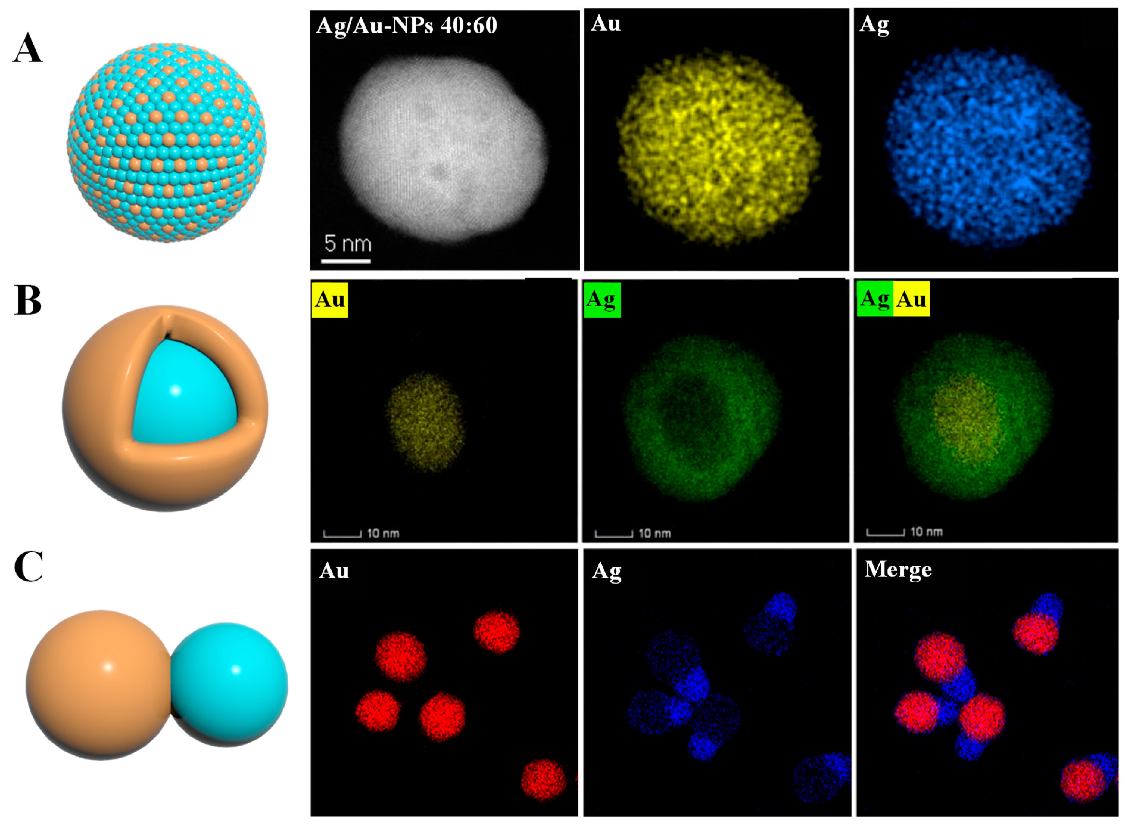
To sum up, as shown in the model structure diagram in Figure 2, where orange represents the Ag element (or Au element) and the light blue represents the Au element (or Ag element), the Au and Ag elements in Au-Ag alloy nanomaterials are distributed in a disordered state on a single nanoparticle. Core–shell nanomaterials have Au and Ag elements distributed in a core–shell state as a whole on a single nanoparticle. Aggregates are equivalent to the coupling of Au nanoparticles and Ag nanoparticles, and the elements are distributed in two regions. The elemental mapping images representing different types of Au-Ag BNPs provided in the literature and presented in Figure 2 show that the distribution positions of the two elements (Au and Ag) constituting the nanoparticles are different.

2.2. Synthesis of Au-Ag BNPs

The effective synthesis of nanoparticles is crucial to their SERS enhancement. Consequently, understanding the common synthesis methods, particularly those for Au-Ag BNPs used in SERS detection, is essential. Currently, the synthesis of Au-Ag BNPs is typically classified into physical, chemical and biological methods. Physical methods offer advantages in the preparation of monodisperse and core–shell structures; however, their high costs and low yields limit large-scale application. Chemical methods are the most effective for regulating the structure of Au-Ag BNPs (such as alloys and core–shell configurations), and they are especially suitable for electrochemical and optical sensing. Biological methods hold promise in antibacterial and biomedical applications due to their environmental friendliness and biocompatibility, though challenges related to size control and repeatability remain to be addressed.

2.2.1. Physical Synthesis Methods

Physical synthesis methods for nanoparticles typically involve breaking bulk materials into fine particles. These methods offer advantages such as a narrow particle size distribution and uniform performance. Laser ablation, a top-down technique, has proven to be cost-effective for producing nanoparticles of various shapes and size distributions within a short time (typically a few minutes) [41]. Pulsed laser ablation can be used to synthesize Au-Ag BNPs in liquid through several preparation pathways. Initially, single-metal colloids (e.g., Ag cubes) are synthesized from ablation targets. The target is then replaced with a different metal (e.g., Au cubes), and ablation occurs under the existing colloid to produce bimetallic nanoparticles. This typically results in core–shell bimetallic nanoparticles, where the core consists of the first ablation material and the shell is composed of the second material. Additionally, Au-Ag BNPs can be synthesized by mixing two different metal colloids and ablating the mixture, resulting in alloy or core–shell bimetallic nanoparticles [42].
Au-Ag BNPs can also be produced via mechanical grinding and ball milling. For example, Murugadoss et al. mechanically mixed metal salts and reducing agents (AgNO3, chitosan and NaOH) in a solid state, then added a concentrated HAuCl3 solution to obtain Au-Ag BNPs [43].
Microwave radiation-assisted synthesis provides certain advantages for the manufacture of BNPs. Using a microwave reactor, Au-Ag BNPs were synthesized by heating a mixture of AgNO3 and NaAuCl4 in ethylene glycol and glycerol [44]. Although this physical method does not involve toxic chemicals, it is not recommended for the synthesis of Au-Ag BNPs due to its high cost, susceptibility to radiation, high temperature, low yield and high energy consumption.

2.2.2. Chemical Methods

Chemical methods involve reactions with precursors and typically include co-reduction, seed-mediated growth, electrochemical displacement reactions, co-precipitation and polyol methods. Each method has distinct advantages and disadvantages, which influence the properties and morphologies of the synthesized Au-Ag BNPs. The co-reduction method simultaneously reduces Au and Ag precursors to form Au-Ag BNPs. Common precursors include HAuCl4 and AgNO3, which are dissolved in either water or an organic solvent, such as ethanol. Reducing agents, such as NaBH4 and trisodium citrate, provide electrons to reduce Au and Ag ions into Au and Ag nanoparticles. By adjusting the precursor concentration and the reducing agent-to-precursor ratio, nanoparticles of varying sizes can be synthesized [45].
The seed-mediated growth method involves two steps: synthesis of seed nanoparticles and the subsequent growth of a second metal on these seeds. For example, small Au NPs can be synthesized by reducing HAuCl4 and then used as seeds. In the second step, these seed nanoparticles are introduced into a solution containing Ag ions, such as AgNO3. Reducing agents facilitate the growth of Ag on the seed surface, resulting in Au-Ag BNPs with a core–shell structure [46].
Electrochemical displacement reactions utilize sacrificial templates or Ag seeds. The template is immersed in a solution containing Au ions, such as HAuCl4. More active Ag atoms are oxidized, releasing electrons, while Au ions are reduced and deposited onto the surface of Ag. This process leads to the gradual substitution of Ag by Au, forming Au-Ag BNPs in which Au replaces the Ag template. The size, shape and composition of the BNPs can be controlled by adjusting reaction conditions, such as the concentration of Au ions and the reaction time [47].
The polyol method uses polyol solvents, such as ethylene glycol, as both solvents and reducing agents. Au and Ag precursors are dissolved in the polyol solvent and heated to a specific temperature, initiating their reduction. The polyol solvent provides electrons to the Au and Ag ions, reducing them into nanoparticles. The reduction process can be controlled by adjusting the reaction temperature and time, resulting in nanoparticles composed of Au and Ag atoms [48].
Chemical methods offer several advantages, including the ability to synthesize a variety of nanostructures (e.g., core–shell, alloys); excellent control over size, shape, properties and surface functionality; and high-yield manufacturing. However, these methods often involve toxic chemicals, produce harmful by-products and are not environmentally friendly.

2.2.3. Biological Green Synthesis Methods

Green synthesis focuses on the use of reagents that are harmless to the environment and emphasizes biological raw materials as the source of reagents for the synthesis of nanomaterials [49]. Alloys, mixtures or core–shell nanoparticles can be generated by reducing or co-reducing precursors with these biological reducing agents. Researchers have reported studies on the synthesis of BNPs using plant-based materials, including palm tree leaf extract [50], piper betle leaf extract [51], banana peel extract [52], etc.
For example, Elemike et al. reported the green synthesis of AgNPs, AuNPs and Ag-Au BNPs using Stigmaphyllon ovatum leaf extract in an aqueous medium [53]. UV-vis spectroscopy revealed surface plasmon resonance bands at 420 nm (AgNPs), 550 nm (AuNPs) and 542 nm (Ag-Au BNPs), with the single band in BNPs indicating nanoalloy formation. TEM analysis showed average particle sizes of 24 nm (AgNPs), 80 nm (AuNPs) and 15 nm (Ag-Au BNPs). Another study by Ganaie et al. demonstrated the eco-friendly synthesis of bimetallic Au-Ag nanoparticles (BNPs) using the invasive plant species Antigonon leptopus [54]. This plant’s extracts, rich in phenolic compounds, flavonoids and proteins, were utilized for reduction and stabilization of the nanoparticles. By adjusting factors like pH, temperature and reactant stoichiometry, the researchers were able to synthesize both nanoalloys and core–shell structures, such as Au-core/Ag-shell particles. These examples highlight the potential of plant-based extracts and green synthesis methods in producing nanoparticles with controlled properties, offering an eco-friendly alternative to traditional chemical approaches.
In conclusion, Au-Ag alloy nanoparticles can all be synthesized through physical, chemical and biological methods. Au-Ag core–shell nanomaterials can be synthesized through physical and chemical methods. Au-Ag aggregates are mainly synthesized through chemical methods. At present, among the three types of synthetic methods, chemical methods are more diverse, have a wider range of applications and are more commonly used for the synthesis of Au-Ag BNPs.

3. Application of Au-Ag SERS Substrates with Bimetallic Synergistic Effects

In the context of food safety, Au-Ag BNPs have shown potential for rapid, sensitive and non-destructive SERS detection of various contaminants and additives. SERS-based sensors using Au-Ag BNPs can detect pesticides, heavy metal ions, harmful microbes, mycotoxins and food additives (Table 1). This ability is crucial for ensuring the safety and quality of food, as it can identify and quantify potentially harmful substances before they reach consumers.

3.1. Detection of Harmful Microbes

Harmful microbes, particularly pathogenic bacteria, can cause food poisoning or be transmitted through food. Numerous types of foodborne pathogenic bacteria can survive and spread in various environments, posing a significant threat to human health [108].
Using Au-Ag BNP substrates and leveraging the characteristic peaks of different microorganisms, along with multivariate statistical analysis, enables the rapid and accurate detection of pathogenic microorganisms such as Escherichia coli and Staphylococcus aureus (S. aureus) [109]. Zhu et al. developed a multifunctional, ultrasensitive SERS film for rapid detection of foodborne pathogens in beef [61]. The substrate was constructed by self-assembling vancomycin (Van)-modified Au@Ag nanoparticles (Au@Ag NPs) onto a polydimethylsiloxane (PDMS) film, integrating the bacterial capture capability of Van with the the Raman enhancement effect of Au@AgNPs. The Au@AgNPs/Van-PDMS film exhibited a high Raman enhancement factor of 1.09 × 10⁵ and good reproducibility. It could effectively capture Clostridium perfringens, Bacillus subtilis and S. aureus isolated from beef, and the SERS spectra of these pathogens showed significant differences. Principal component analysis (PCA) and linear discriminant analysis (LDA) were used for classification, achieving 100% correct qualitative identification by LDA. For quantitative detection, the limit of detection (LOD) for S. aureus in beef was as low as 3 CFU/mL, with a high capture efficiency of 94.97% and a good linear relationship (R2 = 0.96). This method provides an effective means for the rapid and sensitive detection of foodborne pathogens in complex food matrices without the need for in vitro bacterial culture, showcasing great potential for practical applications in food safety monitoring. However, PDMS film may be interfered with by complex matrices.
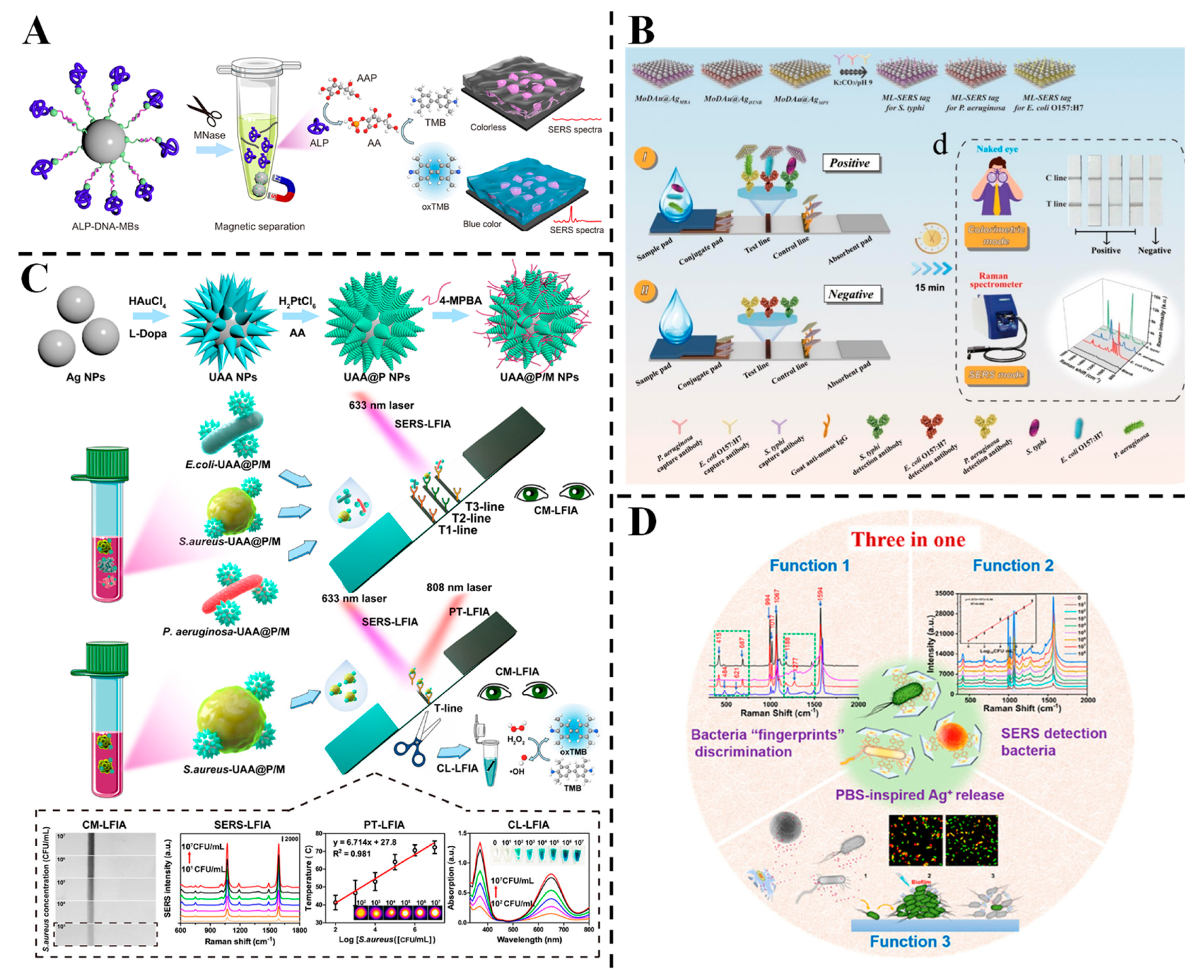
Multimodal signal sensing methods offer complementary advantages, enhancing the reliability of results, and have garnered significant interest in the fields of nanomaterials and sensing [79,110]. Huang et al. designed a close-packed Au@AgPt nanozyme array coupled with a cascade triggering strategy for detecting S. aureus in serum [62] (Figure 3A). The trimetallic nanozymes catalyzed TMB oxidation to generate SERS-active oxTMB, while micrococcal nuclease-secreted bacteria triggered signal attenuation via alkaline phosphatase-mediated reduction. This “on-to-off” strategy achieved LODs of 38 CFU/mL (colorimetric) and 6 CFU/mL (SERS), with linear ranges spanning five orders of magnitude. Shen et al. proposed a 3D membrane-like (ML) tag, MoDAu@Ag, integrated with lateral flow immunoassay (LFIA) for ultrasensitive multiplex pathogen detection [63] (Figure 3B). The tag, which is constructed by loading Au and Ag core–shell nanoparticles (Au@Ag NPs) onto polyethyleneimine (PEI)-modified MoS2 nanosheets, enables the simultaneous detection of P. aeruginosa, S. typhimurium and E. coli O157:H7 using three Raman molecules. Rapid qualitative (colorimetric) and quantitative (SERS) detection of different pathogens was achieved through visual color signal evaluation and characteristic Raman signal measurement within a single detection area. The SERS-encoding LFIA achieved LODs of 30~40 cells/mL across four orders of magnitude, with stable performance in clinical, food and environmental samples. This strategy overcomes the limitations of traditional LFIA, offering a promising platform for on-site, multiplex pathogen diagnosis. Huang et al. introduced multifunctional urchin-shaped Au-Ag@Pt nanoparticles (UAA@P NPs) integrated with LFIA for multimodal bacterial detection [111] (Figure 3C). The UAA@P/M NPs, functionalized with 4-mercaptophenylboronic acid (4-MPBA), enabled ultrasensitive SERS-LFIA, PT-LFIA and CL-LFIA with LODs of 3, 27 and 18 CFU/mL, respectively—330-fold, 37-fold and 55-fold more sensitive than visual CM-LFIA. Partial least-squares discriminant analysis (PLS-DA) accurately differentiated E. coli, S. aureus and P. aeruginosa in spiked blood samples, with recoveries of 90.3~108.8%. These multi-mode sensing strategies have good stability, anti-interference ability and high flexibility, but they are also prone to disadvantages such as more complex material preparation and increased detection processes.
With the continuous advancement of SERS substrate fabrication technology, specific functionalization treatments of precious metal nanoparticles, combined with statistical analysis methods, enable the identification, trace detection and inactivation of pathogenic microorganisms [20]. Zhou et al. reported the development of smart triple-functional Au-Ag-stuffed nanopancakes (AAS-NPs) for simultaneous SERS-based bacterial detection, discrimination and inactivation [64] (Figure 3D). The AAS-NPs were synthesized by Ag-etching Au@Ag NPs with K3[Fe(CN)6], forming unstable Prussian blue analogues to bind bacteria via cyano groups. Using 4-MPBA as both an SERS tag and internal standard, the platform achieved highly sensitive discrimination of E. coli, S. aureus and P. aeruginosa, with a detection limit of 7 CFU mL−1. The SERS sandwich structure (bacteria/4-MPBA/AAS-NPs) provided specific “fingerprint” spectra, while PBS-stimulated AAS-NPs released (Ag+) to kill > 99% of 1 × 10⁵ CFU mL−1 bacteria within 60 min, with antibacterial activity enhanced by 64~72-fold compared to untreated nanoparticles. Although the preparation process may be more complex, intelligent SERS substrates like AAS-NPs, which have multiple functions (identification, detection, inactivation, etc.) and achieve the integration of “detection-processing”, are an important development direction for SERS substrates in the future.

3.2. Detection of Mycotoxins

Mycotoxins are toxic secondary metabolites produced by various fungi and are commonly found in foods such as grains, fruits and vegetables [112,113]. These toxins are particularly prone to production under favorable temperature and humidity conditions during food processing, storage and transportation. Recent advancements in nanotechnology have led to the development of various Au-Ag BNPs substrates, which significantly enhance SERS signals, improve detection limits and make this technology more applicable to real-world samples [114]. Consequently, SERS has emerged as an active research area for the detection of mycotoxins, including aflatoxin, deoxynivalenol (DON), zearalenone (ZEN) and patulin (PAT). Efforts are ongoing to develop novel nanomaterials and methodologies for more sensitive and rapid analysis [115,116].
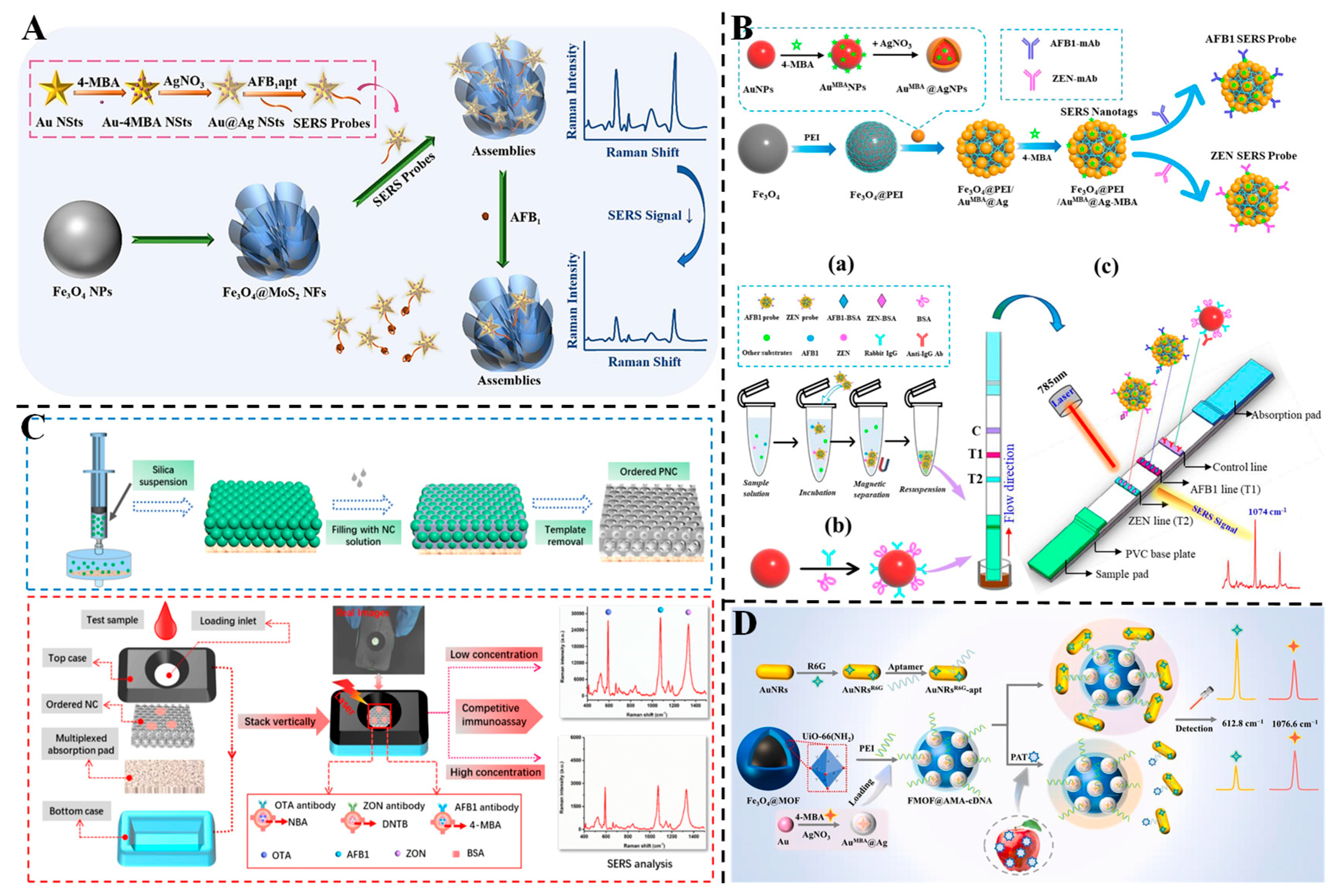
Aflatoxins are a common group of mycotoxins, comprising at least 18 known types. Among them, aflatoxin B1 (AFB1) is the most toxic and carcinogenic, making it a major focus of regulatory concern in agricultural products [117]. To enable ultrasensitive detection of AFB1, Chen et al. developed a three-dimensional (3D) plasmonic SERS aptasensor by integrating Au@Ag bimetallic nanostars (Au@Ag BNSs) with Fe3O4@MoS2 magnetic nanoflowers [69]. As illustrated in Figure 4A, the Au@Ag BNSs, functionalized with 4-mercaptobenzoic acid (4-MBA) and AFB1-specific aptamers, served as SERS probes, while the Fe3O4@MoS2 component enabled magnetic separation and signal enhancement via synergistic plasmonic and chemical effects. The sensor demonstrated a low LOD of 58.9 pg/mL and a linear detection range from 0.1 to 100 ng/mL. In spiked peanut samples, the recovery rates ranged from 95.53% to 98.73%, with RSDs below 5.2%, indicating high specificity and minimal matrix interference. The 3D architecture facilitated efficient signal enrichment and rapid magnetic separation, offering a promising approach for on-site AFB1 detection in food safety applications. Similarly, Tan et al. designed a dual-mode aptasensor combining colorimetric and label-free SERS detection for ultrasensitive quantification of AFB1 [70]. The sensor employed self-assembled Ag@Au IP6 bifunctional nanozymes, whose core–shell structure provided both peroxidase-like activity for TMB–H2O2 color reactions and plasmonic enhancement for SERS signal amplification. The aptasensor leveraged a hybridization chain reaction to amplify signal molecules, achieving an LOD of 0.58 pg/L and a linear range from 2 to 200 pg/L. In spiked red wine samples, recoveries ranged from 97.12% to 101.28% with RSDs within 5%, demonstrating resistance to matrix interference. The dual-mode strategy combined a visual colorimetric readout with quantitative SERS analysis, offering a robust platform for on-site and precise AFB1 monitoring in complex food matrices.
DON contamination not only reduces grain quality but also induces symptoms such as vomiting, anorexia and immunotoxicity and disrupts growth and reproduction [118]. Zhao et al. developed a core–shell–satellite nanoassembly consisting of an AuNR@Ag core, an ultrathin SiO2 layer and AuNP satellites, which served as an SERS immunoprobe for ultrasensitive detection of DON [71]. The anisotropic AuNR@Ag core facilitated strong localized electromagnetic coupling, while the SiO2 layer protected the Raman reporter (4-MBA) and enhanced colloidal stability. The AuNP satellites increased surface coverage and hotspot formation, thereby improving signal amplification. The SERS-LFIA exhibited an LOD of 0.053 fg/mL and a wide linear range from 0.1 fg/mL to 1 μg/mL. In spiked corn and wheat samples, recoveries ranged from 95.90% to 105.83%, with RSDs < 7.67%, demonstrating excellent accuracy and resistance to matrix interference. This strategy integrates LFIA and plasmonic enhancement, offering a robust platform for rapid, quantitative DON detection in complex food matrices, with potential applications for trace mycotoxin analysis in food safety monitoring.
ZEN has been identified as an endocrine disruptor that can impair the development of internal organs, lead to reproductive disorders and cause digestive dysfunction in animals, posing significant economic risks [119]. Humans are also at risk of ZEN exposure through the consumption of contaminated food. Therefore, the effective detection of ZEN is crucial for safeguarding human health [120]. Furthermore, the coexistence of multiple mycotoxins in food is common, and the potential synergistic effects and enhanced toxicity from their accumulation present serious threats to both human health and the economy [121]. Yin et al. developed a magnetic nanocomposite-based SERS-LFIA sensor for the simultaneous detection of AFB1 and ZEN in corn samples [72] (Figure 4B). The core–interlayer–satellite magnetic nanocomposites (Fe3O4@PEI/AuMBA@AgMBA) were employed as dual-functional SERS tags, integrating magnetic enrichment and Raman signal amplification. Under optimal conditions, the detection ranges of AFB1 and ZEN in corn samples were 0.1~10 μg/kg and 4~400 μg/kg, with LODs of 0.095 μg/kg and 1.896 μg/kg, respectively. The recoveries of spiked corn samples ranged from 91.28% to 109.52% for AFB1 and 94.71% to 108.15% for ZEN, with RSDs < 10%. The method showed good agreement with HPLC results and enabled rapid detection within 20 min, demonstrating its potential for on-site, simultaneous monitoring of mycotoxins with high sensitivity, accuracy and practicality. SERS technology offers advantages such as a sharp peak shape, high peak intensity and high sensitivity. When combined with various Raman tag molecules for SERS encoding, it facilitates multiplexed detection at a single site. As shown in Figure 4C, Chen et al. developed an SERS vertical flow assay (VFA) using photonic nitrocellulose (PNC) with an ordered inverse opal structure as the sensing substrate for simultaneous detection of ochratoxin A (OTA), AFB1 and ZEN [73]. Three SERS nanotags (AuNBA@Ag, Au4-MBA@Ag and AuDNTB@Ag) were encoded with distinct Raman reporters for each mycotoxin, enabling multiplexed detection. The slow-photon effect of the PNC substrate enhanced light–matter interaction, boosting SERS signal intensity. The assay achieved LODs of 8.2 fg/mL for OTA, 13.7 fg/mL for AFB1 and 47.6 fg/mL for ZEN, all below European Commission tolerable limits. Spiked recoveries in cereal samples ranged from 89.6% to 107.1%, consistent with ELISA results. The ordered porous structure improved reagent mixing and immune reaction kinetics, demonstrating the potential of the PNC-based VFA for high-throughput, on-site screening of multiple mycotoxins in food safety applications.
PAT causes damage to both the respiratory and urinary systems, resulting in nerve palsy, pulmonary edema and renal failure. It is also teratogenic, carcinogenic and highly toxic to human health [122,123]. Zhou et al. developed a magnetic metal–organic framework (MOF)-based ratiometric SERS aptasensor for the sensitive detection of PAT in apples [74] (Figure 4D). The sensor utilized Fe3O4@UiO-66-NH2 loaded with 4-MBA-labeled Au@Ag NPs as the SERS substrate, and AuNRs modified with rhodamine 6G (R6G) and aptamers as capture probes. The magnetic MOF facilitated efficient separation and enrichment of target analytes, while the dual-layer Raman reporter system ensured stable internal calibration. The SERS intensity ratio exhibited a negative correlation, with PAT concentrations ranging from 0.01 to 100 ng/mL, with an LOD of 0.0465 ng/mL. In spiked apple samples, recoveries ranged from 95.90% to 105.83%, demonstrating high resistance to interference and accuracy. The integration of magnetic separation and ratiometric sensing mitigates matrix effects, highlighting the aptasensor’s potential for practical mycotoxin detection in real food samples.

3.3. Detection of Pesticides

The use of pesticides has significantly enhanced agricultural productivity [124]. However, the widespread application of pesticides in agriculture has also led to serious pesticide residues, which pose inevitable risks to both the ecosystem and human health [125]. Consequently, the development of simple, rapid, sensitive and reliable detection methods for the swift monitoring of pesticide residues in food has become an urgent challenge. SERS technology is widely used in pesticide residue analysis due to its advantages, including non-destructive data acquisition, high sensitivity and fast detection speed [126,127].
Au-Ag alloys nanoparticles exhibit a synergistic effect between Au and Ag and a good SERS enhancement effect. As a result, many pesticide molecules with high SERS sensitivity are typically detected using label-free methods. Cho et al. synthesized silica nanoparticles surface-decorated with Au-Ag alloy nanoparticles (SiO2@AuAg) as stable SERS substrates [80]. Seed-mediated growth was used to control the Au:Ag ratio, optimizing hotspots for enhanced Raman signals. The substrates demonstrated high sensitivity with LODs of 6.95 × 10⁻⁷ M (crystal violet), 5.56 × 10⁻⁷ M (thiram) and 7.14 × 10⁻⁶ M (carbaryl), along with excellent reproducibility (RSD ≈ 8.4% after 3 days). The robust structure combined silica stability with Au-Ag plasmonic effects, enabling reliable detection of trace contaminants in environmental and food samples.
In addition, spectral data often contains noise from the sample substrate. Chemometric algorithms are valuable tools for extracting meaningful spectral information. Deep learning has advanced significantly in the field of chemometrics and has been successfully applied to the self-learning and modeling of spectroscopic data [128]. Li et al. designed Au-Ag octahedral hollow cages (Au-Ag OHCs) using Cu2O templates and coupled them with a convolutional neural network (CNN) for quantifying thiram and pymetrozine in tea [34]. The hollow structure and rough edges of Au-Ag OHCs enhanced electromagnetic fields, while the CNN algorithm improved spectral analysis, achieving LODs of 0.286 μg/kg (thiram) and 29 μg/kg (pymetrozine). The CNN model outperformed partial least squares and extreme learning machine, with correlation values reaching 0.995, demonstrating robust anti-interference capabilities and accuracy comparable to HPLC. The machine learning (especially CNN) in this method significantly improves the ability of complex spectral analysis, but the problems of model lightweighting and on-site deployment need to be solved.
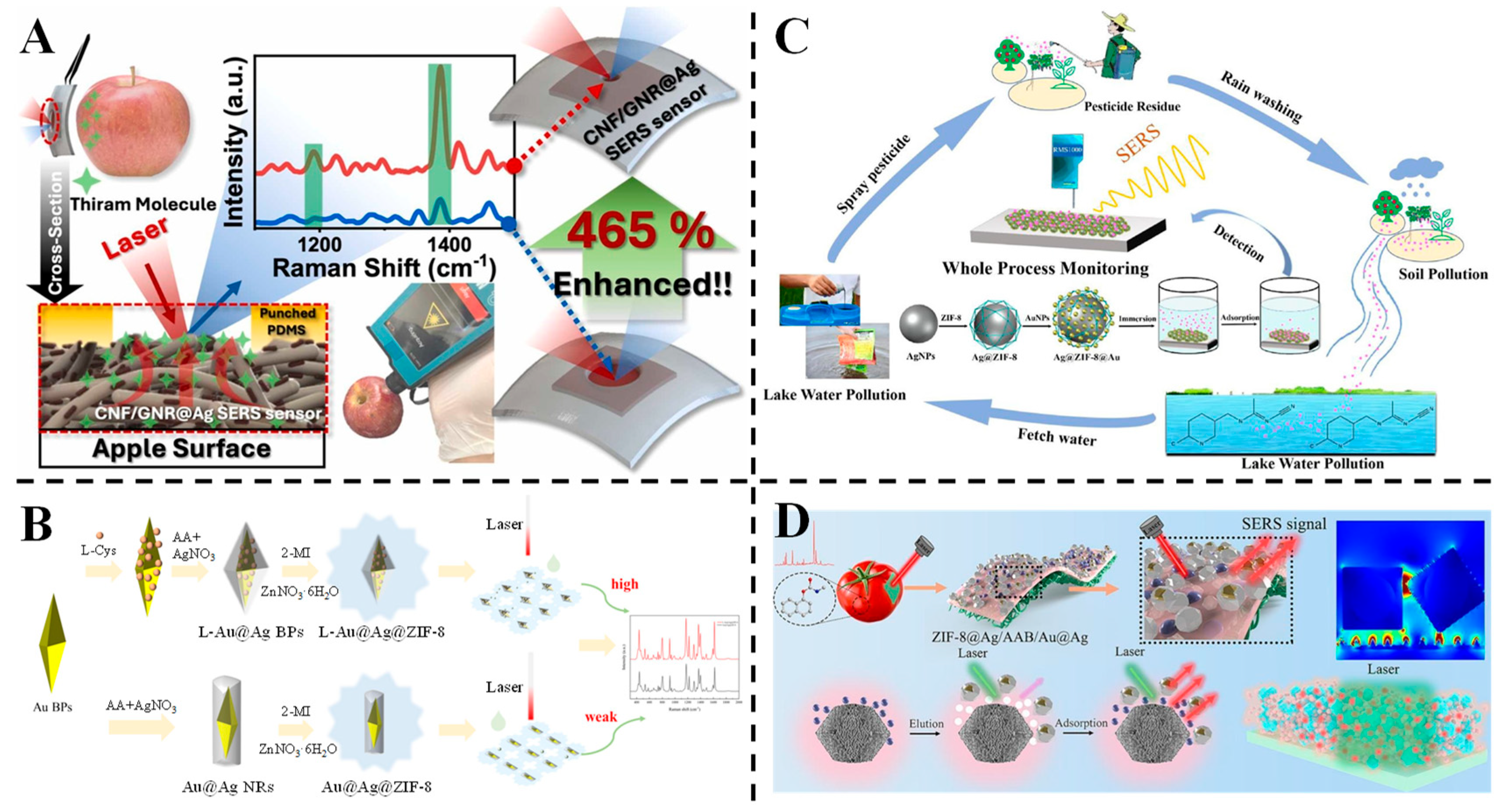
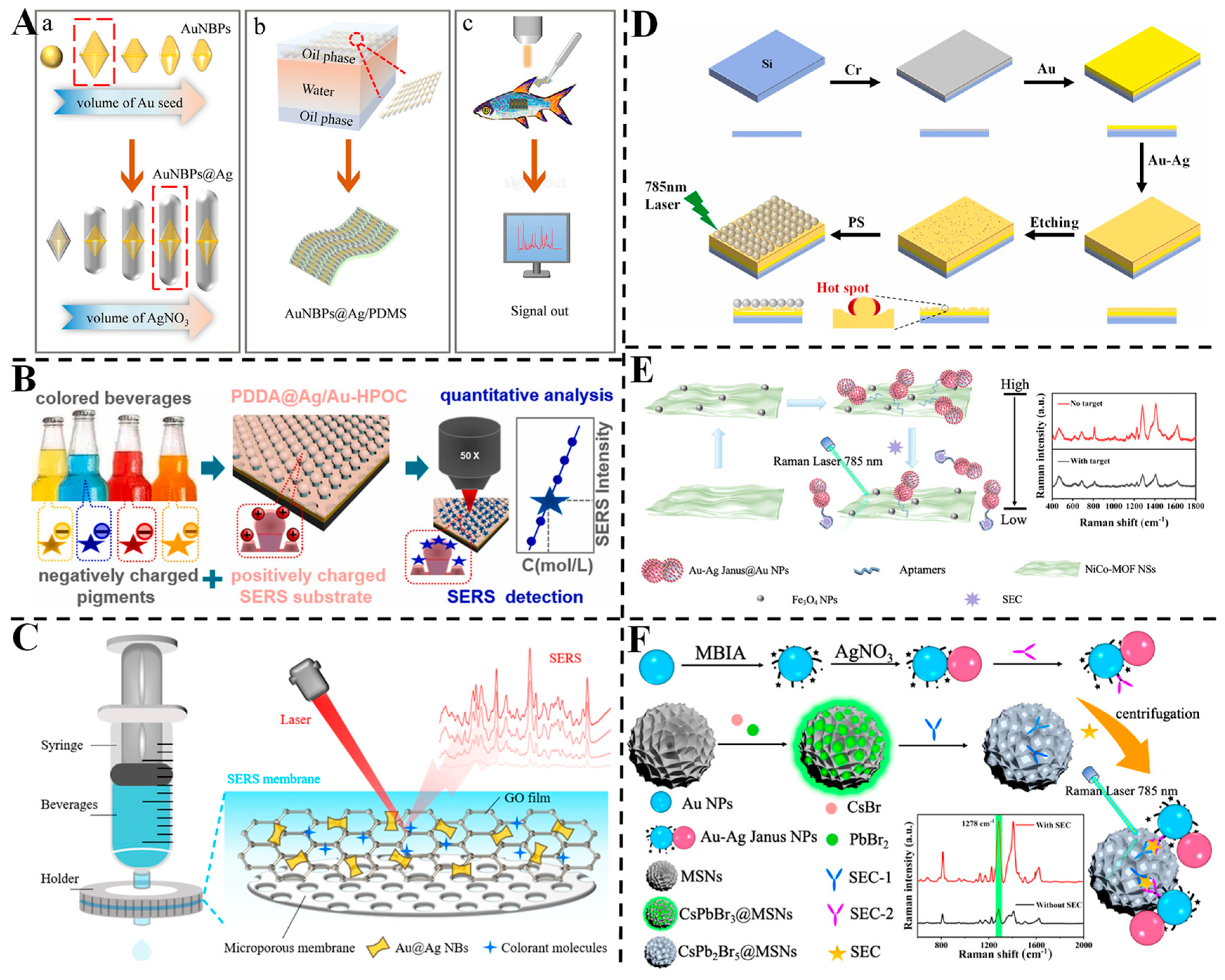
Au-Ag core–shell nanomaterials also have significant Raman enhancement effects and are widely used in pesticide residue detection. For example, Park et al. developed a flexible cellulose nanofiber (CNF)/AuNRs@Ag (Au@Ag NRs) SERS sensor for on-site pesticide detection, utilizing localized evaporation enrichment via hole-punched PDMS [81] (Figure 5A). The hydrophilic CNF matrix and Ag-coated AuNRs created dense hotspots, achieving an ultra-low LOD of 10−11 M for thiram on apple and chili pepper surfaces. The sensor demonstrated a 465% signal enhancement via controlled evaporation, overcoming coffee-ring effects and improving analyte concentration in the detection area. Portable Raman spectroscopy enabled real-time analysis, highlighting its adaptability for non-planar surfaces and complex food matrices. Raveendran and Docoslis developed a two-step method to fabricate Ag-Au core–shell nanostructures on microelectrodes for SERS-based toxicant detection [129]. Ag nanostructures were first grown via electrochemical deposition on microelectrodes functionalized with 11-mercaptoundecanoic acid, followed by a galvanic reaction with HAuCl4 to form the Au shell. The Ag-Au nanostructures exhibited a maximum SERS enhancement factor of 6.51 × 10⁵ and showed superior chemical stability, retaining 93 ± 7.3% of the signal after 24 h in PBS compared to 48 ± 5.0% for Ag nanostructures. Using multivariate analysis with classifiers, they successfully detected and quantified four toxicants (thiram, thiabendazole, malachite green and biphenyl-4-thiol) at 1 ppm with 100% accuracy. Additionally, flexible Ag-Au/PDMS SERS substrates were fabricated, enabling direct detection of thiram on apple peels without sample pretreatment, demonstrating their potential for on-site food and water safety monitoring.
MOFs, with their large surface area, are highly effective in adsorbing target substances [130]. They are increasingly being integrated with precious metal nanomaterials to create composite structures for novel SERS substrates. Yang et al. fabricated a chiral spiny L-Au@Ag@ZIF-8 (L-AAZ) three-layer core–shell SERS substrate for quinalphos detection in tangerines [82] (Figure 5B). The chiral L-Au@Ag bipyramids provided abundant hotspots, while ZIF-8 facilitated selective adsorption and molecular sieving, thereby reducing matrix interference. This substrate achieved a high enhancement factor (3.15 × 10⁵) and a low LOD of 6.56 × 10⁻¹⁰ M for malachite green, with stable performance over 55 days. Density functional theory (DFT) simulations and X-ray photoelectron spectroscopy (XPS) analysis revealed that hydrogen bonding and π-π interactions were primarily responsible for the adsorption process. Real-sample testing demonstrated an LOD of 10 ng/mL in tangerine juice, confirming its potential for sensitive food safety monitoring. Zhang et al. fabricated Ag@ZIF-8@Au nanoparticles as a robust SERS platform for acetamiprid detection [83] (Figure 5C). The core–shell structure integrated ZIF-8’s molecular enrichment capabilities with Ag/Au plasmonic hotspots, achieving an LOD of 9.027 × 10⁻¹⁰ M and a high enhancement factor of 4.3 × 10⁷. The platform exhibited excellent reproducibility (RSD ≤ 7.198%) and stability (RSD = 3.127% over 6 weeks), effectively detecting acetamiprid in complex matrices such as lake water, tea leaves and oranges. The synergistic electromagnetic and chemical enhancements, combined with ZIF-8’s adsorption capability, enabled reliable trace detection, underscoring its potential for on-site applications. Pan et al. constructed a ZIF-8@Ag/AAB/Au@Ag composite SERS paper sensor for carbaryl quantification, leveraging a 3D platform with cellulose paper as the substrate [84] (Figure 5D). ZIF-8 facilitated molecular adsorption, Ag layers enhanced plasmonic hotspots and Au@Ag NPs boosted electromagnetic coupling. The sensor demonstrated a linear range of 0.01~20 µg/mL and a low LOD of 5.72 × 10⁻³ µg/mL, with high reproducibility (RSD = 4.49%) and stability over 2 months. Finite-difference time-domain (FDTD) simulations confirmed enhanced electromagnetic fields at the Ag/Au@Ag NP interface, validating the full-dimensional divergence enhancement strategy for rapid on-site detection in fruits and vegetables. FDTD simulation provides a new idea for optimizing the structure of SERS nanomaterials or confirming the “hotspots” distribution of nanomaterials.

3.4. Detection of Antibiotics

Antibiotics are extensively used in industries such as aquaculture, livestock and poultry production, and agriculture to enhance economic productivity. Consequently, antibiotic residues are frequently detected in various food products, including meat, fish, milk, eggs and fruits. These residues accumulate in the human body through the food chain, potentially causing damage to organs and leading to conditions such as anemia and cardiovascular diseases [131]. Therefore, establishing an effective method for monitoring and analyzing veterinary drug residues in aquatic products is crucial for ensuring food safety.
Au-Ag nanoaggregates create nanoscale gaps that facilitate the generation of strong Raman hotspots, thereby enhancing detection sensitivity. Barveen et al. developed an Ag/Au/AgCl heterostructure via hydrothermal and photoreduction processes, serving as a reusable SERS substrate for ultrasensitive detection of analgesics and antibiotics [89] (Figure 6A). The nanoscale interparticle gaps in the heterostructure generated abundant hotspots, enhancing Raman signals through synergistic electromagnetic and chemical effects. The substrate exhibited wide linear ranges for paracetamol (10−1~10−10 M) and furazolidone (10−1~10−9 M), with detection limits as low as 2.8 × 10⁻¹² M and 1.9 × 10⁻¹¹ M, respectively. It enabled separate and multiplex detection in human urine samples with satisfactory recoveries (80~105%). The AgCl component facilitated photodegradation of adsorbed molecules, allowing the substrate to maintain ~84.2% SERS activity after five recycling tests, highlighting its potential for accurate and sustainable trace detection in complex biological matrices. Jiao et al. constructed aligned TiO2 nanorod arrays (NRAs) decorated with closely interconnected Au/Ag nanoparticles, creating a near-infrared (NIR) SERS active sensor for antibiotic detection in water [90]. The heterostructure exhibited strong absorption in the 400~1300 nm region, enabling ultrasensitive 785 nm laser-excited detection of ciprofloxacin (LOD = 10−9 M) and chloramphenicol (LOD = 10−8 M) in real-world water samples. The unique architecture provided abundant hotspots and efficient charge transfer, enhancing both electromagnetic and chemical contributions to SERS. Leveraging the photocatalytic degradation capability of TiO2, the sensor retained ~84.2% of its SERS activity after five cycles, demonstrating excellent reusability. This work presents a multifunctional NIR-SERS platform with high sensitivity and sustainability for monitoring antibiotic residues in environmental and biological matrices.
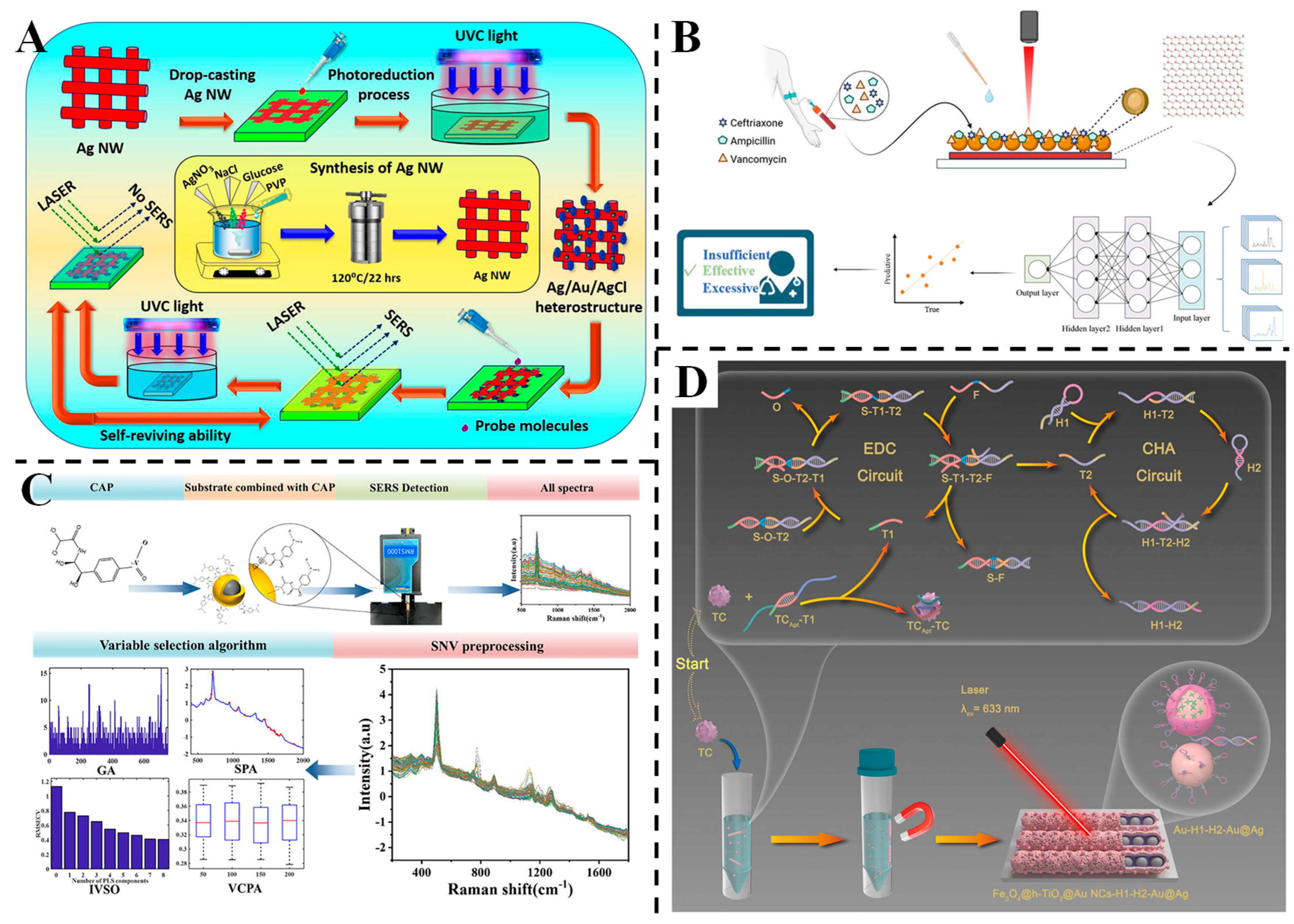
Cao et al. designed a highly sensitive SERS biosensor using WS2/Au@Ag nanocomposites for detecting ceftriaxone, ampicillin and vancomycin in serum, integrated with a 2D-CNN deep learning model for concentration prediction [91] (Figure 6B). The hybrid substrate, combining two-dimensional WS2 and noble metal core–shell nanostructures, achieved an LOD of 10−14 M for R6G and exhibited linear responses for the three antibiotics across 0.5~1000 μg/mL. The 2D-CNN model, which converts SERS spectra into 2D images via the short-time Fourier transform, demonstrated excellent regression performance with R2 values of 0.9993 and 0.9997 for ceftriaxone and ampicillin in mixed serum solutions, surpassing traditional machine learning methods. Yang et al. designed a rapid method for chloramphenicol (CAP) residue detection in tilapia using Ag@Au NPs as SERS nanosensors coupled with chemometric algorithms [92] (Figure 6C). The Ag@Au NPs exhibited a high enhancement factor (2.67 × 10⁶), and the variable combination population analysis-partial least-squares (VCPA-PLS) model with standard normal variate pretreatment achieved an LOD of 1 × 10⁻⁵ μg/mL. The method showed excellent predictive performance, with recoveries ranging from 98 to 104% in real samples, demonstrating its reliability and applicability for sensitive and specific CAP determination in complex food matrices.
Lv et al. constructed a cascade amplification SERS aptasensor for tetracycline (TC) detection using Fe3O4@h-TiO2/Au nanochains and Au@Ag NPs integrated with enzyme-free DNA circuits [93] (Figure 6D). The dual amplification strategy enabled a low LOD of 15.91 pg/mL and a linear range from 0.01 to 100 ng/mL. The magnetic Fe3O4 facilitated easy separation, while the Au@Ag NPs provided strong SERS enhancement. The sensor exhibited excellent specificity and storage stability, with recoveries in real samples (fish, milk and lake water) ranging from 93.2 to 105.3%, showcasing its utility for sensitive and efficient TC analysis in food and environmental safety.

3.5. Detection of Heavy Metals

Heavy metal ions accumulate in organisms through the food chain within the ecological cycle, posing significant harm to animals, plants and humans [132]. In recent decades, the threat posed by food contaminated with heavy metals has raised substantial concern among the public [133]. Once absorbed, these ions can bind to proteins, leading to their inactivation and compromising the health and safety of organisms. Consequently, the rapid, cost-effective and accurate detection of heavy metal ions is crucial for human health monitoring and environmental protection [134].
Xu et al. developed a highly sensitive SERS sensor for ultratrace detection of Cr (VI), utilizing methimazole-functionalized Au@Ag nano-sea urchins (Au@Ag NSUs) integrated with paper tips [94]. The Au@Ag NSUs combined the stability of Au cores with the strong SERS enhancement provided by the Ag shells. The paper tip facilitated gravity-driven analyte enrichment at the tip, significantly amplifying the signal. The redox reaction between methimazole and Cr (VI) modulated the SERS signal intensity, enabling an LOD as low as 0.956 ng/L. The sensor demonstrated excellent reproducibility (RSD ≤ 7.35%) and stability, retaining 92% of its initial signal intensity after 30 days. Analysis of real water samples, including lake and tap water, yielded recoveries between 98.17% and 105.73%, confirming its reliability for environmental monitoring. This work underscores the potential of paper-based SERS platforms for on-site, cost-effective heavy metal analysis.
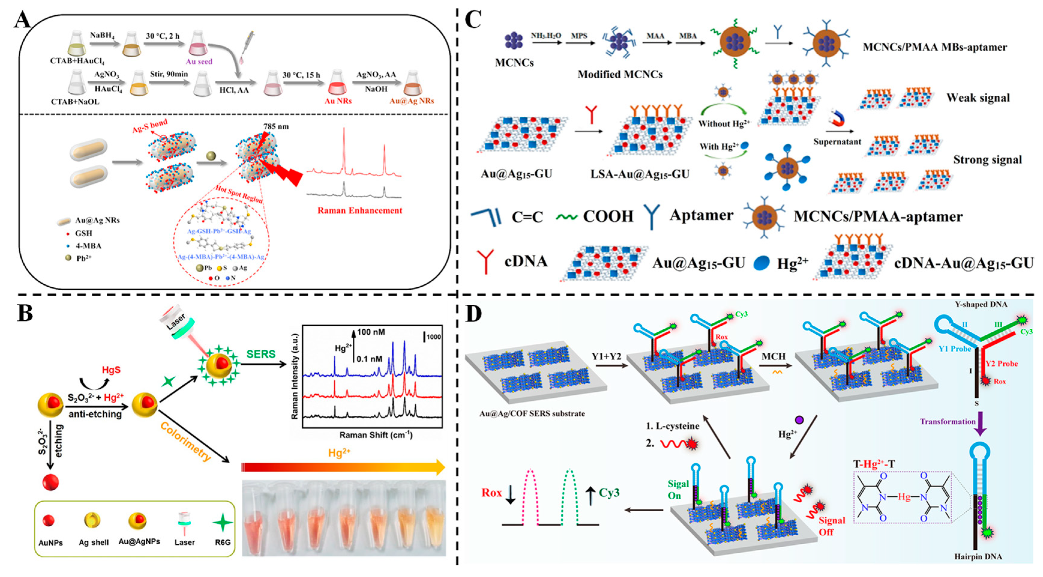
Liu et al. designed an SERS sensor for Pb2+ detection using glutathione and 4-MBA-functionalized Au@Ag core–shell nanorods (Au@Ag NRs) [95] (Figure 7A). The free carboxyl groups of glutathione and 4-MBA chelated Pb2+, inducing nanoparticle aggregation and enhancing “hotspots” for SERS signal amplification. Under optimized conditions, the sensor showed a linear response from 0.5 to 1000 μg/L, with an LOD of 0.021 μg/L and a high enhancement factor (1.0328 × 10⁷). Real samples, including tea powder and glutinous rice flour, exhibited recoveries of 81.31~101.12% and low RSDs (≤9.82%), verifying its accuracy for food safety monitoring. The strategy combines specific chelation with plasmonic enhancement, enabling rapid and sensitive Pb2+ analysis in complex matrices.
Wang et al. proposed a dual-mode strategy for Hg2+ detection using Au@AgNPs whose Ag shells undergo controllable etching by thiosulfate ions [96] (Figure 7B). In the presence of Hg2+, the formation of insoluble HgS suppresses shell etching, restoring the orange colloidal color and enhancing the SERS signal of R6G. The colorimetric method achieved a detection limit of 2 μM (naked eye) and 0.2 μM (UV–vis), while the SERS method offered a far lower limit of 0.1 nM with a linear range of 0.1 nM~1 μM. This dual-mode approach integrates visual screening and ultrasensitive quantification, showcasing versatility for field-based and laboratory applications. Li et al. fabricated a dual-channel biosensor leveraging Au@Ag/graphene-upconversion (Au@Ag-GU) nanohybrids as multifunctional signal indicators for simultaneous SERS and fluorescence detection of Hg2+ [97] (Figure 7C). Magnetite–polymethacrylic acid magnetic beads (MCNCs/PMAA MBs) conjugated with aptamers captured Hg2+, triggering the release of cDNA-Au@Ag-GU into supernatants. The Au@Ag-GU hybrid enhanced both signals: SERS achieved a detection limit of 0.33 ppb, while fluorescence offered 1 ppb, with linear ranges of 0.1~800 nM and 0.001~100 nM, respectively. Spiked tap water and milk samples showed recoveries of 96.0~109.8% with low RSDs (≤4.76%), demonstrating its applicability for food and environmental analysis. The dual-mode design integrates the advantages of high sensitivity and a broad linear range, addressing diverse detection needs. Li et al. introduced a ratiometric SERS sensor based on Au@Ag NPs embedded in covalent organic frameworks (COFs) combined with Y-shaped DNA labeled with two Raman reporters (Cy3 and Rox) [98] (Figure 7D). The Au@Ag/COF substrate provided uniform electromagnetic fields, while the Y-shaped DNA design brought reporters closer to the surface, enhancing signal coupling. In the presence of Hg2+, the formation of T-Hg2+-T complexes increased Cy3 signals (“signal-on”) and decreased Rox signals (“signal-off”), resulting in an LOD of 5.0 × 10⁻¹⁶ M, among the lowest reported for Hg2+. Real samples (river water, tap water and milk) exhibited recoveries of 97.9~104.0% and excellent reproducibility (RSD ≤ 8.0%). This strategy integrates structural design, COF stability and dual-signal correction, offering a robust platform for ultrasensitive heavy metal detection in complex matrices.

3.6. Detection of Other Contaminants

Other food contaminants include food additives, nanoplastics, bacterial toxins, etc. Due to their toxicity, carcinogenicity or mutagenicity, they can cause acute and chronic health problems. Because it can achieve real-time non-destructive testing and has the advantages of high sensitivity and accuracy, SERS technology is used in the detection of these substances. Based on Au-Ag BNPs, the SERS can be functionalized and modified to achieve highly sensitive trace detection of harmful substances, further ensuring food safety.
Niu et al. designed Au nanobipyramid@Ag (Au NBPs@Ag) nanorods with tunable aspect ratios via wet chemical synthesis, optimizing their plasmonic properties for enhanced SERS performance [101] (Figure 8A). By controlling the amount of Ag precursor, nanorods with an optimal aspect ratio of 4.43 demonstrated strong electromagnetic coupling, enabling highly sensitive detection of malachite green. The flexible PDMS-supported substrate, fabricated via interfacial self-assembly, achieved an LOD of 0.1 nM and a wide linear range (0.1 nM to 10 μM). When applied to river bass samples, the substrate exhibited reliable recoveries (89.8~121%) and low RSDs (<15%), highlighting its potential for environmental and food safety monitoring of trace contaminants. Zhang et al. engineered a poly(diallyldimethylammonium chloride)/Ag/Au hybrid plasmonic optical cavity (PDDA@Ag/Au-HPOC) substrate via UV holographic lithography, utilizing electrostatic interactions between positively charged PDDA and negatively charged dyes for enhanced adsorption [102] (Figure 8B). This strategy enabled direct SERS detection of dyes like amaranth and Allura red in unopened beverages without pretreatment, achieving LODs of 0.3022 and 0.2482 mg/L, respectively. The substrate exhibited excellent selectivity (no interference from neutral/positively charged molecules) and stability (signal retention over 9 days, RSD < 12.7%). A linear response (R2 > 0.96) and high recovery rates (95~110%) in real drinks validate its applicability for on-site food additive monitoring. Kong et al. developed a graphene oxide (GO)/Au@Ag nanobone (NB) membrane, combining the enrichment capability of GO with the plasmonic enhancement of Au@Ag for simultaneous extraction and SERS detection of colorants [103] (Figure 8C). The microporous membrane provided a large surface area for dense “hotspots” formation, achieving an LOD of 1.12 × 10⁻⁹ M for R6G and detecting six colorants (including carmine and brilliant blue) at concentrations below regulatory limits. A support vector machine (SVM) model enabled accurate colorant identification (94.8% accuracy), even for those with overlapping Raman peaks. Successful detection in commercial beverages (e.g., energy drinks and bayberry wine) demonstrates its potential for rapid, on-site food safety screening.
Liu et al. presented an easily fabricated Ag core embedded Au film (Ag@Au Film) for direct SERS detection of polystyrene (PS) nanoplastics [104] (Figure 8D). By optimizing the Ag-Au gap via nitric acid etching, the substrate achieved LODs of 25~50 μg/mL for PS particles (50~310 nm) in deionized and river water, with linear ranges up to 6.25 mg/mL (R2 > 0.97). FDTD simulations revealed enhanced electromagnetic fields at the Ag-Au interface, driving signal amplification. The substrate showed good reproducibility (RSD < 8.1%) and detected multiple plastics (PE, PET and PP), offering a simple, sensitive method for environmental microplastic analysis without complex pretreatment.
Xu et al. developed an SERS immunoassay for the sensitive detection of Staphylococcus aureus enterotoxin C (SEC) using Au-Ag Janus nanoparticle (NP)/perovskite composites [106] (Figure 8E). The plasmonic Au-Ag Janus NPs, functionalized with 2-mercaptobenzoimidazole-5-carboxylic acid (MBIA) ligands, exhibited inherent SERS activity. CsPbBr3@mesoporous silica nanomaterials (MSNs) were synthesized and transformed into CsPb2Br5@MSNs in the aqueous phase. Paired SEC antibody–antigen interactions drove the formation of Au-Ag Janus NP–CsPb2Br5@MSN composites, which showed amplified SERS activity due to electromagnetic field enhancement and electron transfer mechanisms. A linear correlation was established between the SERS signals of the composites and the SEC concentration, achieving an LOD of 0.83 pg/mL. This additive-free SERS immunoassay is simple, sensitive and reproducible, demonstrating its potential for food safety monitoring by harnessing the synergistic effects of plasmonic NPs and perovskite materials. Xu et al. reported the tunable preparation of SERS-active Au-Ag Janus@Au NPs for label-free detection of SEC [105] (Figure 8F). By adjusting the pH of the reaction system, solid and hollow Au-Ag Janus@Au NPs were synthesized, with solid structures exhibiting enhanced SERS activity due to strong plasmonic coupling between the Au dots and Au-Ag Janus NPs. The solid Au-Ag Janus@Au NPs showed a 2.27-fold higher SERS intensity than the Au-Ag Janus NPs and a 17.46-fold higher intensity than their hollow counterparts. These NPs were employed as label-free probes in an SERS aptasensor, where aptamer-modified Au-Ag Janus@Au NPs were assembled on NiCo-MOF/Fe3O4 nanosheets. The aptasensor demonstrated a low LOD of 0.55 pg/mL and excellent stability, with recovery rates in milk samples ranging from 94.4% to 98.1%. This strategy offers a sensitive and reliable method for SEC detection in complex food matrices, showcasing the advantages of tunable nanostructures for improved SERS performance.

4. Challenges and Outlook

SERS technology offers several advantages, including rapid detection, high sensitivity, non-destructiveness, minimal sample preparation, immunity to water interference and reduced photobleaching. Au-Ag BNPs are ideal materials for SERS substrates due to the stability of Au and the high enhancement activity of Ag. As a result, this technology has gained significant attention from researchers in the field of food contaminant detection. Its practical application in complex food matrices inevitably faces some challenges and opportunities.

4.1. Challenges

SERS technology using Au-Ag BNPs faces notable challenges in practical food contaminant detection. First, synthesizing Au-Ag BNPs with a uniform morphology and high stability remains problematic. Core–shell nanoparticles often exhibit irregular structures like nanorods or nanostars when prepared via seed growth methods, relying on surfactants such as cetyltrimethyl ammonium bromide (CTAB) that introduce background interference and compromise batch-to-batch repeatability. Although the Ag shell on the outside can achieve a better Raman enhancement effect than the Au shell, it also has disadvantages such as being prone to oxidation, resulting in poor long-term stability. Therefore, a protective layer can be added outside the Ag shell, such as silica, a surfactant, a polymer, etc. [135,136]. Additionally, controlling the Ag shell thickness is equally challenging. If the Ag shell is too thin (<3 nm), the localized surface plasmon resonance effect will be inadequate; if it is too thick (>10 nm), the core “hotspots” will be shielded [137]. Second, complex food matrices pose interference, as large molecules like proteins and polysaccharides adsorb onto nanoparticle surfaces, hindering target binding and decreasing Raman signals, especially in label-free applications [138]. While sample pretreatment could mitigate this issue, it increases time consumption, complicates procedures and may not meet the requirements for on-site testing. Lastly, standardization and commercialization hurdles persist, including a lack of unified substrate preparation and testing protocols across laboratories (reducing data comparability) and the high costs of Raman instrumentation and specialized synthesis of Au-Ag BNPs, restricting accessibility for small enterprises and field use.

4.2. Outlook

In recent years, SERS technology based on Au-Ag BNPs has also ushered in new development opportunities. First, precise control over shell structures is enabling the development of smart substrates. For example, a core–shell–satellite structure with “hotspot networks” can be formed by depositing secondary Ag NPs onto the surface of Au@Ag NPs [77]. MOFs (such as ZIF-8) or graphene coatings can be introduced to increase adsorption sites and form porous/hollow structures [139]. Magnetic composite nano-substrates can be prepared, and SERS can be combined with magnetic separation to achieve rapid enrichment and detection of target substances. The photothermal effect of Au@Ag NPs can inactivate microorganisms, facilitating integrated “detection–disinfection” processes [140]. Second, surface modification with aptamers, antibodies or molecular imprinting enhances anti-interference performance and selectivity. In label-free detection, Raman-active molecules within the Raman-silent spectral window (1800~2800 cm−1) are employed to enhance resistance to optical interference [141]. In addition, the geometric structure of Au-Ag BNPs significantly influences LSPR and electromagnetic field distribution. Generally, an increase in nanoparticle size leads to a redshift in the LSPR peak and an exponential enhancement of the electromagnetic field intensity. Sharp structural features can markedly amplify the local field strength due to the tip effects. Moreover, a smaller interparticle distance results in a stronger electric field, also exhibiting exponential enhancement. The dielectric properties and material composition of the nanoparticles further affect the electromagnetic field intensity. The FDTD simulation results provide valuable guidance for the rational design of SERS substrates and can verify the spatial distribution or variation in electromagnetic fields in practical applications. Furthermore, detection methodologies are advancing through the integration of portable platforms (e.g., paper-based or flexible substrates, and microfluidic chips) for on-site analysis, as well as microarrays for high-throughput screening. Finally, the incorporation of artificial intelligence (AI) algorithms facilitates automated spectral analysis and real-time decision-making, thereby promoting more efficient, scalable and intelligent applications of SERS in food safety. For example, conventional machine learning algorithms (such as PCA, LDA, SVM and random forest) are widely employed for feature extraction and dimensionality reduction. Deep learning (such as CNNs, recurrent neural networks and autoencoders) can achieve end-to-end automatic feature learning, noise reduction, spectral information extraction and accuracy enhancement [142]. The integration of these algorithms with detection hardware (e.g., portable Raman spectrometers, microfluidic devices and flexible wearable sensors) facilitates real-time monitoring and intelligent decision-making.

5. Conclusions

SERS, with its outstanding performance in terms of its ultra-sensitivity, non-destructiveness and capability for molecular fingerprint has been employed in food contaminant detection. The primary strategies for pollutant detection via SERS can be broadly categorized into two methods: label-free detection and labeled detection. The controllable synthesis of efficient SERS-active nanomaterials is essential for the successful application of SERS. Over time, SERS substrates have evolved from simple noble metal-based structures to more advanced composite materials and polymetallic substrates. Au-Ag BNPs exhibit a synergistic enhancement effect, which results in a stronger SERS signal compared to single-component noble metal nanomaterials. To effectively apply SERS for the rapid detection of harmful pollutants, it is crucial to address challenges related to the stability and functionalization of nanoparticles, interference from sample matrices, and the need for on-site portability. Future research should focus on the development of intelligent, integrated substrates and cost-effective, user-friendly SERS detection platforms. Additionally, integrating cutting-edge technologies such as artificial intelligence and microfluidics will help meet the demands for on-site detection.

Author Contributions

Conceptualization, P.Y.; resources, P.Y. and J.C.; writing—original draft preparation, P.Y. and C.S.; writing—review and editing, X.Y., J.C., C.L. and Z.Y.; supervision, P.Y.; funding acquisition, P.Y. and C.S. All authors have read and agreed to the published version of the manuscript.

Funding

The work was supported by the Open Fund of Key Laboratory of Modern Agricultural Equipment and Technology, the Ministry of Education (MAET202308), the Priority Academic Program Development of Jiangsu Higher Education Institutions (grant number PAPD-2023-87), and the project of the Zhenjiang Youth Science and Technology Talent Support Program (ZJRCTJ-2024-038).

Institutional Review Board Statement

Not applicable.

Informed Consent Statement

Not applicable.

Data Availability Statement

The original contributions presented in the study are included in the article; further inquiries can be directed to the corresponding author.

Conflicts of Interest

The authors declare no conflicts of interest.

References

  1. Wang, L.; Huang, X.; Wang, C.; Tian, X.; Chang, X.; Ren, Y.; Yu, S. Applications of surface functionalized Fe3O4 NPs-based detection methods in food safety. Food Chem. 2021, 342, 128343. [Google Scholar] [CrossRef] [PubMed]
  2. Kang, W.; Lin, H.; Jiang, H.; Yao-Say Solomon Adade, S.; Xue, Z.; Chen, Q. Advanced applications of chemo-responsive dyes based odor imaging technology for fast sensing food quality and safety: A review. Compr. Rev. Food Sci. Food Saf. 2021, 20, 5145–5172. [Google Scholar] [CrossRef] [PubMed]
  3. Song, S.-H.; Gao, Z.-F.; Guo, X.; Chen, G.-H. Aptamer-Based Detection Methodology Studies in Food Safety. Food Anal. Methods 2019, 12, 966–990. [Google Scholar] [CrossRef]
  4. Xia, X.; Zhang, B.; Wang, J.; Li, B.; He, K.; Zhang, X. Rapid Detection of Escherichia coli O157:H7 by Loop-Mediated Isothermal Amplification Coupled with a Lateral Flow Assay Targeting the z3276 Genetic Marker. Food Anal. Methods 2022, 15, 908–916. [Google Scholar] [CrossRef]
  5. Zhang, Y.; Li, M.; Cui, Y.; Hong, X.; Du, D. Using of Tyramine Signal Amplification to Improve the Sensitivity of ELISA for Aflatoxin B1 in Edible Oil Samples. Food Anal. Methods 2018, 11, 2553–2560. [Google Scholar] [CrossRef]
  6. Sharma, A.S.; Ali, S.; Sabarinathan, D.; Murugavelu, M.; Li, H.; Chen, Q. Recent progress on graphene quantum dots-based fluorescence sensors for food safety and quality assessment applications. Compr. Rev. Food Sci. Food Saf. 2021, 20, 5765–5801. [Google Scholar] [CrossRef]
  7. Zhang, X.; Zhou, Y.; Huang, X.; Hu, X.; Huang, X.; Yin, L.; Huang, Q.; Wen, Y.; Li, B.; Shi, J.; et al. Switchable aptamer-fueled colorimetric sensing toward agricultural fipronil exposure sensitized with affiliative metal-organic framework. Food Chem. 2023, 407, 135115. [Google Scholar] [CrossRef]
  8. Qin, C.; Guo, W.; Liu, Y.; Liu, Z.; Qiu, J.; Peng, J. A Novel Electrochemical Sensor Based on Graphene Oxide Decorated with Silver Nanoparticles–Molecular Imprinted Polymers for Determination of Sunset Yellow in Soft Drinks. Food Anal. Methods 2017, 10, 2293–2301. [Google Scholar] [CrossRef]
  9. Ma, L.; Yang, X.; Xue, S.; Zhou, R.; Wang, C.; Guo, Z.; Wang, Y.; Cai, J. “Raman plus X” dual-modal spectroscopy technology for food analysis: A review. Compr. Rev. Food Sci. Food Saf. 2025, 24, e70102. [Google Scholar] [CrossRef]
  10. Fleischmann, M.; Hendra, P.J.; McQuillan, A.J. Raman spectra of pyridine adsorbed at a silver electrode. Chem. Phys. Lett. 1974, 26, 163–166. [Google Scholar] [CrossRef]
  11. Ma, L.; Han, E.; Yin, L.; Xu, Q.; Zou, C.; Bai, J.; Wu, W.; Cai, J. Simultaneous detection of mixed pesticide residues based on portable Raman spectrometer and Au@Ag nanoparticles SERS substrate. Food Control 2023, 153, 109951. [Google Scholar] [CrossRef]
  12. Sun, J.; Shi, Z.; Wang, L.; Zhang, X.; Luo, C.; Hua, J.; Feng, M.; Chen, Z.; Wang, M.; Xu, C. Construction of a microcavity-based microfluidic chip with simultaneous SERS quantification of dual biomarkers for early diagnosis of Alzheimer’s disease. Talanta 2023, 261, 124677. [Google Scholar] [CrossRef] [PubMed]
  13. Ge, S.; Chen, G.; Deng, J.; Gu, Y.; Mao, Y.; Zhou, X.; Li, G. Multiplex signal amplification strategy-based early-stage diagnosis of Parkinson’s disease on a SERS-enabled LoC system. Anal. Chim. Acta 2023, 1247, 340890. [Google Scholar] [CrossRef]
  14. Wang, L.; Gao, X.; Han, J.; Yuan, J.; Wang, Z.; Ma, X. Core-shell SERS nanoprobes for ROS detection and imaging during apoptosis. Food Biosci. 2025, 67, 106339. [Google Scholar] [CrossRef]
  15. Zhang, A.; Ding, Z.; Shen, Z.; Yan, Z.; Han, K.; Li, J.; Zhang, M.; Zhang, W. Nano-arrayed Cu2S@MoS2 heterojunction SERS sensor for highly sensitive and visual detection of polystyrene in environmental matrices. Talanta 2025, 292, 127934. [Google Scholar] [CrossRef]
  16. Jiang, L.; Hassan, M.M.; Ali, S.; Li, H.; Sheng, R.; Chen, Q. Evolving trends in SERS-based techniques for food quality and safety: A review. Trends Food Sci. Technol. 2021, 112, 225–240. [Google Scholar] [CrossRef]
  17. Li, H.; Chen, Q.; Ouyang, Q.; Zhao, J. Fabricating a Novel Raman Spectroscopy-Based Aptasensor for Rapidly Sensing Salmonella typhimurium. Food Anal. Methods 2017, 10, 3032–3041. [Google Scholar] [CrossRef]
  18. Kneipp, K.; Kneipp, H.; Kneipp, J. Surface-Enhanced Raman Scattering in Local Optical Fields of Silver and Gold Nanoaggregates from Single-Molecule Raman Spectroscopy to Ultrasensitive Probing in Live Cells. Acc. Chem. Res. 2006, 39, 443–450. [Google Scholar] [CrossRef]
  19. Long, Y.; Li, H.; Du, Z.; Geng, M.; Liu, Z. Confined Gaussian-distributed electromagnetic field of tin(II) chloride-sensitized surface-enhanced Raman scattering (SERS) optical fiber probe: From localized surface plasmon resonance (LSPR) to waveguide propagation. J. Colloid Interface Sci. 2021, 581, 698–708. [Google Scholar] [CrossRef]
  20. Liu, S.; Li, H.; Hassan, M.M.; Zhu, J.; Wang, A.; Ouyang, Q.; Zareef, M.; Chen, Q. Amplification of Raman spectra by gold nanorods combined with chemometrics for rapid classification of four Pseudomonas. Int. J. Food Microbiol. 2019, 304, 58–67. [Google Scholar] [CrossRef]
  21. Wang, J.; Ahmad, W.; Mehedi Hassan, M.; Zareef, M.; Viswadevarayalu, A.; Arslan, M.; Li, H.; Chen, Q. Landing microextraction sediment phase onto surface enhanced Raman scattering to enhance sensitivity and selectivity for chromium speciation in food and environmental samples. Food Chem. 2020, 323, 126812. [Google Scholar] [CrossRef]
  22. Li, H.; Geng, W.; Zheng, Z.; Haruna, S.A.; Chen, Q. Flexible SERS sensor using AuNTs-assembled PDMS film coupled chemometric algorithms for rapid detection of chloramphenicol in food. Food Chem. 2023, 418, 135998. [Google Scholar] [CrossRef] [PubMed]
  23. Yin, L.; You, T.; Arslan, M.; El-Seedi, H.R.; Guo, Z.; Zou, X.; Cai, J. Dual-layers Raman reporter-tagged Au@Ag combined with core-satellite assemblies for SERS detection of Zearalenone. Food Chem. 2023, 429, 136834. [Google Scholar] [CrossRef] [PubMed]
  24. Liu, X.; Ye, Z.; Xiang, Q.; Xu, Z.; Yue, W.; Li, C.; Xu, Y.; Wang, L.; Cao, X.; Zhang, J. Boosting electromagnetic enhancement for detection of non-adsorbing analytes on semiconductor SERS substrates. Chem 2023, 9, 1464–1476. [Google Scholar] [CrossRef]
  25. Lv, Q.; Tan, J.; Wang, Z.; Gu, P.; Liu, H.; Yu, L.; Wei, Y.; Gan, L.; Liu, B.; Li, J.; et al. Ultrafast charge transfer in mixed-dimensional WO3−x nanowire/WSe2 heterostructures for attomolar-level molecular sensing. Nat. Commun. 2023, 14, 2717. [Google Scholar] [CrossRef]
  26. Qiu, B.; Xing, M.; Yi, Q.; Zhang, J. Chiral Carbonaceous Nanotubes Modified with Titania Nanocrystals: Plasmon-Free and Recyclable SERS Sensitivity. Angew. Chem. Int. Ed. 2015, 54, 10643–10647. [Google Scholar] [CrossRef] [PubMed]
  27. Yang, Z.; Lin, Z.; Yang, J.; Wang, J.; Yue, J.; Liu, B.; Jiang, L. Fabrication of porous noble metal nanoparticles based on laser ablation toward water and dealloying for biosensing. Appl. Surf. Sci. 2022, 579, 152130. [Google Scholar] [CrossRef]
  28. Yin, L.; You, T.; El-Seedi, H.R.; El-Garawani, I.M.; Guo, Z.; Zou, X.; Cai, J. Rapid and sensitive detection of zearalenone in corn using SERS-based lateral flow immunosensor. Food Chem. 2022, 396, 133707. [Google Scholar] [CrossRef]
  29. Jiao, S.; Wu, L.; Jiang, H.; Zhang, S.; Han, Y.; Huang, H. A review on SERS-based techniques for mycotoxin detection: From construction to application. TrAC Trends Anal. Chem. 2025, 184, 118120. [Google Scholar] [CrossRef]
  30. Chang, K.; Zhao, Y.; Wang, M.; Xu, Z.; Zhu, L.; Xu, L.; Wang, Q. Advances in metal-organic framework-plasmonic metal composites based SERS platforms: Engineering strategies in chemical sensing, practical applications and future perspectives in food safety. Chem. Eng. J. 2023, 459, 141539. [Google Scholar] [CrossRef]
  31. Jiao, T.; Dong, C.; Zhu, A.; Ahmad, W.; Peng, L.; Wu, X.; Chen, Q.; Wei, J.; Chen, X.; Qin, O.; et al. AFB1-responsive mesoporous silica nanoparticles for AFB1 quantification based on aptamer-regulated release of SERS reporter. Food Chem. 2025, 463, 141417. [Google Scholar] [CrossRef] [PubMed]
  32. Hidayah, A.N.; Triyono, D.; Herbani, Y.; Isnaeni; Saleh, R. The performance of Au-Ag nanoalloys as SERS substrate on silicon wafer in enhancement Raman signal of deltamethrin pesticides. AIP Conf. Proc. 2022, 2652, 020001. [Google Scholar] [CrossRef]
  33. Quyen, T.T.B.; Hwang, B.-J. Novel Ag/Au Nanocubes Modified the Negative/Positive Charge on the Surface and Their Application in Surface-Enhanced Raman Scattering. Procedia CIRP 2016, 40, 551–556. [Google Scholar] [CrossRef]
  34. Li, H.; Luo, X.; Haruna, S.A.; Zareef, M.; Chen, Q.; Ding, Z.; Yan, Y. Au-Ag OHCs-based SERS sensor coupled with deep learning CNN algorithm to quantify thiram and pymetrozine in tea. Food Chem. 2023, 428, 136798. [Google Scholar] [CrossRef]
  35. Li, J.-F.; Zhang, Y.-J.; Ding, S.-Y.; Panneerselvam, R.; Tian, Z.-Q. Core–Shell Nanoparticle-Enhanced Raman Spectroscopy. Chem. Rev. 2017, 117, 5002–5069. [Google Scholar] [CrossRef] [PubMed]
  36. Awiaz, G.; Lin, J.; Wu, A. Recent advances of Au@Ag core–shell SERS-based biosensors. Exploration 2023, 3, 20220072. [Google Scholar] [CrossRef] [PubMed]
  37. Fu, X.; Yin, L.; Zhang, Y.; Zhou, R.; El-Seedi, H.R.; Zou, X.; Gong, Y.; Guo, Z. SERS aptasensor detection of aflatoxin B1 based on silicon-au-ag Janus nanocomposites. Food Chem. 2025, 467, 142325. [Google Scholar] [CrossRef] [PubMed]
  38. Nieto-Argüello, A.; Medina-Cruz, D.; Pérez-Ramírez, Y.S.; Pérez-García, S.A.; Velasco-Soto, M.A.; Jafari, Z.; De Leon, I.; González, M.U.; Huttel, Y.; Martínez, L.; et al. Composition-Dependent Cytotoxic and Antibacterial Activity of Biopolymer-Capped Ag/Au Bimetallic Nanoparticles against Melanoma and Multidrug-Resistant Pathogens. Nanomaterials 2022, 12, 779. [Google Scholar] [CrossRef]
  39. Awiaz, G.; Wu, X.; Zhang, C.; Pan, T.; Xu, X.; Lin, J.; Wu, A. Au@Ag-Au core@double shell SERS bioprobes for high-resolution tumor cells imaging. Chin. J. Anal. Chem. 2023, 51, 100204. [Google Scholar] [CrossRef]
  40. Wang, J.; Luo, D.; Cai, Y.; Li, X.-L.; Chen, H.-Y.; Xu, J.-J. A plasmonic Au-Ag janus nanoprobe for monitoring endogenous hydrogen sulfide generation in living cells. Biosens. Bioelectron. 2022, 213, 114422. [Google Scholar] [CrossRef]
  41. Moram, S.S.B.; Byram, C.; Soma, V.R. Effect of wavelength and liquid on formation of Ag, Au, Ag/Au nanoparticles via picosecond laser ablation and SERS-based detection of DMMP. Beilstein J. Nanotechnol. 2024, 15, 1054–1069. [Google Scholar] [CrossRef] [PubMed]
  42. Alheshibri, M. Fabrication of Au–Ag Bimetallic Nanoparticles Using Pulsed Laser Ablation for Medical Applications: A Review. Nanomaterials 2023, 13, 2940. [Google Scholar] [CrossRef]
  43. Murugadoss, A.; Kai, N.; Sakurai, H. Synthesis of bimetallic gold–silver alloy nanoclusters by simple mortar grinding. Nanoscale 2012, 4, 1280–1282. [Google Scholar] [CrossRef] [PubMed]
  44. Patel, K.; Kapoor, S.; Dave, D.P.; Mukherjee, T. Synthesis of Au, Au/Ag, Au/Pt and Au/Pd nanoparticles using the microwave-polyol method. Res. Chem. Intermed. 2006, 32, 103–113. [Google Scholar] [CrossRef]
  45. Lyu, X.; Liu, Q.; Yuan, Q.; Liang, X.; Chen, Q.; Luo, P.; Yang, Y.; Fang, Z.; Bao, H. Ultrafast synthesis of multi-branched Au/Ag bimetallic nanoparticles at room temperature for photothermal reduction of 4-nitrophenol. J. Catal. 2023, 428, 115174. [Google Scholar] [CrossRef]
  46. Hassan, M.M.; Li, H.; Ahmad, W.; Zareef, M.; Wang, J.; Xie, S.; Wang, P.; Ouyang, Q.; Wang, S.; Chen, Q. Au@Ag nanostructure based SERS substrate for simultaneous determination of pesticides residue in tea via solid phase extraction coupled multivariate calibration. LWT 2019, 105, 290–297. [Google Scholar] [CrossRef]
  47. Carrillo-Torres, R.C.; García-Soto, M.J.; Morales-Chávez, S.D.; Garibay-Escobar, A.; Hernández-Paredes, J.; Guzmán, R.; Barboza-Flores, M.; Álvarez-Ramos, M.E. Hollow Au–Ag bimetallic nanoparticles with high photothermal stability. RSC Adv. 2016, 6, 41304–41312. [Google Scholar] [CrossRef]
  48. Tsuji, M.; Nishio, M.; Jiang, P.; Miyamae, N.; Lim, S.; Matsumoto, K.; Ueyama, D.; Tang, X.-L. Role of chloride ions in the formation of Au@Ag core–shell nanocrystal structures by using a microwave–polyol method. Colloids Surf. A Physicochem. Eng. Asp. 2008, 317, 247–255. [Google Scholar] [CrossRef]
  49. Larrañaga-Tapia, M.; Betancourt-Tovar, B.; Videa, M.; Antunes-Ricardo, M.; Cholula-Díaz, J.L. Green synthesis trends and potential applications of bimetallic nanoparticles towards the sustainable development goals 2030. Nanoscale Adv. 2023, 6, 51–71. [Google Scholar] [CrossRef]
  50. Al-Haddad, J.; Alzaabi, F.; Pal, P.; Rambabu, K.; Banat, F. Green synthesis of bimetallic copper–silver nanoparticles and their application in catalytic and antibacterial activities. Clean Technol. Environ. Policy 2020, 22, 269–277. [Google Scholar] [CrossRef]
  51. Lagashetty, A.; Ganiger, S.K.; Shashidhar. Synthesis, characterization and antibacterial study of Ag–Au Bi-metallic nanocomposite by bioreduction using piper betle leaf extract. Heliyon 2019, 5, e02794. [Google Scholar] [CrossRef]
  52. Newase, S.; Bankar, A.V. Synthesis of bio-inspired Ag–Au nanocomposite and its anti-biofilm efficacy. Bull. Mater. Sci. 2017, 40, 157–162. [Google Scholar] [CrossRef]
  53. Elemike, E.E.; Onwudiwe, D.C.; Nundkumar, N.; Singh, M.; Iyekowa, O. Green synthesis of Ag, Au and Ag-Au bimetallic nanoparticles using Stigmaphyllon ovatum leaf extract and their in vitro anticancer potential. Mater. Lett. 2019, 243, 148–152. [Google Scholar] [CrossRef]
  54. Ganaie, S.U.; Tasneem, A.; Abbasi, S.A. Rapid and green synthesis of bimetallic Au–Ag nanoparticles using an otherwise worthless weed Antigonon leptopus. J. Exp. Nanosci. 2016, 11, 395–417. [Google Scholar] [CrossRef]
  55. Bi, L.; Wang, X.; Cao, X.; Liu, L.; Bai, C.; Zheng, Q.; Choo, J.; Chen, L. SERS-active Au@Ag core-shell nanorod (Au@AgNR) tags for ultrasensitive bacteria detection and antibiotic-susceptibility testing. Talanta 2020, 220, 121397. [Google Scholar] [CrossRef]
  56. Jia, N.; Xiong, Y.; Wang, Y.; Lu, S.; Zhang, R.; Kang, Y.; Du, Y. A novel surface-enhanced Raman scattering method for detecting fish pathogenic bacteria with Fe3O4@PEI nanocomposite and concentrated Au@Ag. J. Raman Spectrosc. 2022, 53, 211–221. [Google Scholar] [CrossRef]
  57. Xu, Y.; He, P.; Ahmad, W.; Hassan, M.M.; Ali, S.; Li, H.; Chen, Q. Catalytic hairpin activated gold-magnetic/gold-core-silver-shell rapid self-assembly for ultrasensitive Staphylococcus aureus sensing via PDMS-based SERS platform. Biosens. Bioelectron. 2022, 209, 114240. [Google Scholar] [CrossRef]
  58. Cai, J.; Lin, Y.; Yu, X.; Yang, Y.; Hu, Y.; Gao, L.; Xiao, H.; Du, J.; Wang, H.; Zhong, X.; et al. Multifunctional AuAg-doping Prussian Blue-based MOF: Enhanced colorimetric catalytic activities and amplified SERS signals for bacteria discrimination and detection. Sens. Actuators B Chem. 2023, 394, 134279. [Google Scholar] [CrossRef]
  59. Nazarovskaia, D.A.; Domnin, P.A.; Gyuppenen, O.D.; Tsiniaikin, I.I.; Ermolaeva, S.A.; Gonchar, K.A.; Osminkina, L.A. Advanced Bacterial Detection with SERS-Active Gold- and Silver-Coated Porous Silicon Nanowires. Bull. Russ. Acad. Sci. Phys. 2023, 87, S41–S46. [Google Scholar] [CrossRef]
  60. Jin, L.; Yang, J.; You, G.; Ge, C.; Cao, Y.; Shen, S.; Wang, D.; Hui, Q. A characteristic bacterial SERS marker for direct identification of Salmonella in real samples assisted by a high-performance SERS chip and a selective culture medium. Spectrochim. Acta Part A Mol. Biomol. Spectrosc. 2023, 301, 122941. [Google Scholar] [CrossRef]
  61. Zhu, Y.; Liu, S.; Zhao, L.; Wang, N.; Li, M.; Liang, D.; Zhao, G.; Liu, W.; Sun, L.; Xu, L.; et al. Preparation of a multifunctional and ultrasensitive Au@AgNPs-Van/PDMS film SERS substrate for rapid detection of foodborne pathogens in beef. Microchem. J. 2023, 193, 109209. [Google Scholar] [CrossRef]
  62. Huang, X.; Yang, Y.; Zhou, H.; Hu, L.; Yang, A.; Jin, H.; Zheng, B.; Pi, J.; Xu, J.; Sun, P.; et al. Coupling of an Au@AgPt nanozyme array with an micrococcal nuclease-specific responsiveness strategy for colorimetric/SERS sensing of Staphylococcus aureus in patients with sepsis. J. Pharm. Anal. 2025, 15, 101085. [Google Scholar] [CrossRef] [PubMed]
  63. Shen, W.; Li, J.; Zheng, S.; Wang, S.; Wang, C.; Yin, J.; Wang, C. 3D membrane-like tag mediated SERS encoding-lateral flow immunoassay for ultrasensitive and multiplex diagnosis of pathogens. Chem. Eng. J. 2025, 514, 163223. [Google Scholar] [CrossRef]
  64. Zhou, S.; Guo, X.; Huang, H.; Huang, X.; Zhou, X.; Zhang, Z.; Sun, G.; Cai, H.; Zhou, H.; Sun, P. Triple-Function Au–Ag-Stuffed Nanopancakes for SERS Detection, Discrimination, and Inactivation of Multiple Bacteria. Anal. Chem. 2022, 94, 5785–5796. [Google Scholar] [CrossRef]
  65. Wu, X.; Yin, L.; Gao, S.; Zhou, R.; Zhang, Y.; Xue, S.; Jayan, H.; El-Seedi, H.R.; Zou, X.; Guo, Z. Core-satellite nanoassembly system with aptamer-conjugated Au@Ag nanoparticles for SERS detection of patulin in apples. Food Control 2024, 159, 110293. [Google Scholar] [CrossRef]
  66. Chen, R.; Sun, Y.; Huo, B.; Mao, Z.; Wang, X.; Li, S.; Lu, R.; Li, S.; Liang, J.; Gao, Z. Development of Fe3O4@Au nanoparticles coupled to Au@Ag core-shell nanoparticles for the sensitive detection of zearalenone. Anal. Chim. Acta 2021, 1180, 338888. [Google Scholar] [CrossRef]
  67. Shao, B.; Ma, X.; Zhao, S.; Lv, Y.; Hun, X.; Wang, H.; Wang, Z. Nanogapped Au(core) @ Au-Ag(shell) structures coupled with Fe3O4 magnetic nanoparticles for the detection of Ochratoxin A. Anal. Chim. Acta 2018, 1033, 165–172. [Google Scholar] [CrossRef]
  68. Chen, P.; Li, C.; Ma, X.; Wang, Z.; Zhang, Y. A surface-enhanced Raman scattering aptasensor for ratiometric detection of aflatoxin B1 based on graphene oxide-Au@Ag core-shell nanoparticles complex. Food Control 2022, 134, 108748. [Google Scholar] [CrossRef]
  69. Chen, P.; Jiang, C.; Wang, Z.; Lian, H.-Z.; Ma, X. 3D plasmonic SERS aptasensor for rapid detection of aflatoxin B1 combined with Au@Ag bimetallic nanostars and Fe3O4@MoS2 magnetic nanoflowers. Microchem. J. 2024, 196, 109598. [Google Scholar] [CrossRef]
  70. Tan, X.; Kang, K.; Zhang, R.; Dong, J.; Wang, W.; Kang, W. An ultrasensitive dual-mode approach for AFB1 detection: Colorimetric/label-free SERS aptasensor based on a self-assembled core-shell structured Ag@Au IP6 bifunctional nanozyme. Sens. Actuators B Chem. 2024, 412, 135854. [Google Scholar] [CrossRef]
  71. Zhao, Z.; Ren, M.; Zhang, W.; Chen, Z.; Zhang, L.; Qu, X.; Shi, J.; Xia, W.; Xu, X.; Yang, Y. SERS-lateral flow immunoassay based on AuNR@Ag@SiO2-AuNP assembly for ultra-sensitive detection of deoxynivalenol in grain. LWT 2024, 212, 117015. [Google Scholar] [CrossRef]
  72. Yin, L.; Cai, J.; Ma, L.; You, T.; Arslan, M.; Jayan, H.; Zou, X.; Gong, Y. Dual function of magnetic nanocomposites-based SERS lateral flow strip for simultaneous detection of aflatoxin B1 and zearalenone. Food Chem. 2024, 446, 138817. [Google Scholar] [CrossRef] [PubMed]
  73. Chen, R.; Hu, J.; Wang, H.; Li, C.; Kang, H.; Chen, Y.; Yang, L.; Tang, X.; Xiong, B.; Zhao, X. Vertical flow immunoassay for multiplex mycotoxins based on photonic nitrocellulose and SERS nanotags. Food Chem. X 2025, 25, 102152. [Google Scholar] [CrossRef]
  74. Zhou, R.; Wu, X.; Xue, S.; Yin, L.; Gao, S.; Zhang, Y.; Wang, C.; Wang, Y.; El-Seedi, H.R.; Zou, X.; et al. Magnetic metal-organic frameworks-based ratiometric SERS aptasensor for sensitive detection of patulin in apples. Food Chem. 2025, 466, 142200. [Google Scholar] [CrossRef]
  75. Chen, Z.; Sun, Y.; Shi, J.; Zhang, W.; Zhang, X.; Huang, X.; Zou, X.; Li, Z.; Wei, R. Facile synthesis of Au@Ag core–shell nanorod with bimetallic synergistic effect for SERS detection of thiabendazole in fruit juice. Food Chem. 2022, 370, 131276. [Google Scholar] [CrossRef]
  76. Zhu, A.; Xu, Y.; Ali, S.; Ouyang, Q.; Chen, Q. Au@Ag nanoflowers based SERS coupled chemometric algorithms for determination of organochlorine pesticides in milk. LWT 2021, 150, 111978. [Google Scholar] [CrossRef]
  77. Zheng, Y.; Yin, L.; Jayan, H.; Jiang, S.; El-Seedi, H.R.; Zou, X.; Guo, Z. In situ self-cleaning PAN/Cu2O@Ag/Au@Ag flexible SERS sensor coupled with chemometrics for quantitative detection of thiram residues on apples. Food Chem. 2025, 473, 143032. [Google Scholar] [CrossRef]
  78. Wang, K.; Li, J. Reliable SERS detection of pesticides with a large-scale self-assembled Au@4-MBA@Ag nanoparticle array. Spectrochim. Acta Part A Mol. Biomol. Spectrosc. 2021, 263, 120218. [Google Scholar] [CrossRef] [PubMed]
  79. Ma, L.; Yang, X.; Yin, L.; Han, E.; Wang, C.; Zhou, R.; Bai, J.; Wang, Y.; Guo, Z.; Cai, J. Rapid dual-modal detection of two types of pesticides in fruits using SERS-based immunoassay. J. Food Compos. Anal. 2024, 136, 106781. [Google Scholar] [CrossRef]
  80. Cho, H.-S.; Yang, C.-H.; Kim, Y.-H.; Kim, Y.J.; Yoo, K.; Shin, M.; Bae, H.-J.; Jun, B.-H. Silica nanoparticles surface-decorated with Au-Ag alloy nanoparticles as highly sensitive, reproducible, and stable surface-enhanced Raman scattering (SERS) substrates. J. Alloys Compd. 2025, 1010, 177523. [Google Scholar] [CrossRef]
  81. Park, M.; Kim, Y.-S.; Kim, S.; Lim, J.Y. Ultra-sensitive, on-site pesticide detection for environmental and food safety monitoring using flexible cellulose nano fiber/Au nanorod@Ag SERS sensor. J. Hazard. Mater. 2025, 487, 137197. [Google Scholar] [CrossRef] [PubMed]
  82. Yang, Q.; Pu, H.; Sun, D.-W. Chiral spiny L-au@ag@ZIF-8 three-layer core-shell SERS substrate for sensitive detection of quinalphos in tangerines. Food Chem. 2025, 485, 144433. [Google Scholar] [CrossRef] [PubMed]
  83. Zhang, D.; He, M.; Qin, C.; Wu, Z.; Cao, M.; Ni, D.; Yu, Z.; Liang, P. A highly effective SERS platform formed by the fabrication of Ag@ZIF-8@Au nanoparticles for rapid detection of acetamiprid in environment. Spectrochim. Acta Part A Mol. Biomol. Spectrosc. 2024, 308, 123754. [Google Scholar] [CrossRef] [PubMed]
  84. Pan, Y.; Zhao, P.; Cai, W.; Fang, H.; Wang, X.; Zhao, J.; Zhu, P.; Yang, H.; Ge, S.; Yu, J. MOF@Ag/AAB/Au@Ag composite matrix full-dimensional divergence effect-based SERS paper sensor for rapid carbaryl quantification. Food Chem. 2025, 472, 142885. [Google Scholar] [CrossRef] [PubMed]
  85. Hassan, M.M.; Wei, S.; Xu, Y.; Zareef, M.; Li, H.; Sayada, J.; Chen, Q. Ascorbate functionalized Au@AgNPs SERS sensor combined random frog-partial least squares for the prediction of chloramphenicol in milk. J. Food Compos. Anal. 2024, 129, 106106. [Google Scholar] [CrossRef]
  86. Yang, J.; Zhang, R.; Liu, J.; Xiong, R.; He, Y.; Luo, X.; Yang, X. Well-ordered Au@Ag NBPs/SiO2 nanoarray for sensitive detection of chloramphenicol via DNAzyme-assisted SERS sensing. Food Chem. 2024, 454, 139806. [Google Scholar] [CrossRef]
  87. Nguyen, M.T.-T.; Le, T.A.; Nguyen, N.T.; Pham-Van, H.; Ngo, T.C. Controlled synthesis of Ag–Au alloy nanoparticles for optimizing surface enhanced Raman scattering-based detection of antibiotic and pesticide residues. Opt. Mater. 2025, 159, 116550. [Google Scholar] [CrossRef]
  88. Mao, S.; Pei, F.; Feng, S.; Hao, Q.; Zhang, P.; Tong, Z.; Mu, X.; Lei, W.; Liu, B. Detection of trace Rhodamine B using stable, uniformity, and reusable SERS substrate based on Ag@SiO2-Au nanoparticles. Colloids Surf. A Physicochem. Eng. Asp. 2023, 657, 130595. [Google Scholar] [CrossRef]
  89. Riswana Barveen, N.; Wang, T.-J.; Chang, Y.-H. Photochemical synthesis of Ag/Au/AgCl heterostructure from Ag nanowires as a reusable SERS substrate for ultrasensitive detection of analgesics and antibiotics. Chem. Eng. J. 2021, 423, 130191. [Google Scholar] [CrossRef]
  90. Jiao, A.; Cui, Q.; Li, S.; Li, H.; Xu, L.; Tian, Y.; Ma, H.; Zhang, M.; Liu, X.; Chen, M. Aligned TiO2 nanorod arrays decorated with closely interconnected Au/Ag nanoparticles: Near-infrared SERS active sensor for monitoring of antibiotic molecules in water. Sens. Actuators B Chem. 2022, 350, 130848. [Google Scholar] [CrossRef]
  91. Cao, Y.; Yang, Y.; Zhao, W.; Liu, H.; Zhang, X.; Chen, H.; Sui, M.; Ma, P. SERS based determination of ceftriaxone, ampicillin, and vancomycin in serum using WS2/Au@Ag nanocomposites and a 2D-CNN regression model. Spectrochim. Acta Part A Mol. Biomol. Spectrosc. 2025, 333, 125850. [Google Scholar] [CrossRef] [PubMed]
  92. Yang, Z.; Zhu, A.; Adade, S.Y.-S.S.; Ali, S.; Chen, Q.; Wei, J.; Chen, X.; Jiao, T.; Chen, Q. Ag@Au core–shell nanoparticle-based surface-enhanced Raman scattering coupled with chemometrics for rapid determination of chloramphenicol residue in fish. Food Chem. 2024, 438, 138026. [Google Scholar] [CrossRef]
  93. Lv, Y.; Qi, S.; Khan, I.M.; Dong, X.; Qin, M.; Yue, L.; Zhang, Y.; Wang, Z. Concatenated dynamic DNA network modulated SERS aptasensor based on gold-magnetic nanochains and Au@Ag nanoparticles for enzyme-free amplification analysis of tetracycline. Anal. Chim. Acta 2023, 1270, 341238. [Google Scholar] [CrossRef]
  94. Xu, R.; Tan, L.; Xu, W.; Xiao, L.; Zheng, Y.; Li, Y.; Lou, Y. Ultrasensitive surface-enhanced Raman scattering sensing of Cr(vi) with a Au@Ag nano-sea urchin paper-tip substrate. Chem. Commun. 2024, 60, 12872–12875. [Google Scholar] [CrossRef] [PubMed]
  95. Liu, M.; Zareef, M.; Zhu, A.; Wei, W.; Li, H.; Chen, Q. SERS-based Au@Ag core-shell nanoprobe aggregates for rapid and facile detection of lead ions. Food Control 2024, 155, 110078. [Google Scholar] [CrossRef]
  96. Wang, J.; Wu, J.; Zhang, Y.; Zhou, X.; Hu, Z.; Liao, X.; Sheng, B.; Yuan, K.; Wu, X.; Cai, H.; et al. Colorimetric and SERS dual-mode sensing of mercury (II) based on controllable etching of Au@Ag core/shell nanoparticles. Sens. Actuators B Chem. 2021, 330, 129364. [Google Scholar] [CrossRef]
  97. Li, H.; Huang, X.; Mehedi Hassan, M.; Zuo, M.; Wu, X.; Chen, Y.; Chen, Q. Dual-channel biosensor for Hg2+ sensing in food using Au@Ag/graphene-upconversion nanohybrids as metal-enhanced fluorescence and SERS indicators. Microchem. J. 2020, 154, 104563. [Google Scholar] [CrossRef]
  98. Li, Y.; Zhou, N.; Yan, J.; Cui, K.; Chu, Q.; Chen, X.; Luo, X.; Deng, X. A dual-signaling surface-enhanced Raman spectroscopy ratiometric strategy for ultrasensitive Hg2+ detection based on Au@Ag/COF composites. Food Chem. 2024, 456, 139998. [Google Scholar] [CrossRef]
  99. Li, H.; Zhong, D.; Yan, S.; Tian, Y.; Lei, H.; Zhao, W.; Wei, F.; Dou, S. Multilayer Au@SiO2@Ag@SiO2 composites enhanced surface Raman scattering for non-destructive trace detection. J. Photochem. Photobiol. A Chem. 2025, 467, 116439. [Google Scholar] [CrossRef]
  100. Liu, S.; Zhu, Y.; Zhao, L.; Li, M.; Liang, D.; Li, M.; Zhao, G.; Ma, Y.; Tu, Q. Characteristic substance analysis and rapid detection of bacteria spores in cooked meat products by surface enhanced Raman scattering based on Ag@AuNP array substrate. Anal. Chim. Acta 2024, 1308, 342616. [Google Scholar] [CrossRef]
  101. Niu, F.; Jiang, P.; Wang, L.; Gao, Y.; Zhu, H.; Gao, N.; Cai, Z.; He, H.; He, Y.; Chang, G. Controllable growth of Au NBPs@Ag nanorods modified PDMS as SERS substrate for rapid detection of malachite green in aquatic products. Microchem. J. 2024, 201, 110569. [Google Scholar] [CrossRef]
  102. Zhang, Y.; Chen, Z.; Jin, J.; Zhou, M.; Zhong, J.-H.; Liu, B. Quantitative SERS detection of weakly adsorbed dye molecules by PDDA@Ag/Au-HPOC substrates. Sens. Actuators B Chem. 2025, 422, 136683. [Google Scholar] [CrossRef]
  103. Kong, L.; Chen, J.; Huang, M. GO/Au@Ag nanobones decorated membrane for simultaneous enrichment and on-site SERS detection of colorants in beverages. Sens. Actuators B Chem. 2021, 344, 130163. [Google Scholar] [CrossRef]
  104. Liu, Y.; Qin, Z.; Jia, X.; Zhou, J.; Li, H.; Wang, X.; Wang, G. Direct detection of polystyrene microspheres by using easily fabricated Ag core embedded Au film SERS substrates with high sensitivity. J. Environ. Chem. Eng. 2024, 12, 113311. [Google Scholar] [CrossRef]
  105. Xu, Y.; Jin, Z.; Zhao, Y. Tunable Preparation of SERS-Active Au–Ag Janus@Au NPs for Label-Free Staphylococcal Enterotoxin C Detection. J. Agric. Food Chem. 2023, 71, 1224–1233. [Google Scholar] [CrossRef]
  106. Xu, Y.; Shi, L.; Jing, X.; Miao, H.; Zhao, Y. SERS-Active Composites with Au–Ag Janus Nanoparticles/Perovskite in Immunoassays for Staphylococcus aureus Enterotoxins. ACS Appl. Mater. Interfaces 2022, 14, 3293–3301. [Google Scholar] [CrossRef]
  107. Duan, N.; Yao, T.; Li, C.; Wang, Z.; Wu, S. Surface-enhanced Raman spectroscopy relying on bimetallic Au–Ag nanourchins for the detection of the food allergen β-lactoglobulin. Talanta 2022, 245, 123445. [Google Scholar] [CrossRef]
  108. Xu, Y.; Mehedi, H.M.; Selva, S.A.; Huanhuan, L.; Chen, Q. Recent advancement in nano-optical strategies for detection of pathogenic bacteria and their metabolites in food safety. Crit. Rev. Food Sci. Nutr. 2023, 63, 486–504. [Google Scholar] [CrossRef]
  109. Zhu, A.; Ali, S.; Jiao, T.; Wang, Z.; Ouyang, Q.; Chen, Q. Advances in surface-enhanced Raman spectroscopy technology for detection of foodborne pathogens. Compr. Rev. Food Sci. Food Saf. 2023, 22, 1466–1494. [Google Scholar] [CrossRef]
  110. Li, W.; Zhang, X.; Shi, Y.; Hu, X.; Wang, X.; Liang, N.; Shen, T.; Zou, X.; Shi, J. A dual-modal biosensor coupling cooperative catalysis strategy for sensitive detection of AFB1 in agri-products. Food Chem. 2023, 426, 136553. [Google Scholar] [CrossRef]
  111. Huang, X.; Chen, L.; Zhi, W.; Zeng, R.; Ji, G.; Cai, H.; Xu, J.; Wang, J.; Chen, S.; Tang, Y.; et al. Urchin-Shaped Au–Ag@Pt Sensor Integrated Lateral Flow Immunoassay for Multimodal Detection and Specific Discrimination of Clinical Multiple Bacterial Infections. Anal. Chem. 2023, 95, 13101–13112. [Google Scholar] [CrossRef]
  112. Ma, S.; Wang, M.; You, T.; Wang, K. Using Magnetic Multiwalled Carbon Nanotubes as Modified QuEChERS Adsorbent for Simultaneous Determination of Multiple Mycotoxins in Grains by UPLC-MS/MS. J. Agric. Food Chem. 2019, 67, 8035–8044. [Google Scholar] [CrossRef]
  113. Xia, X.; Zhang, Y.; Li, M.; Garba, B.; Zhang, Q.; Wang, Y.; Zhang, H.; Li, P. Isolation and characterization of a Bacillus subtilis strain with aflatoxin B1 biodegradation capability. Food Control 2017, 75, 92–98. [Google Scholar] [CrossRef]
  114. Duan, N.; Chang, B.; Zhang, H.; Wang, Z.; Wu, S. Salmonella typhimurium detection using a surface-enhanced Raman scattering-based aptasensor. Int. J. Food Microbiol. 2016, 218, 38–43. [Google Scholar] [CrossRef] [PubMed]
  115. Hassan, M.M.; Zareef, M.; Xu, Y.; Li, H.; Chen, Q. SERS based sensor for mycotoxins detection: Challenges and improvements. Food Chem. 2021, 344, 128652. [Google Scholar] [CrossRef] [PubMed]
  116. He, P.; Mehedi Hassan, M.; Yang, W.; Shi, Z.; Zhou, X.; Xu, Y.; Ouyang, Q.; Chen, Q. Rapid and stable detection of three main mycotoxins in rice using SERS optimized AgNPs@K30 coupled multivariate calibration. Food Chem. 2023, 398, 133883. [Google Scholar] [CrossRef] [PubMed]
  117. Wang, B.; Mahoney, N.E.; Pan, Z.; Khir, R.; Wu, B.; Ma, H.; Zhao, L. Effectiveness of pulsed light treatment for degradation and detoxification of aflatoxin B1 and B2 in rough rice and rice bran. Food Control 2016, 59, 461–467. [Google Scholar] [CrossRef]
  118. Shen, G.; Cao, Y.; Yin, X.; Dong, F.; Xu, J.; Shi, J.; Lee, Y.-W. Rapid and nondestructive quantification of deoxynivalenol in individual wheat kernels using near-infrared hyperspectral imaging and chemometrics. Food Control 2022, 131, 108420. [Google Scholar] [CrossRef]
  119. Wu, S.; Liu, L.; Duan, N.; Li, Q.; Zhou, Y.; Wang, Z. Aptamer-Based Lateral Flow Test Strip for Rapid Detection of Zearalenone in Corn Samples. J. Agric. Food Chem. 2018, 66, 1949–1954. [Google Scholar] [CrossRef]
  120. Luo, L.; Ma, S.; Li, L.; Liu, X.; Zhang, J.; Li, X.; Liu, D.; You, T. Monitoring zearalenone in corn flour utilizing novel self-enhanced electrochemiluminescence aptasensor based on NGQDs-NH2-Ru@SiO2 luminophore. Food Chem. 2019, 292, 98–105. [Google Scholar] [CrossRef]
  121. Gao, S.; Wei, Z.; Zheng, X.; Wang, T.; Huang, X.; Shen, T.; Zhang, D.; Guo, Z.; Zhang, Y.; Zou, X. Multiplexed lateral-flow immunoassays for the simultaneous detection of several mycotoxins in foodstuffs. Trends Food Sci. Technol. 2025, 156, 104858. [Google Scholar] [CrossRef]
  122. Guo, Z.; Chen, P.; Wang, M.; Zuo, M.; El-Seedi, H.R.; Chen, Q.; Shi, J.; Zou, X. Rapid enrichment detection of patulin and alternariol in apple using surface enhanced Raman spectroscopy with coffee-ring effect. LWT 2021, 152, 112333. [Google Scholar] [CrossRef]
  123. Xue, S.; Yin, L.; Gao, S.; Zhou, R.; Zhang, Y.; Jayan, H.; El-Seedi, H.R.; Zou, X.; Guo, Z. A film-like SERS aptasensor for sensitive detection of patulin based on GO@Au nanosheets. Food Chem. 2024, 441, 138364. [Google Scholar] [CrossRef] [PubMed]
  124. Marimuthu, M.; Xu, K.; Song, W.; Chen, Q.; Wen, H. Safeguarding food safety: Nanomaterials-based fluorescent sensors for pesticide tracing. Food Chem. 2025, 463, 141288. [Google Scholar] [CrossRef]
  125. Zhou, J.-W.; Zou, X.-M.; Song, S.-H.; Chen, G.-H. Quantum Dots Applied to Methodology on Detection of Pesticide and Veterinary Drug Residues. J. Agric. Food Chem. 2018, 66, 1307–1319. [Google Scholar] [CrossRef]
  126. Xu, Y.; Kutsanedzie, F.Y.H.; Hassan, M.; Zhu, J.; Ahmad, W.; Li, H.; Chen, Q. Mesoporous silica supported orderly-spaced gold nanoparticles SERS-based sensor for pesticides detection in food. Food Chem. 2020, 315, 126300. [Google Scholar] [CrossRef] [PubMed]
  127. Hassan, M.M.; Zareef, M.; Jiao, T.; Liu, S.; Xu, Y.; Viswadevarayalu, A.; Li, H.; Chen, Q. Signal optimized rough silver nanoparticle for rapid SERS sensing of pesticide residues in tea. Food Chem. 2021, 338, 127796. [Google Scholar] [CrossRef]
  128. Deng, J.; Ni, L.; Bai, X.; Jiang, H.; Xu, L. Simultaneous analysis of mildew degree and aflatoxin B1 of wheat by a multi-task deep learning strategy based on microwave detection technology. LWT 2023, 184, 115047. [Google Scholar] [CrossRef]
  129. Raveendran, J.; Docoslis, A. Detection and quantification of toxicants in food and water using Ag–Au core-shell fractal SERS nanostructures and multivariate analysis. Talanta 2021, 231, 122383. [Google Scholar] [CrossRef]
  130. Zhang, Z.; Zhang, Y.; Wang, C.; Liu, X.; El-Seedi, H.R.; Gómez, P.L.; Alzamora, S.M.; Zou, X.; Guo, Z. Enhanced composite Co-MOF-derived sodium carboxymethyl cellulose visual films for real-time and in situ monitoring fresh-cut apple freshness. Food Hydrocoll. 2024, 157, 110475. [Google Scholar] [CrossRef]
  131. Marimuthu, M.; Arumugam, S.S.; Sabarinathan, D.; Li, H.; Chen, Q. Metal organic framework based fluorescence sensor for detection of antibiotics. Trends Food Sci. Technol. 2021, 116, 1002–1028. [Google Scholar] [CrossRef]
  132. Jiang, H.; Lin, H.; Lin, J.; Yao-Say Solomon Adade, S.; Chen, Q.; Xue, Z.; Chan, C. Non-destructive detection of multi-component heavy metals in corn oil using nano-modified colorimetric sensor combined with near-infrared spectroscopy. Food Control 2022, 133, 108640. [Google Scholar] [CrossRef]
  133. Guo, Z.; Chen, P.; Yosri, N.; Chen, Q.; Elseedi, H.R.; Zou, X.; Yang, H. Detection of Heavy Metals in Food and Agricultural Products by Surface-enhanced Raman Spectroscopy. Food Rev. Int. 2023, 39, 1440–1461. [Google Scholar] [CrossRef]
  134. Zhang, K.; Kwadzokpui, B.A.; Adade, S.Y.-S.S.; Lin, H.; Chen, Q. Quantitative and qualitative detection of target heavy metals using anti-interference colorimetric sensor Array combined with near-infrared spectroscopy. Food Chem. 2024, 459, 140305. [Google Scholar] [CrossRef]
  135. Haidar, I.; Day, A.; Martino, U.; Chevillot-Biraud, A.; Félidj, N.; Boubekeur-Lecaque, L. Bottom-up assembly of Au@Ag plasmonic nanocrystals: Issues to be addressed to achieve a good SERS substrate. Appl. Mater. Today 2019, 15, 462–471. [Google Scholar] [CrossRef]
  136. Sun, M.; Li, B.; Liu, X.; Chen, J.; Mu, T.; Zhu, L.; Guo, J.; Ma, X. Performance enhancement of paper-based SERS chips by shell-isolated nanoparticle-enhanced Raman spectroscopy. J. Mater. Sci. Technol. 2019, 35, 2207–2212. [Google Scholar] [CrossRef]
  137. Wang, H.; Levin, C.S.; Halas, N.J. Nanosphere Arrays with Controlled Sub-10-nm Gaps as Surface-Enhanced Raman Spectroscopy Substrates. J. Am. Chem. Soc. 2005, 127, 14992–14993. [Google Scholar] [CrossRef]
  138. Guo, Z.; Wu, X.; Jayan, H.; Yin, L.; Xue, S.; El-Seedi, H.R.; Zou, X. Recent developments and applications of surface enhanced Raman scattering spectroscopy in safety detection of fruits and vegetables. Food Chem. 2024, 434, 137469. [Google Scholar] [CrossRef]
  139. Wang, H.; Wang, J.; Chen, H.; Fu, X.; Fu, J.; Huang, Y.; Zhang, Y. Tailoring SERS sensitivity in AgNWs-ZIF-67 substrates: Effects of MOF thickness and probe-pore size matching. Talanta 2025, 287, 127624. [Google Scholar] [CrossRef]
  140. Chen, Z.; Sun, Y.; Zhang, X.; Shen, Y.; Khalifa, S.A.M.; Huang, X.; Shi, J.; Li, Z.; Zou, X. Green and sustainable self-cleaning flexible SERS base: Utilized for cyclic-detection of residues on apple surface. Food Chem. 2024, 441, 138345. [Google Scholar] [CrossRef]
  141. Xu, N.; Sun, D.; Sun, H.; Ding, Q.; Zong, J.; Qu, Y.; Hong, M.; Tong, K. The applications of bio-orthogonal Raman labels for visualizing lipids in eukaryotic cells. TrAC Trends Anal. Chem. 2025, 183, 118099. [Google Scholar] [CrossRef]
  142. Al-Shaebi, Z.; Akdeniz, M.; Ahmed, A.O.; Altunbek, M.; Aydin, O. Breakthrough Solution for Antimicrobial Resistance Detection: Surface-Enhanced Raman Spectroscopy-based on Artificial Intelligence. Adv. Mater. Interfaces 2023, 2300664. [Google Scholar] [CrossRef]
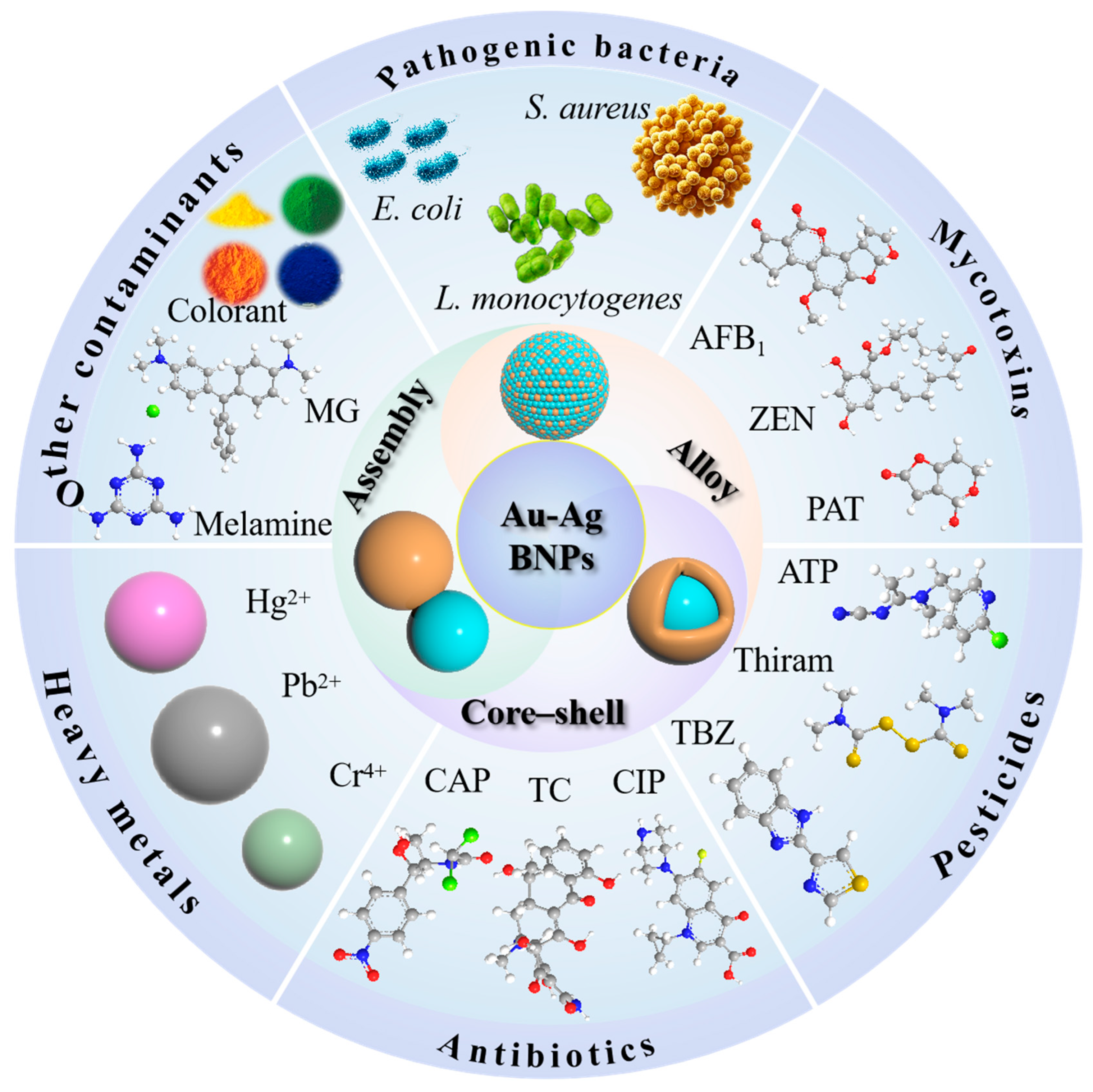
Figure 1. Schematic diagram of the types of SERS substrates based on Au-Ag bimetallic nanoparticles (Au-Ag BNPs) and their applications in food contaminants.
Foods 14 02109 g001
Figure 2. The types of Au-Ag BNPs and schematic diagrams of their model structures (orange and light blue respectively represent one of the Ag and Au elements). (A) Energy dispersive X-ray spectroscopy mapping of individual Ag/Au-NPs with atomic ratios (Ag:Au = 40:60) [38]. (B) The corresponding Au and Ag elemental mapping images of the Au@Ag NPs [39]. (C) Element mapping images of the Au-Ag JNPs [40].
Foods 14 02109 g002
Figure 3. (A) Colorimetric/SERS sensing for micrococcal nuclease (MNase)-responsive detection [62]. (B) Schematic of the preparation of three kinds of immuno-MoDAu@Ag SERS tags and MoDAu@Ag-based SERS encoding-LFIA for simultaneous detection of P. aeruginosa, E. coli O157:H7 and S. typhi [63]. (C) Schematic illustration of the synthesis process for multifunctional UAA@P/M and the procedures of UAA@P/M-integrated LFIA for multimodal bacterial detection [111]. (D) Schematic diagram of the SERS sandwich structure made of bacteria/SERS tags/AAS-NPs, used for specific bacterial identification, sensitive detection and reliable bacterial inactivation [64].
Foods 14 02109 g003
Figure 4. (A) Schematic illustration of the developed 3D SERS aptasensor based on Au-4MBA@Ag NSts-AFB1apt- Fe3O4@MoS2 NFs assemblies for AFB1 detection [69]. (B) Schematic diagrams of the preparation of two SERS probes and the SERS-LFIA test strip sensing process for simultaneous detection of two mycotoxins [72]. (C) SERS VFA biosensor based on an ordered PNC membrane and SERS nanotags for multiplex mycotoxin detection [73]. (D) Schematic diagram of the preparation process and sensing principle of magnetic MOFs-based ratiometric SERS aptasensor [74].
Foods 14 02109 g004
Figure 5. (A) Schematic diagram of detection of pesticides on apple surfaces using CNF/GNR@Ag SERS substrate [81]. (B) Schematic overview of the synthesis of AAZ and L-AAZ and the working principle of the SERS sensor for quinalphos detection [82]. (C) Illustration of the fabrication of Ag@ZIF-8@Au and its application in SERS detection of acetamiprid [83]. (D) Schematic diagram of paper-based SERS sensor quantification of carbaryl [84].
Foods 14 02109 g005
Figure 6. (A) Schematic representation of the synthesis procedure, the SERS sensing and the self-reviving ability for the SERS substrate based on the Ag/Au/AgCl heterostructure [89]. (B) Schematic diagram of detection and analysis of three antibiotics [91]. (C) Schematic diagram of chloramphenicol detection [92]. (D) Schematic diagram of TC detection based on aptamer recognition and cascade DNA network amplification Raman aptasensor [93].
Foods 14 02109 g006
Figure 7. (A) Process of preparing Au@Ag NRs and schematic illustration of the detection of Pb2+ based on the aggregates of the Au@Ag NR probe [95]. (B) Principle diagram of the colorimetric and SERS dual-mode probe for determination of Hg2+ based on controllable etching unmodified Au@Ag NPs [96]. (C) Dual-channel biosensor based on Au@Ag15-GU nanohybrids for detection of Hg2+ [97]. (D) Schematic illustration of the dual-signaling SERS ratiometric platform for Hg2+ detection [98].
Foods 14 02109 g007
Figure 8. (A) Illustration of the preparation of Au NBPs@Ag/PDMS as SERS substrate for detecting contaminants in river bass [101]. (B) The detection process for weakly adsorbed dye molecules based on PDDA@Ag/Au-HPOC substrate [102]. (C) Schematic diagram of the enrichment procedure of colorants in beverages and SERS detection combined with GO/Au@Ag NBs membrane [103]. (D) Synthetic scheme of preparation procedure of the Ag@Au film SERS substrate [104]. (E) Schematic illustration of the SERS-active Au-Ag Janus@Au NP-engineered SERS aptasensor for the detection of SEC [105]. (F) Schematic illustration of plasmonic Au-Ag Janus NP/perovskite composite-engineered SERS immunoassay for SEC detection [106].
Foods 14 02109 g008
Table 1. Typical research progress on the detection of food contaminants by SERS technology.
ContaminantsPlasmonic NanostructuresDetection MethodExtra Technology or Functional MoleculesLimit of DetectionReferences
E. coliAu@Ag NRsLabeledAntibody102 CFU/mL[55]
E. piscicida, E. coli, V. anguillarum, V. harveyi and P. plecoglossicidaAu@Ag NPsLabel-freeSeparation and enrichment of magnetic materialsClassification;
105 CFU/mL (E. piscicida)
[56]
S. aureusAu-assisted magnetic nanoparticles; Au@Ag-ATPLabeledHybridization chain reaction, aptamer0.25 CFU/mL[57]
E. coli and S. aureusAuAg@PB MOFLabel-free4-MPBA-functionalized substrate, dual-modal sensing42 CFU/mL (E. coli); 45 CFU/mL (S. aureus)[58]
Listeria innocuaAuAg@pSiNWsLabel-free/1.14 × 104 CFU/mL[59]
SalmonellaAu-Ag NPs/SiLabel-free/1 CFU/mL[60]
Clostridium perfringens , Bacillus subtilis and S. aureusAu@AgNPs/Van-PDMSLabel-freeMachine learning algorithms3 CFU/mL (S. aureus)[61]
S. aureusAu@AgPt nanozyme arrayLabel-free (indirect)Dual mode38 CFU/mL (colorimetric); 6 CFU/mL (SERS)[62]
P. aeruginosa, S. typhimurium and E. coliMoDAu@AgLabeledAntibody, LFIA29 cells/mL; 34 cells/mL; 40 cells/mL[63]
E. coli, S. aureus and P. aeruginosaAu-Ag-stuffed nanopancakesLabeled (internal standard)Triple-functional substrates7 CFU/mL[64]
PATAu@AgMBA NPs; AuNSs-cDNALabeledAptamer0.0281 ng/mL[65]
ZENAuMBA@AgMBA NPsLabeledAntibody3 μg/kg[23]
ZENAuDTNB@Ag NPsLabeledAptamer0.001 ng/mL[66]
OTAAuMBA@AuAg NPsLabeledAptamer0.004 ng/mL[67]
AFB1Au-Ag Janus NPsLabeledAptamer0.5 pg/mL[37]
AFB1AuMBA@Ag NPs; ITO-Au-GOLabeledAptamer0.1 pg/mL[68]
AFB1Au@Ag bimetallic nanostarsLabeledAptamer58.9 pg/mL[69]
AFB1Ag@Au IP6 bifunctional nanozymesLabeledDual mode,
aptamer
0.58 pg/L[70]
DONAuNR@Ag@SiO2-AuNPLabeledAntibody, LFIA0.053 fg/mL[71]
AFB1 and ZENFe3O4@PEI/AuMBA@AgMBALabeledAntibody, LFIA0.095 μg/kg (AFB1); 1.896 μg/kg (ZEN)[72]
OTA, AFB1 and ZENAuNBA@Ag, Au4-MBA@Ag and AuDNTB@AgLabeledAntibody, vertical flow immunoassay8.2 fg/mL (OTA), 13.7 fg/mL (AFB1), 47.6 fg/mL (ZEN)[73]
PATAu@Ag NPs, MOF and AuNRsLabeledAptamer0.0465 ng/mL[74]
ThiabendazoleAu@Ag NRsLabel-free/0.032 mg/kg[75]
Thiram and acetamipridAu@Ag NPsLabel-freeSimultaneous detection0.076 μM (thiram); 1.22 μM (acetamiprid)[11]
2,4-D and imidaclopridAu@Ag nanoflowersLabel-freeChemometric algorithms2.98 μg/L (2,4-D); 5.5 μg/L (imidacloprid)[76]
ThiramPAN/Cu2O@Ag/Au@AgNPsLabel-freeDeep learning algorithm0.02 mg/L[77]
Thiram and thiabendazoleAu@4-MBA@Ag arrayLabel-freeRatio Raman0.38 μg/L (thiram); 25 μg/L (thiabendazole)[78]
Acetamiprid and carbendazimAuMBA@AgMBA NPsLabeledLFIA0.27 μg/kg (acetamiprid); 1.71 μg/kg (carbendazim)[79]
Crystal violet, thiram and carbarylSiO2@AuAgLabel-free/6.95 × 10−7 M (crystal violet); 5.56 × 10⁻⁷ M (thiram); 7.14 × 10⁻⁶ M (carbaryl)[80]
Thiram and pymetrozineAu-Ag octahedral hollow cagesLabel-freeMachine learning algorithms0.286 μg/kg (thiram); 29 μg/kg (pymetrozine)[34]
ThiramCellulose nanofiber/AuNRs@AgLabel-free/10−11 M[81]
Malachite greenChiral spiny L-Au@Ag@ZIF-8Label-free/6.56 × 10−10 M[82]
AcetamipridAg@ZIF-8@Au nanoparticlesLabel-free/9.027 × 10−10 M[83]
CarbarylZIF-8@Ag/AAB/Au@AgLabel-free/5.72 × 10−3 µg/mL[84]
ChloramphenicolAscorbate-functionalized Au@Ag NPsLabel-freeChemometric algorithms2.73 × 10−5 μg/mL[85]
ChloramphenicolAu@Ag NBPs/SiO2 nanoarrayLabeledDNA enzyme amplification strategy6.42 × 10−13 M[86]
Amoxicillin and fenobucarbAg-Au alloy nanoparticlesLabel-free//[87]
RhBAg@SiO2-Au NPsLabel-free/5 × 10−9 M[88]
Paracetamol and furazolidoneAg/Au/AgCl heterostructureLabel-free/2.8 × 10−12 M (paracetamol); 1.9 × 10⁻¹¹ M (furazolidone)[89]
Ciprofloxacin and chloramphenicolTiO2/Au/Ag nanorod arraysLabel-free/10−9 M (ciprofloxacin); 10−8 M (chloramphenicol)[90]
R6GWS2/Au@Ag nanocompositesLabel-freeDeep learning algorithm10−14 M[91]
ChloramphenicolAu@Ag NPsLabel-freechemometric algorithms1 × 10−5 μg/mL[92]
TetracyclineFe3O4@h-TiO2/Au nanochains and Au@Ag NPsLabeledAptamer, cascade amplification15.91 pg/mL[93]
Cr (VI)Au@Ag nano-sea urchinsLabeledMethimazole-functionalized0.956 ng/L[94]
Pb2+Au@Ag NRsLabeledGlutathione and 4-MBA-functionalized0.021 μg/L[95]
Hg2+Au@Ag NPsLabeledColorimetric/SERS dual-mode, etching Ag shell2 μM (naked eye); 0.2 nM(UV-vis); 0.1 nM (SERS)[96]
Hg2+Au@Ag/graphene-upconversion nanohybridsLabeledFluorescence/SERS dual-mode, aptamer0.33 ppb (SERS); 1 ppb (fluorescence)[97]
Hg2+Au@Ag/COFLabeledY-shaped DNA-functionalized5.0 × 10−16 M[98]
R6G, thiram, melamine and piroxicamAu@SiO2@Ag@SiO2 compositesLabel-free/10−9 M (R6G); 10−6 M (thiram); 10−3 M (melamine); 10−3 M (piroxicam);[99]
Bacteria sporesAg@AuNP arrayLabel-freeChemometric algorithms10 CFU/mL[100]
Malachite greenAu nanobipyramid@AgLabel-free/0.1 nM[101]
Amaranth and allura redPDDA/Ag/Au hybrid plasmonic optical cavityLabel-free/0.3022 mg/L (amaranth); 0.2482 mg/L (allura red)[102]
Six colorantsGO/Au@Ag nanobonesLabel-freeMachine learning algorithms/[103]
Polystyrene nanoplasticsAg@Au filmLabel-free/25 (310 nm), 50 (50, 70 nm) μg/mL[104]
SECAu-Ag Janus NPsLabeledMBIA-functionalized, antibody0.55 pg/mL[105]
SECAu-Ag Janus NPsLabeledMBIA-functionalized, aptamer0.83 pg/mL[106]
β-lactoglobulinAu-Ag nanourchinsLabeledAptamer0.07 ng/mL[107]
E. coli, Escherichia coli; E. piscicida, Edwardsiella piscicida; V. anguillarum, Vibrio anguillarum; V. harveyi, Vibrio harveyi; P. plecoglossicida, Pseudomonas plecoglossicida; S. aureus, Staphylococcus aureus; AuAg@PB MOF, AuAg-doping Prussian blue analogue-based metal–organic framework; 4-MPBA, 4-mercaptophenylboronic acid; AuAg@pSiNWs, gold- and silver-coated porous silicon nanowires; P. aeruginosa, Pseudomonas aeruginosa; S. typhimurium, Salmonella typhimurium; 2,4-D, 2,4-dichlorophenoxyacetic acid; PAN, polyacrylonitrile; LFIA, lateral flow immunoassay; PAT, patulin; ZEN, zearalenone; OTA, ochratoxin A; AFB1, Aflatoxin B1. DON, deoxynivalenol; AAB, artificial antibody; COF, covalent organic framework; PDDA, poly(diallyldimethylammonium chloride); GO, graphene oxide; SEC, Staphylococcus aureus enterotoxin C; MBIA, 2-mercaptobenzoimidazole-5-carboxylic acid.
Disclaimer/Publisher’s Note: The statements, opinions and data contained in all publications are solely those of the individual author(s) and contributor(s) and not of MDPI and/or the editor(s). MDPI and/or the editor(s) disclaim responsibility for any injury to people or property resulting from any ideas, methods, instructions or products referred to in the content.