Article

Germination and Heat Resistance of Parageobacillus and Geobacillus spp. Spores

Department of Animal Production and Food Science, Faculty of Veterinary, AgriFood Institute of Aragon (IA2), University of Zaragoza—CITA, Miguel Servet 177, 50013 Zaragoza, Spain
*
Author to whom correspondence should be addressed.
Foods 2025, 14(12), 2061; https://doi.org/10.3390/foods14122061
Submission received: 6 May 2025 / Revised: 3 June 2025 / Accepted: 4 June 2025 / Published: 11 June 2025

Abstract

Geobacillus and Parageobacillus spores are major spoilage agents in thermally treated, shelf-stable foods, particularly milk products, due to their high heat resistance. This study aimed to investigate how spore purification, maturation time, and sporulation temperature influence the germination and heat resistance of P. thermoglucosidasius, G. thermodenitrificans, and G. stearothermophilus spores, with the goal of improving the reliability of microbial risk assessment. All three species germinate efficiently in milk, likely triggered by lactose and glucose. Ethanol-treated spores during purification germinated without heat activation, while water-washed spores required it. At least four days of maturation were needed for efficient germination, though extending maturation to seven days led to strain-dependent changes in heat resistance: it increased in G. thermodenitrificans, decreased in P. thermoglucosidasius, and remained stable in G. stearothermophilus. Sporulation at 55 °C consistently favored germination at the same revival temperature. G. stearothermophilus reached the highest heat resistance at 55 °C, whereas the other species were more resistant when sporulated at 60 °C. These findings underscore the importance of standardizing spore-preparation protocols, as key parameters such as purification, maturation time, and sporulation temperature critically affect spore properties relevant to food stability.

1. Introduction

The genus Geobacillus and the recently reclassified Parageobacillus consist of spore-forming bacteria characterized by an optimal growth temperature ranging between 45 °C and 65 °C [1,2]. Despite their thermophilic trait, these bacteria are ubiquitous in nature and therefore can easily enter the food chain [1,3,4]. The Geobacillus and Parageobacillus species are common contaminants in mildly acidic self-stable foods, particularly ultra-high-temperature (UHT) processed or sterilized milk and milk powders [5,6]. Among them, G. stearothermophilus is one of the most prevalent bacteria in dairy products [7,8,9], although other species such as G. thermodenitrificans and P. thermoglucosidasius are also frequently isolated [10,11,12,13]. This high prevalence likely originates from spores produced in biofilms on manufacturing surfaces maintained at elevated temperatures, such as heat exchangers and evaporation sections, rather than contaminated raw milk [6,14]. Spores of Geobacillus and Parageobacillus are extremely high-heat resistant and can survive commercial thermal treatments [12,15,16,17]. The germination and subsequent growth of surviving spores during storage under warm conditions, which are becoming increasingly common due to climate change [16,18], along with the proteolytic and lipolytic enzymes produced during vegetative growth under processing conditions, can lead to off-flavors and coagulation in dairy products and their final applications [5,14,19,20].
Thermal processing conditions are often optimized to eliminate sufficient levels of thermophilic spores, particularly G. stearothermophilus, while minimizing negative effects on product quality [21]. Additionally, G. stearothermophilus spores are widely used as biological indicators for sterilization validation, not only in food processing but also in the medical and pharmaceutical industries [22]. These validation processes often involve challenge tests to assess heat resistance and/or germination in end products using spores synthetically produced under laboratory conditions.
Sporulation medium composition, incubation temperature, maturation time, and purification techniques are known to significantly influence both germination and heat resistance in model mesophilic Bacillus spp. spores [23,24,25,26,27,28]. However, the impact of these factors on Parageobacillus and Geobacillus spp. spores, whose germination and resistance properties widely differ from mesophilic Bacillus spp. spores [29,30], remains largely unexplored. Addressing this knowledge gap is essential for designing effective thermal processing strategies that accurately assess the risk of spore survival without compromising food quality.
The aim of this study was to evaluate the effects of spore-purification methods, maturation time, and sporulation temperature on the germination and heat resistance of P. thermoglucosidasius (DSM 2542), G. thermodenitrificans (DSM 465), and G. stearothermophilus (ATCC 12980) spores, after confirming that these three strains are representative candidates for milk spoilage.

2. Materials and Methods

2.1. Obtention and Purification of Spore Suspensions

The strains P. thermoglucosidasius DSM 2542, G. thermodenitrificans DSM 465, and G. stearothermophilus ATCC 12980 were provided by the Bacillus Genetic Stock Center (Columbus, OH, USA). The strains were stored at −80 °C in 2TY broth (Sigma-Aldrich, St. Louis, MO, USA) supplemented with 25% glycerol (Panreac, Barcelona, Spain). For revitalization, cells were streaked on Tryptone Soya Agar (Oxoid, Basingstoke, UK) supplemented with 0.6% yeast extract (Oxoid) (TSAYE) and incubated at 55 °C for 24 h.
For sporulation, a single colony was inoculated into a 250 mL flask containing 20 mL of 2TY broth and was incubated at 55 °C overnight with shaking (130 rpm; Heidolph Promax 1010, Schwabach, Germany). Afterwards, a volume of 200 µL from the culture was inoculated into a 500 mL Erlenmeyer flask containing 50 mL of liquid TYE sporulation medium. The composition of this medium was optimized to reach the maximum sporulation yield in the three species, and it was composed of 0.4% tryptone (VWR International Chemicals, Radnor, PA, USA), 0.4% yeast extract, 1.13 mM CaCl2 dihydrate (VWR International), 0.033 mM MnSO4 monohydrate (Carlo Erba Reagents GmbH, Milan, Italy), 1 mM MgSO4 heptahydrate (Panreac), 0.04 mM FeSO4 heptahydrate (Sigma-Aldrich), and 80 mM HEPES of pH 7.0 (Sigma-Aldrich). As reference conditions, the sporulation cultures were incubated at 55 °C with a magnetic stirring (250 rpm; MIXdrive 6, 2mag, Munich, Germany) for 4 d. To study the effect of maturation time, the spores were also harvested at 1, 2, or 7 d of incubation at 55 °C, and to study the effect of incubation temperature, sporulation was also performed at 50 °C and 60 °C for 4 d. The sporulation yield was monitored over time by phase-contrast microscopy (Nikon Eclipse E400, Tokyo, Japan), calculating the proportion of bright-phase spores over total cells, and by plate counts, estimating the difference in the number of CFU/mL before and after heat treatment at 80 °C for 15 min applied in an Eppendorf thermoblock (LABNET International, Edison, NJ, USA).
The spores were harvested by centrifugation at 3345× g (Gyrozen 1736R, CIC Controltecnica, Madrid, Spain) for 20 min at 4 °C, and the spores from the pellets were purified using four different procedures:
-
Protocol 1: four consecutive washes with distilled water [30];
-
Protocol 2: one wash with distilled water followed by ethanol treatment (50%, v/v; SAEQSA, Zaragoza, Spain) for 1 h at 25 °C and four washes with distilled water [31];
-
Protocol 3: four washes with distilled water followed by Tween 80 treatment (0.01%, v/v; Sigma-Aldrich) for 1 h at 25 °C and four washes with distilled water [32];
-
Protocol 4: one wash with distilled water followed by three washes with 0.1% (v/v) Tween 80, ethanol treatment (50%, v/v) for 1 h at 25 °C, three washes with 0.01% Tween 80, and four washes with distilled water [33].
None of the procedures included gradient density centrifugation due to the low levels of cell debris observed after each purification procedure and the fact that it may reduce heat resistance [25].
After each method, the spore purity (99% bright-phase spores) was verified by phase-contrast microscopy and the suspensions were kept at −20 °C until usage. To assess biological variability, three different spore populations were obtained at each environmental condition.

2.2. Germination Assays

Germination was monitored by the reduction in optical density at 600 nm (OD600) resulting from the release of dipicolinic acid (DPA) and the rehydration of spores. The germination of spore samples adjusted to an OD600 of 0.4–0.6 was induced by a GR-saturating concentration of glucose (100 mM), lactose (100 mM), or casein hydrolysate (2%, w/v; Merck Millipore, Rahway, NJ, USA) in 25 mM HEPES buffer (pH 7.4). Where indicated, the spores were heat-treated at 100 °C for 30 min in an Eppendorf thermoblock and then incubated on ice for 15 min before exposure to nutrients [34]. OD600 was measured using a multiwell plate reader (CLARIOstar Plus, BMG, Ortenberg, Germany), which automatically recorded data every 3 min. Each reading was preceded by 30 s of shaking to prevent spore sedimentation. Samples were routinely germinated for 4 h at 55 °C, but also at 50 °C and 60 °C when evaluating the interaction between sporulation and germination temperatures. Germination curves were constructed using the percentage of OD600 fall (ODt/OD0 × 100, where OD0 and ODt represent the initial value and the value measured at further incubation times, respectively).
At the end of the spectrophotometric assays, the percentage of germinated spores was determined by phase-contrast microscopy. A total of 100 to 150 individuals per sample were examined and categorized as either dormant (phase-bright cells) or germinated spores (phase-dark and -grey cells). The lower and upper limits of quantification for germinated spores were approximately 5.0% and 97.0%, respectively.
In addition, germination and growth was assessed in UHT whole, skim, or whole lactose-free milk (DIA Supermercados, Madrid, Spain). The spores were inoculated in an Eppendorf containing 250 µL of milk to a final concentration of ca. 3 × 10⁸ CFU/mL, and the samples were incubated at 55 °C in an Eppendorf thermoblock (LABNET International, Edison, NJ, USA) with shaking (300 rpm). Germination was estimated by the evolution of the plate count after heat treatment (80 °C, 15 min) over time (Log(N0/Nt), where N0 and Nt represent the number of survivors in CFU/mL at time 0 and after different intervals, respectively.
For each spore preparation and germination condition, we obtained germination curves from at least three different biological replicates.

2.3. Thermal Treatments

Heat treatments were conducted using a thermoresistometer TR-SC [35]. This piece of apparatus consists of a 450 mL stainless steel chamber equipped with an electric heater, a cooling coil, and a stirring mechanism to ensure the uniform distribution of inoculum and temperature. Nitrogen gas at a pressure of 2.0 bars was introduced into the treatment chamber to achieve temperatures exceeding 100 °C and facilitate sample extraction over time. Once the target temperature was stabilized (±0.1 °C), spores were introduced into the treatment chamber containing McIlvaine citrate-phosphate buffer at pH 7.0 [36], achieving a final concentration of approximately 5 × 10⁴ CFU/mL. Subsequently, 0.2 mL samples were taken at different intervals of time, and viability was assessed according to the procedure described below. For each spore preparation condition and treatment temperature, we obtained three inactivation curves from different biological replicates.

2.4. Determination of Viability

Viability was determined by pour-plating in TSAYE; plates were incubated at 55 °C for 48 h. Longer incubation times did not affect the survival counts. The plate counts were obtained using an automatic colony counting system by using image analysis. The limit of quantification was 1.5 × 10² CFU/mL for thermal inactivation assays and 3 × 10² CFU/mL for growth assays.

2.5. Modeling of Heat Inactivation Curves

Inactivation curves were constructed by plotting the survival fraction (Log(Nt/N0)) against treatment time. As most survival curves displayed a shoulder phase, the data were fitted to the Log-linear + shoulder equation proposed by Geeraerd, et al. [37] (Equation (1)), using the GInaFiT Excel tool [38] (KU Leuven, Leuven, Belgium). In this model, N0 is the initial cell concentration (CFU/mL), Nt is the cell concentration (CFU/mL) at a specific time t, Sl (shoulder length, min) is the time required to reach the exponential inactivation phase, and Kmax (inactivation rate, min−1) is the slope of the exponential portion of the survival curve. The GInaFiT Version 1.6 software also provides the coefficient of determination (R2) and the root mean square error (RMSE) to evaluate the goodness of fit.
L o g   N t = L o g   N 0 K m a x   t L n 10 + L o g e x p K m a x   S l 1 + ( e x p K m a x   S l 1 ) e x p K m a x   t
For comparison, we calculated the time needed for 3 Log reductions (3DT) for each treatment temperature. To evaluate the impact of treatment temperature on heat resistance, thermal death time (TDT) curves were generated by plotting Log 3DT values against temperature. z values, which represent the number of degrees required to decrease the 3DT value by one Log unit, were calculated as the inverse of the slope of the TDT curves.

2.6. Statistical Analysis

Two-way ANOVA with Tukey’s and Sidak’s multiple comparisons test and unpaired parametric t-test were performed using GraphPad PRISM 8.4.2 (GraphPad Software Inc., San Diego, CA, USA). Statistical significance was considered when the p value was ≤0.05. The data presented in the figures represent averages and standard deviations derived from a minimum of three biological replicates.

3. Results and Discussion

3.1. Germination in Milk Products

We investigated spore germination of P. thermoglucosidasius, G. thermodenitrificans, and G. stearothermophilus strains in whole milk, skim milk, and whole lactose-free milk at 55 °C to assess their potential as representative food-spoilage agents (Figure 1). P. thermoglucosidasius spores germinated the fastest, reaching an average of 0.62 Log CFU/mL of germinated cells (75.9% germination) after 15 min, and 1.17 Log CFU/mL (93.3%) after 30 min across all of the milk products. G. stearothermophilus spores germinated at a similar rate to P. thermoglucosidasius in whole and lactose-free milk (ca. 0.68 Log CFU/mL, 79.2%, at 15 min, and 1.05 Log CFU/mL, 91.0%, at 30 min). In skim milk, the germination of G. stearothermophilus spores was slower, reaching only 0.53 Log CFU/mL (70.3%) at 30 min, and increasing to 1.12 Log CFU/mL (92.5%) at 45 min. G. thermodenitrificans spores germinated more slowly, requiring 90 min in whole milk and 180 min in skim and lactose-free milk to exceed 90.0% germination. After 7 h, the germination efficiency was similar (p > 0.05) among the three strains in whole and skim milk (on average 1.37 Log CFU/mL, 95.7%). In lactose-free milk, P. thermoglucosidasius and G. thermodenitrificans reached the highest germination efficiency (1.78 Log CFU/mL, 98.5%), which was significantly (p ≤ 0.05) higher than that of G. stearothermophilus (1.26 Log CFU/mL, 94.5%). Extending incubation to 24 h did not improve germination (p > 0.05). It is important to note that P. thermoglucosidasius and, to a lesser extent, G. stearothermophilus proliferated in the three milk products, while G. thermodenitrificans could not grow (Figure S1). However, lactose-free milk contaminated with G. thermodenitrificans exhibited signs of spoilage (coagulation) after 24 h, as did the P. thermoglucosidasius and G. stearothermophilus samples, suggesting that some germinated spores may resume metabolic activity.
To identify potential germinants, we explored germination in response to milk containing carbohydrates (lactose and glucose, the latter as a lactose breakdown product) and casein hydrolysate (an amino acid source) by measuring the OD600 decrease over 4 h at 55 °C. As shown in Figure 2, the three strains germinated in glucose and lactose, but not in the amino acid mixture. Only the P. thermoglucosidasius spores showed a significant OD600 decrease, but to a lower level than in both sugars. This is in contrast to the typical germination of Bacillus and Clostridium spp. spores, which germinate efficiently in the presence of certain L-amino acids [39,40]. Closer examination of the germination efficiency at the end of the 4 h assays by phase-contrast microscopy revealed that P. thermoglucosidasius exhibited a significantly (p ≤ 0.05) higher proportion of phase-dark spores than the other two species in both lactose and glucose, and that all three species germinated between 1.2- and 1.7-fold more efficiently (p ≤ 0.05) in glucose than in lactose.
Overall, P. thermoglucosidasius DSM 2542, G. thermodenitrificans DSM 465, and G. stearothermophilus ATCC 12980 spores germinated efficiently in milk products, likely triggered by lactose or, in lactose-free milk, by glucose. Interestingly, although G. stearothermophilus ATCC 12980 spores responded to lactose, this strain lacks the genetic capacity for lactose usage [41]. Similarly, many P. thermoglucosidasius isolates are unable to metabolize lactose [42,43]. However, lactose metabolism may not be essential for growth in milk. Although G. stearothermophilus ATCC 12980 and G. thermodenitrificans DSM 465 exhibited limited or no growth, respectively, in the tested dairy products (Figure S1), previous studies have demonstrated that the same strains can adhere, form biofilms, and sporulate on milk contact surfaces [10,44]. Thus, the ability of both Geobacillus strains to grow and sporulate in milk may depend on the physical state of the culture, in addition to variations in milk composition [45,46].

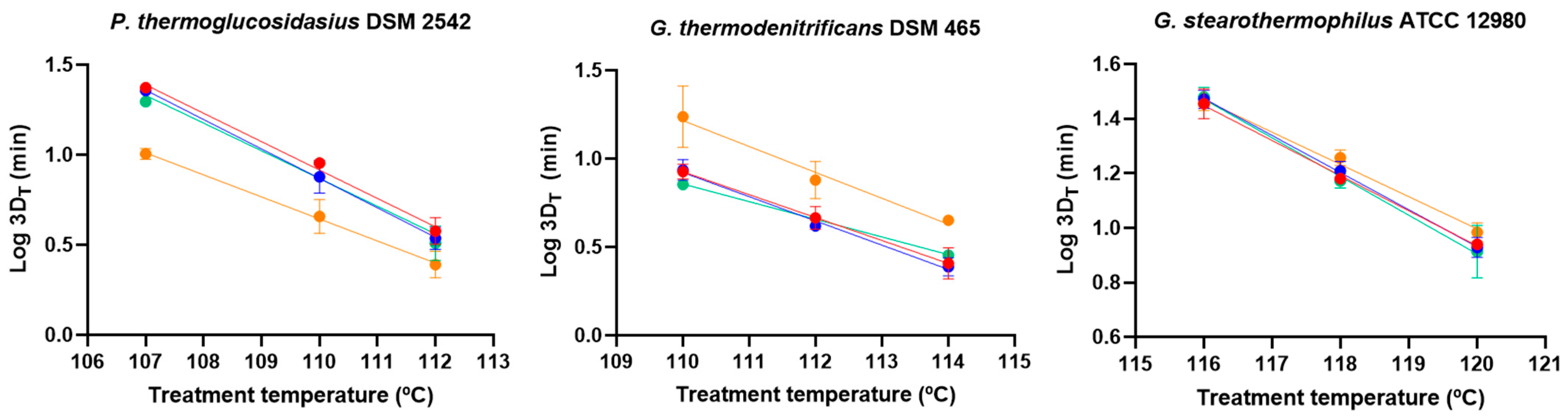
3.2. Effect of Purification Method on Germination and Heat Resistance

We evaluated the effect of different spore-purification methods (for simplicity, protocol 1—water washes, protocol 2—ethanol treatment, protocol 3—Tween (0.01%) treatment, protocol 4—Tween (0.1%) washes followed by ethanol treatment and Tween (0.01%) washes) on the germination and heat inactivation kinetics of P. thermoglucosidasius, G. thermodenitrificans, and G. stearothermophilus sporulated at 55 °C for 4 d. It is important to note that none of the methods affected spore viability, as confirmed by growth on the TSAYE plate. The proportion of germinated spores after 4 h exposure to glucose or lactose, as major inducers of germination, is shown in Figure 3A. Most notably, the germination efficiency remained ≤5.0% in all populations washed only with water, but all samples subjected to ethanol treatment before water washes responded to both glucose and lactose. Additionally, the purification method had particular effects on germination in certain strains and nutrient conditions. P. thermoglucosidasius spores treated with Tween or subjected to Tween washes with intermediate ethanol treatment germinated in both nutrients, but the fraction of germinated spores in glucose was significantly higher (p ≤ 0.05) in ethanol-treated samples compared to those treated with Tween-based methods. G. stearothermophilus spores purified with Tween washes and intermediate ethanol treatment germinated only in lactose, achieving an efficiency comparable (p > 0.05) to those treated with ethanol treatment followed by water washes.
The effect of heat-activation treatment (100 °C, 30 min), previously shown to reduce the germination initiation time and increase germination efficiency in G. stearothermophilus NGB101 [34], was also examined (Figure 3B). Unlike non-activated spores, only heat-activated spores washed with distilled water or treated with Tween germinated in glucose and lactose, while those treated with ethanol followed by either water or Tween washes did not germinate (≤5.0%). In P. thermoglucosidasius and G. thermodenitrificans, no significant (p > 0.05) differences were found in germination efficiency between heat-activated spores purified with water washes or Tween treatment, regardless of the nutrient. However, in G. stearothermophilus, Tween-treated spores exhibited a 1.4-fold and 2.3-fold higher (p ≤ 0.05) proportion of germinated spores in glucose and lactose, respectively, compared to water-washed spores. Interestingly, P. thermoglucosidasius was the only strain in which both non-activated and heat-activated spores treated with Tween responded to nutrients. Heat activation slightly increased germination efficiency 1.2-fold (p ≤ 0.05; Figure 3A,B) and doubled the germinigucose but had no effect in lactose.
In conclusion, the purification method determines the response of P. thermoglucosidasius, G. thermodenitrificans, and G. stearothermophilus spores to nutrients and the effectiveness of prior heat activation. In general, and except for some particularities in specific nutrients and strains, spores purified with ethanol treatment followed by water washes germinated without requiring heat activation, whereas heat treatment was strictly necessary for spores prepared without ethanol exposure. Ethanol treatment (50%) is commonly used for spore purification to kill vegetative cells [31,33]. On the other hand, ethanol exposure (ca. 20–80%) has also been reported to activate germination in certain Bacillus and Clostridium species [47,48,49]. Our results suggest that ethanol purification can activate Parageobacillus and Geobacillus spores for nutrient germination, yielding efficiency comparable to heat-activated spores purified with just water washes (Figure 3).
Another intriguing finding was that ethanol-treated spores failed to germinate after heat-activation treatment. It has been suggested that sublethal ethanol and heat treatments individually enhance germination by conformational changes in essential proteins that unblock or enhance their function, with GRs thought to be the primary target in the case of heat activation, or by altering the properties of the inner membrane where such proteins reside [48,50,51]. However, ethanol treatment at an elevated temperature can damage spores, likely by disrupting spore permeability, including a reduction in inner-membrane viscosity [52,53,54]. In addition, the combined heat and ethanol treatment impairs the germination of surviving spores, presumably due to the denaturation of some critical proteins such as cortex lytic enzymes, as observed in C. perfringens and B. thuringiensis [48,55]. Thus, it can be hypothesized that transient exposure to ethanol may alter inner membrane or critical germination proteins, favoring spore germination while rendering them sensitive to subsequent heat perturbation.
The effect of the spore-purification method on heat resistance was assessed by obtaining survival curves of P. thermoglucosidasius, G. thermodenitrificans, and G. stearothermophilus spores prepared using the four purification protocols at a temperature appropriate for each strain (112 °C, 114 °C, and 120 °C, respectively). Table 1 displays the heat inactivation parameters, Kmax and Sl, along with the 3DT values. While the purification method did not affect G. stearothermophilus heat resistance, P. thermoglucosidasius and G. thermodenitrificans spores exposed to Tween (with or without intermediate ethanol treatment) exhibited higher (p ≤ 0.05) 3DT values than spores purified by the methods excluding this compound. However, these differences were minor compared to other methodological factors studied, such as maturation time and sporulation temperature (see below). Despite the fact that Tween compounds are commonly used in spore preparation to kill vegetative cells and prevent spore aggregation [32,56], to the best of our knowledge there is little information on the effect of these chemicals on spore structure. In vegetative cells, growth in media supplemented with Tween 80 or Tween 20 provides protection against acidity, high pressure, and freeze-drying [57,58,59], likely due to the incorporation of the oleic acid moiety into the cell membrane, altering its properties [57,59]. In addition, Tween compounds provide thermal stability to proteins [60,61]. Since damage to proteins related to energy metabolism—likely embedded in the inner membrane—is a key process responsible for the heat inactivation of spores, and inner membrane properties have been shown to affect heat resistance [62,63,64], it is plausible that Tween exposure may change inner membrane characteristics, thereby increasing thermal stability and thereby enhancing spore survival.
From a practical standpoint, the purification procedure of Parageobacillus and Geobacillus spores influences resistance and especially germination. The substantial impact on germination may help explain discrepancies in the literature regarding the ability of spores from the same strain to germinate in specific nutrients and the need for thermal activation [30,34,65]. However, other methodological factors, such as sporulation medium composition, the germination assay method, and the intensity of thermal treatment, may also contribute. For subsequent research, the spores were routinely purified using ethanol treatment followed by water washes, as this method rendered the spore activated for germination, avoiding the need for heat activation and without affecting heat resistance.

3.3. Effect of Maturation Time on Germination and Heat Resistance

To investigate the effect of maturation time on germination and heat resistance, spores cultured at 55 °C were collected after 1, 2, 4, or 7 d of incubation. Please note that the maximum spore yield and spore release from sporangia occurred at 24 h, with the total spore counts and viability remaining unchanged over time. Reain (Figure 4), the P. thermoglucosidasius spores harvested on d 1 did not germinate (≤5.0%) after 4 h exposure to lactose, while those collected between d 2 and 7 germinated in glucose and lactose to reach a similar (p > 0.05) efficiency. More striking was the effect of maturation time on the germination of the Geobacillus spp. spores. None of the spores collected on d 1 and 2 responded to glucose, whereas spores incubated for 4 to 7 d germinated to an equal (p > 0.05) extent. Moreover, only G. thermodenitrificans and G. stearothermophilus spores collected on d 4 were able to germinate in lactose. The requirement of a maturation period after spore release in the spent sporulation medium to detect population-wide germination, with the necessary duration depending on the strain and type of germinant, appears to be a unique characteristic of Parageobacillus and Geobacillus spores. In B. subtilis, the effect of maturation time has been observed at the single-cell level [26,66], but this effect may vary depending on the composition and physical state of the sporulation medium, spore age, and incubation temperature [26,67].
Maturation time also influenced heat resistance at the reference treatment temperature set for each strain (112 °C, 114 °C, and 120 °C for P. thermoglucosidasius, G. thermodenitrificans, and G. stearothermophilus, respectively). To examine the thermal dependence of this effect, inactivation kinetics were evaluated at additional temperatures (Table S1), from which TDT curves were constructed using 3DT values (Figure 5). P. thermoglucosidasius spores incubated for 7 d exhibited lower (p ≤ 0.05) 3DT values than those collected on d 1 and 2, with greater differences observed at lower treatment temperatures: 7 d mature spores were ca. 1.5-fold, 2.0-fold, and 2.3-fold more sensitive (p ≤ 0.05) than the youngest spores when treated at 112 °C, 110 °C, and 107 °C, respectively (Figure 5, Table S1). Additionally, 4 d mature spores showed lower (p ≤ 0.05) 3D110 °C and 3D107 °C values than 1 d mature spores but significantly higher heat resistance than 7 d mature spores. Consequently, z values estimated from the TDT curves increased with harvest time but not significantly (p > 0.05, Table 2). In contrast, G. thermodenitrificans spores collected on d 7 exhibited higher (p ≤ 0.05) 3DT values than the younger spores at all of the tested temperatures (114 °C, 112 °C, and 110 °C; Figure 5, Table S1). The heat resistance of G. stearothermophilus spores remained unchanged with maturation time at all temperatures, except for a minor increase (1.2-fold, p ≤ 0.05) in 3D118 °C values in 7 d mature spores compared to 1 and 4 d mature spores (Figure 5, Table S1). z values for both G. thermodenitrificans and G. stearothermophilus did not correlate with changes in maturation time (Table 2).
The observed increase in heat resistance with maturation time in the spent medium for G. thermodenitrificans aligns with findings for mesophilic Bacillus spp. spores [66,67,68,69,70]. This effect, together with changes in germination behavior, has been linked to increased cross-linking between coat proteins [66,69,71], with this structure playing a crucial role in both spore properties [72,73]. However, it remains unclear whether these changes in spore behavior are directly due to alterations in the coat structure itself, or rather to indirect effects on other components, such as the inner membrane properties or the state of embedded germination- and heat resistance-related proteins [26,74]. Additionally, Camilleri et al. [70] reported that core wet density increased with spore age, suggesting that reduced water content may also contribute to the development of heat resistance in mature spores. Further research is necessary to elucidate the relationship between changes in heat resistance with spore age and modifications in the coat, inner membrane, water content, and other structural components in each species.
Since P. thermoglucosidasius and both Geobacillus strains required at least 2 and 4 d of incubation, respectively, to germinate in lactose, and given that extending the harvest time from 4 to 7 d affected heat resistance—either increasing or decreasing it, particularly in P. thermoglucosidasius and G. thermodenitrificans spores—the maturation time for all strains was standardized to 4 d for subsequent investigations.

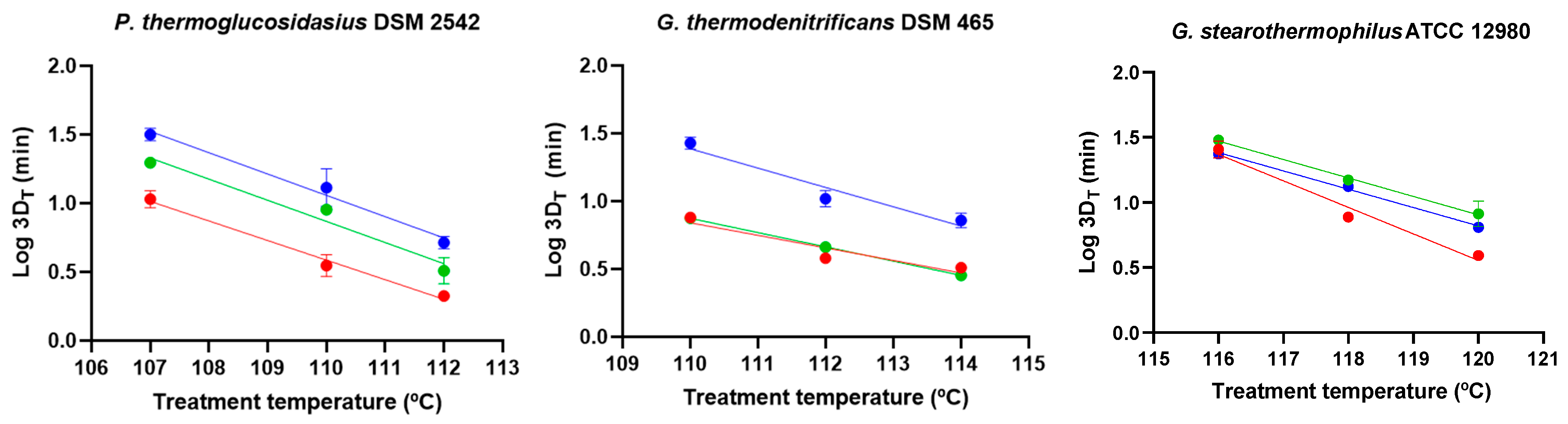
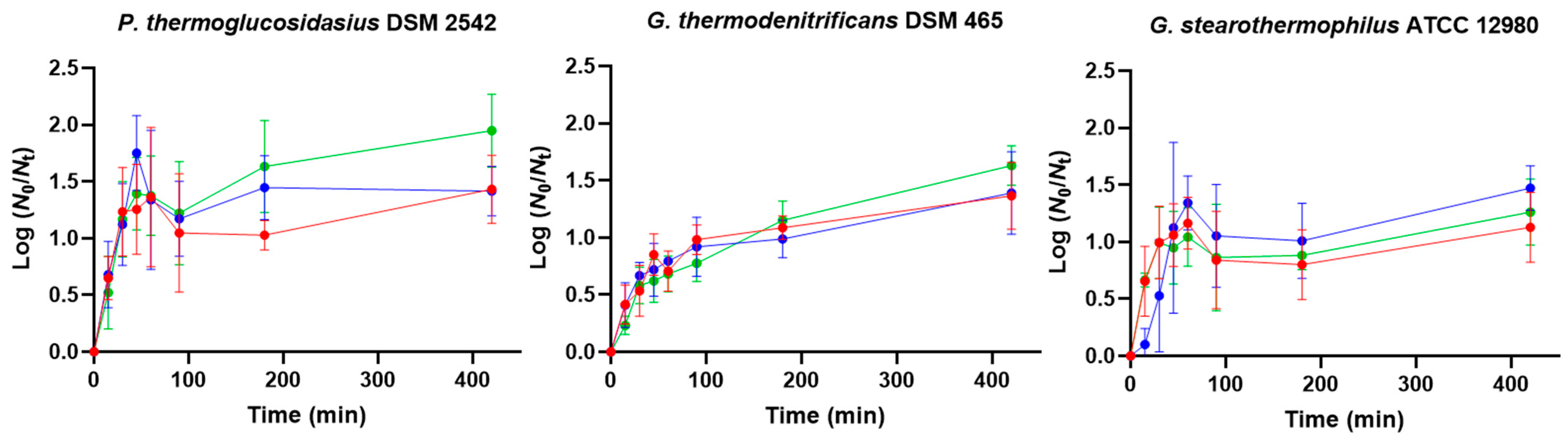
3.4. Effect of Sporulation Temperature on Germination and Heat Resistance

To examine the impact of sporulation temperature, spores were produced at 50 °C, 55 °C, and 60 °C. The sporulation rate and spore release were minimally affected by temperature changes, ensuring consistent maturation times. Although sporulation temperature did not affect spore viability, it did influence germination efficiency of the first germinating individuals in glucose and lactose when revived at 55 °C (Figure 6). Only spores produced at 55 °C exhibited a response (>5.0%) after 4 h exposure to glucose or lactose, except for P. thermoglucosidasius spores prepared at 50 °C and 60 °C in glucose. In this scenario, the germination efficiency of P. thermoglucosidasius spores produced at 55 °C and 60 °C exceeded 97.0%, while spores cultured at 50 °C reached a lower extent (84.0%).
As the effect of sporulation temperature may depend on the germination temperature [75,76], the response to glucose and lactose was also tested at 50 °C and 60 °C (Figure 6). Lowering the germination temperature from 55 °C to 50 °C significantly (p ≤ 0.05) decreased the efficiency of all P. thermoglucosidasius spores in glucose, while the differential effect of sporulation temperature vanished. It also inhibited (≤ 5.0%) the germination of P. thermoglucosidasius spores in lactose and Geobacillus spp. spores in both nutrients. Increasing the germination temperature from 55 °C to 60 °C enhanced the germination of P. thermoglucosidasius spores produced at 50 °C in glucose. It also stimulated the response of P. thermoglucosidasius spores prepared at 50 °C and 60 °C in lactose, with the fraction of germinated spores produced at 50 °C being approximately 1.4-fold higher (p ≤ 0.05) than in the other two populations. No germination (≤5.0%) was detected at 60 °C in G. thermodenitrificans spores, including those produced at 55 °C. Overall, sporulation temperature is a key factor influencing germination, with effects that vary depending on the strain, germinant type, and germination temperature. Generally, the three species exhibited the highest germination efficiency when sporulated and germinated at a moderate thermophilic temperature (55 °C). In B. subtilis, changes in germination kinetics induced by variations in sporulation temperature have been attributed to differences in active GR or GerD levels [27], alterations in the coat structure [72,77,78] and/or variations in the inner membrane properties [79,80]. For P. thermoglucosidasius and G. stearothermophilus, the germination of spores produced at temperatures lower or higher than 55 °C was enhanced by increasing the germination temperature in the presence of either lactose or glucose, but not in G. thermodenitrificans. The increase in germination efficiency with incubation temperature observed in P. thermoglucosidasius and G. stearothermophilus spores has also been reported for other Bacillus spp., where it has been hypothesized that elevated temperatures enhance molecular motion and membrane permeability, thus facilitating germination [81]. More research is needed to fully elucidate the effects of sporulation temperature on germination in mesophilic Bacillus spp. and, by extension, Parageobacillus and Geobacillus spp. spores.
As the sporulation temperature was expected to significantly influence heat resistance, inactivation curves were obtained at different treatment temperatures, and the TDT curve was generated from 3DT values for each strain (Figure 7, Table S2). The 3DT values of P. thermoglucosidasius spores increased progressively (p ≤ 0.05) with sporulation temperature across all treatment temperatures (Figure 7, Table S2), following similar (p > 0.05) z values (Table 2). In G. thermodenitrificans, spores produced at 60 °C exhibited the greatest (p ≤ 0.05) resistance at all treatment temperatures (Figure 7, Table S2). The z values of G. thermodenitrificans spores prepared at 60 °C were significantly (p ≤ 0.05) lower than those sporulated at 50 °C (Table 2). Conversely, G. stearothermophilus spores produced at 55 °C displayed the highest (p ≤ 0.05) heat resistance at all temperatures, and spores prepared at 60 °C showed greater (p ≤ 0.05) resistance at 120 °C and 118 °C than those sporulated at 50 °C (Figure 7, Table S1). In agreement z values of spores produced at 50 °C were lower than those sporulated at 55 °C and 60 °C.
Figure 7. Thermal death time (TDT) curves, plotting 3DT values against treatment temperature, for P. thermoglucosidasius DSM 2542, G. thermodenitrificans DSM 465, and G. stearothermophilus ATCC 12980 spores prouced at different temperatures: 50 °C (red dots), 55 °C (green dots), and 60 °C (blue dots). Sporulation was performed for 4 d, and spores were purified using ethanol treatment followed by water washes (protocol 2). Values in the figures correspond to averages and standard deviations calculated from three biological replicates.
Foods 14 02061 g007
In general, heat resistance in spore-forming bacteria, including thermophilic spores, increases with rising sporulation temperature up to a certain threshold, beyond which it either plateaus or declines [82,83,84]. Indeed, it is well established that the specific temperature range in which heat resistance positively correlates with sporulation temperature varies between species and strains [29,84,85]. The findings of this study are consistent with this trend. In G. stearothermophilus, spores produced at 55 °C exhibited greater heat resistance than those sporulated at 50 °C or 60 °C, which aligns with the results of Mtimet et al. [82], who reported that the highest heat resistance for the same strain (ATCC 12980) occurs at sporulation temperatures near 57 °C. In contrast, spores of P. thermoglucosidasius and G. thermodenitrificans showed increased heat resistance when the sporulation temperature increased from 50 °C or 55 °C to 60 °C. Further experimental work is needed to determine whether additional increases in sporulation temperature led to a peak in heat resistance, followed by a decline in these two strains. Leguérinel et al. [29] reported that the rate of increase in heat resistance with sporulation temperature (i.e., the increase in sporulation temperature required to change DT values 10-fold) in G. stearothermophilus ATCC 12980 spores was higher than in other strains of B. subtilis, B. cereus, and B. licheniformis. It would be worthwhile investigating whether the magnitude of the dependence of heat resistance on sporulation temperature is similar in thermophilic microorganisms such as P. thermoglucosidasius and G. thermodenitrificans.
According to our results, the inactivation temperature should be carefully considered when comparing the effects of sporulation conditions on different strains. While variations in sporulation temperature did not affect the z value of P. thermoglucosidasius spores, it did influence those of G. stearothermophilus and G. thermodenitrificans spores. It is important to note that other researchers did not observe a dependency of the z value of G. stearothermophilus spores, including the strain used in this study, on sporulation temperature [29,86]. This discrepancy may be attributed to the influence of other environmental factors on the interaction between heat resistance and sporulation temperature, such as sporulation medium and treatment medium composition [21,85,86,87,88], as well as differences in the calculation methods used for heat-resistance parameters. Regarding the latter, z values can change when varying the Log reduction target in non-linear inactivation kinetics due to the differing weight of the shoulder length and inactivation rate [38]. Moreover, the relationship between shoulder length and inactivation rate depends on the sporulation temperature [86].
Therefore, sporulation temperature modulates heat resistance, with the extent of this effect varying depending on the strain and the treatment temperature. Heat resistance increased with the sporulation temperature in P. thermoglucosidasius and G. thermodenitrificans, while in G. stearothermophilus, the highest resistance was observed at the moderate sporulation temperature. The increased heat resistance with sporulation temperature in mesophilic Bacillus spp. has been linked to reduced core water content, elevated mineral and DPA levels, and/or structural modifications in the cortex and coat [69,73,84,89,90,91]. Therefore, investigating these characteristics in thermophilic spores could provide valuable insights.
Altogether, based on previous studies, mainly on model Bacillus spp. spores, it can be speculated that changes in coat structure, inner membrane properties, and/or reduced core water content may underlie the increased heat resistance associated with maturation time in G. thermodenitrificans, as well as the enhanced resistance observed with sporulation temperatures up to 60 °C in both G. thermodenitrificans and P. thermoglucosidasius. Additionally, it can be hypothesized that shorter maturation times and lower sporulation temperatures may alter the levels of active GRs and/or other structures, such as the coat and inner membrane, making spores less responsive to nutrients, despite ethanol treatment during purification. This suggests that such activation treatment alone may be insufficient to overcome other critical alterations affecting spore germination. Further research is needed to mechanistically elucidate the spore components driving behavioral changes under each studied factor, as well as to understand the variability in these effects among different strains. In addition, it would be interesting to investigate the interactions between purification, maturation time, and sporulation temperature at both phenotypic and mechanistic levels.

4. Conclusions

To the best of our knowledge, this is the first study to systematically examine how sporulation conditions, specifically temperature and maturation time, as well as spore-purification methods influence the germination and heat resistance of various Parageobacillus and Geobacillus species. Although these factors have been extensively studied in Bacillus spp., limited information is available for thermophilic spore-formers. Thus, our findings provide new insights into the behavior of Parageobacillus and Geobacillus spp. spores, which is particularly relevant given the anticipated increase in food spoilage caused by thermophilic spores due to climate change, as well as the growing interest in exploiting these bacteria in the biotechnology industry.
This study demonstrates that the spore purification method, maturation time, and sporulation temperature influence spore germination and heat resistance. These parameters should be carefully considered when designing challenge tests for research or industrial validation purposes. Based on representative strains of P. thermoglucosidasius, G. thermodenitrificans, and G. stearothermophilus, we propose general recommendations for spore preparation. However, it should be noted that the impact of each factor may vary depending on additional methodological conditions, such as the composition of the sporulation medium and spore storage conditions, as well as intraspecific variations.
For germination studies, ethanol purification followed by water washes is recommended, as it promotes efficient nutrient-induced germination without requiring heat activation. A minimum maturation period of 4 d in spent sporulation medium is necessary to obtain germination-competent spores. Optimal germination was observed when spores were produced at 55 °C and germinated at the same temperature. However, the limited germination response of P. thermoglucosidasius and G. stearothermophilus spores produced at suboptimal sporulation temperatures (50 °C or 60 °C) could be partially offset by increasing the germination temperature from 55 °C to 60 °C.
For heat resistance assays, the purification method had no effect, but increasing the maturation time had strain-specific outcomes: resistance increased in G. thermodenitrificans, decreased in P. thermoglucosidasius, and remained stable in G. stearothermophilus. The sporulation temperature also had strain-dependent effects: G. stearothermophilus spores were most resistant at 55 °C, while P. thermoglucosidasius and G. thermodenitrificans showed highest resistance when sporulated at 60 °C.

Supplementary Materials

The following supporting information can be downloaded at: https://www.mdpi.com/article/10.3390/foods14122061/s1, Figure S1. Growth curves of P. thermoglucosidasius DSM 2542, G. thermodenitrificans DSM 465, and G. stearothermophilus ATCC 12980 spores in whole milk (●), skim milk (●), and whole lactose-free milk (●) at 55 °C. Data in the figures correspond to mean values and standard deviations calculated from three biological replicates. The dotted line represents the limit of quantification (3 × 10² CFU/mL); Table S1. Heat resistance parameters (Sl, Kmax, and 3DT) of P. thermoglucosidasius DSM 2542, G. thermodenitrificans DSM 465, and G. stearothermophilus ATCC 12980 spores with different maturation times (1, 2, 4, or 7 d). Sporulation was performed at 55 °C and spores were purified using ethanol treatment followed by water washes (protocol 2). Data in brackets represent the standard deviations of the mean values calculated from three biological replicates. Different letters indicate statistically significant differences (p ≤ 0.05) in resistance parameters among spores of different ages within each strain and treatment temperature; Table S2. Heat resistance parameters (Sl, Kmax, and 3DT) of P. thermoglucosidasius DSM 2542, G. thermodenitrificans DSM 465, and G. stearothermophilus ATCC 12980 spores produced at different temperatures (50 °C, 55 °C, and 60 °C). Spores were collected at day 4 and purified using ethanol treatment followed by water washes (protocol 2). Data in brackets represent the standard deviations of the mean values calculated from three biological replicates. Different letters indicate statistically significant differences (p ≤ 0.05) in resistance parameters among spores prepared at different temperatures within each strain and treatment temperature.

Author Contributions

Conceptualization, M.S., S.C. and E.G.; methodology, M.S. and E.G.; formal analysis, M.S., S.C. and E.G.; investigation, M.S.; resources, E.G.; data curation, M.S.; writing—original draft preparation, M.S. and E.G.; writing—review and editing, M.S., S.C. and E.G.; visualization, M.S.; supervision, S.C. and E.G.; project administration, E.G.; funding acquisition, E.G. All authors have read and agreed to the published version of the manuscript.

Funding

This research was funded by Agencia Estatal de Investigación (MCIN/AEI/10.13039/501100011033), grant number PID2023-148505OB-I00, and a Ph.D. scholarship from the Government of Aragón (to M.S.).

Institutional Review Board Statement

Not applicable.

Informed Consent Statement

Not applicable.

Data Availability Statement

The original contributions presented in the study are included in the article/Supplementary Material, further inquiries can be directed to the corresponding author.

Acknowledgments

The authors would also like to acknowledge “Servicio General de Apoyo a la Investigación-SAI” (University of Zaragoza) for developing the automatic colony.

Conflicts of Interest

The authors declare no conflicts of interest. The funders had no role in the design of the study; in the collection, analyses, or interpretation of data; in the writing of the manuscript; or in the decision to publish the results.

References

  1. Najar, I.N.; Nagendra, T. A systematic review of the genera Geobacillus and Parageobacillus: Their evolution, current taxonomic status and major applications. Microbiology 2020, 166, 800–816. [Google Scholar] [CrossRef]
  2. Aliyu, H.; Lebre, P.; Blom, J.; Cowan, D.; De Maayer, P. Phylogenomic re-assessment of the thermophilic genus Geobacillus. Syst. Appl. Microbiol. 2016, 39, 527–533. [Google Scholar] [CrossRef]
  3. Zeigler, D.R. The Geobacillus paradox: Why is a thermophilic bacterial genus so prevalent on a mesophilic planet? Microbiology 2014, 160, 1–11. [Google Scholar] [CrossRef]
  4. Iacumin, L.; Pellegrini, M.; Colautti, A.; Orecchia, E.; Comi, G. Microbial Characterization of Retail Cocoa Powders and Chocolate Bars of Five Brands Sold in Italian Supermarkets. Foods 2022, 11, 2753. [Google Scholar] [CrossRef]
  5. Burgess, S.A.; Lindsay, D.; Flint, S.H. Thermophilic bacilli and their importance in dairy processing. Int. J. Food Microbiol. 2010, 144, 215–225. [Google Scholar] [CrossRef]
  6. André, S.; Vallaeys, T.; Planchon, S. Spore-forming bacteria responsible for food spoilage. Res. Microbiol. 2017, 168, 379–387. [Google Scholar] [CrossRef]
  7. Sadiq, F.A.; Li, Y.; Liu, T.; Flint, S.; Zhang, G.; He, G. A RAPD based study revealing a previously unreported wide range of mesophilic and thermophilic spore formers associated with milk powders in China. Int. J. Food Microbiol. 2016, 217, 200–208. [Google Scholar] [CrossRef]
  8. Yuan, D.-D.; Liu, G.-C.; Ren, D.-Y.; Zhang, D.; Zhao, L.; Kan, C.-P.; Yang, Y.-Z.; Ma, W.; Li, Y.; Zhang, L.-B. A survey on occurrence of thermophilic bacilli in commercial milk powders in China. Food Control 2012, 25, 752–757. [Google Scholar] [CrossRef]
  9. Delaunay, L.; Cozien, E.; Gehannin, P.; Mouhali, N.; Mace, S.; Postollec, F.; Leguerinel, I.; Mathot, A.-G. Occurrence and diversity of thermophilic sporeformers in French dairy powders. Int. Dairy J. 2021, 113, 104889. [Google Scholar] [CrossRef]
  10. Karaca, B.; Karakaya, A.B.; Ozcan, B.; Coleri Cihan, A. Rapid detection of Geobacillus and Anoxybacillus species by quantitative qPCR (qPCR) in commercial dairy products. J. Food Saf. 2022, 42, e12964. [Google Scholar] [CrossRef]
  11. Karaca, B.; Buzrul, S.; Coleri Cihan, A. Anoxybacillus and Geobacillus biofilms in the dairy industry: Effects of surface material, incubation temperature and milk type. Biofouling 2019, 35, 551–560. [Google Scholar] [CrossRef]
  12. Sadiq, F.A.; Flint, S.; He, G. Microbiota of milk powders and the heat resistance and spoilage potential of aerobic spore-forming bacteria. Int. Dairy J. 2018, 85, 159–168. [Google Scholar] [CrossRef]
  13. Manachini, P.L.; Mora, D.; Nicastro, G.; Parini, C.; Stackebrandt, E.; Pukall, R.; Fortina, M.G. Bacillus thermodenitrificans sp. nov., nom. rev. Int. J. Syst. Evol. Microbiol. 2000, 50 Pt 3, 1331–1337. [Google Scholar] [CrossRef]
  14. Murphy, S.C.; Martin, N.H.; Barbano, D.M.; Wiedmann, M. Influence of raw milk quality on processed dairy products: How do raw milk quality test results relate to product quality and yield? J. Dairy Sci. 2016, 99, 10128–10149. [Google Scholar] [CrossRef]
  15. Pujol, L.; Albert, I.; Magras, C.; Johnson, N.B.; Membré, J.M. Probabilistic exposure assessment model to estimate aseptic-UHT product failure rate. Int. J. Food Microbiol. 2015, 192, 124–141. [Google Scholar] [CrossRef]
  16. Koutsoumanis, K.P.; Misiou, O.D.; Kakagianni, M.N. Climate change threatens the microbiological stability of non-refrigerated foods. Food Res. Int. 2022, 162, 111990. [Google Scholar] [CrossRef]
  17. Peñalver-Soto, J.L.; Garre, A.; Aznar, A.; Fernández, P.S.; Egea, J.A. Dynamics of Microbial Inactivation and Acrylamide Production in High-Temperature Heat Treatments. Foods 2021, 10, 2535. [Google Scholar] [CrossRef]
  18. Kakagianni, M.; Koutsoumanis, K.P. Mapping the risk of evaporated milk spoilage in the Mediterranean region based on the effect of temperature conditions on Geobacillus stearothermophilus growth. Food Res. Int. 2018, 111, 104–110. [Google Scholar] [CrossRef]
  19. Kakagianni, M.; Gougouli, M.; Koutsoumanis, K.P. Development and application of Geobacillus stearothermophilus growth model for predicting spoilage of evaporated milk. Food Microbiol. 2016, 57, 28–35. [Google Scholar] [CrossRef]
  20. Jha, S.; Singh, N.; Anand, S. Occurrence of aerobic bacterial endospores in dried dairy ingredients. Int. J. Dairy Technol. 2023, 76, 1025–1029. [Google Scholar] [CrossRef]
  21. Champidou, C.; Ellouze, M.; Haddad, N.; Membré, J.-M. Modeling Geobacillus stearothermophilus spores inactivation in plant-based drinks to design UHT processing. Food Res. Int. 2025, 201, 115518. [Google Scholar] [CrossRef]
  22. Feurhuber, M.; Neuschwander, R.; Taupitz, T.; Frank, C.; Hochenauer, C.; Schwarz, V. Mathematically modelling the inactivation kinetics of Geobacillus stearothermophilus spores: Effects of sterilization environments and temperature profiles. Phys. Med. 2022, 13, 100046. [Google Scholar] [CrossRef]
  23. Bressuire-Isoard, C.; Broussolle, V.; Carlin, F. Sporulation environment influences spore properties in Bacillus: Evidence and insights on underlying molecular and physiological mechanisms. FEMS Microbiol. Rev. 2018, 42, 614–626. [Google Scholar] [CrossRef]
  24. Setlow, P. Observations on Research with Spores of Bacillales and Clostridiales Species. J. Appl. Microbiol. 2019, 126, 348–358. [Google Scholar] [CrossRef]
  25. Juneja, V.K.; Osoria, M.; Altuntas, E.G.; Taneja, N.K.; Thakur, S.; Kumar, G.D.; Setlow, P. Effects of spore purity on the wet heat resistance of Clostridium perfringens, Bacillus cereus and Bacillus subtilis spores. Food Res. Int. 2024, 177, 113904. [Google Scholar] [CrossRef]
  26. Swarge, B.; Nafid, C.; Vischer, N.; Kramer, G.; Setlow, P.; Brul, S. Investigating Synthesis of the MalS Malic Enzyme during Bacillus subtilis Spore Germination and Outgrowth and the Influence of Spore Maturation and Sporulation Conditions. mSphere 2020, 5, e00464-20. [Google Scholar] [CrossRef]
  27. Ramirez-Peralta, A.; Zhang, P.; Li, Y.-Q.; Setlow, P. Effects of Sporulation Conditions on the Germination and Germination Protein Levels of Bacillus subtilis Spores. Appl. Environ. Microbiol. 2012, 78, 2689–2697. [Google Scholar] [CrossRef]
  28. Li, L.; Jin, J.; Hu, H.; Deveau, I.F.; Foley, S.L.; Chen, H. Optimization of sporulation and purification methods for sporicidal efficacy assessment on Bacillus spores. J. Ind. Microbiol. Biotechnol. 2022, 49, kuac014. [Google Scholar] [CrossRef]
  29. Leguérinel, I.; Couvert, O.; Mafart, P. Modelling the influence of the incubation temperature upon the estimated heat resistance of heated Bacillus spores. Lett. Appl. Microbiol. 2006, 43, 17–21. [Google Scholar] [CrossRef]
  30. Georget, E.; Kushman, A.; Callanan, M.; Ananta, E.; Heinz, V.; Mathys, A. Geobacillus stearothermophilus ATCC 7953 spore chemical germination mechanisms in model systems. Food Control 2015, 50, 141–149. [Google Scholar] [CrossRef]
  31. Koransky, J.R.; Allen, S.D.; Dowell, V.R., Jr. Use of ethanol for selective isolation of sporeforming microorganisms. Appl. Environ. Microbiol. 1978, 35, 762–765. [Google Scholar] [CrossRef]
  32. Abhyankar, W.; Beek, A.T.; Dekker, H.; Kort, R.; Brul, S.; de Koster, C.G. Gel-free proteomic identification of the Bacillus subtilis insoluble spore coat protein fraction. Proteomics 2011, 11, 4541–4550. [Google Scholar] [CrossRef] [PubMed]
  33. Begyn, K.; Kim, T.D.; Heyndrickx, M.; Michiels, C.; Aertsen, A.; Rajkovic, A.; Devlieghere, F. Directed evolution by UV-C treatment of Bacillus cereus spores. Int. J. Food Microbiol. 2020, 317, 108424. [Google Scholar] [CrossRef] [PubMed]
  34. Zhou, T.; Dong, Z.; Setlow, P.; Li, Y.-Q. Kinetics of Germination of Individual Spores of Geobacillus stearothermophilus as Measured by Raman Spectroscopy and Differential Interference Contrast Microscopy. PLoS ONE 2013, 8, e74987. [Google Scholar] [CrossRef]
  35. Condón, S.; Arrizubieta, M.J.; Sala, F.J. Microbial heat determinations by the multipoint system with the thermorresistometer TR-SC Improvement of this methodology. J. Microbiol. Methods 1993, 18, 357–366. [Google Scholar] [CrossRef]
  36. Dawson, R.M.C.; Elliot, D.C.; Elliot, W.H.; Jones, K.M. (Eds.) pH, buffers and physiological media. In Data for Biochemical Research, 2nd ed.; Clarendon Press: Oxford, UK, 1974. [Google Scholar]
  37. Geeraerd, A.H.; Herremans, C.H.; Van Impe, J.F. Structural model requirements to describe microbial inactivation during a mild heat treatment. Int. J. Food Microbiol. 2000, 59, 185–209. [Google Scholar] [CrossRef]
  38. Valdramidis, V.; Bernaerts, K.; Van Impe, J.; Geeraerd, A. An Alternative Approach to Non-Log-Linear Thermal Microbial Inactivation: Modelling the Number of Log Cycles Reduction with Respect to Temperature. Food Technol. Biotechnol. 2005, 43, 321–327. [Google Scholar]
  39. Paredes-Sabja, D.; Setlow, P.; Sarker, M.R. Germination of spores of Bacillales and Clostridiales species: Mechanisms and proteins involved. Trends Microbiol. 2011, 19, 85–94. [Google Scholar] [CrossRef]
  40. Setlow, P. Summer Meeting 2013—When the Sleepers Wake: The Germination of Spores of Bacillus Species. J. Appl. Microbiol. 2013, 115, 1251–1268. [Google Scholar] [CrossRef]
  41. Burgess, S.A.; Flint, S.H.; Lindsay, D.; Cox, M.P.; Biggs, P.J. Insights into the Geobacillus stearothermophilus species based on phylogenomic principles. BMC Microbiol. 2017, 17, 140. [Google Scholar] [CrossRef] [PubMed]
  42. Zhao, Y.; Caspers, M.P.; Abee, T.; Siezen, R.J.; Kort, R. Complete genome sequence of Geobacillus thermoglucosidans TNO-09.020, a thermophilic sporeformer associated with a dairy-processing environment. J. Bacteriol. 2012, 194, 4118. [Google Scholar] [CrossRef] [PubMed]
  43. Zhao, Y.; Kumar, M.; Caspers, M.P.M.; Nierop Groot, M.N.; Van Der Vossen, J.M.B.M.; Abee, T. Short communication: Growth of dairy isolates of Geobacillus thermoglucosidans in skim milk depends on lactose degradation products supplied by Anoxybacillus flavithermus as secondary species. J. Dairy Sci. 2018, 101, 1013–1019. [Google Scholar] [CrossRef] [PubMed]
  44. Kumar, M.; Flint, S.; Palmer, J.; Chanapha, S.; Hall, C. Influence of Incubation Temperature and Total Dissolved Solids on Biofilm and Spore Formation by Dairy Isolates of Geobacillus stearothermophilus. Appl. Environ. Microbiol. 2021, 87, e02311-20. [Google Scholar] [CrossRef]
  45. Wang, T.; Flint, S.; Palmer, J. Magnesium and calcium ions: Roles in bacterial cell attachment and biofilm structure maturation. Biofouling 2019, 35, 959–974. [Google Scholar] [CrossRef]
  46. Wang, T.; Flint, S.; Palmer, J. Heterogeneous response of Geobacillus stearothermophilus biofilms to calcium. Int. Dairy J. 2021, 116, 104961. [Google Scholar] [CrossRef]
  47. Hyatt, M.T.; Levinson, H.S. Effect of sugars and other carbon compounds on germination and postgerminative development of Bacillus megaterium spores. J. Bacteriol. 1964, 88, 1403–1415. [Google Scholar] [CrossRef]
  48. Craven, S.E.; Blankenship, L.C. Activation and injury of Clostridium perfringens spores by alcohols. Appl. Environ. Microbiol. 1985, 50, 249–256. [Google Scholar] [CrossRef]
  49. Kim, J.; Foegeding, P.M. Effects of heat-, CaCl2- and ethanol-treatments on activation of Bacillus spores*. J. Appl. Bacteriol. 1990, 69, 414–420. [Google Scholar] [CrossRef]
  50. Luu, S.; Cruz-Mora, J.; Setlow, B.; Feeherry, F.E.; Doona, C.J.; Setlow, P. The Effects of Heat Activation on Bacillus Spore Germination, with Nutrients or under High Pressure, with or without Various Germination Proteins. Appl. Environ. Microbiol. 2015, 81, 2927–2938. [Google Scholar] [CrossRef]
  51. Berg, R.W.; Sandine, W.E. Activation of bacterial spores. A Review. J. Food Prot. 1970, 33, 435–441. [Google Scholar]
  52. Setlow, B.; Loshon, C.A.; Genest, P.C.; Cowan, A.E.; Setlow, C.; Setlow, P. Mechanisms of killing spores of Bacillus subtilis by acid, alkali and ethanol. J. Appl. Microbiol. 2002, 92, 362–375. [Google Scholar] [CrossRef] [PubMed]
  53. Cronin, U.P.; Wilkinson, M.G. Monitoring changes in germination and permeability of bacillus cereus endospores following chemical, heat and enzymatic treatments using flow cytometry. J. Rapid Methods Autom. Microbiol. 2008, 16, 164–184. [Google Scholar] [CrossRef]
  54. Loison, P.; Gervais, P.; Perrier-Cornet, J.M.; Kuimova, M.K. Effect of ethanol perturbation on viscosity and permeability of an inner membrane in Bacillus subtilis spores. Biochim. Biophys. Acta 2016, 1858, 2060–2069. [Google Scholar] [CrossRef] [PubMed]
  55. Wang, G.; Chen, H.; Wang, X.; Peng, L.; Peng, Y.; Li, Y.Q. Probing the germination kinetics of ethanol-treated Bacillus thuringiensis spores. Appl. Opt. 2017, 56, 3263–3269. [Google Scholar] [CrossRef]
  56. Zhou, K.X.; Li, N.; Christie, G.; Wilson, D.I. Assessing the Impact of Germination and Sporulation Conditions on the Adhesion of Bacillus Spores to Glass and Stainless Steel by Fluid Dynamic Gauging. J. Food Sci. 2017, 82, 2614–2625. [Google Scholar] [CrossRef]
  57. Corcoran, B.M.; Stanton, C.; Fitzgerald, G.F.; Ross, R.P. Growth of probiotic lactobacilli in the presence of oleic acid enhances subsequent survival in gastric juice. Microbiology 2007, 153, 291–299. [Google Scholar] [CrossRef] [PubMed]
  58. Hansen, M.L.; Petersen, M.A.; Risbo, J.; Hümmer, M.; Clausen, A. Implications of modifying membrane fatty acid composition on membrane oxidation, integrity, and storage viability of freeze-dried probiotic, Lactobacillus acidophilus La-5. Biotechnol. Prog. 2015, 31, 799–807. [Google Scholar] [CrossRef]
  59. Reitermayer, D.; Kafka, T.A.; Lenz, C.A.; Vogel, R.F. Interrelation between Tween and the membrane properties and high pressure tolerance of Lactobacillus plantarum. BMC Microbiol. 2018, 18, 72. [Google Scholar] [CrossRef]
  60. Nikiforidis, C.V.; Kiosseoglou, V. The Role of Tween in Inhibiting Heat-Induced Destabilization of Yolk-Based Emulsions. Food Hydrocoll. 2007, 21, 1310–1318. [Google Scholar] [CrossRef]
  61. Porter, W.R.; Staack, H.; Brandt, K.; Manning, M.C. Thermal stability of low molecular weight urokinase during heat treatment. I. Effects of protein concentration, pH and ionic strength. Thromb. Res. 1993, 71, 265–279. [Google Scholar] [CrossRef]
  62. Kanaan, J.; Murray, J.; Higgins, R.; Nana, M.; DeMarco, A.M.; Korza, G.; Setlow, P. Resistance properties and the role of the inner membrane and coat of Bacillus subtilis spores with extreme wet heat resistance. J. Appl. Microbiol. 2022, 132, 2157–2166. [Google Scholar] [CrossRef] [PubMed]
  63. Korza, G.; DePratti, S.; Fairchild, D.; Wicander, J.; Kanaan, J.; Shames, H.; Nichols, F.C.; Cowan, A.; Brul, S.; Setlow, P. Expression of the 2Duf protein in wild-type Bacillus subtilis spores stabilizes inner membrane proteins and increases spore resistance to wet heat and hydrogen peroxide. J. Appl. Microbiol. 2023, 134, lxad040. [Google Scholar] [CrossRef]
  64. Yu, B.; Kanaan, J.; Shames, H.; Wicander, J.; Aryal, M.; Li, Y.; Korza, G.; Stanley, B.; Kramer, G.; Li, Y.-Q.; et al. Identification and characterization of new proteins crucial for bacterial spore resistance and germination. Front. Microbiol. 2023, 14, 1161604. [Google Scholar] [CrossRef]
  65. Salvador, M.; Yruela, I.; Lau, M.S.H.; Minton, N.P.; Condón, S.; Gayán, E. Identification and Role of Germinant Receptors in the Revival of Geobacillus and Parageobacillus Spores. In Proceedings of the 11th European Spores Conference, Cambridge, UK, 8–10 April 2025. [Google Scholar]
  66. Abhyankar, W.; Pandey, R.; Ter Beek, A.; Brul, S.; De Koning, L.J.; De Koster, C.G. Reinforcement of Bacillus subtilis spores by cross-linking of outer coat proteins during maturation. Food Microbiol. 2015, 45, 54–62. [Google Scholar] [CrossRef]
  67. Ghosh, S.; Korza, G.; Maciejewski, M.; Setlow, P. Analysis of Metabolism in Dormant Spores of Bacillus Species by 31P Nuclear Magnetic Resonance Analysis of Low-Molecular-Weight Compounds. J. Bacteriol. 2015, 197, 992–1001. [Google Scholar] [CrossRef]
  68. Setlow, P.; Christie, G. New Thoughts on an Old Topic: Secrets of Bacterial Spore Resistance Slowly Being Revealed. Microbiol. Mol. Biol. Rev. 2023, 87, e00080-22. [Google Scholar] [CrossRef] [PubMed]
  69. Sanchez-Salas, J.-L.; Setlow, B.; Zhang, P.; Li, Y.-Q.; Setlow, P. Maturation of Released Spores Is Necessary for Acquisition of Full Spore Heat Resistance during Bacillus subtilis Sporulation. Appl. Environ. Microbiol. 2011, 77, 6746–6754. [Google Scholar] [CrossRef]
  70. Camilleri, E.; Korza, G.; Green, J.; Yuan, J.; Li, Y.-Q.; Caimano, M.J.; Setlow, P. Properties of Aged Spores of Bacillus subtilis. J. Bacteriol. 2019, 201, e00231-19. [Google Scholar] [CrossRef] [PubMed]
  71. Ursem, R.; Swarge, B.; Abhyankar, W.R.; Buncherd, H.; De Koning, L.J.; Setlow, P.; Brul, S.; Kramer, G. Identification of Native Cross-Links in Bacillus subtilis Spore Coat Proteins. J. Proteome Res. 2021, 20, 1809–1816. [Google Scholar] [CrossRef]
  72. Isticato, R.; Lanzilli, M.; Petrillo, C.; Donadio, G.; Baccigalupi, L.; Ricca, E. Bacillus subtilis builds structurally and functionally different spores in response to the temperature of growth. Environ. Microbiol. 2020, 22, 170–182. [Google Scholar] [CrossRef]
  73. Ghosh, S.; Setlow, B.; Wahome, P.G.; Cowan, A.E.; Plomp, M.; Malkin, A.J.; Setlow, P. Characterization of spores of Bacillus subtilis that lack most coat layers. J. Bacteriol. 2008, 190, 6741–6748. [Google Scholar] [CrossRef] [PubMed]
  74. Cowan, A.E.; Olivastro, E.M.; Koppel, D.E.; Loshon, C.A.; Setlow, B.; Setlow, P. Lipids in the inner membrane of dormant spores of Bacillus species are largely immobile. Proc. Natl. Acad. Sci. USA 2004, 101, 7733–7738. [Google Scholar] [CrossRef]
  75. Garcia, D.; Der Voort, M.V.; Abee, T. Comparative analysis of Bacillus weihenstephanensis KBAB4 spores obtained at different temperatures. Int. J. Food Microbiol. 2010, 140, 146–153. [Google Scholar] [CrossRef]
  76. Freire, V.; Condón, S.; Gayán, E. Impact of sporulation temperature on germination of Bacillus subtilis spores under optimal and adverse environmental conditions. Food Res. Int. 2024, 182, 114064. [Google Scholar] [CrossRef] [PubMed]
  77. Abhyankar, W.R.; Kamphorst, K.; Swarge, B.N.; Van Veen, H.; Van Der Wel, N.N.; Brul, S.; de Koster, C.G.; de Koning, L.J. The Influence of Sporulation Conditions on the Spore Coat Protein Composition of Bacillus subtilis Spores. Front. Microbiol. 2016, 7, 1636. [Google Scholar] [CrossRef]
  78. Saggese, A.; Barletta, G.D.G.; Vittoria, M.; Donadio, G.; Isticato, R.; Baccigalupi, L.; Ricca, E. CotG Mediates Spore Surface Permeability in Bacillus subtilis. mBio 2022, 13, e02760-22. [Google Scholar] [CrossRef] [PubMed]
  79. Luo, Y.; Korza, G.; DeMarco, A.M.; Kuipers, O.P.; Li, Y.Q.; Setlow, P. Properties of spores of Bacillus subtilis with or without a transposon that decreases spore germination and increases spore wet heat resistance. J. Appl. Microbiol. 2021, 131, 2918–2928. [Google Scholar] [CrossRef]
  80. Planchon, S.; Dargaignaratz, C.; Levy, C.; Ginies, C.; Broussolle, V.; Carlin, F. Spores of Bacillus cereus strain KBAB4 produced at 10 °C and 30 °C display variations in their properties. Food Microbiol. 2011, 28, 291–297. [Google Scholar] [CrossRef]
  81. Trunet, C.; Mtimet, N.; Mathot, A.G.; Postollec, F.; Leguerinel, I.; Couvert, O.; Broussolle, V.; Carlin, F.; Coroller, L. Suboptimal Bacillus licheniformis and Bacillus weihenstephanensis Spore Incubation Conditions Increase Heterogeneity of Spore Outgrowth Time. Appl. Environ. Microbiol. 2020, 86, e02061-19. [Google Scholar] [CrossRef]
  82. Mtimet, N.; Trunet, C.; Mathot, A.-G.; Venaille, L.; Leguérinel, I.; Coroller, L.; Couvert, O. Modeling the behavior of Geobacillus stearothermophilus ATCC 12980 throughout its life cycle as vegetative cells or spores using growth boundaries. Food Microbiol. 2015, 48, 153–162. [Google Scholar] [CrossRef]
  83. Baril, E.; Coroller, L.; Couvert, O.; Leguérinel, I.; Postollec, F.; Boulais, C.; Carlin, F.; Mafart, P. Modeling heat resistance of Bacillus weihenstephanensis and Bacillus licheniformis spores as function of sporulation temperature and pH. Food Microbiol. 2012, 30, 29–36. [Google Scholar] [CrossRef] [PubMed]
  84. Palop, A.; Mañas, P.; Condón, S. Sporulation temperature and heat resistance of Bacillus spores: A review. J. Food Saf. 1999, 19, 57–72. [Google Scholar] [CrossRef]
  85. Wells-Bennik, M.H.J.; Janssen, P.W.M.; Klaus, V.; Yang, C.; Zwietering, M.H.; Den Besten, H.M.W. Heat resistance of spores of 18 strains of Geobacillus stearothermophilus and impact of culturing conditions. Int. J. Food Microbiol. 2019, 291, 161–172. [Google Scholar] [CrossRef] [PubMed]
  86. Ruiz, V.; Alonso, R.; Mañas, P.; Condón, S.; Condón-Abanto, S. Geobacillus stearothermophilus STCC4517 spore suspensions showed survival curves with shoulder phenomena independent of sporulation temperature and pH, whose duration was an exponential function of treatment temperature. Food Microbiol. 2022, 104, 103969. [Google Scholar] [CrossRef]
  87. Sala, F.J.; Ibarz, P.; Palop, A.; Raso, J.; Condón, S. Sporulation Temperature and Heat Resistance of Bacillus subtilis at Different pH Values. J. Food Prot. 1995, 58, 239–243. [Google Scholar] [CrossRef]
  88. Kumar, M. A Study of the Abiotic Factors Influencing the Biofilm and Spore Formation of Dairy Isolates of Geobacillus stearothermophilus and Characterisation of Spores Based on Their Heat Resistance. Ph.D. Thesis, Massey University, Palmerston North, New Zealand, 2021. [Google Scholar]
  89. Beaman, T.C.; Gerhardt, P. Heat resistance of bacterial spores correlated with protoplast dehydration, mineralization, and thermal adaptation. Appl. Environ. Microbiol. 1986, 52, 1242–1246. [Google Scholar] [CrossRef]
  90. Melly, E.; Setlow, P. Heat Shock Proteins Do Not Influence Wet Heat Resistance of Bacillus subtilis Spores. J. Bacteriol. 2001, 183, 779–784. [Google Scholar] [CrossRef]
  91. Bressuire-Isoard, C.; Bornard, I.; Henriques, A.O.; Carlin, F.; Broussolle, V. Sporulation Temperature Reveals a Requirement for CotE in the Assembly of both the Coat and Exosporium Layers of Bacillus cereus Spores. Appl. Environ. Microbiol. 2016, 82, 232–243. [Google Scholar] [CrossRef]
Figure 1. Germination kinetics of P. thermoglucosidasius DSM 2542, G. thermodenitrificans DSM 465, and G. stearothermophilus ATCC 12980 spores in whole milk (red dots), skim milk (blue dots), and whole lactose-free milk (green dots) at 55 °C. Spores were produced at 55 °C for 4 d and purified using ethanol treatment followed by water washes (protocol 2). Germination was determined by the decrease in plate counts after mild heat treatment (80 °C, 15 min) over time (Log(N0/Nt)). Data in the figures correspond to mean values and standard deviations calculated from three biological replicates.
Foods 14 02061 g001
Figure 2. Germination kinetics of P. thermoglucosidasius DSM 2542, G. thermodenitrificans DSM 465, and G. stearothermophilus ATCC 12980 spores in casein hydrolysate (green dots), glucose (red dots), or lactose (blue dots) at 55 °C. Spores were produced at 55 °C for 4 d and purified using ethanol treatment followed by water washes (protocol 2). Germination was determined by the percentage of OD600 fall over time (ODt/OD0 × 100). Data in the figures correspond to mean values and standard deviations calculated from three biological replicates. The dotted line represents the mean OD600 decrease in samples with no germinants added.
Foods 14 02061 g002
Figure 3. Germination efficiency of non-heat-activated (A) and heat-activated (B) spores of P. thermoglucosidasius DSM 2542, G. thermodenitrificans DSM 465, and G. stearothermophilus ATCC 12980 purified using different methods (protocol 1—water, protocol 2—ethanol, protocol 3—Tween, protocol 4—Tween + ethanol) after 4 h exposure to glucose (purple bars) or lactose (blue bars) at 55 °C. Sporulation was performed at 55 °C for 4 d. The dotted lines indicate the lower and upper limits of quantification (≤5.0% and ≥97.0%, respectively). Data in the figures correspond to mean values and standard deviations calculated from three biological replicates. Letters above each bar allow data to be statistically compared. There are significant differences (p ≤ 0.05) among spore-purification methods within each strain and germinant when they do not share the same letter.
Foods 14 02061 g003
Figure 4. Germination efficiency of non-heat-activated spores of P. thermoglucosidasius DSM 2542, G. thermodenitrificans DSM 465, and G. stearothermophilus ATCC 12980 with different maturation times (1, 2, 4, or 7 d) after 4 h exposure to glucose (purple bars) or lactose (blue bars) at 55 °C. Sporulation was performed at 55 °C and spores were purified using ethanol treatment followed by water washes (protocol 2). The dotted lines indicate the lower and upper limits of quantification (≤5.0% and ≥97.0%, respectively). Values in the figures correspond to mean values and standard deviations calculated from three biological replicates. Letters above each bar allow data to be statistically compared. There are significant differences (p ≤ 0.05) among spores with different maturation time within each strain and germinant when they do not share the same letter.
Foods 14 02061 g004
Figure 5. Thermal death time (TDT) curves, plotting 3DT values against treatment temperature, for P. thermoglucosidasius DSM 2542, G. thermodenitrificans DSM 465, and G. stearothermophilus ATCC 12980 spores with different maturation times: 1 d (red dots), 2d (blue dots), 4 d (green dots), and 7 d (orange dots). Sporulation was performed at 55 °C and spores were purified using ethanol treatment followed by water washes (protocol 2). Values in the figures correspond to averages and standard deviations calculated from three biological replicates.
Foods 14 02061 g005
Figure 6. Germination efficiency of non-heat-activated spores of P. thermoglucosidasius DSM 2542, G. thermodenitrificans DSM 465, and G. stearothermophilus ATCC 12980 produced at different temperatures (50 °C, purple bars; 55 °C, blue bars; or 60 °C, green bars) after 4 h exposure to glucose (A) or lactose (B) at germination temperatures of 50 °C, 55 °C, or 60 °C. Sporulation was performed for 4 d, and spores were purified using ethanol treatment followed by water washes (protocol 2). The dotted lines indicate the lower and upper limits of quantification (≤5.0% and ≥97.0%). Values in the figures correspond to averages and standard deviations calculated from three biological replicates. Letters above each bar allow data to be statistically compared. There are significant differences (p ≤ 0.05) among spores produced at different temperatures at each germination temperature, strain, and germinant when they do not share the same lowercase letter, while there are significant differences (p ≤ 0.05) among germination temperatures within spores produced at each sporulation temperature, strain, and germinant when they do not share the same capital letter.
Foods 14 02061 g006
Table 1. Heat resistance parameters (Sl, Kmax, and 3DT) of P. thermoglucosidasius DSM 2542, G. thermodenitrificans DSM 465, and G. stearothermophilus ATCC 12980 spores purified by different methods (protocol 1—water, protocol 2—ethanol, protocol 3—Tween, protocol 4—Tween + ethanol). Sporulation was performed at 55 °C for 4 d. Data in brackets represent the standard deviations of the mean values calculated from three biological replicates.
StrainTreatment Temperature (°C)Purification MethodSl (min)Kmax (min−1)3DT (min)R2RMSE
P. thermoglucosidasius DSM 2542112Water0.57 a (0.08)2.47 a (0.58)3.35 a (0.40)0.9900.125
Ethanol0.12 b (0.07)2.29 ab (0.64)3.23 a (0.75)0.9880.151
Tween0.27 ab (0.53)1.61 b (0.21)4.57 b (0.57)0.9700.414
Tween + ethanol1.13 c (0.23)1.88 b (0.09)4.81 b (0.07)0.9730.351
G. thermodenitrificans DSM 465114Water1.21 a (0.10)4.61 a (1.09)2.72 a (0.28)0.9880.133
Ethanol1.25 a (0.16)4.33 a (0.20)2.85 a (0.11)0.9810.280
Tween0.51 a (0.99)2.87 b (0.88)3.41 b (0.38)0.9890.136
Tween + ethanol0.32 a (0.75)1.97 b (0.43)4.47 b (1.23)0.9820.148
G. stearothermophilus ATCC 12980120Water2.32 a (0.32)1.90 a (0.35)6.12 a (0.82)0.9840.145
Ethanol2.15 a (0.86)1.16 a (0.02)8.18 a (1.77)0.9570.248
Tween2.05 a (0.36)1.27 a (0.35)7.57 a (1.03)0.9650.333
Tween + ethanol2.23 a (0.56)1.22 a (0.07)7.91 a (0.83)0.9830.145
a, b, c Letters allow data to be statistically compared. In each resistant parameter, there are significant differences (p ≤ 0.05) among spore-purification methods within each strain when they do not share the same letter.
Table 2. z values calculated from TDT curves (Figure 5 nr P. thermoglucosidasius DSM 2542, G. thermodenitrificans DSM 465, and G. stearothermophilus ATCC 12980 spores produced at different maturation times (1, 2, 4, or 7 d) and temperatures (50 °C, 55 °C, or 60 °C). Sporulation was performed at 55 °C and spores were purified using ethanol treatment followed by water washes (protocol 2). Data in brackets represent the standard deviations of the mean values calculated from three biological replicates.
StrainMaturation Time (d)Sporulation Temperatura (°C)z (°C)R2
P. thermoglucosidasius DSM 25421556.22 a (0.69)0.962
2556.12 a (0.33)0.979
4556.54 a (0.92)0.964
7558.22 a (1.23)0.990
4507.03 a (0.59)0.974
4606.48 a (0.83)0.958
G. thermodenitrificans DSM 4651557.86 ab (1.73)0.990
2557.36 a (1.23)0.986
4559.78 bc (0.88)0.996
7556.98 a (1.70)0.948
45010.83 c (0.63)0.877
4607.03 a (0.54)0.914
G. stearothermophilus ATCC 129801558.21 a (0.41)0.993
2557.38 a (0.67)0.990
4557.08 a (1.10)0.958
7558.47 a (0.49)0.992
4505.07 b (0.49)0.925
4607.11 a (0.21)0.992
a, b, c Letters allow data to be statistically compared. There are significant differences (p ≤ 0.05) among spores of different maturation times and sporulation temperatures within the same strain when they do not share the same letter.
Disclaimer/Publisher’s Note: The statements, opinions and data contained in all publications are solely those of the individual author(s) and contributor(s) and not of MDPI and/or the editor(s). MDPI and/or the editor(s) disclaim responsibility for any injury to people or property resulting from any ideas, methods, instructions or products referred to in the content.

