Review

Food Allergenicity Evaluation Methods: Classification, Principle, and Applications

1
State Key Laboratory of Food Science and Resources, Nanchang University, Nanchang 330047, China
2
School of Medicine, Macau University of Science and Technology, Macau 999078, China
3
School of Food Science and Technology, Nanchang University, Nanchang 330047, China
4
Jiangxi Province Key Laboratory of Food Allergy, Nanchang University, Nanchang 330047, China
*
Author to whom correspondence should be addressed.
Foods 2025, 14(12), 2005; https://doi.org/10.3390/foods14122005
Submission received: 30 April 2025 / Revised: 27 May 2025 / Accepted: 30 May 2025 / Published: 6 June 2025
(This article belongs to the Section Food Quality and Safety)

Abstract

The incidence of food allergies is increasing annually, emerging as a significant global concern for food safety and public health. To prevent allergic reactions, patients are advised to avoid ingesting allergenic foods. However, completely avoiding contact with such foods in real life is often challenging. Therefore, the development of reliable allergenicity evaluation methods is essential to assist food allergy patients in avoiding exposure to allergenic foods. These evaluation methods include mass spectrometry-based methods, bioinformatics predictions, simulated digestion, enzyme-linked immunosorbent assays, cell-based models, and animal models. Each method operates on distinct principles and is suited for specific evaluation contexts. This review systematically summarizes the pathogenesis of food allergies and details the principles and practical applications of common allergenicity evaluation methods. By highlighting recent studies in food allergenicity evaluation, we aim to deepen the understanding of allergenicity assessment and offer an overview with perspectives on its enhancement.

Graphical Abstract

1. Introduction

Food allergy is defined as a pathological immune response triggered by the ingestion of specific foods, which can affect multiple organs, including the gastrointestinal tract, skin, and respiratory system [1,2]. Approximately 90% of food allergies are attributable to proteins present in certain foods, with common allergenic sources including milk, eggs, peanuts, tree nuts, fish, shellfish, and wheat [1]. The rapid development of logistics and the globalization of food supply chains have significantly increased the variety of foods available; however, this diversification has also been associated with a consistent rise in the incidence of food allergies. A cross-sectional survey on the prevalence of food allergies among children under 18 years of age in the United States reported a prevalence rate of 7.6% in 2018, a notable increase from 3.9% in 2007 and 3.3% in 1997 [3,4]. Similarly, a report from the United Kingdom documented a rapid rise in food allergy prevalence among children under 15 years of age, reflecting an annual increase of 6.6% from 1998 to 2018 [5]. In China, epidemiological data from the Chongqing province indicated that the prevalence of food allergies among children rose from 3.5% in 1999 to 7.7% in 2009, and further to 11.1% in 2019 [6].
Currently, there is no definitive “cure” for food allergies. For individuals with mild symptoms, antihistamines and corticosteroids can be administered judiciously to alleviate allergic reactions [7]. In contrast, patients experiencing severe symptoms require epinephrine in addition to antihistamines and corticosteroids for both prevention and treatment [7]. Despite these emergency measures, strict avoidance of allergenic foods remains essential for preventing allergic reactions in both mild and severe cases. Consequently, the evaluation of allergenicity in food components and the labeling of potential allergens in food products have become crucial strategies to prevent accidental ingestion of allergenic foods by sensitized individuals, and to mitigate the risk of allergic reactions [8]. To date, several methods have been developed for evaluating the allergenicity of food components, including bioinformatics-based predictions, mass spectrometry, in vivo- and in vitro-simulated digestion, enzyme-linked immunosorbent assays (ELISA), cell models, and animal models. Each method operates on distinct principles and focuses on different aspects of allergenicity evaluation. Although numerous studies have been published regarding the evaluation of food allergenicity, a comprehensive review of the methods employed for this purpose is currently lacking.
As previously described, this review aims to summarize the mechanisms underlying food allergies and the fundamental principles of food allergenicity assessment methods. Additionally, we provide a comprehensive overview of recent applications and advancements in these allergenicity assessment techniques for food allergens. This review aims to provide new perspectives and inspire further research and applications in the evaluation of food allergenicity.

2. The Mechanisms Underlying Food Allergy Development

Food allergies can be classified into three subtypes based on the involvement of allergen-specific IgE antibodies: IgE-mediated food allergy, non-IgE-mediated food allergy, and mixed-mediated food allergy. Among these, the pathogenesis of non-IgE-mediated and mixed-mediated food allergies remains less well-defined. Potential mechanisms may involve an imbalance of T cells and related cytokines [9,10,11], overactivation of granulocytes [12,13], and dysfunction of immune tolerance [14]. In contrast, the mechanisms underlying IgE-mediated food allergy are comparatively well-established and will be systematically presented later. Furthermore, the fundamental principle of allergenicity evaluation is constructed based largely on allergenic epitopes involved in IgE-mediated responses. Accordingly, existing methods for assessing food allergenicity predominantly rely on these IgE-associated principles [15,16]. Therefore, this review will focus on summarizing the mechanisms underlying IgE-mediated food allergies.

2.1. Allergenic Epitope and Food Allergy

Allergenic epitopes serve as the structural basis for the binding of food allergens to antibodies and are the primary initiators of food allergic reactions. Based on their spatial structures, allergenic epitopes can be classified into linear and conformational epitopes [17,18]. Linear epitopes consist of sequentially arranged amino acid residues, whereas conformational epitopes are formed by discontinuous amino acid residues that are spatially adjacent. Furthermore, allergenic epitopes can also be categorized based on the types of receptor cells they bind to, distinguishing between B-cell epitopes and T-cell epitopes [18,19]. B-cell epitopes encompass both linear and conformational types, while T-cell epitopes are predominantly linear. Among B-cell epitopes, linear epitopes that bind to IgE and IgG are the major sensitizing epitopes involved in allergic reactions [18,20]. Following digestion and absorption, if allergenic epitopes from food allergens remain intact and are recognized by the immune system, they may trigger food allergic reactions [21].

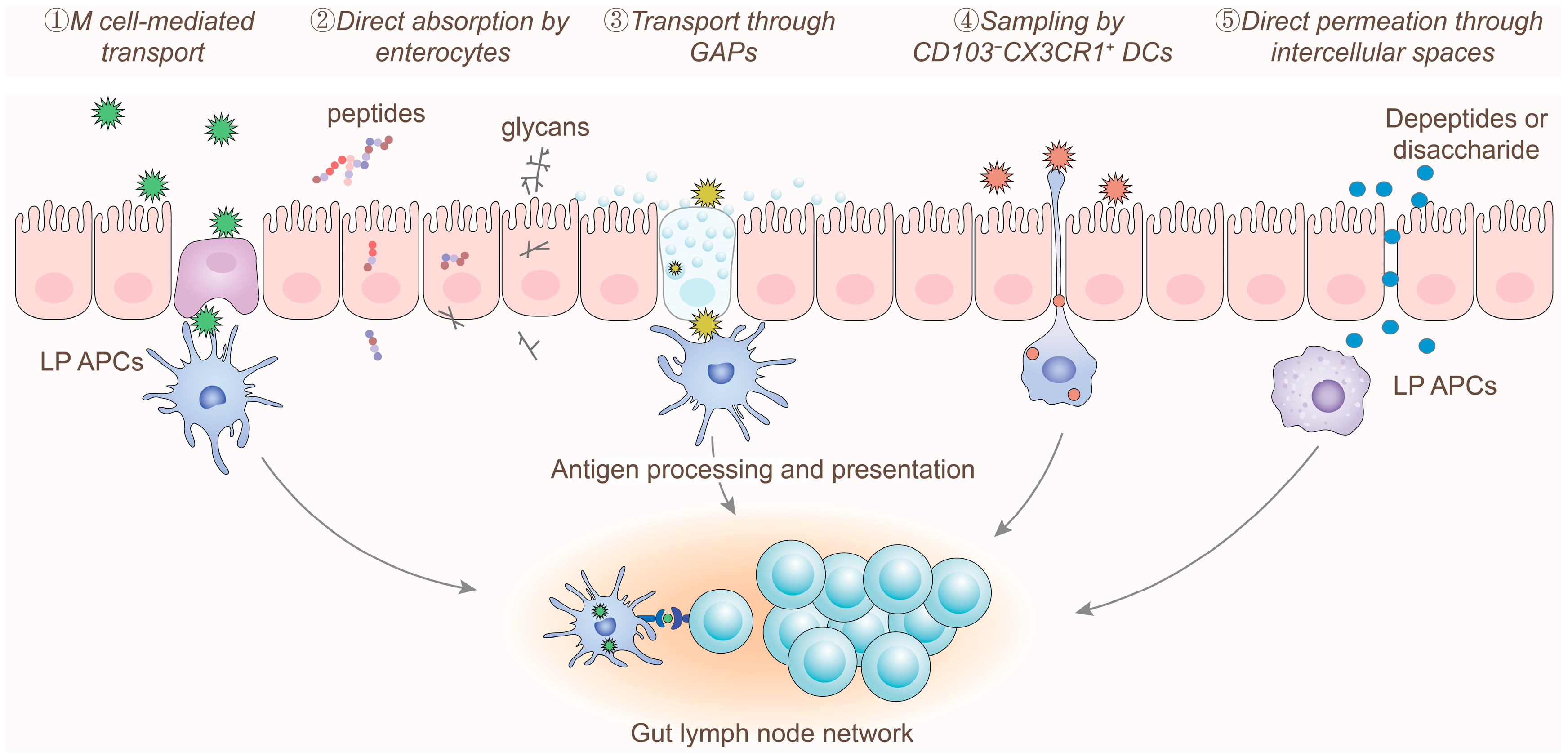
2.2. Digestion and Absorption of Allergens

Food allergens are predominantly proteins. Upon entering the digestive system, the gastrointestinal enzymatic cleavage disrupts the secondary and tertiary structures of food allergen proteins, resulting in their degradation into low-allergenicity peptides [21,22]. This process effectively helps prevent the induction of an allergic immune response in the host [21,22]. However, food processing techniques such as thermal treatment, irradiation, and chemical processing, as well as modifications involving enzymes and bioactive substances, can alter the digestibility of food allergens. These changes may inhibit complete enzymatic degradation, leading to the retention of food components containing allergenic epitopes and consequently increasing the risk of triggering allergic immune responses in the host [21,23,24,25].
After digestion and degradation, food allergens that reach the small intestine can be recognized and presented to the immune system through several mechanisms (Figure 1) [26,27,28]: (1) M cell-mediated transport: Soluble protein aggregates are initially processed, absorbed, and transported by M cells located in the follicle-associated epithelium. Subsequently, these antigens are recognized and presented to the immune system by antigen-presenting cells (APCs) within the lymphoid follicles. (2) Direct absorption by enterocytes: Amino acids, oligopeptides, and glycans with molecular weights less than 1500 Da can be directly absorbed and utilized by enterocytes for transport. (3) Goblet cell-associated antigen passage: Soluble protein particles and peptides can be absorbed and transported to the basal layer via GAPs. These antigens are then recognized, processed, and presented to naïve T cells and antigen-specific regulatory T cells (Tregs) by CD103+ DCs, which are essential for maintaining the immune activity of intestinal Tregs [29,30]. (4) Sampling by CD103CX3CR1+ DCs: CD103CX3CR1+ DCs located beneath the intestinal epithelium form trans-epithelial dendrites that actively sample low molecular weight protein particles and microbial antigens from the intestinal lumen, thereby maintaining tolerance to commensal bacteria and certain water-soluble antigens [31]. (5) Direct permeation through intercellular spaces: Some amino acids and peptides with molecular weights less than 600 Da can directly permeate through intercellular spaces into the subepithelial region, from where they are transported by blood vessels and lymphatic vessels.
Under physiological conditions, large food allergens that retain their allergenic epitopes are primarily transported through M cells and GAPs to induce immune tolerance [26,27,28]. During this process, endogenous components such as mucins, antimicrobial peptides, and secondary bile acids can be co-transported with allergenic proteins, which further diminishes their sensitizing potential [32,33]. In contrast, smaller molecular allergens that can be absorbed by enterocytes or permeate through intercellular spaces exhibit lower allergenicity and are less likely to provoke an allergic immune response in the host [28,34]. Therefore, various lifestyle factors, including high-fat diets, alcohol consumption, irregular sleep patterns, the use of antibiotics, and intestinal inflammation or infection, can compromise the integrity of the intestinal barrier. This disruption increases the likelihood of leakage of large molecular food allergens across the intestinal barrier, thereby elevating the risk of developing food allergies [35,36,37].

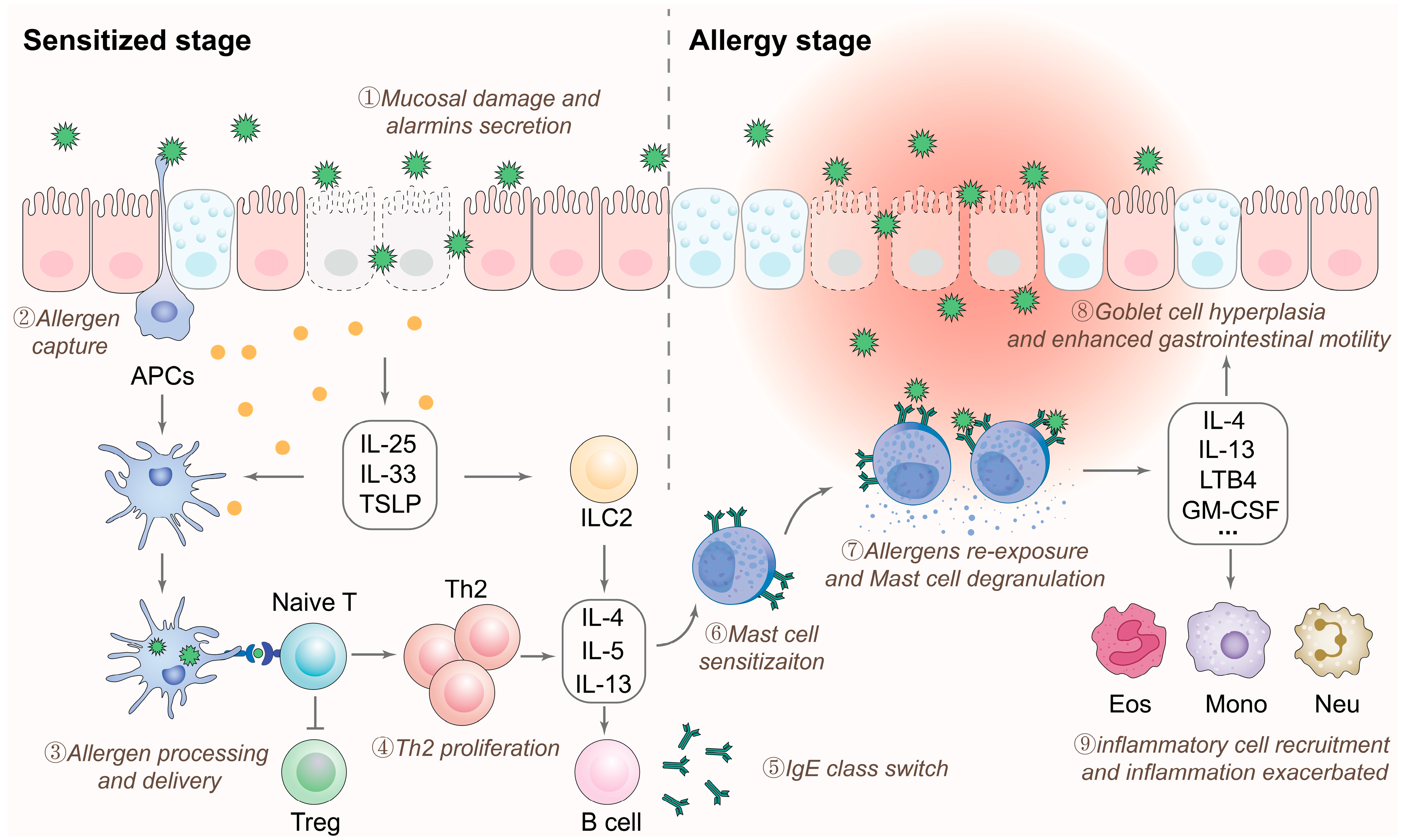
2.3. Immune Responses to Allergens

Following translocation across the intestinal barrier, food allergens that retain their allergenic epitopes can be recognized by the immune system. This recognition results in the release of bioactive mediators from effector cells, which can induce tissue damage and lead to clinical symptoms. The progression of food allergies can be divided into two phases: the sensitization phase and the effector phase (Figure 2) [38,39,40].
(1) Sensitization phase: Upon entering the host, allergens penetrate the compromised intestinal epithelial barrier and are subsequently captured and processed by APCs, such as macrophages and DCs. Concurrently, in response to epithelial-derived alarmins, including IL-25, IL-33, and thymic stromal lymphopoietin (TSLP), APCs upregulate the expression of OX40 ligand. Meanwhile, group 2 innate lymphoid cells (ILC2) increase the secretion of type 2 cytokines, such as IL-4, IL-5, and IL-13. These factors collectively promote the differentiation of type 2 helper T cells (Th2 cells). Subsequently, activated Th2 cells produce cytokines such as IL-4, IL-5, IL-9, and IL-13, which induce specific B cells to undergo class switching to IgE-secreting plasma cells, resulting in the generation of large amounts of allergen-specific IgE antibodies. These IgE antibodies then attach to the high-affinity IgE receptor (FcεRI) on the surfaces of mast cells and basophils via their Fc regions, thereby sensitizing the host.
(2) Effector phase: Upon re-exposure of a sensitized host to the same allergen, the allergen binds to allergen-specific IgE receptors on the surface of mast cells and basophils, leading to the cross-linking and clustering of multiple FcεRI receptors. This interaction transmits activation signals intracellularly through the β and γ chains of the FcεRI receptor, engaging downstream signaling pathways that activate the cells. The activation results in the release of preformed and newly synthesized bioactive mediators, including histamine, serotonin, leukotrienes, platelet-activating factor, tryptase, heparin, and prostaglandin D2, which further leads to the manifestation of various allergic symptoms.

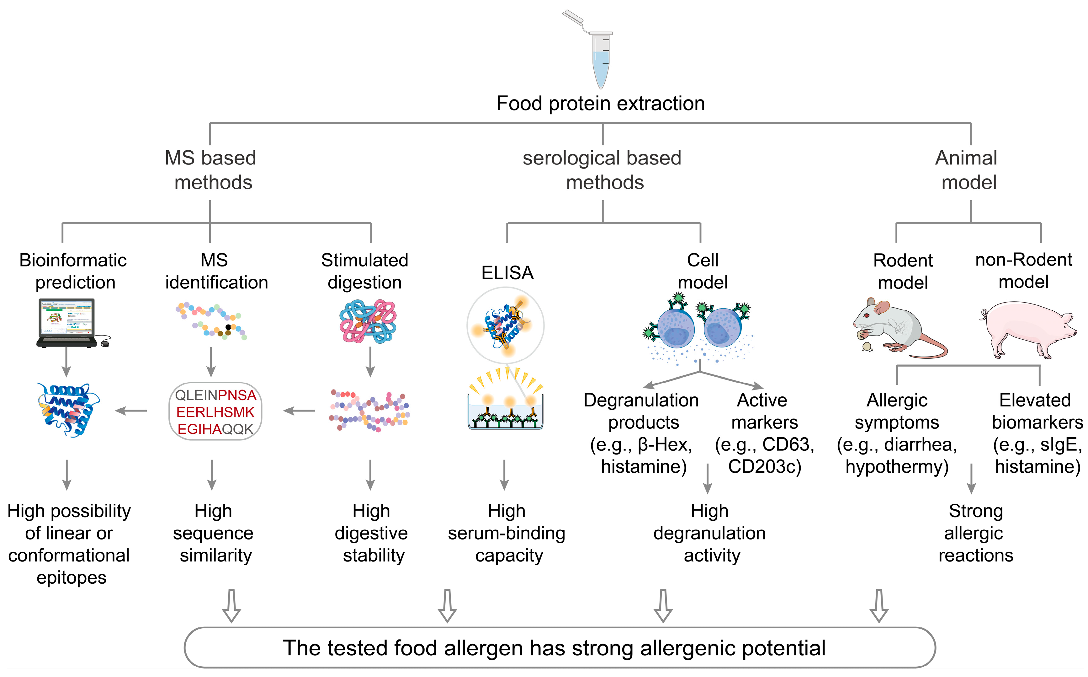
3. Assessment Methods for the Allergenicity of Food Components

As previously described, food allergens undergo a series of enzymatic digestion and trans-epithelial transport processes before being recognized by the immune system. The likelihood of triggering an allergic immune response is influenced by the molecular structure of residual allergens and the nature and quantity of allergenic epitopes [18,20,21]. Similarly, various assessment methods have been developed to evaluate the allergenicity of food allergens by mimicking the immune presentation processes (Figure 3). The principles and recent advancements of these assessment methods are summarized in Table 1.

3.1. Mass Spectrometry-Based Methods

The fundamental principle of mass spectrometry (MS) involves ionizing a sample to generate molecules or molecular fragments with varying charges. These ions are then accelerated by an electric field and directed into a mass analyzer, where they are separated based on differences in their mass-to-charge ratios under the combined influence of electric and magnetic fields. The resulting MS analysis produces characteristic peptide fragments or fingerprint spectra. By comparing these characteristic data with information from established protein databases, accurate identification of protein types and allergenic epitopes can be achieved [41,42]. Historically, the application of early MS techniques in protein and peptide analysis was somewhat limited due to the polar nature, low volatility, and thermal instability of most proteins and peptides. However, the introduction of soft ionization techniques, such as Matrix-Assisted Laser Desorption/Ionization and Electrospray Ionization, has significantly broadened the application of mass spectrometry in these fields.
The fundamental advantage of MS in evaluating food allergenicity is its capability to analyze proteins based on their amino acid sequences or peptides generated through enzymatic digestion, rather than on higher-order protein structures, which makes it an indirect method for supporting food allergenicity assessment. This characteristic allows MS to effectively mitigate the potential impacts of food processing on detection results [42,43]. Additionally, the automated and standardized workflow of MS enhances the sensitivity and stability of detection, thereby ensuring the reliability and reproducibility of the results. Currently, MS can be utilized for both qualitative and quantitative detection of food allergenic components. Qualitative detection is typically achieved through techniques such as peptide mass fingerprinting and peptide–fragment fingerprinting [41,44,45], while quantitative detection often employs methods like isotope labeling and selected reaction monitoring [41,46].
Table 1. Comparison of established food allergenicity evaluation methods.
MethodsDetection PrincipleAdvantagesShortcomingsApplicable RangePredictive Accuracy
Mass
spectrometry
[41,42,43]
Comparing the peptide segments with known epitopes that can be recognized by T/B cellsThe procedure is relatively simple and allows for quick resultsLarge-scale instruments and a reliable database are required, and only linear epitope information can be
obtained
Suitable for almost all allergenic foodsRelatively low
Bioinformatic
prediction
[47,48,49,50]
Comparing peptide segments with allergic epitopes and making predictions of
potential linear and conformational epitopes
Computational simulation can identify a wide range of potential epitopesThis process demands proficient operational skills and a reliable database; otherwise, inaccurate results may occurSuitable for almost all allergenic foodsRelatively low
Simulated
digestion
[15,21,22]
Detecting the
structure, molecular weight, and allergic epitopes in residual
digestion products
It can reflect the effects of different digestion
conditions and
interactions
between food components and allergic epitopes after digestion
The operation is
relatively complex, and the experimental results may not reflect the
actual situation
accurately
Suitable for almost all allergenic foodsRelatively low
ELISA
examination
[51,52,53]
Detecting the allergic epitopes based on the antigen–antibody
interaction
Can quickly
determine the components
responsible for
potential allergic reactions in
patients
Obtaining suitable
serum can be
challenging
Suitable for
allergenic foods tested with
human or animal serum
Medium
Cell model
[54,55]
The interaction
between allergens and allergen-specific IgE can induce mast cell or basophil degranulation
Can accurately
determine the presence of
substances that trigger mast cell or basophil activation
Obtaining suitable
serum can be
challenging
Suitable for
allergenic foods tested with
human or animal serum
Relatively high
Animal model
[56,57,58]
Sensitizing animals with allergens and
observing changes in allergic symptoms and biomarkers after
allergen challenge
The experimental results best
capture the actual allergenic
potential of the tested food
components through multiple aspects
The experiment is costly, time-consuming, and may involve ethical issuesSuitable for
allergenic foods tested with protein reference material
Relatively high
MS has been successfully employed to evaluate the allergenicity of food components in dairy products, shrimp, meat, and other food items (Table 2). Monaci et al. [59] utilized Liquid Chromatography-Quadrupole-Time of Flight Mass Spectrometry to assess milk allergens in thermally processed foods, successfully identifying specific peptides associated with milk allergens. These peptides can serve as markers for preliminary screening and validation of target allergens in future studies, providing a foundation for related research. Stasio et al. [60] extracted peanut components using SDS-PAGE and identified their allergenicity using a Quadrupole-Orbitrap Mass Spectrometer. In the MS spectra, the ten most intense ion peaks were selected for fragmentation analysis. The spectral data were then converted into protein identification results by integrating instrument analysis software with the NCBI database, thereby assessing the allergenic potential of the peanut proteins.
Pilolli et al. [61] developed a relatively rapid sample processing procedure involving ultrasound-assisted Size Exclusion Chromatography for extraction and purification and constructed a multidimensional chromatographic analysis system. Utilizing this system, they established an efficient and streamlined analytical workflow, successfully identifying the allergenicity from eggs, milk, soy, hazelnut, and peanut components in complex food matrices through Selected Reaction Monitoring analysis. Despite the advantages listed above, MS cannot detect conformational epitopes and heavily depends on sequence homology to known allergens. Moreover, it cannot assess the allergenic potential of novel proteins. The stringent requirements for allergen extraction and purification, combined with the high costs of instrument setup, have further limited the widespread application of MS in food allergenicity assessment.

3.2. Bioinformatic Prediction

Bioinformatics is an interdisciplinary field that integrates biology, mathematics, and computer science, with the goal of analyzing and interpreting vast amounts of biological data. Its application in assessing food allergenicity relies on the principle of homology analysis. Specifically, the amino acid sequence of the protein under investigation is compared against internationally recognized databases of known allergen sequences to predict the likelihood of cross-reactivity between the protein of interest and established allergens [47,48,49,50].
In bioinformatics-based allergenicity assessments, the construction of a comprehensive and robust database of known allergens is essential. Currently, Allergome is the largest global allergen database, containing over 10,000 allergen sequence entries. In addition to this resource, several specialized databases are available, including the allergen search engine AllergenOnline (http://www.allergenonline.org/, accessed on 25 May 2025), the allergen nomenclature database ALLERGEN NOMENCLATURE (https://www.allergen.org/, accessed on 25 May 2025), the allergen protein family database ALLFAM (https://www.meduniwien.ac.at/allfam/, accessed on 25 May 2025), and the allergen structure databases SDAP (https://fermi.utmb.edu/, accessed on 25 May 2025) and COMPARE (https://db.comparedatabase.org/, accessed on 25 May 2025). These databases provide extensive data support and analytical tools for allergen research, facilitating a deeper understanding of allergen characteristics and their sensitization mechanisms.
In recent years, advancements in the understanding of allergens, alongside the continuous accumulation of related data, have led to the establishment and refinement of bioinformatics technologies for predicting the allergenicity of food components. For instance, FUGUE can predict the three-dimensional structures of proteins from their sequences and identify homologous proteins with low sequence similarity through combined sequence and structural similarity analyses [62]. The Allermatch system utilizes sequence alignment techniques to predict protein allergenicity [63]. Additionally, AlgPred integrates multiple prediction tools into a unified framework, facilitating effective predictions of novel allergens and their epitopes [64].
In terms of specific applications, Hu et al. [65] utilized DNAMAN software to predict novel IgE-binding epitopes of the bovine milk allergen BLG and identified four key amino acids: A34, A37, R40, and V. Garino et al. [66] employed the Allergome database and the BLAST (version 1.3.0) sequence alignment tool in conjunction with seven different allergenicity prediction software packages. Through sequence and domain analyses, they ultimately evaluated the allergenicity of 28 allergenic lipid transfer proteins across various species.
Despite these advantages, the use of bioinformatics in predicting food allergenicity has several limitations. Firstly, the accuracy of prediction models is limited by the quality and quantity of available allergen data [47]. Incomplete or inaccurate data can lead to unreliable predictions. Furthermore, the experience of the operator may also affect the results obtained. Secondly, predicting the allergenic potential of complex mixtures or processed foods remains challenging due to the dynamic nature of protein interactions and modifications. The development of artificial intelligence and improvement of allergen databases may help overcome these limitations [67].

3.3. Simulated Digestion

The simulated digestion method for allergenicity assessment involves recreating in vivo digestive conditions by mimicking the physiological characteristics of humans. This approach employs relevant digestive enzyme systems to evaluate the digestive and absorption properties of samples in vitro [21,22,68]. Allergenic proteins that remain intact and immunogenic after processing and gastrointestinal digestion, and prior to entering the immune system, may possess potential allergenicity. Generally, the greater the digestive stability of a protein, the higher its allergenic potential; conversely, lower digestive stability is associated with reduced allergenicity [21,22]. Based on variations in digestive conditions, the simulated digestion method can be further categorized into two approaches: static in vitro-simulated digestion and dynamic in vitro-simulated digestion.
The static simulated digestion system can be divided into three main components based on the digestive phases: the oral phase, the gastric and small intestinal phase, and the large intestinal fermentation phase [69]. Given the rapid onset of most food allergic reactions and the distribution characteristics of the human lymphatic immune system, the evaluation of food allergenicity typically focuses on simulating the digestive processes of the mouth, stomach, and duodenum, with less emphasis on the large intestinal fermentation phase [69,70]. This approach offers several advantages, including operational simplicity and broad applicability.
In assessing the potential allergenicity of food allergens, commonly used static in vitro digestion methods are primarily based on standardized protocols proposed by international organizations, such as the United States Pharmacopeia (USP) and INFOGEST. The INFOGEST method, first introduced by Minekus et al. in 2014, aims to enhance the reproducibility and comparability of digestion results across different laboratories through standardized experimental conditions and procedures [69]. Subsequently, Brodkorb et al. optimized this method, developing the INFOGEST 2.0 in vitro static digestion protocol [71]. This updated version provides more detailed descriptions of digestive fluids, procedural steps, and equipment, as well as specific guidelines for enzyme activity assays, sample collection, and processing. Currently, the INFOGEST 2.0 method has been widely applied in studies evaluating the potential allergenicity of various food allergens [72,73,74].
Dynamic in vitro-simulated digestion represents an innovative approach that integrates a range of dynamic changes, including food intake, secretion of digestive fluids, gastrointestinal emptying, peristalsis, and absorption across the gastrointestinal mucosa. This method maximizes the simulation of physiological conditions within the human or animal gastrointestinal tract. Compared to traditional static in vitro-simulated digestion methods, dynamic approaches address their limitations by minimizing manual operations and maintaining consistent simulated digestion conditions, effectively reducing experimental errors arising from operational variability or fluctuations in digestion conditions [21,22].
Moreover, dynamic in vitro digestion models can replicate digestive conditions across various physiological or health states, including those of infants, adults, individuals with gastrointestinal diseases, allergic patients, and diabetic patients. This capability is crucial for obtaining critical data, such as the threshold levels of food allergens [21,22,69,71]. Unlike static digestion processes, dynamic conditions influence the digestive stability of proteins and their interactions with other food components, which may significantly impact the evaluation of the allergenic potential of digested products and the kinetic behavior of digestion-resistant peptides. Consequently, dynamic in vitro digestion offers a more accurate representation of food allergen digestion.
For instance, Prodic et al. investigated the allergenic potential of digestion-resistant peptides released during the simulated gastric digestion of peanuts [75]. Their study revealed that peptides with strong allergenic potential retained continuous epitope sequences, primarily originating from the 2S albumin in peanuts, findings that are closely consistent with observations in real-world research scenarios. Despite this, the limitations of simulated digestion in completely reflecting real ingestion conditions in humans limit its applications in broad food allergenicity evaluation.
Table 2. Applications of allergenicity evaluation methods on food components.
MethodsFood MatrixExperimental DesignEpitope FeaturesReference
Mass
spectrometry-based
methods
LC-MS/MSCookiesEstablishing a fast sample-handling procedure and using the signature peptide sequences to evaluate the allergenic potential.Linear epitope[61]
LC-MS/MSInsectsProteins extracted from cricket and black soldier fly were sequenced and their cross-reactivity to allergens from shrimps was examined.Linear epitope[76]
RPLC-ESI-HRMSChocolateUtilizing the signature peptide sequences to search for and align the target allergens.Linear epitope[77]
LC-QQQ-MSMeat-based foodstuffs like cooked meat and sausagesUtilizing the signature peptide and its isotope-labeled peptide as standards and internal standards, respectively.Linear epitope[78]
LC-MS/MSShrimpsThe signature peptides were confirmed and synthesized as the quantitative peptides, and the relative isotope-labeled internal standards were used in the quantitative analysis.Linear epitope[79]
Bioinformatic predictionMachine learningMicroalgaeDIA-NN was used to create a peptide spectrum library from PBQC DIA-MS data, with the “Deep learning-based spectra” option enabled to predict potential allergenicity.Linear epitope[80]
Database-based searching and
prediction
MilkUtilizing the UniProt protein database to predict key homology sequences and use DNAMAN software to identify allergic sequence alignments.Linear epitope[65]
Database-based searching and
prediction
Egg productsThe linear epitope of potential allergens was predicted based on computational approaches including Immunomedicine Group, ABCpred, BCEpred, BepiPred-3.0 Server, and DNAStar.Linear epitope[81]
Machine learningSesameUtilizing AlphaFold2 to predict the 3D structure of potential allergens based on known sequences and evaluate the accuracy of the predicted results using Psi/Phi Ramachandran plots and the WHATCHECK Complete suite.Conformational epitope[82]
Machine learningFruits and nutsPeptides similar to known IgE epitopes can be identified with the property distance tool, and conformational epitopes can be identified by the cross–react method, both based on the SDAP webserver.Conformational epitope[83]
Simulated
digestion
Static simulated
digestion
PeanutsVerifying the correlation between allergenicity and resistance to proteolysis by pepsin.Linear epitope[75]
Static simulated
digestion
MilkVerifying the contribution of protein phosphorylation to food allergenicity before and after simulated digestion.Linear epitope[68]
Dynamic simulated digestionShrimpsExamining the effects of digestion stability on food allergenicity during dynamic digestion.Conformational epitope[84]
Dynamic simulated digestionMilkEvaluating the effects of thermal processing and lactosylation on digestibility and allergenicity.Conformational epitope[85]
ELISA
examination
Indirect ELISAInsectsEvaluating the risk of food allergy from eating Gryllus bimaculatus in patients allergic to shrimps, using ELISA and IgE crosslinking luciferase assays.Conformational epitope[86]
Sandwich ELISACrustacean productsDesigning a system using polyclonal and monoclonal antibodies to capture and recognize potential allergens.Linear epitope and Conformational epitope[87]
Sandwich ELISAFish productsPolyclonal antibodies were raised against parvalbumins from 13 fish species of seven fish orders, selected for molecular diversity and immunoreactivity.Conformational epitope[88]
Sandwich ELISAAlmondsCombining sandwich ELISA with lateral flow immunoassay to identify potential allergens.Conformational epitope[89]
Cell modelHuman peripheral blood basophilsPeanutsAssessing diagnostic application of natural and recombinant allergens by BAT and identifying IgE-binding epitopes using oligopeptide microarrays.Linear epitope[90]
KU812 cell modelSoymilkUtilizing a sensitized cell model to evaluate the changes in spatial structure and sequences after bacterial fermentation.Linear epitope[91]
LAD2 cell modelShrimpsThe LAD2 cell degranulation assay was used to assess the binding affinity and antigenicity of the allergenic epitopes.Linear epitope[92]
RBL-2H3 cell modelMealwormThe RBL-2H3 cell model was used to evaluate the allergenicity of mealworm tropomyosin in comparison with shrimp and chicken tropomyosin.Conformational epitope[93]
Animal modelBALB/c mice modelSoybean mealUtilizing the BALB/c mice model to determine whether solid-state fermentation could degrade major allergens and reduce the potential allergenicity of soybean meal.Linear and conformational epitope[94]
C3H/HeNCrl mice modelMilkUsing the C3H/HeNCrl mice model to examine the susceptibility of potential allergens.Conformational epitope[95]
BN rat modelEgg and potato componentsUtilizing the BN rat model to determine the potential allergenicity of novel proteins in genetically modified food.Conformational epitope[96]
Pig modelSoybean productsUsing the pig model to evaluate the allergenicity of soybean protein after
deglycosylation and pepsin digestion.
Linear and conformational epitope[97]
Monkey modelTransgenic riceEvaluating the allergenicity of transgenic crops by examining the hematological, biochemical, pathological, and histopathological changes in sensitized monkeys.Linear epitope[98]

3.4. ELISA

This method is based on the specific antigen-antibody reaction and primarily evaluates the allergenic potential of target components by measuring their binding to specific IgE in the serum of allergic patients [51,52]. ELISA is commonly used to detect specific epitopes in food components, thereby reflecting the structural integrity of antigens, but it does not directly measure allergenicity. Given the limited availability of serum from allergic individuals, researchers often use specific IgE serum derived from immunized animals as an alternative for serological experiments.
The ELISA assay is one of the most widely used methods for evaluating food allergenicity, with a variety of commercially available ELISA kits on the market. Common types of ELISA include sandwich ELISA, competitive/inhibition ELISA, direct ELISA, and indirect ELISA, each characterized by distinct features and suitability for different testing conditions [53].
Sandwich ELISA is particularly effective for analyzing complex samples, as it does not require purification of the target antigen prior to measurement, while still achieving high sensitivity and specificity. Competitive/inhibition ELISA is suitable for detecting small molecular weight antigens, measuring the concentration of antigens or antibodies in the sample by analyzing interference with the expected signal output. Direct ELISA, which omits the use of a secondary antibody and the signal amplification step, is appropriate for analyzing immune responses to antigens, but has relatively lower detection sensitivity. Indirect ELISA involves coating the antigen onto a microplate, where the unlabeled primary antibody binds to the specific antigen during detection, followed by the application of an enzyme-conjugated secondary antibody specific to the host species of the primary antibody, thereby completing the measurement of the target [53,99,100].
This far, numerous studies have reported the use of ELISA technology to assess the allergenicity of food components. For instance, He et al. developed several ELISA-based detection methods for evaluating the allergenic potential of components such as ALA, BLG, and casein in food matrices [86,101,102]. Similarly, Arun et al. employed sandwich ELISA to assess the allergenic potential of peanut allergens, including Ara h 1, Ara h 2, Ara h 3, Ara h 6, and Ara h 8, in food matrices [103]. A substantial number of application examples have been well summarized in the literature [104], and thus will not be reiterated here.

3.5. Cell Model

In recent years, cell culture technology has been widely applied in molecular biology, genetics, immunology, and other fields, evolving into a significant biotechnological tool with notable achievements [54]. In the evaluation of food allergenicity, mast cells and basophils are the most commonly used cellular models, primarily due to their critical roles as effector cells in allergic reactions [54,55,105]. Upon activation, mast cells can induce bronchial spasms and increase vascular permeability, leading to clinical symptoms by synthesizing and releasing various mediators, such as histamine and leukotrienes [55]. Additionally, mast cells release multiple cytokines that further promote the development and progression of allergic diseases [105]. Therefore, by co-incubating the food allergenic components under investigation with sensitized mast cells or basophils, and detecting the levels of effector molecules in the culture system, researchers can assess the allergenic potential of the test food components [54]. These methods offer a controllable, stable, and reproducible platform for investigating allergic reactions. Their use enables efficient screening of food allergens and evaluation of their sensitizing potential in an ethically sound and practically advantageous manner compared to in vivo experiments. Consequently, these methods hold significant value and promise for predicting human allergic responses in food allergenicity assessment.
Currently, commonly used cell lines for food allergenicity assessment include Rat Basophilic Leukemia 2H3 (RBL-2H3) and human mast cell lines, such as Human Mast Cell Line 1 (HMC-1) and Laboratory of Allergic Diseases 2 (LAD2) [54,106]. Additionally, there are reports of using bone marrow-derived mast cells for evaluation. However, these cells require 4 to 6 weeks to mature. The prolonged culture period, high material consumption, and associated costs limit their application in food allergen detection [107]. Consequently, commercially available cell lines like RBL-2H3, HMC-1, and LAD2 are more frequently employed for evaluating food allergenicity. For example, Jiang et al. [108] successfully identified the major fish allergen parvalbumin by detecting fluorescence signals in RBL-2H3 cells. Similarly, Vogel et al. [109] utilized a humanized RBL-2H3 cell model to analyze and identify the allergenicity of peanut and hazelnut proteins in complex food matrices.
Furthermore, the Basophil Activation Test (BAT) has gained widespread application for detecting IgE-mediated hypersensitivity reactions [110,111]. This method assesses the expression of activation markers on the surface of basophils and the presence of factors in the supernatant to evaluate allergic reactions. The BAT method has been extensively used in related studies. For instance, Sabato et al. [112] confirmed the high sensitivity of basophils to food allergens through BAT experiments. Additionally, Schwager et al. [90] employed the BAT method to evaluate the allergenicity of natural and recombinant peanut oleosin, finding that roasted peanuts significantly enhanced the allergenicity of this protein.

3.6. Animal Model

Animal models play a crucial role in assessing food allergen sensitizing potential. They serve as a bridge between in vitro assessment and real human allergic reactions, and they can closely mimic the human immune response to allergens. By exposing animals to allergens through various routes—such as oral administration, inhalation, or skin contact—similar to human exposure, researchers can observe and measure immune responses and clinical symptoms. The allergenic potential can further be evaluated by detecting specific IgE and IgG antibody levels, as well as assessing allergic inflammation through changes in cytokine profiles. Additionally, animal models provide valuable insights into the mechanisms underlying allergic sensitization and development [56,113]. However, due to inherent differences between animals and humans, results should be interpreted with caution. Appropriate species and strain selection, along with well-designed experiments, are essential to ensure relevance to human biology. Despite these limitations, animal models remain indispensable in allergenicity assessment because they offer in vivo data that closely approximates human allergic reactions [56,113].
Rodents are often the preferred choice for constructing food allergy animal models due to their physiological and genetic similarities to humans [57]. Mice, in particular, are widely used in research because of their unique advantages, including a rich array of genetic resources and high compatibility with genetic manipulations, such as forward genetics, reverse genetics, transgenic technology, and targeted mutations or gene knock-in/knock-out [114]. The extensive application of these genetic engineering techniques in mouse models has led to the development of many genetically modified humanized mouse models, which are valuable tools for studying human diseases. For example, Jimenez-Saiz et al. utilized various mouse strains (e.g., C57BL/6, Rag2-/-γc+, (Igh-7tmlLed) 25, BCL6fl/fl) to elucidate the exposure routes of human food allergens, providing a theoretical basis for potential therapeutic approaches [113]. Additionally, Burton et al. successfully established a humanized adaptive immune system mouse model (huNSG mouse) capable of simulating IgE-mediated food allergic reactions by engrafting human tissue-derived mast cells, thus offering a novel platform for studying IgE-mediated allergic responses [115]. Furthermore, mice have a short breeding cycle (approximately 18–20 days), are cost-effective, and are easy to manage, making them an ideal choice for disease model research [58].
Commonly used experimental rodent strains in food allergenicity evaluation include mouse strains such as C57BL/6, C3H/HeJ, DBA2, and BALB/c, as well as rat strains like BN [57]. Among these, Balb/c mice, which tend to differentiate toward Th2 cells in their immune response, have been extensively utilized in allergy research. For instance, Fotschki et al. [116] sensitized Balb/c mice by intraperitoneal injection and assessed the allergenic potential of food components such as BLG and ALA by measuring specific IgE levels in mouse serum. Wavrin et al. [117] evaluated the allergenic potential of the peanut allergen Arah 1 by exposing Balb/c mice to the allergen through contact, inhalation, and oral administration.
In addition to rodents, other animal models, such as pigs, dogs, and monkeys, are also widely employed for food allergenicity evaluation. These non-rodent models are considered more representative in biomedical research due to their closer genetic relationship with humans and their ability to better simulate human physiological and pathological processes. For example, Li et al. [118] utilized a pig model to evaluate the allergenicity of genetically modified milk containing recombinant human lactoferrin. By monitoring indicators such as the incidence of diarrhea, humoral immune responses, Th2 cell responses, and intestinal mucosal damage, the study demonstrated that the genetically modified milk did not induce allergic reactions. However, despite their similarities to humans in terms of organ size, physiological structure, disease progression, and histological features, the use of these larger mammals in food allergenicity evaluation is limited due to economic and time constraints.

4. Conclusions and Perspectives

This review comprehensively clarifies the mechanisms of food allergies and summarizes the methods for assessing food component allergenicity, covering the principles and applications of various techniques. Although these methods differ in their mechanisms, all are developed based on allergen epitope information. By analyzing the residual allergen epitopes and their quantitative and qualitative changes, we can preliminarily predict a component’s allergenic potential.
Future research should develop evaluation methods that better align with food allergy mechanisms. For mass spectrometry, technological advancements are expected to boost detection efficiency and throughput. Meanwhile, more powerful data-processing software and algorithms will aid in the in-depth mining of allergen information, such as modification sites and conformational changes. This will enhance our understanding of allergens’ characteristics and sensitization patterns. In addition, gathering and updating data on allergen amino acid sequences, 3D structures, and antigenic epitopes helps refine existing allergen databases. Integrating various protein properties, such as sequence similarity, domains, and physicochemical characteristics, into advanced predictive algorithms and models can enhance the accuracy and reliability of allergenicity predictions and minimize both false-positive and false-negative rates.
Simulated digestion methods must refine their conditions to better replicate the human digestive environment, factoring in elements like digestive fluid composition and concentration, pH, temperature, and enzyme activity. This will enhance the accuracy of in vitro results in reflecting in vivo conditions. Additionally, integrating simulated digestion with advanced detection techniques such as mass spectrometry and immunoassays allows for real-time monitoring and analysis of allergens and their degradation products as digestion occurs. This approach yields valuable insights into sensitization behavior and allergenicity, establishing a reliable foundation for assessing the digestive stability and allergenic potential of food allergens.
ELISA has high sensitivity and specificity for allergen detection. However, there is still room for improvement. Future research could focus on developing more specific antibodies to minimize cross-reactivity with non-allergenic proteins, thereby reducing false-positive results. Furthermore, the development of multiplex ELISA assays that can simultaneously detect multiple allergens in a single sample is highly anticipated. This would not only improve the efficiency of food allergenicity testing but also reduce costs and resource consumption.
Cell and animal models must be further optimized and standardized to better emulate human immunological and physiological responses, and to expand their application in studying disease mechanisms and evaluating treatments. Additionally, it is also crucial to consider economic and time costs, so that food allergenicity assessments can truly benefit patients, enhance their well-being, and alleviate the health and economic burdens on both patients and society. Looking ahead, the development of more precise, efficient, and cost-effective food allergenicity evaluation methods is anticipated. Such methods would provide stronger scientific support for the diagnosis, treatment, and prevention of food allergies.

Author Contributions

H.Z. wrote the main manuscript. X.C. reviewed and revised the manuscript. X.L. was responsible for the concept design and reviewed the manuscript. All authors have read and agreed to the published version of the manuscript.

Funding

This work was supported by the National Key Research and Development Program of China (No. 2024YFF1105905), the Key Project of Natural Science Foundation of Jiangxi Province, China (No. 20232ACB205022), the Jiangxi Nutrition and Health Management Medical Research Institute Cultivation Project (No. 2022-PYXM-05), and the Special Project for Research and Development in Key Areas of Guangdong Province (No. 2022B0202030002).

Institutional Review Board Statement

Not applicable.

Informed Consent Statement

Not applicable.

Data Availability Statement

No new data were created or analyzed in this study. Data sharing is not applicable to this article.

Conflicts of Interest

All authors declare that they have no relevant conflicts of interest.

Abbreviation

APCsAntigen-presenting cellsLAD2Laboratory of Allergic Diseases 2
BATBasophil activation testLPLamina propria
β-Hexβ-hexosaminidaseLTB4Leukotriene B4
DCsDendritic cellsMonoMonocytes
ELISAEnzyme-linked immunosorbent assaysMSMass spectrometry
EosEosinophilsNeuNeutrophils
FcεRIhigh-affinity IgE receptorRBL-2H3Rat Basophilic Leukemia 2H3
GAPsGoblet cell-associated antigen passagesTh2 cellsT helper 2 cells
GM-CSFStimulating factorTregsRegulatory T cells
HMC-1Human Mast Cell Line 1TSLPThymic stromal lymphopoietin
ILC2sLymphoid cells

References

  1. Sampath, V.; Abrams, E.M.; Adlou, B.; Akdis, C.; Akdis, M.; Brough, H.A.; Chan, S.; Chatchatee, P.; Chinthrajah, R.S.; Cocco, R.R. Food allergy across the globe. J. Allergy Clin. Immunol. 2021, 148, 1347–1364. [Google Scholar] [CrossRef] [PubMed]
  2. Sindher, S.B.; Long, A.; Chin, A.R.; Hy, A.; Sampath, V.; Nadeau, K.C.; Chinthrajah, R.S. Food allergy, mechanisms, diagnosis and treatment: Innovation through a multi-targeted approach. Allergy 2022, 77, 2937–2948. [Google Scholar] [CrossRef] [PubMed]
  3. Gupta, R.S.; Warren, C.M.; Smith, B.M.; Blumenstock, J.A.; Jiang, J.; Davis, M.M.; Nadeau, K.C. The public health impact of parent-reported childhood food allergies in the United States. Pediatrics 2018, 142, e20181235. [Google Scholar] [CrossRef] [PubMed]
  4. Branum, A.M.; Lukacs, S.L. Food allergy among children in the United States. Pediatrics 2009, 124, 1549–1555. [Google Scholar] [CrossRef]
  5. Conrado, A.B.; Ierodiakonou, D.; Gowland, M.H.; Boyle, R.J.; Turner, P.J. Food anaphylaxis in the United Kingdom: Analysis of national data, 1998–2018. BMJ 2021, 372, n251. [Google Scholar] [CrossRef]
  6. Ma, Z.; Chen, L.; Xian, R.; Fang, H.; Wang, J.; Hu, Y. Time trends of childhood food allergy in China: Three cross-sectional surveys in 1999, 2009, and 2019. Pediatr. Allergy Immunol. 2021, 32, 1073–1079. [Google Scholar] [CrossRef]
  7. Sicherer, S.H.; Sampson, H.A. Food allergy: A review and update on epidemiology, pathogenesis, diagnosis, prevention, and management. J. Allergy Clin. Immunol. 2018, 141, 41–58. [Google Scholar] [CrossRef]
  8. Renz, H.; Allen, K.J.; Sicherer, S.H.; Sampson, H.A.; Lack, G.; Beyer, K.; Oettgen, H.C. Food allergy. Nat. Rev. Dis. Prim. 2018, 4, 17098. [Google Scholar] [CrossRef]
  9. Berin, M.C.; Lozano-Ojalvo, D.; Agashe, C.; Baker, M.G.; Bird, J.A.; Nowak-Wegrzyn, A. Acute FPIES reactions are associated with an IL-17 inflammatory signature. J. Allergy Clin. Immunol. 2021, 148, 895–901.e6. [Google Scholar] [CrossRef]
  10. Morita, H.; Nomura, I.; Orihara, K.; Yoshida, K.; Akasawa, A.; Tachimoto, H.; Ohtsuka, Y.; Namai, Y.; Futamura, M.; Shoda, T. Antigen-specific T-cell responses in patients with non–IgE-mediated gastrointestinal food allergy are predominantly skewed to TH2. J. Allergy Clin. Immunol. 2013, 131, 590–592.e6. [Google Scholar] [CrossRef]
  11. Bol-Schoenmakers, M.; Rezende, M.M.; Bleumink, R.; Boon, L.; Man, S.; Hassing, I.; Fiechter, D.; Pieters, R.; Smit, J. Regulation by intestinal γδ T cells during establishment of food allergic sensitization in mice. Allergy 2011, 66, 331–340. [Google Scholar] [CrossRef] [PubMed]
  12. Simon, D.; Cianferoni, A.; Spergel, J.; Aceves, S.; Holbreich, M.; Venter, C.; Rothenberg, M.; Terreehorst, I.; Muraro, A.; Lucendo, A. Eosinophilic esophagitis is characterized by a non-IgE-mediated food hypersensitivity. Allergy 2016, 71, 611–620. [Google Scholar] [CrossRef] [PubMed]
  13. Pecora, V.; Prencipe, G.; Valluzzi, R.; Dahdah, L.; Insalaco, A.; Cianferoni, A.; De Benedetti, F.; Fiocchi, A. Inflammatory events during food protein-induced enterocolitis syndrome reactions. Pediatr. Allergy Immunol. 2017, 28, 464–470. [Google Scholar] [CrossRef]
  14. Chinthrajah, R.S.; Hernandez, J.D.; Boyd, S.D.; Galli, S.J.; Nadeau, K.C. Molecular and cellular mechanisms of food allergy and food tolerance. J. Allergy Clin. Immunol. 2016, 137, 984–997. [Google Scholar] [CrossRef]
  15. EFSA Panel on Genetically Modified Organisms (GMO Panel). Scientific Opinion on the assessment of allergenicity of GM plants and microorganisms and derived food and feed. EFSA J. 2010, 8, 1700. [Google Scholar] [CrossRef]
  16. Dramburg, S.; Hilger, C.; Santos, A.F.; de Las Vecillas, L.; Aalberse, R.C.; Acevedo, N.; Aglas, L.; Altmann, F.; Arruda, K.L.; Asero, R. EAACI molecular allergology user’s guide 2.0. Pediatr. Allergy Immunol. 2023, 34, e13854. [Google Scholar] [CrossRef]
  17. Zhou, F.; He, S.; Sun, H.; Wang, Y.; Zhang, Y. Advances in epitope mapping technologies for food protein allergens: A review. Trends Food Sci. Technol. 2021, 107, 226–239. [Google Scholar] [CrossRef]
  18. Kamath, S.D.; Bublin, M.; Kitamura, K.; Matsui, T.; Ito, K.; Lopata, A.L. Cross-reactive epitopes and their role in food allergy. J. Allergy Clin. Immunol. 2023, 151, 1178–1190. [Google Scholar] [CrossRef]
  19. Liu, C.; Sathe, S.K. Food allergen epitope mapping. J. Agric. Food Chem. 2018, 66, 7238–7248. [Google Scholar] [CrossRef]
  20. Lee, A.S.E.; Suprun, M.; Sampson, H. Epitope-based IgE assays and their role in providing diagnosis and prognosis of food allergy. J. Allergy Clin. Immunol. Pract. 2023, 11, 2983–2988. [Google Scholar] [CrossRef]
  21. Sun, N.; Liu, Y.; Liu, K.; Wang, S.; Liu, Q.; Lin, S. Gastrointestinal fate of food allergens and its relationship with allergenicity. Compr. Rev. Food Sci. Food Saf. 2022, 21, 3376–3404. [Google Scholar] [CrossRef] [PubMed]
  22. Bøgh, K.L.; Madsen, C.B. Food allergens: Is there a correlation between stability to digestion and allergenicity? Crit. Rev. Food Sci. Nutr. 2016, 56, 1545–1567. [Google Scholar] [CrossRef] [PubMed]
  23. Vanga, S.K.; Singh, A.; Raghavan, V. Review of conventional and novel food processing methods on food allergens. Crit. Rev. Food Sci. Nutr. 2017, 57, 2077–2094. [Google Scholar] [CrossRef] [PubMed]
  24. Liu, Q.; Lin, S.; Sun, N. How does food matrix components affect food allergies, food allergens and the detection of food allergens? A systematic review. Trends Food Sci. Technol. 2022, 127, 280–290. [Google Scholar] [CrossRef]
  25. Yang, H.; Qu, Y.; Gao, Y.; Sun, S.; Ding, R.; Cang, W.; Wu, R.; Wu, J. Role of the dietary components in food allergy: A comprehensive review. Food Chem. 2022, 386, 132762. [Google Scholar] [CrossRef]
  26. Zhang, X.; Chen, X.; Wang, Z.; Meng, X.; Hoffmann-Sommergruber, K.; Cavallari, N.; Wu, Y.; Gao, J.; Li, X.; Chen, H. Goblet cell-associated antigen passage: A gatekeeper of the intestinal immune system. Immunology 2023, 170, 1–12. [Google Scholar] [CrossRef]
  27. Newberry, R.D.; Hogan, S.P. Intestinal epithelial cells in tolerance and allergy to dietary antigens. J. Allergy Clin. Immunol. 2021, 147, 45–48. [Google Scholar] [CrossRef]
  28. Perrier, C.; Corthesy, B. Gut permeability and food allergies. Clin. Exp. Allergy 2011, 41, 20–28. [Google Scholar] [CrossRef]
  29. Noah, T.K.; Knoop, K.A.; McDonald, K.G.; Gustafsson, J.K.; Waggoner, L.; Vanoni, S.; Batie, M.; Arora, K.; Naren, A.P.; Wang, Y.-H. IL-13–induced intestinal secretory epithelial cell antigen passages are required for IgE-mediated food-induced anaphylaxis. J. Allergy Clin. Immunol. 2019, 144, 1058–1073.e1053. [Google Scholar] [CrossRef]
  30. Knoop, K.A.; Kulkarni, D.H.; McDonald, K.G.; Gustafsson, J.K.; Davis, J.E.; Floyd, A.N.; Newberry, R.D. In vivo labeling of epithelial cell–associated antigen passages in the murine intestine. Lab Anim. 2020, 49, 79–88. [Google Scholar] [CrossRef]
  31. Niess, J.H.; Brand, S.; Gu, X.; Landsman, L.; Jung, S.; McCormick, B.A.; Vyas, J.M.; Boes, M.; Ploegh, H.L.; Fox, J.G. CX3CR1-mediated dendritic cell access to the intestinal lumen and bacterial clearance. Science 2005, 307, 254–258. [Google Scholar] [CrossRef] [PubMed]
  32. Parrish, A.; Boudaud, M.; Kuehn, A.; Ollert, M.; Desai, M.S. Intestinal mucus barrier: A missing piece of the puzzle in food allergy. Trends Mol. Med. 2022, 28, 36–50. [Google Scholar] [CrossRef]
  33. Liang, J.; Zheng, B.; Zhang, Y.; Zeng, H. Food allergy and gut microbiota. Trends Food Sci. Technol. 2023, 140, 104141. [Google Scholar] [CrossRef]
  34. Breiteneder, H.; Mills, E.C. Molecular properties of food allergens. J. Allergy Clin. Immunol. 2005, 115, 14–23. [Google Scholar] [CrossRef] [PubMed]
  35. Nowak-Wegrzyn, A.; Szajewska, H.; Lack, G. Food allergy and the gut. Nat. Rev. Gastroenterol. Hepatol. 2017, 14, 241–257. [Google Scholar] [CrossRef]
  36. Lee, K.H.; Song, Y.; Wu, W.; Yu, K.; Zhang, G. The gut microbiota, environmental factors, and links to the development of food allergy. Clin. Mol. Allergy 2020, 18, 5. [Google Scholar] [CrossRef]
  37. Poto, R.; Fusco, W.; Rinninella, E.; Cintoni, M.; Kaitsas, F.; Raoul, P.; Caruso, C.; Mele, M.C.; Varricchi, G.; Gasbarrini, A. The role of gut microbiota and leaky gut in the pathogenesis of food allergy. Nutrients 2023, 16, 92. [Google Scholar] [CrossRef]
  38. Santos, A.F.; Riggioni, C.; Agache, I.; Akdis, C.A.; Akdis, M.; Alvarez-Perea, A.; Alvaro-Lozano, M.; Ballmer-Weber, B.; Barni, S.; Beyer, K. EAACI guidelines on the diagnosis of IgE-mediated food allergy. Allergy 2023, 78, 3057–3076. [Google Scholar] [CrossRef]
  39. Tedner, S.G.; Asarnoj, A.; Thulin, H.; Westman, M.; Konradsen, J.R.; Nilsson, C. Food allergy and hypersensitivity reactions in children and adults—A review. J. Intern. Med. 2022, 291, 283–302. [Google Scholar] [CrossRef]
  40. Zhang, X.; Chen, X.; Meng, X.; Wu, Y.; Gao, J.; Chen, H.; Li, X. Extracellular adenosine triphosphate: A new gateway for food allergy mechanism research? Food Chem. 2024, 141821. [Google Scholar] [CrossRef]
  41. Hu, W.; Zhang, X.; Shen, Y.; Meng, X.; Wu, Y.; Tong, P.; Li, X.; Chen, H.; Gao, J. Quantifying allergenic proteins using antibody-based methods or liquid chromatography–mass spectrometry/mass spectrometry: A review about the influence of food matrix, extraction, and sample preparation. Compr. Rev. Food Sci. Food Saf. 2024, 23, e70029. [Google Scholar] [CrossRef] [PubMed]
  42. Carrera, M.; Abril, A.G.; Pazos, M.; Calo-Mata, P.; Villa, T.G.; Barros-Velázquez, J. Proteins and peptides: Proteomics approaches for food authentication and allergen profiling. Curr. Opin. Food Sci. 2024, 101172. [Google Scholar] [CrossRef]
  43. Birse, N.; Burns, D.T.; Walker, M.J.; Quaglia, M.; Elliott, C.T. Food allergen analysis: A review of current gaps and the potential to fill them by matrix-assisted laser desorption/ionization. Compr. Rev. Food Sci. Food Saf. 2023, 22, 3984–4003. [Google Scholar] [CrossRef] [PubMed]
  44. Oeo-Santos, C.; Mas, S.; Benedé, S.; López-Lucendo, M.; Quiralte, J.; Blanca, M.; Mayorga, C.; Villalba, M.; Barderas, R. A recombinant isoform of the Ole e 7 olive pollen allergen assembled by de novo mass spectrometry retains the allergenic ability of the natural allergen. J. Proteom. 2018, 187, 39–46. [Google Scholar] [CrossRef]
  45. Bogdanov, B.; Smith, R.D. Proteomics by FTICR mass spectrometry: Top down and bottom up. Mass Spectrom. Rev. 2005, 24, 168–200. [Google Scholar] [CrossRef]
  46. Bianco, M.; Ventura, G.; Calvano, C.D.; Losito, I.; Cataldi, T.R. Food allergen detection by mass spectrometry: From common to novel protein ingredients. Proteomics 2023, 23, 2200427. [Google Scholar] [CrossRef]
  47. Wang, C.; Wang, Y.; Liu, G.; Fu, L. Food allergomics based on high-throughput and bioinformatics technologies. Food Res. Int. 2020, 130, 108942. [Google Scholar] [CrossRef]
  48. Barre, A.; Pichereaux, C.; Simplicien, M.; Burlet-Schiltz, O.; Benoist, H.; Rougé, P. A proteomic-and bioinformatic-based identification of specific allergens from edible insects: Probes for future detection as food ingredients. Foods 2021, 10, 280. [Google Scholar] [CrossRef]
  49. Yang, Y.; He, X.; Li, F.; He, S.; Liu, M.; Li, M.; Xia, F.; Su, W.; Liu, G. Animal-derived food allergen: A review on the available crystal structure and new insights into structural epitope. Compr. Rev. Food Sci. Food Saf. 2024, 23, e13340. [Google Scholar] [CrossRef]
  50. Sharma, N.; Patiyal, S.; Dhall, A.; Pande, A.; Arora, C.; Raghava, G.P. AlgPred 2.0: An improved method for predicting allergenic proteins and mapping of IgE epitopes. Brief. Bioinform. 2021, 22, bbaa294. [Google Scholar] [CrossRef]
  51. Sheng, K.; Jiang, H.; Fang, Y.; Wang, L.; Jiang, D. Emerging electrochemical biosensing approaches for detection of allergen in food samples: A review. Trends Food Sci. Technol. 2022, 121, 93–104. [Google Scholar] [CrossRef]
  52. Morais, S.; Tortajada-Genaro, L.A.; Maquieira, Á.; Martinez, M.-Á.G. Biosensors for food allergy detection according to specific IgE levels in serum. TrAC Trends Anal. Chem. 2020, 127, 115904. [Google Scholar] [CrossRef]
  53. Peng, P.; Liu, C.; Li, Z.; Xue, Z.; Mao, P.; Hu, J.; Xu, F.; Yao, C.; You, M. Emerging ELISA derived technologies for in vitro diagnostics. TrAC Trends Anal. Chem. 2022, 152, 116605. [Google Scholar] [CrossRef]
  54. Huang, J.; Liu, C.; Wang, Y.; Wang, C.; Xie, M.; Qian, Y.; Fu, L. Application of in vitro and in vivo models in the study of food allergy. Food Sci. Hum. Wellness 2018, 7, 235–243. [Google Scholar] [CrossRef]
  55. Oettgen, H.C. Mast cells in food allergy: Inducing immediate reactions and shaping long-term immunity. J. Allergy Clin. Immunol. 2023, 151, 21–25. [Google Scholar] [CrossRef]
  56. Castan, L.; Bøgh, K.L.; Maryniak, N.Z.; Epstein, M.M.; Kazemi, S.; O’Mahony, L.; Bodinier, M.; Smit, J.J.; Van Bilsen, J.H.; Blanchard, C. Overview of in vivo and ex vivo endpoints in murine food allergy models: Suitable for evaluation of the sensitizing capacity of novel proteins? Allergy 2020, 75, 289–301. [Google Scholar] [CrossRef]
  57. Liu, T.; Navarro, S.; Lopata, A.L. Current advances of murine models for food allergy. Mol. Immunol. 2016, 70, 104–117. [Google Scholar] [CrossRef]
  58. Larsen, J.M.; Bøgh, K.L. Animal models of allergen-specific immunotherapy in food allergy: Overview and opportunities. Clin. Exp. Allergy 2018, 48, 1255–1274. [Google Scholar] [CrossRef]
  59. Monaci, L.; Nørgaard, J.V.; van Hengel, A.J. Feasibility of a capillary LC/ESI-Q-TOF MS method for the detection of milk allergens in an incurred model food matrix. Anal. Methods 2010, 2, 967–972. [Google Scholar] [CrossRef]
  60. Di Stasio, L.; Picariello, G.; Mongiello, M.; Nocerino, R.; Canani, R.B.; Bavaro, S.; Monaci, L.; Ferranti, P.; Mamone, G. Peanut digestome: Identification of digestion resistant IgE binding peptides. Food Chem. Toxicol. 2017, 107, 88–98. [Google Scholar] [CrossRef]
  61. Pilolli, R.; De Angelis, E.; Monaci, L. Streamlining the analytical workflow for multiplex MS/MS allergen detection in processed foods. Food Chem. 2017, 221, 1747–1753. [Google Scholar] [CrossRef] [PubMed]
  62. Shi, J.; Blundell, T.L.; Mizuguchi, K. FUGUE: Sequence-structure homology recognition using environment-specific substitution tables and structure-dependent gap penalties. J. Mol. Biol. 2001, 310, 243–257. [Google Scholar] [CrossRef] [PubMed]
  63. Fiers, M.W.; Kleter, G.A.; Nijland, H.; Peijnenburg, A.A.; Nap, J.P.; Van Ham, R.C. Allermatch™, a webtool for the prediction of potential allergenicity according to current FAO/WHO Codex alimentarius guidelines. BMC Bioinform. 2004, 5, 133. [Google Scholar] [CrossRef] [PubMed]
  64. Saha, S.; Raghava, G.P.S. AlgPred: Prediction of allergenic proteins and mapping of IgE epitopes. Nucleic Acids Res. 2006, 34, W202–W209. [Google Scholar] [CrossRef]
  65. Hu, W.; Xie, F.; Wu, Y.; Meng, X.; Yang, A.; Wu, Z.; Gao, J.; Li, X.; Chen, H. Identification and Validation of Key Amino Acids in IgE Linear Epitopes of β-Lactoglobulin: Comparison of Recognition Patterns of Chinese Bovine Milk-Allergic Sera with Different Symptoms. J. Agric. Food Chem. 2025, 73, 5537–5547. [Google Scholar] [CrossRef]
  66. Garino, C.; Coïsson, J.D.; Arlorio, M. In silico allergenicity prediction of several lipid transfer proteins. Comput. Biol. Chem. 2016, 60, 32–42. [Google Scholar] [CrossRef]
  67. Wang, L.; Niu, D.; Zhao, X.; Wang, X.; Hao, M.; Che, H. A comparative analysis of novel deep learning and ensemble learning models to predict the allergenicity of food proteins. Foods 2021, 10, 809. [Google Scholar] [CrossRef]
  68. Chen, W.-M.; Shao, Y.-H.; Wang, Z.; Liu, J.; Tu, Z.-C. Simulated in vitro digestion of α-lactalbumin modified by phosphorylation: Detection of digestive products and allergenicity. Food Chem. 2022, 372, 131308. [Google Scholar] [CrossRef]
  69. Minekus, M.; Alminger, M.; Alvito, P.; Ballance, S.; Bohn, T.; Bourlieu, C.; Carrière, F.; Boutrou, R.; Corredig, M.; Dupont, D. A standardised static in vitro digestion method suitable for food—An international consensus. Food Funct. 2014, 5, 1113–1124. [Google Scholar] [CrossRef]
  70. Xavier, A.A.; Mariutti, L.R. Static and semi-dynamic in vitro digestion methods: State of the art and recent achievements towards standardization. Curr. Opin. Food Sci. 2021, 41, 260–273. [Google Scholar] [CrossRef]
  71. Brodkorb, A.; Egger, L.; Alminger, M.; Alvito, P.; Assunção, R.; Ballance, S.; Bohn, T.; Bourlieu-Lacanal, C.; Boutrou, R.; Carrière, F. INFOGEST static in vitro simulation of gastrointestinal food digestion. Nat. Protoc. 2019, 14, 991–1014. [Google Scholar] [CrossRef] [PubMed]
  72. Prodić, I.; Krstić Ristivojević, M.; Smiljanić, K. Antioxidant properties of protein-rich plant foods in gastrointestinal digestion—Peanuts as our antioxidant friend or foe in allergies. Antioxidants 2023, 12, 886. [Google Scholar] [CrossRef] [PubMed]
  73. Prodić, I.; Smiljanić, K.; Simović, A.; Radosavljević, J.; Ćirković Veličković, T. Thermal processing of peanut grains impairs their mimicked gastrointestinal digestion while downstream defatting treatments affect digestomic profiles. Foods 2019, 8, 463. [Google Scholar] [CrossRef] [PubMed]
  74. Ménard, O.; Lesmes, U.; Shani-Levi, C.; Calahorra, A.A.; Lavoisier, A.; Morzel, M.; Rieder, A.; Feron, G.; Nebbia, S.; Mashiah, L. Static in vitro digestion model adapted to the general older adult population: An INFOGEST international consensus. Food Funct. 2023, 14, 4569–4582. [Google Scholar] [CrossRef]
  75. Prodic, I.; Stanic-Vucinic, D.; Apostolovic, D.; Mihailovic, J.; Radibratovic, M.; Radosavljevic, J.; Burazer, L.; Milcic, M.; Smiljanic, K.; van Hage, M. Influence of peanut matrix on stability of allergens in gastric-simulated digesta: 2S albumins are main contributors to the IgE reactivity of short digestion-resistant peptides. Clin. Exp. Allergy 2018, 48, 731–740. [Google Scholar] [CrossRef]
  76. Karnaneedi, S.; Johnston, E.B.; Bose, U.; Juhász, A.; Broadbent, J.A.; Ruethers, T.; Jerry, E.M.; Kamath, S.D.; Limviphuvadh, V.; Stockwell, S. The allergen profile of two edible insect species—Acheta domesticus and Hermetia illucens. Mol. Nutr. Food Res. 2024, 68, 2300811. [Google Scholar] [CrossRef]
  77. Bianco, M.; Calvano, C.D.; Ventura, G.; Losito, I.; Cataldi, T.R. Determination of hidden milk allergens in meat-based foodstuffs by liquid chromatography coupled to electrospray ionization and high-resolution tandem mass spectrometry. Food Control 2022, 131, 108443. [Google Scholar] [CrossRef]
  78. Ho, C.-W.; Hsu, J.-L.; Chen, S.-H.; Liaw, E.-T.; Liu, S.-S.; Huang, E.S.; Chen, Y.-K.; Huang, C.-C.J.; Yu, H.-S. Development and validation of mass spectrometry-based method for detecting shrimp allergen tropomyosin. LWT 2021, 152, 112367. [Google Scholar] [CrossRef]
  79. Fan, S.; Ma, J.; Liu, Z.; Ning, Y.; Cao, M.; Li, Q.; Zhang, Y. Determination of egg and milk allergen in food products by liquid chromatography-tandem mass spectrometry based on signature peptides and isotope-labeled internal standard. Food Sci. Hum. Wellness 2023, 12, 728–736. [Google Scholar] [CrossRef]
  80. Hamzelou, S.; Belobrajdic, D.; Juhász, A.; Brook, H.; Bose, U.; Colgrave, M.L.; Broadbent, J.A. Nutrition, allergenicity and physicochemical qualities of food-grade protein extracts from Nannochloropsis oculate. Food Chem. 2023, 424, 136459. [Google Scholar] [CrossRef]
  81. Wang, T.; Zhang, L.; Raghavan, V.; Liu, Y.; Wang, J. Analysis of epitopes and structural responses in egg allergen Gal d 1 using bioinformatic tools and molecular dynamics simulation. Curr. Res. Food Sci. 2025, 101048. [Google Scholar] [CrossRef]
  82. He, S.; Gao, K.; Pan, T.; Wu, Y.; Di, D.; Li, X.; Sun, H.; Zhang, Y. Exploring the allergenic potential of sesame oleosins: Isolation and bioinformatics analysis. Int. J. Biol. Macromol. 2024, 280, 135997. [Google Scholar] [CrossRef] [PubMed]
  83. Negi, S.S.; Schein, C.H.; Braun, W. The updated Structural Database of Allergenic Proteins (SDAP 2.0) provides 3D models for allergens and incorporated bioinformatics tools. J. Allergy Clin. Immunol. Glob. 2023, 2, 100162. [Google Scholar] [CrossRef] [PubMed]
  84. Guo, Y.; Luo, C.; Xu, L.; Lin, H.; Li, Z.; Pavase, T.R.; Wu, Y.; Chen, Y. Comparison of digestibility and potential allergenicity of raw shrimp (Litopenaeus vannamei) extracts in static and dynamic digestion systems. Food Chem. 2021, 345, 128831. [Google Scholar] [CrossRef] [PubMed]
  85. Gasparini, A.; Benedé, S.; Tedeschi, T.; Sforza, S.; Recio, I.; Miralles, B. In vitro simulated semi-dynamic gastrointestinal digestion: Evaluation of the effects of processing on whey proteins digestibility and allergenicity. Food Funct. 2022, 13, 1593–1602. [Google Scholar] [CrossRef]
  86. Kamemura, N.; Sugimoto, M.; Tamehiro, N.; Adachi, R.; Tomonari, S.; Watanabe, T.; Mito, T. Cross-allergenicity of crustacean and the edible insect Gryllus bimaculatus in patients with shrimp allergy. Mol. Immunol. 2019, 106, 127–134. [Google Scholar] [CrossRef]
  87. Zhao, J.; Li, Y.; Li, R.; Timira, V.; Dasanayaka, B.P.; Zhang, Z.; Zhang, J.; Lin, H.; Li, Z. Evaluation of poly-and monoclonal antibody-based sandwich enzyme-linked immunosorbent assay (ELISA) for their performance to detect crustacean residues in processed foods. Food Control 2022, 138, 108983. [Google Scholar] [CrossRef]
  88. Liang, J.; Taylor, S.L.; Baumert, J.; Lee, N.A. Development of a sensitive sandwich ELISA with broad species specificity for improved fish allergen detection. Food Chem. 2022, 396, 133656. [Google Scholar] [CrossRef]
  89. Civera, A.; Galan-Malo, P.; Segura-Gil, I.; Mata, L.; Tobajas, A.P.; Sánchez, L.; Pérez, M.D. Development of sandwich ELISA and lateral flow immunoassay to detect almond in processed food. Food Chem. 2022, 371, 131338. [Google Scholar] [CrossRef]
  90. Schwager, C.; Kull, S.; Behrends, J.; Röckendorf, N.; Schocker, F.; Frey, A.; Homann, A.; Becker, W.-M.; Jappe, U. Peanut oleosins associated with severe peanut allergy—Importance of lipophilic allergens for comprehensive allergy diagnostics. J. Allergy Clin. Immunol. 2017, 140, 1331–1338.e1338. [Google Scholar] [CrossRef]
  91. Lu, Q.; Zuo, L.; Wu, Z.; Li, X.; Tong, P.; Wu, Y.; Fan, Q.; Chen, H.; Yang, A. Characterization of the protein structure of soymilk fermented by Lactobacillus and evaluation of its potential allergenicity based on the sensitized-cell model. Food Chem. 2022, 366, 130569. [Google Scholar] [CrossRef] [PubMed]
  92. Fu, L.; Wang, J.; Ni, S.; Wang, C.; Wang, Y. Identification of allergenic epitopes and critical amino acids of major allergens in Chinese shrimp (Penaeus chinensis) by immunoinformatics coupled with competitive-binding strategy. J. Agric. Food Chem. 2018, 66, 2944–2953. [Google Scholar] [CrossRef] [PubMed]
  93. Klueber, J.; Costa, J.; Randow, S.; Codreanu-Morel, F.; Verhoeckx, K.; Bindslev-Jensen, C.; Ollert, M.; Hoffmann-Sommergruber, K.; Morisset, M.; Holzhauser, T. Homologous tropomyosins from vertebrate and invertebrate: Recombinant calibrator proteins in functional biological assays for tropomyosin allergenicity assessment of novel animal foods. Clin. Exp. Allergy 2020, 50, 105–116. [Google Scholar] [CrossRef]
  94. Yang, A.; Zuo, L.; Cheng, Y.; Wu, Z.; Li, X.; Tong, P.; Chen, H. Degradation of major allergens and allergenicity reduction of soybean meal through solid-state fermentation with microorganisms. Food Funct. 2018, 9, 1899–1909. [Google Scholar] [CrossRef] [PubMed]
  95. Huang, M.; Shao, H.; Zhang, X.; Yang, F.; Wang, J.; Tan, S.; Chen, H.; Li, X. Comparison of cow’s milk allergy models highlighted higher humoral and Th2 immune responses in BALB/c than C3H/HeNCrl mice. Food Chem. Toxicol. 2024, 184, 114315. [Google Scholar] [CrossRef]
  96. Jia, X.-D.; Li, N.; Wu, Y.-N.; Yang, X.-G. Studies on BN rats model to determine the potential allergenicity of proteins from genetically modified foods. World J. Gastroenterol. 2005, 11, 5381. [Google Scholar] [CrossRef]
  97. Fu, C.J.; Jez, J.M.; Kerley, M.S.; Allee, G.L.; Krishnan, H.B. Identification, characterization, epitope mapping, and three-dimensional modeling of the α-subunit of β-conglycinin of soybean, a potential allergen for young pigs. J. Agric. Food Chem. 2007, 55, 4014–4020. [Google Scholar] [CrossRef]
  98. Domon, E.; Takagi, H.; Hirose, S.; Sugita, K.; Kasahara, S.; Ebinuma, H.; Takaiwa, F. 26-Week oral safety study in macaques for transgenic rice containing major human T-cell epitope peptides from Japanese cedar pollen allergens. J. Agric. Food Chem. 2009, 57, 5633–5638. [Google Scholar] [CrossRef]
  99. Wu, L.; Li, G.; Xu, X.; Zhu, L.; Huang, R.; Chen, X. Application of nano-ELISA in food analysis: Recent advances and challenges. TrAC Trends Anal. Chem. 2019, 113, 140–156. [Google Scholar] [CrossRef]
  100. Ahmed, S.; Ning, J.; Peng, D.; Chen, T.; Ahmad, I.; Ali, A.; Lei, Z.; Abu bakr Shabbir, M.; Cheng, G.; Yuan, Z. Current advances in immunoassays for the detection of antibiotics residues: A review. Food Agric. Immunol. 2020, 31, 268–290. [Google Scholar] [CrossRef]
  101. He, S.; Xiong, M.; Li, L.; Yan, Y.; Li, J.; Feng, Z.; Li, Y.; Zhao, J.; Dong, Y.; Li, X. One-step purification of IgE epitope-specific antibody using immunomagnetic beads and highly sensitive detection of bovine β-lactoglobulin for the prediction of milk allergenicity in foods. J. Agric. Food Chem. 2023, 71, 14068–14078. [Google Scholar] [CrossRef] [PubMed]
  102. He, S.; Li, X.; Wu, Y.; Wu, S.; Wu, Z.; Yang, A.; Tong, P.; Yuan, J.; Gao, J.; Chen, H. Highly sensitive detection of bovine β-lactoglobulin with wide linear dynamic range based on platinum nanoparticles probe. J. Agric. Food Chem. 2018, 66, 11830–11838. [Google Scholar] [CrossRef]
  103. Pandey, A.K.; Varshney, R.K.; Sudini, H.K.; Pandey, M.K. An improved enzyme-linked immunosorbent assay (ELISA) based protocol using seeds for detection of five major peanut allergens Ara h 1, Ara h 2, Ara h 3, Ara h 6, and Ara h 8. Front. Nutr. 2019, 6, 68. [Google Scholar] [CrossRef] [PubMed]
  104. Aquino, A.; Conte-Junior, C.A. A systematic review of food allergy: Nanobiosensor and food allergen detection. Biosensors 2020, 10, 194. [Google Scholar] [CrossRef] [PubMed]
  105. Galli, S.J.; Gaudenzio, N.; Tsai, M. Mast cells in inflammation and disease: Recent progress and ongoing concerns. Annu. Rev. Immunol. 2020, 38, 49–77. [Google Scholar] [CrossRef]
  106. Passante, E.; Frankish, N. The RBL-2H3 cell line: Its provenance and suitability as a model for the mast cell. Inflamm. Res. 2009, 58, 737–745. [Google Scholar] [CrossRef]
  107. Akula, S.; Paivandy, A.; Fu, Z.; Thorpe, M.; Pejler, G.; Hellman, L. How relevant are bone marrow-derived mast cells (BMMCs) as models for tissue mast cells? A comparative transcriptome analysis of BMMCs and peritoneal mast cells. Cells 2020, 9, 2118. [Google Scholar] [CrossRef]
  108. Jiang, D.; Jiang, H.; Ji, J.; Sun, X.; Qian, H.; Zhang, G.; Tang, L. Mast-cell-based fluorescence biosensor for rapid detection of major fish allergen parvalbumin. J. Agric. Food Chem. 2014, 62, 6473–6480. [Google Scholar] [CrossRef]
  109. Vogel, L.; Lüttkopf, D.; Hatahet, L.; Haustein, D.; Vieths, S. Development of a functional in vitro assay as a novel tool for the standardization of allergen extracts in the human system. Allergy 2005, 60, 1021–1028. [Google Scholar] [CrossRef]
  110. Santos, A.F.; Alpan, O.; Hoffmann, H.J. Basophil activation test: Mechanisms and considerations for use in clinical trials and clinical practice. Allergy 2021, 76, 2420–2432. [Google Scholar] [CrossRef]
  111. Hemmings, O.; Kwok, M.; McKendry, R.; Santos, A.F. Basophil activation test: Old and new applications in allergy. Curr. Allergy Asthma Rep. 2018, 18, 77. [Google Scholar] [CrossRef] [PubMed]
  112. Sabato, V.; Van Hengel, A.; De Knop, K.; Verweij, M.; Hagendorens, M.; Bridts, C.; De Clerck, L.; Schiavino, D.; Stevens, W.; Ebo, D. Human basophils: A unique biological instrument to detect the allergenicity of food. J. Investig. Allergol. Clin. Immunol. Off. Organ Int. Assoc. Asthmology (INTERASMA) Soc. Latinoam. Alerg. Inmunol./Int. Assoc. Asthmology-Barc. 2011, 21, 179–184. [Google Scholar]
  113. Jiménez-Saiz, R.; Chu, D.K.; Mandur, T.S.; Walker, T.D.; Gordon, M.E.; Chaudhary, R.; Koenig, J.; Saliba, S.; Galipeau, H.J.; Utley, A. Lifelong memory responses perpetuate humoral TH2 immunity and anaphylaxis in food allergy. J. Allergy Clin. Immunol. 2017, 140, 1604–1615.e1605. [Google Scholar] [CrossRef] [PubMed]
  114. Justice, M.J.; Siracusa, L.D.; Stewart, A.F. Technical approaches for mouse models of human disease. Dis. Models Mech. 2011, 4, 305–310. [Google Scholar] [CrossRef]
  115. Burton, O.T.; Stranks, A.J.; Tamayo, J.M.; Koleoglou, K.J.; Schwartz, L.B.; Oettgen, H.C. A humanized mouse model of anaphylactic peanut allergy. J. Allergy Clin. Immunol. 2017, 139, 314–322.e9. [Google Scholar] [CrossRef]
  116. Fotschki, J.; Szyc, A.; Laparra, J.; Markiewicz, L.; Wróblewska, B. Immune-modulating properties of horse milk administered to mice sensitized to cow milk. J. Dairy Sci. 2016, 99, 9395–9404. [Google Scholar] [CrossRef]
  117. Wavrin, S.; Bernard, H.; Wal, J.-M.; Adel-Patient, K. Cutaneous or respiratory exposures to peanut allergens in mice and their impacts on subsequent oral exposure. Int. Arch. Allergy Immunol. 2014, 164, 189–199. [Google Scholar] [CrossRef]
  118. Li, Q.; Hu, W.; Zhao, J.; Wang, J.; Dai, Y.; Zhao, Y.; Meng, Q.; Li, N. Supplementation transgenic cow’s milk containing recombinant human lactoferrin enhances systematic and intestinal immune responses in piglets. Mol. Biol. Rep. 2014, 41, 2119–2128. [Google Scholar] [CrossRef]
Figure 1. Mechanisms for food allergen delivery. Allergens can migrate from the intestinal lumen to the subepithelial layer through various pathways: large soluble proteins mainly utilize M cell-mediated transcytosis and goblet cell-associated antigen passages (GAPs), or they are captured by CD103CXCR1+ dendritic cells (DCs) located beneath the epithelial layer. Conversely, small-molecular-weight particles can directly enter enterocytes via absorption or permeate through intercellular spaces. Abbreviation: LP: lamina propria; APCs: antigen-presenting cells.
Foods 14 02005 g001
Figure 2. Mechanisms of IgE-mediated food allergy. Damage to the intestinal epithelial barrier upon exposure to food allergens allows allergen penetration and induces the release of alarmins. This activates antigen-presenting cells (APCs) and group 2 innate lymphoid cells (ILC2s), promoting naïve T cell differentiation into Th2 cells. Further, Th2 cytokines from ILC2s and Th2 cells induce IgE class switching in B cells. Allergen-specific IgE then binds to FcεRI receptors on mast cells, leading to mast cell sensitization. Upon re-exposure to the same allergens, sensitized mast cells can rapidly recognize the incoming allergens via IgE antibodies and initiate the degranulation process, releasing cytokines and chemokines that drive epithelial hyperplasia, inflammatory cell recruitment, and allergic inflammation. Abbreviations: Eos: eosinophils; Mono: monocytes; Neu: neutrophils; Treg: regulatory T cell; TSLP: thymic stromal lymphopoietin; GM-CSF: granulocyte–macrophage colony-stimulating factor; LTB4: leukotriene B4.
Foods 14 02005 g002
Figure 3. The classification and framework of allergenicity assessment methods. Allergenicity assessment methods can be classified into three categories: MS-based methods (e.g., MS identification, bioinformatic prediction, and simulated digestion), serology-based methods (e.g., ELISA and cell models), and animal-based methods (e.g., rodent and non-rodent models). MS-based methods detect allergen sequences to predict allergic epitopes. Serology-based methods rely on antigen–antibody reactions and depend heavily on the availability of allergen-specific sera. Animal-based models replicate sensitization routes to generate in vivo data reflecting allergenicity and sensitization mechanisms similar to those in humans. Each positive result indicates that the tested food allergen has the potential to induce an allergic reaction. Abbreviations: β-Hex; sIgE: allergen-specific IgE.
Foods 14 02005 g003
Disclaimer/Publisher’s Note: The statements, opinions and data contained in all publications are solely those of the individual author(s) and contributor(s) and not of MDPI and/or the editor(s). MDPI and/or the editor(s) disclaim responsibility for any injury to people or property resulting from any ideas, methods, instructions or products referred to in the content.

