Article

Exogenous L-Cysteine and Its Transport Through CtaP Play a Role in Biofilm Formation, Swimming Motility, and Swarming Motility of Listeria monocytogenes

by
Mahide Muge Yilmaz Topcam
,
Nattanicha Prayoonwiwat
,
Carolina Bruschi
and
Kimon Andreas G. Karatzas
*
Department of Food and Nutritional Sciences, School of Chemistry, Food and Pharmacy, University of Reading, Reading RG6 6AD, UK
*
Author to whom correspondence should be addressed.
Foods 2025, 14(11), 1845; https://doi.org/10.3390/foods14111845
Submission received: 6 April 2025 / Revised: 9 May 2025 / Accepted: 16 May 2025 / Published: 22 May 2025
(This article belongs to the Section Food Quality and Safety)

Abstract

:
Listeria monocytogenes is of a significant concern for the food industry, largely due to its ability to form biofilms. Flagellar motility and environmental factors are crucial for biofilm formation. Cysteine is an important compound affecting the behavior of this bacterium; therefore, we investigated its role in growth, biofilm formation and motility of L. monocytogenes 10403S through a mutant in cysteine uptake (ΔctaP). Basal defined media (DM) and L-cysteine-supplemented DM were used. Biofilm formation was promoted by L-cysteine supplementation in both wild type (WT) and ΔctaP. Lower biofilm formation of ΔctaP compared to WT indicates the significance of the cysteine transporter and cysteine uptake. A negative correlation was found between growth and biofilm formation, especially in the presence of high L-cysteine concentrations. Motility experiments showed that as the L-cysteine concentration increased, the swarming motility of WT decreased. Furthermore, swimming motility of WT was enhanced with L-cysteine supplementation, while the swimming motility of ΔctaP remained unaffected. To evaluate the role of cysteine and CtaP in biofilm formation and motility, transcriptome analysis, comparing WT and ΔctaP in basal and L-cysteine-supplemented (1.57 and 3.67 mM) DM, was conducted at 37 °C. The investigation of biofilm-related genes explained the role of ctaP and revealed induced expression of flagella and chemotaxis genes by L-cysteine.

1. Introduction

Listeria monocytogenes is a Gram-positive bacterium that can thrive in both aerobic and facultative anaerobic conditions. It is the third most harmful zoonotic pathogen that is responsible for listeriosis, affecting humans and animals through contaminated food [1,2]. L. monocytogenes has emerged as a frequent contaminant in foods, posing a threat primarily to consumers and the food industry. Upon ingestion, this pathogen can cross epithelial barriers, invade cells, and replicate inside them by manipulating virulence factors and spread through the body of the host, causing the serious disease listeriosis [3]. Although listeriosis outbreaks were first reported in the 1980s, most major incidents have occurred over the past two decades, with an increasing frequency worldwide [4,5,6,7]. Recent surveillance reports confirm that listeriosis remains a significant global health concern, particularly in vulnerable populations. Data from the European Food Safety Authority (EFSA) and the U.S. Centers for Disease Control and Prevention (CDC) show a steady number of confirmed cases in recent years [4,8], highlighting the significance of food safety practices and continued monitoring.
Numerous listeriosis outbreaks have been linked to milk and dairy products, meat and meat-based items, and fresh produce [2,9] that contain high concentrations of cysteine [10,11]. Its ability to withstand diverse environmental factors contributes to its presence and persistence in foods resulting in cases and outbreaks of listeriosis. This is mostly attributed to the rapid biofilm formation on various surfaces within food processing environments and some strains have developed resistance to different disinfectants [2]. Biofilms are communities of microorganisms that adhere to surfaces and undergo significant changes as they transition from a planktonic state to biofilm communities [12]. The capacity of L. monocytogenes to survive and persist for extended periods in food industry equipment and environments, especially under challenging conditions, is likely linked to its ability to form biofilms. Therefore, the latter is a key focus of research in food safety [13].
Flagellar motility plays a significant role in the survival of L. monocytogenes outside the host, which is particularly important for nutrient acquisition through chemotaxis, and it facilitates biofilm formation, contributing to bacterial persistence in the environment [14,15,16]. Bacterial motility and biofilm formation are influenced by environmental factors, including various chemical, physical, and biological stimuli. Bacteria capable of self-movement have the advantage of navigating towards nutrients, or away from harmful substances, enabling them to actively seek environments optimal for growth and form complex communities that enhance their survival [17]. Among the defined motility behaviors, swimming and swarming motility are the only ones directly related to flagella [9]. The availability and abundance of environmental nutrients influence motility, biofilm formation, and colonization [18,19,20,21]. L. monocytogenes strains were able to form higher biofilms in a nutrient-limited environment than in nutrient-rich environments [19,20]. While many organisms are non-motile in minimal media [18], supplementation with amino acids such as glutamate, aspartate, histidine, and proline can induce swarming motility [22].
Moreover, nutrient availability and quorum sensing (QS) work together to influence the biofilm formation of L. monocytogenes [23]. While there is limited research specifically addressing the role of cysteine and its metabolism in L. monocytogenes biofilm formation, various studies suggest potential links. L. monocytogenes possesses several genes encoding transporters to fulfil nutrient requirements. The Ctp complex, comprising CtaP and permeases CtpP1 and CtpP2, is one of the cysteine transporters in L. monocytogenes and it plays a role in growth, virulence, and attachment to host cells [24,25]. However, the role of cysteine and its transport via any channel remains unclear.
Given the scarcity of data on L. monocytogenes, insights from other bacterial species can provide useful information. The CymR regulon, which is responsible for the cysteine metabolism gene expression based on cysteine availability, affects environmental signals and other QS activities [26]. Loss of CymR was found to decrease biofilm formation of Staphylococcus aureus [27], suggesting a broader role for cysteine metabolism in biofilm development. Furthermore, cysteine biosynthesis is essential for swarming motility but not swimming motility of various bacteria, including Salmonella typhimurium and Serratia marcescens [28,29]. This requirement is associated with phospholipase activity and a decrease in the transcription of the flagellar regulator genes flhD and fliA [29]. Additionally, cysteine biosynthesis and transport of exogenous cysteine into the cell affect biofilm formation. For example, the mutation in the cj0025c gene encoding a cysteine transporter in Campylobacter jejuni leads to reduced motility and biofilm formation [30]. These findings highlight the importance of cysteine metabolism in bacterial behavior, raising the possibility that similar mechanisms may be at play in L. monocytogenes.
While the role of environmental factors in the biofilm formation and motility of L. monocytogenes has been increasingly recognized [13], the effect of cysteine remains poorly understood. Despite evidence from other bacterial species such as Staphylococcus aureus, Salmonella typhimurium, Serratia marcescens, and Campylobacter jejuni [27,28,29,30] suggesting that cysteine metabolism and transport influence biofilm and motility, there is a lack of studies directly investigating this in L. monocytogenes. Notably, the role of cysteine transporter CtaP on biofilm formation and flagellar motility in L. monocytogenes remains unexplored. Therefore, in the present study, we aimed to investigate the effect of extracellular cysteine (including different concentrations) and the role of its transporter CtaP on biofilm formation and both swimming and swarming motility of L. monocytogenes 10403S. To further elucidate the underlying mechanisms, we also conducted a transcriptomic analysis to understand how exogenous cysteine and its transport affect these characteristics in L. monocytogenes.

2. Materials and Methods

2.1. Bacterial Strains and Growth Conditions

L. monocytogenes 10403S WT and its isogenic ΔctaP mutant were used in this study (Table 1). Stock cultures were stored at −80 °C in 7% (v/v) dimethyl sulfoxide (DMSO; Sigma-Aldrich Dorset, UK). Prior to the experiments, stock cultures were streaked onto brain heart infusion (BHI) agar (Neogen, UK) and incubated overnight at 37 °C. Three single colonies were transferred into 3 mL of BHI broth and were incubated at 37 °C overnight with shaking at 120 RPM. Five μg/mL erythromycin (Sigma-Aldrich, Dorset, UK) was added into BHI broth for ΔctaP.

2.2. Determination of Planktonic Growth

In this work, we aimed to look at the effects of different concentrations of L-cysteine in Defined media (DM). However, L. monocytogenes is unable to grow without L-cysteine and, therefore, defined media (DM) prepared according to Amezaga et al., 1995 [32]. Subsequently, whenever we mention DM, we refer to the basal version containing the above concentration of cysteine (0.82 mM). In addition to the above, different versions of DM containing gradually increasing final L-cysteine concentrations of 1.57, 3.67, 6.51, and 12.21 mM were prepared. These L-cysteine concentrations were achieved by volumetric adjustment (v/v) of a L-cysteine hydrochloride monohydrate (Sigma-Aldrich, Dorset, UK) stock solution (10 μg/mL), resulting in multiple concentrations of the actual concentration present in DM (0.82 mM) to assess dose-dependent effects. L. monocytogenes strains were initially grown in 3 mL BHI broth (Neogen, UK) at 37 °C overnight as mentioned in Section 2.1. Subsequently, 20 µL of overnight cultures were transferred into 230 µL of DM and L-cysteine-supplemented DMs in a 96-well plate. Negative controls without L. monocytogenes were included for each L-cysteine concentration. After placing the lids on top of the plates, parafilm was used to seal the edges. Plates were incubated at 30 °C and 37 °C for 24 h and cell turbidity was measured using a microtiter plate reader (FLUOstar Omega, BMG Labtech, Ortenberg, Germany), at an optical density of 595 nm (OD595) for the determination of growth. The average OD595 value from the controls on each microtiter plate was subtracted from the average OD595 value of each test strain on the same plate for blank correction. Each assay was repeated three times for each strain and the mean OD595 values along with standard deviations were calculated. All experiments were performed in three biological and three technical replicates.

2.3. Determination of Biofilm Formation

The biofilm formed by the cultures described in Section 2.2 was used for the following step. Subsequently, after 24 h of growth, supernatants were removed by pipetting from each well. Wells were rinsed three times with 250 µL sterilized distilled water. Plates were dried in an upside-down position for 30 min. Then, staining was conducted with the use of 200 µL of 0.1% crystal violet (CV). Plates were covered with parafilm and incubated at room temperature for 45 min. The rinsing step was repeated three times with 250 µL sterilized distilled water. Two hundred and ten µL of 95% ethanol were added to each well and plates were incubated at 4 °C for 30 min for CV stabilization. Two hundred µL of each well was transferred into a sterile 96 well-plate to be measured using a microtiter plate reader (FLUOstar Omega, BMG Labtech, Ortenberg, Germany) at an optical density of 595 nm (OD595 nm). The average OD595 value from the controls on each microtiter plate was subtracted from the average OD595 value of each test strain on the same plate for blank correction. Each assay was repeated three times for each strain and the mean OD595 values along with standard deviations were calculated. All experiments were repeated thrice in terms of biological replicates, and for each biological replicate, three technical replicates were also used.

2.4. Cell Motility Assay: Defined Media Supplemented with Different Concentrations of L-Cysteine with Agar

To conduct motility assays, agar was added to distilled water at concentrations of 0.4% for swarming motility and 0.3% for swimming motility, then sterilized in glass bottles. After sterilization, bottles containing agar were placed in a hot water bath (42 °C) to keep it liquid until preparation of the motility plates. Defined media agar (DM) was prepared as previously described (Section 2.2) with the addition of 5 mg/mL glucose and 169 μg/mL L-glutamic acid monosodium salt hydrate (Sigma-Aldrich, Dorset, UK). Ten μg/mL L-cysteine hydrochloride monohydrate stock solution (Sigma-Aldrich, Dorset, UK) was used to prepare semi-solid agar plates and supplemented with gradually increasing cysteine concentrations (v/v) with a final concentration of 1.57 mM, 3.67 mM, 6.51 mM, and 12.21 mM.
Twenty ml of each semi-solid DM containing different L-cysteine concentrations were poured into plates and left to solidify. Overnight cultures were stabbed onto the plates. Motility was assessed under aerobic conditions for 3 days and motility diameters were measured in centimeters (cm). To evaluate the effect of temperature-dependent behavior, focusing on environmental conditions, plates were incubated at 20, 25, 30, and 37 °C. The latter temperature was included as it is also the temperature of many warm-blooded animals.

2.5. RNA Sequencing (RNA-Seq) Sample Preparation and Analysis of L. monocytogenes 10403S WT and ΔctaP in DMs

Overnight cultures of L. monocytogenes 10403S WT and ΔctaP were inoculated (1% v/v) in 20 mL DM or DM supplemented with 1.57 and 3.67 mM cysteine. Cultures were incubated at 37 °C for 17 h. After growth, 16.66 mL of cultures were mixed with 3.33 mL phenol/ethanol (5/95%) solution. Cell suspensions were centrifuged at 5000× g for 10 min (4 °C). The supernatant was discarded and the cell pellets were frozen at –80 °C until further processing. RNA was isolated with RNeasy Mini Kit (Qiagen, Manchester, UK) and possible contamination with genomic DNA was eliminated through Turbo DNA-free™ Kit (Quiagen, Manchester, UK). RNA purity was assessed using the NanoPhotometerR spectrophotometer (IMPLEN, Westlake Village, CA, USA). All samples passed quality control (the A260/A280 Ratio is around 2.0 for pure RNA and the A260/A230 Ratio is 2.0 for contamination) and were further processed. RNA sequencing (RNA-Seq) and data analysis were performed by Novogene (Hong Kong, China).
To prepare RNA samples, 3 µg of RNA was used in total. Sequencing libraries were created using the NEBNext® Ultra™ Directional RNA Library Prep Kit for Illumina® (NEB, Ipswich, MA, USA), with unique index codes assigned to each sample. mRNA was isolated from the total RNA using poly-T oligo-attached magnetic beads, while rRNA was removed with a specialized kit. The RNA was fragmented using divalent cations at high temperatures in NEBNext First Strand Synthesis Reaction Buffer (5X). First-strand cDNA was synthesized using a random hexamer primer and M-MuLV Reverse Transcriptase (RNaseH), followed by second-strand cDNA synthesis using DNA Polymerase I and RNase H, with dUTP replacing dTTP. The resulting overhangs were converted into blunt ends using exonuclease and polymerase. After adding adenine to the 3′ ends, the NEBNext Adaptor with a hairpin loop structure was ligated to prepare the fragments for hybridization. cDNA fragments of about 150–200 bp were selected and purified using the AMPure XP system (Beckman Coulter, Beverly, CA, USA). Then, 3 µL of USER Enzyme (NEB, Ipswich, MA, USA) were added to the adaptor-ligated cDNA, incubated for 15 min, and then at 95 °C for 5 min. PCR amplification was carried out with Phusion High-Fidelity DNA polymerase, Universal PCR primers, and an Index Primer. The amplified library was purified using the AMPure XP system and its quality was assessed with the Agilent Bioanalyzer 2100 system.
For differential gene expression analysis, WT and ΔctaP were grown in DM and DM supplemented with cysteine (1.57 mM and 3.67 mM) under anaerobic conditions to study how cysteine transport and external cysteine affect L. monocytogenes gene expression.

2.6. Statistical Analysis

All experiments were run in triplicate and results were assessed using paired Student’s T-test and Tukey on Minitab Statistical Software (Version 21.3, Minitab LLC, State College, PA, USA). A p-value lower than 0.05 denotes statistically significant results.
Pearson’s correlation coefficient (r) was calculated to assess the relationship between biofilm formation and growth. Correlation coefficient r ranges from −1 to 1, where the r value close to 1 indicates a strong positive correlation, close to −1 indicates a strong negative correlation, and near 0 suggests no correlation. Statistically, significance was set at p < 0.05.
Statistical analysis for transcriptomic analysis, including three replicates from each group, was performed using the DESeq R package (version 1.18.0), applying a negative binomial distribution model to identify differentially expressed genes. The p-values were adjusted using the Benjamini–Hochberg method and genes with an adjusted p-value below 0.05 were considered differentially expressed.

3. Results

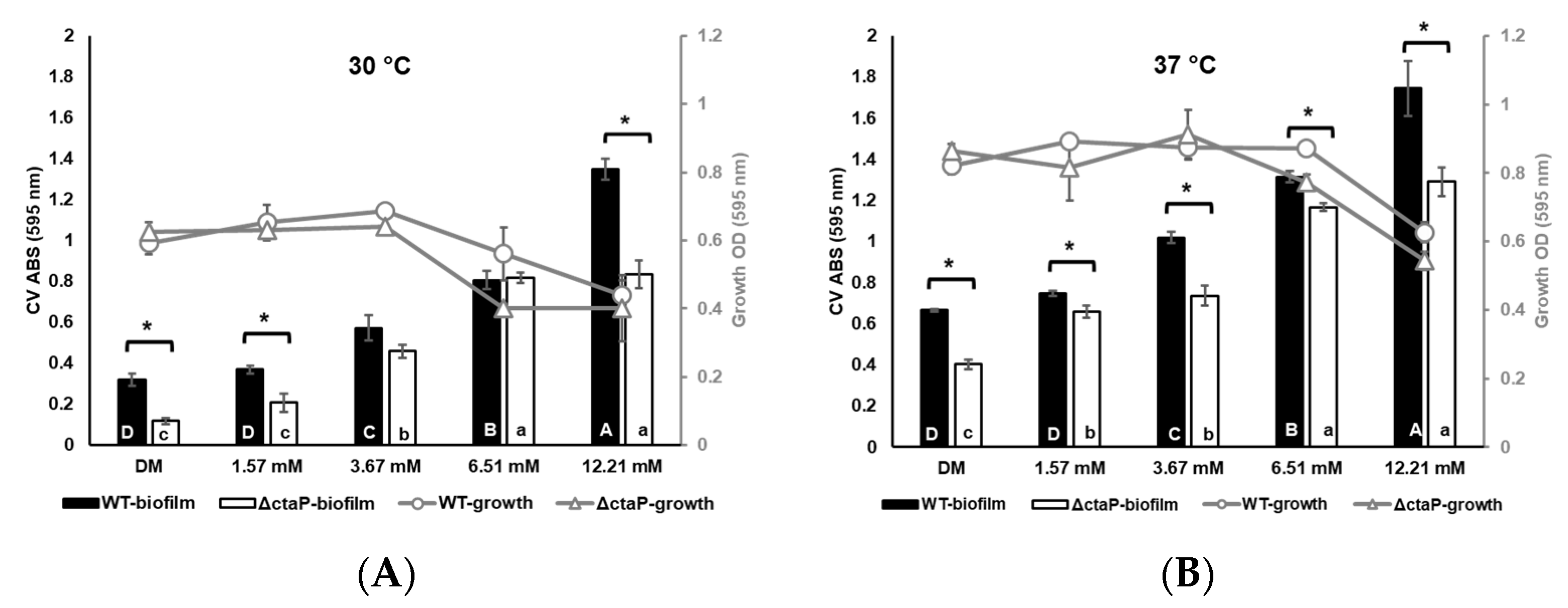
3.1. Planktonic Growth at the End of the 24 h Period

The final OD595 of growth was measured before CV staining. Low L-cysteine supplementation slightly promoted the growth of WT and ΔctaP (p > 0.05; Figure 1) at the end of 24 h. On the other hand, the growth of both WT and ΔctaP in the presence of higher concentrations of L-cysteine (6.51 and 12.21 mM) resulted in lower OD595 (p < 0.05; Figure 1). The difference between OD595 values of WT and ΔctaP in DMs was not significant at both 30 and 37 °C (p > 0.05; Figure 1). However, both WT and ΔctaP had higher OD at 37 °C than at 30 °C (p < 0.05; Figure 1).

3.2. Biofilm Formation at the End of the 24 h Period

Following the determination of growth, biofilm formation of 10403S WT and ΔctaP was also investigated in different cysteine concentrations (DM; 0.82 mM and cysteine supplementation with a final concentration of 1.57, 3.67, 6.51 and 12.21 mM) at 30 and 37 °C. As the temperature increased, the biofilm formation enhanced (Figure 1A,B). Meanwhile, the biofilm formation patterns for both WT and ΔctaP were found to be similar at both 30 and 37 °C.
The lowest biofilm formation of WT was observed in basal DM compared to L-cysteine-supplemented DMs (with 0.320 ABS595 at 30 °C and 0.664 ABS595 at 37 °C; p < 0.05; Figure 1A,B). Supplementation of DM with 1.57 mM L-cysteine did not affect the biofilm formation of WT (p > 0.05) in comparison to basal DM (at both temperatures). Nonetheless, the other L-cysteine concentrations (3.67 mM, 6.51 mM, and 12.21 mM) promoted biofilm formation significantly (p < 0.05; Figure 1A,B). ΔctaP formed less biofilm than the WT in all the L-cysteine supplementation levels (p < 0.05; Figure 1). Similar to WT, increased L-cysteine concentrations resulted in higher biofilm formation for ΔctaP (p < 0.05; Figure 1A,B).

3.3. Relationship Between Growth and Biofilm Formation of L. monocytogenes

Pearson correlation analysis was used to further understand the relationship between growth and biofilm formation in the presence of different L-cysteine concentrations. The correlation analysis of biofilm formation and growth data showed a significant negative correlation for both WT and ΔctaP at 30 and 37 °C (−0.72 and −0.68 for WT and −0.87 and −0.70 for ΔctaP; p < 0.05; Table 2). When analyzing growth conditions separately, the correlation between growth and biofilm formation of WT in basal DM and 1.57 mM L-cysteine had a positive correlation at both 30 °C and 37 °C (p > 0.05; Table 2). On the other hand, L-cysteine supplementation resulted in a strong negative correlation (with −0.99, p < 0.05; Table 2) between bacterial growth and biofilm formation in WT in the presence of 3.67 mM L-cysteine at 30 °C. The same pattern for WT was also observed at 37 °C; however, all the correlations were weak (p > 0.05; Table 2). The correlation between growth and biofilm formation of ΔctaP in basal DM was found to be positive but not statistically significant (p > 0.05; Table 2). L-cysteine supplementation, regardless of the concentration, resulted in a negative correlation between growth and biofilm formation for ΔctaP at 30 °C (p > 0.05; Table 2). However, a strong negative correlation between growth and biofilm formation was observed specifically in 12.21 mM L-cysteine (p < 0.05; Table 2). Controversially, at 37 °C, 12.21 mM L-cysteine resulted in a strong positive correlation for ΔctaP with 0.99 (p < 0.05; Table 2).

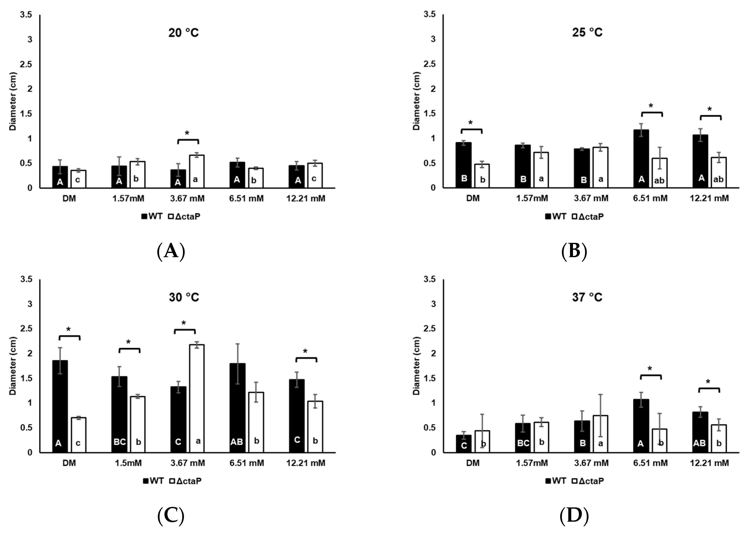
3.4. Effect of Cysteine on Swarming and Swimming Motility of L. monocytogenes 10403S WT and ΔctaP

Swarming and swimming motility of L. monocytogenes 10403S were investigated in DM supplemented with gradually increasing concentrations of L-cysteine. To investigate the role of temperature on swimming and swarming motility, we incubated cells at 20, 25, 30, and 37 °C. The highest swarming motility was observed at 30 °C for WT, followed by 25, 20, and 37 °C, respectively (Figure 2). The motility of WT was relatively higher at 37 °C compared to 20 °C (p > 0.05; Figure 2A,D).
With the addition of low-level L-cysteine (1.57 and 3.67 mM) in the environment, the swarming motility of L. monocytogenes 10403S WT decreased significantly at 30 °C (p < 0.05; Figure 2C). High levels of L-cysteine (6.51 and 12.21 mM) led to higher swarming motility compared to 1.57 and 3.67 mM of L-cysteine supplementation (p > 0.05; Figure 2C). Yet, swarming motility zones on those high levels of L-cysteine were still smaller than in basal DM (p < 0.05; Figure 2C). The same behavior was observed at 25 °C (Figure 2B). While L-cysteine supplementation did not significantly influence the swarming motility of WT at 20 °C, increased motility was observed at 37 °C (Figure 2D).
We also investigated the effect of cysteine on swimming motility. L-cysteine supplementation did not affect the swimming motility behavior of WT at 20 °C (p > 0.05; Figure 3A). Also, supplementation with 1.57 mM L-cysteine did not affect the swimming motility of WT compared to that of basal DM at 25 °C and 30 °C. At those temperatures, concentrations of L-cysteine higher than 1.57 mM increased the swimming motility significantly (p < 0.05; Figure 3B,C). Similarly to swarming motility, a slightly increased swimming motility was observed with L-cysteine supplementation at 37 °C (Figure 3D).
The 10403S ΔctaP was investigated to find out the role of cysteine intake in motility. ΔctaP showed significantly decreased motility compared to WT on both swimming and swarming plates. With the addition of L-cysteine in DM, 10403S WT showed proportionally higher swarming motility than ΔctaP (p < 0.05; Figure 2). The 10403S ΔctaP showed the highest swarming motility on DM agar with a final concentration of 3.67 mM L-cysteine, with a 2.2 cm at 30 °C (p < 0.05; Figure 2C). Although higher levels of L-cysteine (6.51 mM and 12.21 mM) reduced swarming motility compared to plates with 3.67 mM L-cysteine, the ΔctaP exhibited increased swarming motility with additional L-cysteine at 30 °C (p < 0.05; Figure 2C). On the other hand, L-cysteine supplementation, regardless of the concentration, did not affect the swimming motility of ΔctaP at 20 °C and 30 °C (p > 0.05; Figure 3A,B). Although the swimming motility of ΔctaP at 25 °C showed a significant drop in DM with 1.57 mM L-cysteine (p < 0.05; Figure 3B), similar swimming motility was observed on other L-cysteine concentrations tested (p > 0.05; Figure 3B). L-cysteine supplementation increased swimming motility of ΔctaP at 37 °C (p < 0.05; Figure 3D); however, 6.51 mM L-cysteine had no effect (p > 0.05; Figure 3D).

3.5. Transcriptomic Analysis

Transcriptomic analyses comparing (i) L. monocytogenes 10403S WT vs. ΔctaP in basal DM, (ii) WT grown in basal DM vs. DM with 1.57 mM L-cysteine and (iii) WT grown in basal DM vs. 3.67 mM L-cysteine were conducted. The summary of the transcriptome assembly statistics is shown in the supplementary data (Table S1). The error rate of a single base location sequencing was less than 1% in all groups. The Q2 and Q3 were equal to or higher than 97% or 93%, respectively. Supplementation with 1.57 mM L-cysteine resulted in the upregulation of 1485 and downregulation of 1450 genes in WT compared to basal DM (Figure S1). Moreover, with the 3.67 mM L-cysteine supplementation, 1122 genes were upregulated and 1165 genes were downregulated in WT compared to basal DM (Figure S1). The deletion of ctaP resulted in the upregulation of 835 genes and the downregulation of 602 genes in basal DM (Figure S1). Genes involved in biofilm formation, flagellar assembly (KEGG; lmt02040), and bacterial chemotaxis (KEGG; lmt02030) were investigated to assess their roles in biofilm formation and motility.
Genes previously associated with biofilm formation in L. monocytogenes [33,34] were investigated (Table S2). hly (encoding virulence factor listeriolysin O [35]), bdlA (responsible for biofilm detachment), dltA, dltB (encoding D-alanylation of extracellular lipoteichoic acids), and phoR (regulatory gene) were some of the genes that showed significantly altered expression in all compared conditions (padj < 0.05; Table 3). In addition, the expression of the CymR regulon, TcyKLMN transporter complex, and luxS (S-Ribosylhomocysteinase) was also investigated due to their role in cysteine metabolism.
(i) 
Comparison of ΔctaP vs. WT in Basal DM
In ΔctaP, several biofilm-related genes exhibited significant expression changes compared to WT (padj < 0.05; Table 3). While hly was significantly downregulated (padj < 0.05; Table 3), cymR and its regulon (TcyKLMN complex genes) were significantly upregulated (padj < 0.05; Table 4). The opp operon (oppBCD), responsible for oligopeptide transport, was significantly downregulated in ΔctaP compared to WT in basal DM (padj < 0.05; Table 4). Moreover, the loss of ctaP did not affect the transcription of luxS in basal DM (padj > 0.05; Table 4).
In terms of chemotaxis, cheR (Table S3), motA, and motB were upregulated (padj < 0.05; Table 5). Interestingly, ΔctaP did not show significant changes in the transcription of flagellar assembly genes compared to WT in basal DM.
(ii) 
Comparison of WT in Basal DM vs. DM with 1.57 mM L-cysteine
L-cysteine supplementation (1.57 mM) led to significant changes in biofilm formation and motility-related genes. The key biofilm-related gene bdlA (involved in biofilm detachment) and the virulence-associated gene hly were downregulated (Table 3). dltA, dltB (involved in the D-alanylation of extracellular lipoteichoic acids) and the regulatory gene phoR were also downregulated (padj < 0.05; Table 3). The CymR regulon was downregulated (padj < 0.05; Table 4). The luxS gene was also significantly downregulated (padj < 0.05; Table 4).
From the flagellar assembly pathway, motA, motB (Table 5), flgF, and flhA were significantly upregulated, while flgB, fliS, rpoD, flgE, and flgL were significantly downregulated (padj < 0.05; Table S3). Moreover, cheA and genes encoding flagella motor switch proteins (Table 5) were significantly downregulated (padj < 0.05).
(iii) 
Comparison of WT in Basal DM vs. DM with 3.67 mM L-cysteine
The CymR regulon and the TcyKLMN operon remained downregulated in WT with 3.67 mM L-cysteine supplementation compared to basal DM (padj < 0.05; Table 4). The opp operon showed further repression (padj < 0.05; Table 4). All the flagellar assembly genes mentioned above were downregulated in WT with the 3.67 mM L-cysteine supplementation compared to basal DM (padj < 0.05; Table S3). Moreover, all bacterial chemotaxis genes were significantly downregulated (padj < 0.05; some of them presented in Table S3).

4. Discussion

It is known that nutrient availability affects the biofilm formation of microorganisms, including L. monocytogenes. Although research directly examining the role of cysteine and its metabolism in L. monocytogenes biofilm formation is limited, several studies indicate possible connections. For instance, Xayarath et al., 2009, have demonstrated the involvement of the CtaP cysteine transporter in the adhesion of L. monocytogenes to the host cells [24]. Also, the effect of cysteine biosynthesis on biofilm formation [27,30] and the swimming and swarming motility [28,29,36] of various bacteria has been reported previously. However, the specific impact of cysteine and its transport on biofilm formation and motility has never been studied in L. monocytogenes before. Therefore, in this study, the role of extracellular cysteine and its transport on biofilm formation, swarming and swimming motility of L. monocytogenes was investigated.
The significantly lower biofilm formation of ΔctaP compared to WT in all media (Figure 1A,B) clearly indicates that ctaP plays a critical role in the biofilm formation of L. monocytogenes, regardless of temperature. Our data indicate that while L-cysteine supplementation did not significantly affect the growth at lower concentrations (1.57 mM and 3.67 mM), higher concentrations (6.51 mM and 12.21 mM) led to reduced growth but significantly enhanced biofilm formation at both 30 °C and 37 °C (p < 0.05; Figure 1). Notably, biofilm formation increased as growth decreased, resulting in a negative correlation. These negative correlations between biofilm formation and growth have also been confirmed in L. monocytogenes previously [37], particularly in a cysteine-rich environment. One possible explanation is that high concentrations of L-cysteine may induce cell stress or toxicity [38], leading to increased cell death and the release of intracellular components, including extracellular DNA (eDNA) [39,40] which can facilitate adhesion and biofilm stability and formation [41,42], despite the reduced bacterial growth [43]. Although the dose-dependent effect of L-cysteine on eDNA in fermenters has been shown [44], we did not measure eDNA in the present study and we plan to assess this in further investigations aiming to identify the mechanism(s) underlying the enhanced biofilm formation observed at higher cysteine concentrations. However, as a first approach in the present study, we conducted a transcriptomic analysis at lower concentrations.
Biofilm formation is important for the survival and resilience of L. monocytogenes in the environment. However, environmental temperatures can be relatively high during the summer, while they are consistently high in tropical regions, where 40% of the human population of the Earth resides [45]. Higher temperatures have been shown to induce stronger biofilm formation, with biofilms acting as a protective barrier that provides a stable habitat for survival and metabolism. Raw materials containing biofilms enter production lines and could be a source of contamination and lead to the formation of further biofilms in production facilities [45,46,47,48]. Recent increases in listeriosis cases have been linked to cross-contamination in food facilities, with biofilm formation playing a key role [49]. Park et al., 2022, found that biofilm formation of L. monocytogenes varies among strains, with some showing strong biofilm production at 37 °C, which may enhance adhesion to epithelial cells and contribute to human infections [50]. Our findings show that the highest biofilm formation for both WT and ΔctaP was observed at 37 °C and L-cysteine supplementation enhanced biofilm formation of those strains. These results align with previous studies reporting optimal biofilm formation at this temperature [50,51,52]. Therefore, we conducted a transcriptomic analysis at 37 °C comparing WT and ΔctaP in the presence of different L-cysteine concentrations to examine the differential gene expression patterns and explore the relationship between biofilm formation, providing insights into the role of CtaP under varying cysteine levels.
Our transcriptomic data showed that bdlA, a gene crucial for biofilm detachment, was downregulated significantly in the WT with 1.57 and 3.67 mM L-cysteine supplementation (padj < 0.05; Table 3). Currently, no studies have explored the relationship between bdlA and intracellular c-di-GMP concentrations (cyclic di-guanylate monophosphate, a second messenger known to regulate biofilm formation and motility [53]) in L. monocytogenes. However, in the Gram-negative bacterium Pseudomonas aeruginosa, loss of bdlA has been associated with increased adhesion and elevated c-di-GMP levels [54]. The c-di-GMP regulatory pathways play a crucial role in determining whether bacteria will adopt a motile or sessile lifestyle [53] by activating the biosynthesis of exopolysaccharides [55]. Higher levels of c-di-GMP increase the adhesion of cells [56,57]. Therefore, the downregulation of bdlA (padj < 0.05; Table 3) could potentially lead to higher c-di-GMP levels [54] and enhanced attachment, resulting in higher biofilm formation. While this has been demonstrated in Pseudomonas aeruginosa [54], further investigation is needed to confirm whether a similar mechanism occurs in L. monocytogenes.
Biofilm formation stimulator VEG gene (lmo0189) was significantly upregulated with 3.67 mM L-cysteine (padj < 0.05; Table 3) but was not expressed significantly in the presence of 1.57 mM L-cysteine (padj > 0.05). hly, encoding the main virulence factor listeriolysin O (LLO), plays a role in biofilm formation [35]. Similarly, hly was significantly upregulated in the presence of 3.67 mM L-cysteine (padj < 0.05; Table 3), supporting previous findings on the promoting effects of cysteine on hly expression and general virulence of L. monocytogenes [58,59,60]. These data provide further insight into the mechanisms underlying the enhanced biofilm formation observed in WT strains grown in media supplemented with 1.57 mM and 3.67 mM L-cysteine.
Our data show that both dltA and dltB were significantly downregulated in ΔctaP (padj < 0.05; Table 3). These genes are required for the D-alanylation of extracellular lipoteichoic acids, which is essential for maintaining proper surface charge, attachment and biofilm formation [34]. In addition to this, L-cysteine supplementation of ΔctaP did not affect the transcription of these genes (Table S5). All the above suggest that ctaP plays a role in the expression of dltA/dltB, through an unknown regulatory mechanism. Overall, the downregulation of dltA, dltB, and regulatory gene phoR (responsible for the biofilm formation of L. monocytogenes [34]) could explain the lower biofilm formation of ΔctaP compared to WT. This aligns with previous findings indicating that CtaP influences the adherence of L. monocytogenes to host cells [24,25]. Additionally, recent studies indicate that ctaP expression is specifically induced in sessile L. monocytogenes cells but not in planktonic cells [23], further supporting its role in biofilm formation.
Cysteine supplementation restored the biofilm formation of ΔctaP, although the biofilm levels remained lower than those observed in WT (Figure 1B). This is likely due to the cysteine uptake by alternative transporters, such as the TcyKLMN complex, which is controlled by CymR [60], in response to cysteine levels [26]. CymR regulon was upregulated significantly (padj < 0.05; Table 4) in ΔctaP in basal DM, but L-cysteine supplementation led to its downregulation (padj < 0.05; Table S4). Accordingly, loss of ctaP upregulated all tcy genes significantly in basal DM (padj < 0.05; Table 4), highlighting the compensatory role of the TcyKLMN complex under cysteine-limiting conditions [60]. Our transcriptomic data also revealed that cysteine supplementation of ΔctaP downregulated the opp operon (padj < 0.05; Table S4), which is responsible for the transport of cysteine-containing peptides [58,61]. The exact role of TcyKLMN complex genes in the presence of various cysteine concentrations still requires further investigation. However, the expression of the CymR regulon indicates that cysteine is transported through other channels in ΔctaP cells, resulting in increased biofilm formation. Our findings suggest that the CymR regulon is responsive to high cysteine availability while work in S. aureus has shown that it plays a role in biofilm formation [27]. It remains to be investigated if CymR plays a role in biofilm formation in L. monocytogenes, possibly through the regulation of genes such as dltA/B.
Additionally, the downregulation of luxS, which contributes to cysteine biosynthesis by recycling S-adenosylmethionine (SAM) to homocysteine [26], due to L-cysteine supplementation in both WT and ΔctaP (padj < 0.05; Table 4), might lead to decreased cysteine biosynthesis. Since a mutation in luxS has been shown to enhance the biofilm formation of L. monocytogenes [62,63,64], the downregulation of luxS with cysteine supplementation might contribute to increased biofilm formation. CymR regulon interacts with the PlcRa system to further influence signaling molecule production and QS activities. LuxS is responsible for synthesizing QS molecules, AI-2 [26]. Therefore, the presence of exogenous cysteine may impact this process by altering the cysteine biosynthesis pathway, as it provides an external source of cysteine, potentially reducing the requirement for the recycling of SAM through luxS [62,63,64]. Yet, luxS is not the main determinant in biofilm formation, although it contributes to it [65,66,67].
Flagellar motility, both swimming and swarming [37], is crucial for the survival of L. monocytogenes outside the host, contributing to nutrient acquisition through chemotaxis and biofilm formation. These characteristics enhance the bacterium’s persistence in the environment [14,15,16]. Bacterial surface motility, driven by flagella, plays a key role in the effectiveness of initial colonization and early invasion of mammalian host cells [16]. Therefore, the swimming and swarming motility of L. monocytogenes strains were investigated in the presence of different L-cysteine concentrations.
In L. monocytogenes, flagellar motility plays a significant role in the colonization on both internal and external surfaces and this capability is influenced by temperature [14,16,51]. The motility behavior of L. monocytogenes 10403S WT and ΔctaP on DM agar plates were tested. Swimming and swarming motility of L. monocytogenes showed similar behavior at different temperatures (20 °C, 25 °C, and 30 °C), with the highest motility observed at 30 °C. Notably, motility at 37 °C was still comparable to that at 20 °C, consistent with previous studies [65,68,69]. At ambient temperatures (22–28 °C), L. monocytogenes possesses flagella and exhibits motility; however, at the physiological temperature of mammals (37 °C), it loses its flagella and therefore its motility to avoid detection by the immune system [70,71]. However, Gao et al., 2024 [65], have found that motility-related genes motA and motB showed significantly high expression at 37 °C in different species of L. monocytogenes. Moreover, L. monocytogenes isolates from food and clinical sources were able to swim and swarm at 37 °C. At the same time, they were also able to form higher amounts of biofilm [65]. Our data revealed that motA and motB significantly upregulated (padj < 0.05; Table 5) in ΔctaP compared to WT in basal DM. Moreover, L-cysteine supplementation of WT resulted in the upregulation of those genes significantly (padj < 0.05; Table 5). This aligns with previous findings, where L. monocytogenes species capable of swimming and swarming at 37 °C also exhibited high motA and motB expression and biofilm formation [65].
The swarming motility of WT was reduced with L-cysteine supplementation, while ΔctaP showed enhanced swarming motility under similar conditions (Figure 2B,C). Although transcriptomic data did not show significant changes in flagella assembly gene transcription in ΔctaP compared to WT in basal DM, the upregulation of flhA and flhB in WT with L-cysteine supplementation (padj < 0.05; Table 5) points to a concentration-dependent effect of L-cysteine on flagellin-related genes. flhB is known to be essential for the transition from hook assembly to flagellin [72]. The downregulation of flhB in WT with 3.67 mM L-cysteine may explain the observed reduction in swarming motility. These data suggest that L-cysteine affects the motility of WT in a concentration-dependent manner by interfering with flagellin assembly. On the other side, ΔctaP appears to bypass this effect, possibly due to the involvement of other cysteine transporters.
Chemotaxis genes cheA and cheY in ΔctaP (vs. WT) remained unchanged in basal DM. Moreover, L-cysteine supplementation of ΔctaP did not affect the expression of these genes significantly (Table S5). These data might also help to explain why the swimming motility of ΔctaP remained unchanged. In contrast, these genes were significantly downregulated in WT with the L-cysteine supplementation, including both concentrations (padj < 0.05; Table 5), in compliance with the enhanced swimming motility of WT. Chemotaxis allows bacteria to navigate towards attractants or away from repellents by adjusting flagellar rotation. When encountering an attractant, bacteria reduce CheA activity, leading to decreased phosphorylation of CheB and CheY proteins, promoting smooth swimming and more directed movement towards the attractant [73,74], which is consistent with the enhanced motility observed in WT following cysteine supplementation.
It is worth mentioning that the motility zones observed at 37 °C could be a growth wave [75] that is triggered by the optimum growth temperature and more nutrient availability with L-cysteine addition. Even though flagellin production is minimal at 37 °C, there is also a possibility that its expression is influenced by factors beyond temperature [76]. Since osmolarity is known to play a key role in flagellin regulation [77], L-cysteine might affect motility through osmotic shifts or other regulatory pathways. In line with this, the concentration-dependent regulation of flagella assembly and chemotaxis genes, including flhA, flhB, cheY, and cheA, was observed in our study (Table 5).
Our transcriptomic data show that in L. monocytogenes, cysteine uptake through ctaP and exogenous L-cysteine (concentration-dependent; Table 5) affect phospholipase activity. plcA and plcB were downregulated in ΔctaP compared to WT. The downregulation of plcA in ΔctaP might contribute to the decrease in motility of the mutant strain. Interestingly, plcA was upregulated in WT in response to 3.57 mM L-cysteine supplementation (Table 5). However, according to the literature, upregulation of plcA in WT is expected to result in an increased swarming motility (at 30 °C) [29]. This contradicts our swarming motility findings (Figure 2C) but aligns with swimming motility (Figure 3C). This highlights a potentially complex relationship between plcA-mediated phospholipase activity and different motility modes. Although we also evaluated phospholipase activity on ALOA agar, the slight reduction observed in ΔctaP compared to WT was not statistically significant (Figure S2). Further work, such as overexpression studies or direct activity assays, will be important to clarify the functional consequences of plcA regulation on the motility behaviors of L. monocytogenes.

5. Conclusions

This study highlights the effect of cysteine and its transport on biofilm formation and motility of L. monocytogenes. Cysteine transporter CtaP, which is required for the growth and virulence of L. monocytogenes, is also necessary for biofilm formation, swimming, and swarming motility in a concentration-dependent manner. An enhanced biofilm formation was observed for both WT and ΔctaP under increased L-cysteine concentrations. The transcriptomic data support the increased biofilm formation in WT, while the presence of alternative cysteine or peptide transporters may contribute to the enhanced biofilm formation observed in ΔctaP. The different L-cysteine concentrations affected the swimming and swarming motility of L. monocytogenes in opposite ways, underscoring the complexity of its regulatory role in bacterial movement. Moreover, we show that at 37 °C, where L. monocytogenes loses flagella, L-cysteine supplementation influences the expression of flagella assembly and chemotaxis genes in a concentration-dependent manner. In cysteine-rich environments, increased biofilm formation and motility may provide a survival advantage by enhancing surface colonization, nutrient acquisition, and resistance to environmental stressors. This adaptability is critical for L. monocytogenes persistence in food production settings and within the host. These findings contribute valuable insights for future research on bacterial survival, proliferation, and virulence in both industrial and host environments.

Supplementary Materials

The following supporting information can be downloaded at: https://www.mdpi.com/article/10.3390/foods14111845/s1, Figure S1: Volcano plots for (A) L. monocytogenes 10403S WT vs. ΔctaP in basal DM, (B) WT grown in basal DM vs. DM with 1.57 mM L-cysteine, and (C) WT grown in basal DM vs. 3.67 mM L-cysteine under anaerobic conditions; Figure S2: Phospholipase activity of L. monocytogenes 10403S WT and ΔctaP on ALOA plates (Trafalgar Scientific, UK), incubated at 37 °C. Zones from three biological replicates were measured, and average diameters and standard deviations are presented; Table S1: Summary of RNA-seq alignment; Table S2: Transcription of biofilm formation-related genes in WT grown in 1.57 mM L-cysteine-containing DM and 3.67 mM L-cysteine-containing DM compared to non-supplemented DM, and in ΔctaP compared to WT grown in DM under anaerobic conditions; Table S3: Transcription of flagella and chemotaxis genes in WT grown in 1.57 mM L-cysteine-containing DM and 3.67 mM L-cysteine-containing DM compared to non-supplemented DM, and in ΔctaP compared to WT grown in DM under anaerobic conditions; Table S4: Transcription of the genes related to cysteine transport and quorum sensing in ΔctaP compared to WT in DM, in 1.57 mM L-cysteine-containing DM and 3.67 mM L-cysteine-containing DM compared to non-supplemented DM under anaerobic conditions; Table S5: Transcription of dltA, dltB, cheA and cheY in ΔctaP compared to WT in DM, in 1.57 mM L-cysteine-containing DM and 3.67 mM L-cysteine-containing DM compared to non-supplemented DM under anaerobic conditions

Author Contributions

Conceptualization, M.M.Y.T. and K.A.G.K.; methodology, M.M.Y.T. and C.B.; software, M.M.Y.T.; validation, M.M.Y.T. and K.A.G.K.; formal analysis, M.M.Y.T., C.B., and N.P.; investigation, M.M.Y.T.; resources, M.M.Y.T. and K.A.G.K.; data curation, M.M.Y.T.; writing—original draft preparation, M.M.Y.T. and C.B.; writing—review and editing, K.A.G.K.; visualization, M.M.Y.T.; supervision, K.A.G.K.; project administration, K.A.G.K.; funding acquisition, M.M.Y.T. All authors have read and agreed to the published version of the manuscript.

Funding

This research was funded by the Turkish Embassy of Higher Education Scholarship Programme YLSY and the Royal Thai Government Scholarship. The APC was funded by the University of Reading.

Data Availability Statement

The data generated and analyzed during this study are available from the main author (M.M.Y.T.) or the corresponding author (K.A.K.) upon reasonable request.

Acknowledgments

This project is sponsored by the Turkish Embassy of Higher Education Scholarship Programme YLSY, 2016 (Mahide Muge Yilmaz Topcam) and Royal Thai Government Scholarships (Nattanicha Prayoonwiwat). Listeria monocytogenes 10403S ΔctaP was gifted by Nancy E. Freitag, Washington University, St Louis, MO, USA.

Conflicts of Interest

The authors declare no conflicts of interest. The funders had no role in the design of the study; in the collection, analyses, or interpretation of data; in the writing of the manuscript; or in the decision to publish the results.

Abbreviations

The following abbreviations are used in this manuscript:
BHIBrain heart infusion
CVCrystal violet
c-di-GMPCyclic di-guanylate monophosphate
DMSODimethyl sulfoxide
DMDefined media
eDNAExtracellular DNA
SAMS-adenosylmethionine
QSQuorum sensing

References

  1. Quereda, J.J.; Morón-García, A.; Palacios-Gorba, C.; Dessaux, C.; García-del Portillo, F.; Pucciarelli, M.G.; Ortega, A.D. Pathogenicity and virulence of Listeria monocytogenes: A trip from environmental to medical microbiology. Virulence 2021, 12, 2509–2545. [Google Scholar] [CrossRef] [PubMed]
  2. Ravindhiran, R.; Sivarajan, K.; Sekar, J.N.; Murugesan, R.; Dhandapani, K. Listeria monocytogenes an emerging pathogen: A comprehensive overview on listeriosis, virulence determinants, detection, and anti-listerial interventions. Microb. Ecol. 2023, 86, 2231–2251. [Google Scholar] [CrossRef] [PubMed]
  3. Matereke, L.T.; Okoh, A.I. Listeria monocytogenes virulence, antimicrobial resistance, and environmental persistence: A review. Pathogens 2020, 9, 528. [Google Scholar] [CrossRef]
  4. European Food Safety Authority (EFSA); European Centre for Disease Prevention and Control (ECDC). The European Union Summary Report on Trends and Sources of Zoonoses, Zoonotic Agents and Food-Borne Outbreaks in 2017; EFSA: Parma, Italy, 2018. [Google Scholar]
  5. Su, Y.; Liu, A.; Zhu, M.J. Mapping the landscape of listeriosis outbreaks (1998–2023): Trends, challenges, and regulatory responses in the United States. Trends Food Sci. Technol. 2024, 154, 104750. [Google Scholar] [CrossRef]
  6. Fernández-Martínez, N.F.; Ruiz-Montero, R.; Briones, E.; Baños, E.; Rodríguez-Alarcón, L.G.S.M.; Chaves, J.A.; Abad, R.; Varela, C.; Lorusso, N. Listeriosis outbreak caused by contaminated stuffed pork, Andalusia, Spain, July to October 2019. Eurosurveillance 2022, 27, 2200279. [Google Scholar] [CrossRef]
  7. Lachmann, R.; Halbedel, S.; Adler, M.; Becker, N.; Allerberger, F.; Holzer, A.; Boone, I.; Falkenhorst, G.; Kleta, S.; Al Dahouk, S.; et al. Nationwide outbreak of invasive listeriosis associated with consumption of meat products in health care facilities, Germany, 2014–2019. Clin. Microbiol. Infect. 2021, 27, 1035.e1–1035.e5. [Google Scholar] [CrossRef]
  8. Centers for Disease Control and Prevention (CDC). Listeria (Listeriosis) Surveillance. CDC. Available online: https://www.cdc.gov/listeria/outbreaks/index.html (accessed on 20 April 2025).
  9. Dos Reis, J.O.; Vieira, B.S.; Cunha Neto, A.; Castro, V.S.; de Souza Figueiredo, E.E. Antimicrobial Resistance of Listeria monocytogenes from Animal Foods to First- and Second-Line Drugs in the Treatment of Listeriosis from 2008 to 2021: A Systematic Review and Meta-Analysis. Can. J. Infect. Dis. Med. Microbiol. 2022, 2022, 1351983. [Google Scholar] [CrossRef]
  10. Demirkol, O.; Adams, C.; Ercal, N. Biologically Important Thiols in Various Vegetables and Fruits. J. Agric. Food Chem. 2004, 52, 8151–8154. [Google Scholar] [CrossRef] [PubMed]
  11. Larsson, S.C.; Håkansson, N.; Wolk, A. Dietary Cysteine and Other Amino Acids and Stroke Incidence in Women. Stroke 2015, 46, 922–926. [Google Scholar] [CrossRef]
  12. O’Toole, G.; Kaplan, H.B.; Kolter, R. Biofilm formation as microbial development. Annu. Rev. Microbiol. 2000, 54, 49–79. [Google Scholar] [CrossRef]
  13. Da Silva, E.P.; De Martinis, E.C.P. Current knowledge and perspectives on biofilm formation: The case of Listeria monocytogenes. Appl. Microbiol. Biotechnol. 2013, 97, 957–968. [Google Scholar] [CrossRef] [PubMed]
  14. Kamp, H.D.; Higgins, D.E. A protein thermometer controls temperature-dependent transcription of flagellar motility genes in Listeria monocytogenes. PLoS Pathog. 2011, 7, e1002153. [Google Scholar] [CrossRef] [PubMed]
  15. Dietrich, C.; Heuner, K.; Brand, B.C.; Hacker, J.; Steinert, M. Flagellum of Legionella pneumophila positively affects the early phase of infection of eukaryotic host cells. Infect. Immun. 2001, 69, 2116–2122. [Google Scholar] [CrossRef] [PubMed]
  16. O’Neil, H.S.; Marquis, H. Listeria monocytogenes flagella are used for motility, not as adhesins, to increase host cell invasion. Infect. Immun. 2006, 74, 6675–6681. [Google Scholar] [CrossRef]
  17. Mattingly, A.E.; Weaver, A.A.; Dimkovikj, A.; Shrout, J.D. Assessing travel conditions: Environmental and host influences on bacterial surface motility. J. Bacteriol. 2018, 200, e00014-18. [Google Scholar] [CrossRef]
  18. Harshey, R.M. Bacterial motility on a surface: Many ways to a common goal. Annu. Rev. Microbiol. 2003, 57, 249–273. [Google Scholar] [CrossRef]
  19. Cherifi, T.; Jacques, M.; Quessy, S.; Fravalo, P. Impact of nutrient restriction on the structure of Listeria monocytogenes biofilm grown in a microfluidic system. Front. Microbiol. 2017, 8, 864. [Google Scholar] [CrossRef]
  20. Liu, Y.; Zhu, H.; Dou, X.; Jia, K.; Panagou, E.Z.; Zhang, H.; Dong, Q. The influence of nutrients on biofilm formation of an ST87 strain of Listeria monocytogenes. LWT 2024, 191, 115658. [Google Scholar] [CrossRef]
  21. Folsom, J.P.; Siragusa, G.R.; Frank, J.F. Formation of biofilm at different nutrient levels by various genotypes of Listeria monocytogenes. J. Food Sci. 2006, 69, 826–834. [Google Scholar] [CrossRef]
  22. Kohler, T.; Curty, L.K.; Barja, F.; Van Delden, C.; Pechère, J.C. Swarming of Pseudomonas aeruginosa is dependent on cell-to-cell signaling and requires flagella and pili. J. Bacteriol. 2000, 182, 5990–5996. [Google Scholar] [CrossRef]
  23. Lee, Y.J.; Wang, C. Links between S-adenosylmethionine and Agr-based quorum sensing for biofilm development in Listeria monocytogenes EGD-e. MicrobiologyOpen 2020, 9, e1015. [Google Scholar] [CrossRef] [PubMed]
  24. Xayarath, B.; Marquis, H.; Port, G.C.; Freitag, N.E. Listeria monocytogenes CtaP is a multifunctional cysteine transport-associated protein required for bacterial pathogenesis. Mol. Microbiol. 2009, 74, 956–973. [Google Scholar] [CrossRef] [PubMed]
  25. Vaval Taylor, D.M.; Xayarath, B.; Freitag, N.E. Two permeases associated with the multifunctional CtaP cysteine transport system in Listeria monocytogenes play distinct roles in pathogenesis. Microbiol. Spectr. 2023, 11, e03317-22. [Google Scholar] [CrossRef] [PubMed]
  26. Huillet, E.; Gohar, M. Quorum sensing in Bacillus cereus in relation to cysteine metabolism and the oxidative stress response. In Stress and Environmental Regulation of Gene Expression and Adaptation in Bacteria; Wiley: Hoboken, NJ, USA, 2016; pp. 1242–1251. [Google Scholar]
  27. Soutourina, O.; Poupel, O.; Coppée, J.Y.; Danchin, A.; Msadek, T.; Martin-Verstraete, I. CymR, the master regulator of cysteine metabolism in Staphylococcus aureus, controls host sulfur source utilization and plays a role in biofilm formation. Mol. Microbiol. 2009, 73, 194–211. [Google Scholar] [CrossRef]
  28. Turnbull, A.L.; Surette, M.G. L-Cysteine is required for induced antibiotic resistance in actively swarming Salmonella enterica serovar Typhimurium. Microbiology 2008, 154, 3410–3419. [Google Scholar] [CrossRef]
  29. Anderson, M.T.; Mitchell, L.A.; Mobley, H.L.T. Cysteine biosynthesis controls Serratia marcescens phospholipase activity. J. Bacteriol. 2017, 199, e0077617. [Google Scholar] [CrossRef]
  30. Man, L.; Dale, A.L.; Klare, W.P.; Cain, J.A.; Sumer-Bayraktar, Z.; Niewold, P.; Solis, N.; Cordwell, S.J. Proteomics of Campylobacter jejuni growth in deoxycholate reveals Cj0025c as a cystine transport protein required for wild-type human infection phenotypes. Mol. Cell. Proteom. 2020, 19, 1263–1280. [Google Scholar] [CrossRef]
  31. Karatzas, K.A.G.; Brennan, O.; Heavin, S.; Morrissey, J.; O’Byrne, C.P. Intracellular accumulation of high levels of γ-aminobutyrate by Listeria monocytogenes 10403S in response to low pH: Uncoupling of γ-aminobutyrate synthesis from efflux in a chemically defined medium. Appl. Environ. Microbiol. 2010, 76, 3529–3537. [Google Scholar] [CrossRef]
  32. Amezaga, M.R.; Davidson, I.; McLaggan, D.; Verheul, A.; Abee, T.; Booth, I.R. The role of peptide metabolism in the growth of Listeria monocytogenes ATCC 23074 at high osmolarity. Microbiology 1995, 141, 41–49. [Google Scholar] [CrossRef]
  33. Gou, H.; Cao, Q.; Wang, Z.; Liu, Y.; Sun, Y.; Wei, H.; Xue, H. Transcriptomic analysis of Listeria monocytogenes biofilm formation at different times. Can. J. Vet. Res. 2023, 87, 59–66. [Google Scholar]
  34. Alonso, A.N.; Perry, K.J.; Regeimbal, J.M.; Regan, P.M.; Higgins, D.E. Identification of Listeria monocytogenes determinants required for biofilm formation. PLoS ONE. 2014, 9, e0113696. [Google Scholar] [CrossRef] [PubMed]
  35. Li, R.; Liang, Q.; Tian, S.; Zhang, Y.; Liu, S.; Ou, Q.; Chen, Z.; Wang, C. Hemolysin function of Listeria is related to biofilm formation: Transcriptomics analysis. Vet. Res. 2022, 53, 113. [Google Scholar] [CrossRef] [PubMed]
  36. Turnbull, A.L.; Surette, M.G. Cysteine biosynthesis, oxidative stress, and antibiotic resistance in Salmonella typhimurium. Res. Microbiol. 2010, 161, 643–650. [Google Scholar] [CrossRef]
  37. Fan, Y.; Qiao, J.; Lu, Z.; Fen, Z.; Tao, Y.; Lv, F.; Zhao, H.; Zhang, C.; Bie, X. Influence of different factors on biofilm formation of Listeria monocytogenes and the regulation of cheY gene. Food Res. Int. 2020, 137, 109405. [Google Scholar] [CrossRef] [PubMed]
  38. Sørensen, M.A.; Pedersen, S. Cysteine, even in low concentrations, induces transient amino acid starvation in Escherichia coli. J. Bacteriol. 1991, 173, 5244–5246. [Google Scholar] [CrossRef]
  39. Kadam, S.R.; den Besten, H.M.W.; van der Veen, S.; Zwietering, M.H.; Moezelaar, R.; Abee, T. Diversity assessment of Listeria monocytogenes biofilm formation: Impact of growth condition, serotype, and strain origin. Int. J. Food Microbiol. 2013, 165, 259–264. [Google Scholar] [CrossRef]
  40. Zetzmann, M.; Okshevsky, M.; Endres, J.; Sedlag, A.; Caccia, N.; Auchter, M.; Waidmann, M.S.; Desvaux, M.; Meyer, R.L.; Riedel, C.U. DNase-sensitive and -resistant modes of biofilm formation by Listeria monocytogenes. Front. Microbiol. 2015, 6, 1428. [Google Scholar] [CrossRef]
  41. Okshevsky, M.; Meyer, R.L. The role of extracellular DNA in the establishment, maintenance, and perpetuation of bacterial biofilms. Crit. Rev. Microbiol. 2015, 41, 341–352. [Google Scholar] [CrossRef]
  42. Harmsen, M.; Lappann, M.; Knøchel, S.; Molin, S. Role of extracellular DNA during biofilm formation by Listeria monocytogenes. Appl. Environ. Microbiol. 2010, 76, 2271–2279. [Google Scholar] [CrossRef]
  43. Ivanova, L.A.; Egorov, V.V.; Zabrodskaya, Y.A.; Shaldzhyan, A.A.; Baranchikov, A.Y.; Tsvigun, N.V.; Lykholay, A.N.; Yapryntsev, A.D.; Lebedev, D.V.; Kulminskaya, A.A. Matrix is everywhere: Extracellular DNA is a link between biofilm and mineralization in Bacillus cereus planktonic lifestyle. NPJ Biofilms Microbiomes 2023, 9, 9. [Google Scholar] [CrossRef]
  44. Wen, H.Q.; Xing, D.F.; Xie, G.J.; Yin, T.M.; Ren, N.Q.; Liu, B.F. Enhanced photo-fermentative hydrogen production by synergistic effects of formed biofilm and added L-cysteine. Renew. Energy 2019, 139, 643–650. [Google Scholar] [CrossRef]
  45. Nyenje, M.E.; Green, E.; Ndip, R.N. Evaluation of the effect of different growth media and temperature on the suitability of biofilm formation by Enterobacter cloacae strains isolated from food samples in South Africa. Molecules 2013, 18, 9582–9593. [Google Scholar] [CrossRef] [PubMed]
  46. Zhao, X.; Zhao, F.; Wang, J.; Zhong, N. Biofilm formation and control strategies of foodborne pathogens: Food safety perspectives. Royal Soc. Chem. 2017, 58, 36670–36683. [Google Scholar] [CrossRef]
  47. Yin, W.; Wang, Y.; Liu, L.; He, J. Biofilms: The microbial ‘protective clothing’ in extreme environments. Int. J. Mol. Sci. 2019, 20, 3423. [Google Scholar] [CrossRef]
  48. Parrilli, E.; Tutino, M.L.; Marino, G. Biofilm as an adaptation strategy to extreme conditions. Springer Sci. Bus. Media Dtschl. GmbH. 2022, 33, 527–536. [Google Scholar] [CrossRef]
  49. Galié, S.; García-Gutiérrez, C.; Miguélez, E.M.; Villar, C.J.; Lombó, F. Biofilms in the food industry: Health aspects and control methods. Front. Microbiol. 2018, 9, 898. [Google Scholar] [CrossRef]
  50. Park, M.; Kim, J.; Horn, L.; Haan, J.; Strickland, A.; Lappi, V.; Boxrud, D.; Hedberg, C.; Ryu, S.; Jeon, B. Sugar modification of wall teichoic acids determines serotype-dependent strong biofilm production in Listeria monocytogenes. Microbiol. Spectr. 2022, 10, e0276922. [Google Scholar] [CrossRef]
  51. Di Bonaventura, G.; Piccolomini, R.; Paludi, D.; D’Orio, V.; Vergara, A.; Conter, M.; Ianieri, A. Influence of temperature on biofilm formation by Listeria monocytogenes on various food-contact surfaces: Relationship with motility and cell surface hydrophobicity. J. Appl. Microbiol. 2008, 104, 1552–1561. [Google Scholar] [CrossRef]
  52. Doijad, S.P.; Barbuddhe, S.B.; Garg, S.; Poharkar, K.V.; Kalorey, D.R.; Kurkure, N.V.; Rawool, D.B.; Chakraborty, T. Biofilm-forming abilities of Listeria monocytogenes serotypes isolated from different sources. PLoS ONE 2015, 10, e0137046. [Google Scholar] [CrossRef]
  53. Verstraeten, N.; Braeken, K.; Debkumari, B.; Fauvart, M.; Fransaer, J.; Vermant, J.; Michiels, J. Living on a surface: Swarming and biofilm formation. Trends Microbiol. 2008, 16, 496–506. [Google Scholar] [CrossRef]
  54. Morgan, R.; Kohn, S.; Hwang, S.H.; Hassett, D.J.; Sauer, K. BdlA, a chemotaxis regulator essential for biofilm dispersion in Pseudomonas aeruginosa. J. Bacteriol. 2006, 188, 7335–7343. [Google Scholar] [CrossRef] [PubMed]
  55. Köseoğlu, V.K.; Heiss, C.; Azadi, P.; Topchiy, E.; Güvener, Z.T.; Lehmann, T.E.; Miller, K.W.; Gomelsky, M. Listeria monocytogenes exopolysaccharide: Origin, structure, biosynthetic machinery and c-di-GMP-dependent regulation. Mol. Microbiol. 2015, 96, 728–743. [Google Scholar] [CrossRef]
  56. Valentini, M.; Filloux, A. Biofilms and cyclic di-GMP (c-di-GMP) signaling: Lessons from Pseudomonas aeruginosa and other bacteria. J. Biol. Chem. 2016, 291, 12547–12555. [Google Scholar] [CrossRef] [PubMed]
  57. Purcell, E.B.; McKee, R.W.; Courson, D.S.; Garrett, E.M.; McBride, S.M.; Cheney, R.E.; Tamayo, R. A nutrient-regulated cyclic diguanylate phosphodiesterase controls Clostridium difficile biofilm and toxin production during the stationary phase. Infect. Immun. 2017, 85, e01012-17. [Google Scholar] [CrossRef]
  58. Krypotou, E.; Scortti, M.; Grundström, C.; Oelker, M.; Luisi, B.F.; Sauer-Eriksson, A.E.; Vazquez-Boland, J. Control of bacterial virulence through the peptide signature of the habitat. Cell Rep. 2019, 26, 1815–1827. [Google Scholar] [CrossRef]
  59. Xayarath, B.; Alonzo, F.; Freitag, N.E. Identification of a peptide-pheromone that enhances Listeria monocytogenes escape from host cell vacuoles. PLoS Pathog. 2015, 11, e1004707. [Google Scholar] [CrossRef] [PubMed]
  60. Brenner, M.; Friedman, S.; Haber, A.; Livnat-Levanon, N.; Borovok, I.; Sigal, N.; Lewinson, O.; Herskovits, A.A. Listeria monocytogenes TcyKLMN cystine/cysteine transporter facilitates glutathione synthesis and virulence gene expression. mBio 2022, 13, e0044822. [Google Scholar] [CrossRef]
  61. Berude, J.C.; Kennouche, P.; Reniere, M.L.; Portnoy, D.A. Listeria monocytogenes utilizes glutathione and limited inorganic sulfur compounds as sources of essential cysteine. Infect. Immun. 2024, 92, e00422-23. [Google Scholar] [CrossRef] [PubMed]
  62. Sela, S.; Frank, S.; Belausov, E.; Pinto, R. A mutation in the luxS gene influences Listeria monocytogenes biofilm formation. Appl. Environ. Microbiol. 2006, 72, 5653–5658. [Google Scholar] [CrossRef]
  63. Challan Belval, S.; Gal, L.; Margiewes, S.; Garmyn, D.; Piveteau, P.; Guzzo, J. Assessment of the roles of LuxS, S-ribosyl homocysteine, and autoinducer 2 in cell attachment during biofilm formation by Listeria monocytogenes EGD-e. Appl. Environ. Microbiol. 2006, 72, 2644–2650. [Google Scholar] [CrossRef]
  64. Ju, X.; Li, J.; Zhu, M.; Lu, Z.; Lv, F.; Zhu, X.; Bie, X. Effect of the luxS gene on biofilm formation and antibiotic resistance by Salmonella serovar Dublin. Food Res. Int. 2018, 107, 385–393. [Google Scholar] [CrossRef] [PubMed]
  65. Gao, B.; Cai, H.; Xu, B.; Yang, F.; Dou, X.; Dong, Q.; Yan, H.; Bu, X.; Li, Z. Growth, biofilm formation, and motility of Listeria monocytogenes strains isolated from food and clinical samples located in Shanghai (China). Food Res. Int. 2024, 184, 114232. [Google Scholar] [CrossRef] [PubMed]
  66. Bonsaglia, E.C.R.; Silva, N.C.C.; Fernades Júnior, A.; Araújo Júnior, J.P.; Tsunemi, M.H.; Rall, V.L.M. Production of biofilm by Listeria monocytogenes in different materials and temperatures. Food Control. 2014, 35, 386–391. [Google Scholar] [CrossRef]
  67. Jiang, X.; Jiang, C.; Yu, T.; Jiang, X.; Ren, S.; Kang, R.; Qiu, S. Benzalkonium chloride adaptation increases expression of the Agr system, biofilm formation, and virulence in Listeria monocytogenes. Front. Microbiol. 2022, 13, 856274. [Google Scholar] [CrossRef] [PubMed]
  68. Ondrusch, N.; Kreft, J. Blue and Red Light Modulates SigB-Dependent Gene Transcription, Swimming Motility and Invasiveness in Listeria monocytogenes. PLoS ONE 2011, 6, e16151. [Google Scholar] [CrossRef]
  69. Gorski, L.; Palumbo, J.D.; Mandrell, R.E. Attachment of Listeria monocytogenes to radish tissue is dependent upon temperature and flagellar motility. Appl. Environ. Microbiol. 2003, 69, 258–266. [Google Scholar] [CrossRef]
  70. Peel, M.; Donachie, W.; Shaw, A. Temperature-dependent expression of flagella of Listeria monocytogenes studied by electron microscopy, SDS-PAGE, and Western blotting. Microbiology 1988, 134, 2171–2178. [Google Scholar] [CrossRef]
  71. Abdulkadieva, M.M.; Sysolyatina, E.V.; Vasilieva, E.V.; Litvinenko, V.V.; Kalinin, E.V.; Zhukhovitsky, V.G.; Shevlyagina, N.V.; Andreevskaya, S.G.; Stanishevskyi, Y.M.; Vasiliev, M.M.; et al. Motility provides specific adhesion patterns and improves Listeria monocytogenes invasion into human HEp-2 cells. PLoS ONE 2023, 18, e0290842. [Google Scholar] [CrossRef]
  72. Minamino, T. Hierarchical protein export mechanism of the bacterial flagellar type III protein export apparatus. FEMS Lett. 2018, 365, fny117. [Google Scholar] [CrossRef]
  73. Dons, L.; Olsen, J.E.; Rasmussen, O.F. Characterization of two putative Listeria monocytogenes genes encoding polypeptides homologous to the sensor protein CheA and the response regulator CheY of chemotaxis. DNA Seq. 1994, 4, 301–311. [Google Scholar] [CrossRef]
  74. Bischoff, D.S.; Bourret, R.B.; Kirsch, M.L.; Ordal, G.W. Purification and characterization of Bacillus subtilis CheY. Biochemistry 1993, 32, 9256–9261. [Google Scholar] [CrossRef] [PubMed]
  75. Croze, O.A.; Ferguson, G.P.; Cates, M.E.; Poon, W.C.K. Migration of chemotactic bacteria in soft agar: Role of gel concentration. Biophys. J. 2011, 101, 525–534. [Google Scholar] [CrossRef] [PubMed]
  76. Dons, L.; Eriksson, E.; Jin, Y.; Rottenberg, M.E.; Kristensson, K.; Larsen, C.N.; Bresciani, J.; Olsen, J.E. Role of flagellin and the two-component CheA/CheY system of Listeria monocytogenes in host cell invasion and virulence. Infect. Immun. 2004, 72, 3237–3244. [Google Scholar] [CrossRef]
  77. Sánchez-Clemente, R.; Igeño, M.I.; Población, A.G.; Guijo, M.I.; Merchán, F.; Blasco, R. Study of pH changes in media during bacterial growth of several environmental strains. Proceedings 2018, 2, 1297. [Google Scholar] [CrossRef]
Figure 1. Biofilm formation (bars) and growth (lines) of WT (black bars and round markers) and ΔctaP (white bars and triangle markers) in DMs supplemented with different L-cysteine concentrations were measured after 24 h of growth at 30 °C (A) and 37 °C (B). Bars and lines are calculated from three identical biological replicates and error bars represent the standard deviation. Different upper cases show a significant difference between WT strains in DMs (p < 0.05). Different lower cases show a significant difference between ΔctaP strains in DMs (p < 0.05). Asterisks indicate a significant difference between WT and ΔctaP strains in the same media (p < 0.05).
Foods 14 01845 g001
Figure 2. Swarming motility of WT (black bars) and ΔctaP (white bars) cells in DM plates supplemented with different L-cysteine concentrations after 3 days of incubation at 20 °C (A), 25 °C (B), 30 °C (C), and 37 °C (D). Bars calculated from three biological replicates and error bars represent the standard deviation. Different upper cases show a significant difference between WT strains in DMs (p < 0.05). Different lower cases show a significant difference between ΔctaP strains in DMs (p < 0.05). Asterisks indicate a significant difference between WT and ΔctaP strains in the same media (p < 0.05).
Foods 14 01845 g002
Figure 3. Swimming motility of WT (black bars) and ΔctaP (white bars) cells in DM plates supplemented with different L-cysteine concentrations after 3 days of incubation at 20 °C (A), 25 °C (B), 30 °C (C), and 37 °C (D). Bars calculated from three biological replicates and error bars represent standard deviation. Different upper cases show a significant difference between WT strains in DMs (p < 0.05). Different lower cases show a significant difference between ΔctaP strains in DMs (p < 0.05). Asterisks indicate a significant difference between WT and ΔctaP strains in the same media (p < 0.05).
Foods 14 01845 g003
Table 1. L. monocytogenes strains used in this work.
StrainRelevant PropertiesReference Source
10403SSerotype ½ a, wild type[31]
10403S Δlmo013510403S with Δlmo0135::erm
(ΔctaP::erm) deletion
[24]
Table 2. The relationship between growth and biofilm formation of L. monocytogenes 10403S WT and ΔctaP at 30 °C and 37 °C.
Temperature (30 °C)Temperature (37 °C)
ConditionWT Correlation (r)ΔctaP Correlation (r)WT Correlation (r)ΔctaP Correlation (r)
Overall−0.72698 *−0.86986 *−0.68798 *−0.70016 *
DM0.1820.870.857−0.207
1.57 mM0.424−0.7210.96−0.519
3.67 mM−0.998 *−0.84−0.7030.591
6.51 mM−0.41−0.9890.9420.521
12.21 mM0.714−0.998 *0.7840.997 *
* Asterisks indicate a significant difference (padj < 0.05).
Table 3. Transcription of biofilm formation-related genes in WT grown in 1.57 mM L-cysteine-containing DM and 3.67 mM L-cysteine-containing DM compared to non-supplemented DM and in ΔctaP compared to WT grown in DM under anaerobic conditions.
Gene SymbolLocus TagGene ID for 10403SLog2 Fold-Change
WT in 1.57 mM vs. DM
Log2 Fold-Change
WT in 3.67 mM vs. DM
Log2 Fold-Change
ΔctaP vs. WT in DM
Gene Description
bdlAlmo1699LMRG_RS08590−3.11 *−1.48 *−1.17Methyl-accepting chemotaxis protein
dltA LMRG_RS04905−2.19 *+0.62 *−1.50 *D-alanine--poly (phosphoribitol) ligase subunit DltA
dltBlmo0973LMRG_RS04900−2.30 *−0.03−1.78 *D-alanyl-lipoteichoic acid biosynthesis protein DltB
NININovel+0.11+4.21 *+0.56Biofilm formation stimulator VEG
lmo0189LMRG_RS00910−0.56+4.15 *+0.63Veg family protein
hlylmo0202LMRG_RS00975−0.45+7.38 *−1.34 *Cholesterol-dependent cytolysin listeriolysin O
phoRlmo2500LMRG_RS12690−0.81*−0.56 *−0.73 *Alkaline phosphatase synthesis sensor protein PhoR
* Asterisks indicate a significant difference (padj < 0.05). “−“ indicates downregulation while “+” indicates upregulation. NI: Not identified.
Table 4. Transcription of the genes related to cysteine transport and quorum sensing in ΔctaP compared to WT in DM and WT in 1.57 mM L-cysteine-containing DM and 3.67 mM L-cysteine-containing DM compared to non-supplemented DM under anaerobic conditions.
Gene SymbolLocus TagGene ID for 10403SLog2 Fold-Change
WT in 1.57 mM vs. DM
Log2 Fold-Change
WT in 3.67 mM vs. DM
Log2 Fold-Change
ΔctaP vs. WT in DM
Gene Description
tcyKlmo2349LMRG_RS11840+2.02 *−1.88 *+2.55 *Amino acid ABC transporter substrate-binding protein
tcyLlmo2348LMRG_RS11835+2.71 *−2.23 *+1.54 *Amino acid ABC transporter permease
tcyMlmo2347LMRG_RS11830+1.69 *−6.05+1.46 *Amino acid ABC transporter permease
tcyNlmo2346LMRG_RS11825+2.02 *−2.87 *+1.60 *Amino acid ABC transporter ATP-binding protein
CymRlmo1515LMRG_RS07540−0.77 *+0.86 *+0.58 *Rrf2 family transcriptional regulator
oppAlmo0152LMRG_RS00730−3.69 *−2.42 *+3.15Peptide ABC transporter substrate-binding protein PF00496: bacterial extracellular solute-binding proteins, family 5 middle
oppBlmo2195LMRG_RS11135−3.01 *−1.82 *−0.74 *ABC transporter permease
oppC LMRG_RS11130−2.23 *−1.68 *−0.45ABC transporter permease
oppD LMRG_RS11125−2.85 *−0.94 *−0.08ABC transporter ATP-binding protein
oppFlmo2192LMRG_RS11120−2.64 *−1.21 *+0.04ATP-binding cassette domain-containing protein
luxSlmo1288LMRG_RS06405−3.45 *−0.47 *+0.08S-ribosylhomocysteine lyase
* Asterisks indicate a significant difference (padj < 0.05). “−” indicates downregulation while “+” indicates upregulation.
Table 5. Transcription of flagella assembly, chemotaxis, and phospholipase genes in WT grown in 1.57 mM L-cysteine-containing DM and 3.67 mM L-cysteine-containing DM compared to non-supplemented DM and in ΔctaP compared to WT in DM under anaerobic conditions.
Gene SymbolLocus TagGene ID for 10403SLog2 Fold-Change
WT in 1.57 mM vs. DM
Log2 Fold-Change
WT in 3.67 mM vs. DM
Log2 Fold-Change
ΔctaP vs. WT in DM
Gene Description
flhAlmo0680LMRG_RS03415+2.34 *+1.10 *+0.77Flagellar biosynthesis protein FlhA
flhBlmo0679LMRG_RS03410+1.26 *−2.99 *+0.04Flagellar biosynthesis protein FlhB
cheAlmo0692LMRG_RS03475−3.51 *−0.72 *−1.51Chemotaxis protein CheA
cheYlmo0691LMRG_RS03470−2.89 *−0.36−1.35Chemotaxis protein CheY
motAlmo0685LMRG_RS03440+2.75 *−1.89 *+1.27Flagellar motor stator protein MotA
motBlmo0686LMRG_RS03445+3.39 *−1.60 *+1.47Flagellar motor protein MotB
plcAlmo0201LMRG_RS00970−1.42 *+5.40 *−1.15 *Phosphatidylinositol-specific phospholipase C
plcBlmo0205LMRG_RS00990−0.18+4.65 *−0.74Phosphatidylcholine phospholipase C
* Asterisks indicate a significant difference (padj < 0.05). “−” indicates downregulation while “+” indicates upregulation.
Disclaimer/Publisher’s Note: The statements, opinions and data contained in all publications are solely those of the individual author(s) and contributor(s) and not of MDPI and/or the editor(s). MDPI and/or the editor(s) disclaim responsibility for any injury to people or property resulting from any ideas, methods, instructions or products referred to in the content.

