Article

Proteomic Analysis of Bifidobacterium animalis AR668 and AR668-R1 Under Aerobic Culture

by
Yaping Liu
1,
Xiaoxiao Zhao
1,
Miao Yang
1,
Xin Song
1,
Guangqiang Wang
1,
Yongjun Xia
1,
Liang Zhao
2,
Zhiqiang Xiong
1,* and
Lianzhong Ai
1,3
1
Shanghai Engineering Research Center of Food Microbiology, School of Health Science and Engineering, University of Shanghai for Science and Technology, Shanghai 200093, China
2
College of Food Science and Nutritional Engineering, China Agricultural University, Beijing 100083, China
3
School of Agriculture and Biology, Shanghai Jiao Tong University, Shanghai 200240, China
*
Author to whom correspondence should be addressed.
Foods 2025, 14(10), 1766; https://doi.org/10.3390/foods14101766
Submission received: 25 March 2025 / Revised: 2 May 2025 / Accepted: 13 May 2025 / Published: 16 May 2025
(This article belongs to the Section Food Microbiology)

Abstract

:
Bifidobacterium animalis is a widely used probiotic with significant health benefits, but its application is limited by oxygen sensitivity. Our laboratory previously developed an oxygen-tolerant B. animalis AR668-R1 using adaptive laboratory evolution under aerobic culture, but the molecular mechanism remains unclear. In this work, compared to the wild-type parental strain B. animalis AR668, 212 upregulated and 390 downregulated proteins were identified in AR668-R1 under aerobic conditions through comparative proteomic analysis. Enrichment analysis of the differentially expressed proteins between AR668 and AR668-R1 identified the potential oxygen-tolerant related pathways, including the translation process, transmembrane transport system, and carbohydrate metabolism. Furthermore, five potential oxygen-tolerance proteins (DapE, Mth2, MutT, Eno, and MsrAB) were validated by RT-qPCR that may contribute to the aerobic growth of AR668-R1. Through gene overexpression validation, Mth2 (7,8-dihydro-8-oxoguanine triphosphatase) was found to enhance the growth of AR668-R1 by 19.8% compared to the empty plasmid control under aerobic conditions. Our finding provides valuable insights into the oxygen-tolerant mechanisms of B. animalis at the protein level.

1. Introduction

Bifidobacterium animalis, a Gram-positive, anaerobic bacterium, is widely recognized for beneficial effects on human health, particularly the regulation of the gut microbiota. This probiotic strain is characterized by its ability to ferment carbohydrates producing lactic acid and acetic acid, which contributes to the maintenance of a healthy intestinal environment [1]. The physiological properties of B. animalis include the ability to adhere to intestinal epithelial cells, modulate immune responses, and produce antimicrobial compounds that inhibit the growth of pathogenic bacteria [2]. These characteristics make B. animalis a valuable component of functional foods and dietary supplements aimed at promoting gut health. In the food industry, B. animalis has been extensively utilized in the production of fermented dairy products such as yogurt, cheese, and kefir [3], which enhance food nutritional value and sensory properties, such as texture and flavor [4]. Additionally, B. animalis has been incorporated into non-dairy products, including infant formula, cereals, and beverages, to provide probiotic benefits to a wider range of consumers [5]. Despite the widespread application, a significant limitation of B. animalis is sensitivity to oxygen, which restricts viability and functionality in aerobic environments. This oxygen sensitivity poses challenges for the storage, distribution, and incorporation of B. animalis into non-fermented foods and supplements, where exposure to oxygen is inevitable.
The oxidative stress response in model organisms such as E. coli is well characterized, depending primarily on the OxyR/SoxRS regulatory systems and classical reactive oxygen species (ROS)-scavenging enzymes including catalase and superoxide dismutase [6]. However, Bifidobacterium species are notably lacking in these conserved oxidative defense mechanisms [7,8]. Recent studies revealed that B. breve UCC2003 employs an alternative stress response strategy mediated by a coordinated regulatory network. This network harbors ClgR (stress-responsive transcriptional regulator), LexA (the DNA damage response regulator), and HrcA and HspR (heat shock protein regulators) [9]. For hydrogen peroxide detoxification, B. bifidum primarily relies on alkyl hydroperoxide reductase (Ahp) and the thioredoxin reductase-AhpC (TrxR-AhpC) antioxidant system [10]. Furthermore, oxygen tolerance is facilitated by NADH oxidase (NOX) and HemN-like oxygen reductase activity [11]. Notably, B. longum LTBL16 could enhance oxidative resistance through the upregulation of peroxidases and the activation of the NOX-dependent FoxO signaling pathway [12]. These findings collectively demonstrated that Bifidobacterium species develop diverse and specialized mechanisms to mitigate oxidative stress.
However, despite these intrinsic defense systems, most bifidobacteria remain highly sensitive to oxygen exposure, which severely limits their viability during industrial production and food applications where aerobic conditions are unavoidable. To address this limitation, adaptive laboratory evolution (ALE) strategies for selecting oxygen-tolerant strains have been extensively employed to engineer starter cultures with enhanced robustness for industrial food fermentation processes [13]. Wild-type parental strain B. animalis AR668 was isolated from infant feces [14]. The oxygen-tolerant strain B. animalis AR668-R1 was obtained after the ALE adaptation of AR668 [15] (Figure S1). We previously employed genomic and phenotypic analyses to elucidate the potential oxygen tolerance mechanism in B. animalis AR668-R1, revealing that membrane proteins and ABC transporter are involved in conferring oxygen tolerance of B. animalis. However, there is still a need for a comprehensive understanding of the molecular changes that occur during oxygen adaptation [16]. Here, this study aimed to identify potential oxygen-tolerant proteins in B. animalis AR668-R1 using proteomic analysis, which provides valuable insights into the oxygen-tolerant mechanism by which bifidobacteria cope with oxidative stress at the protein level.

2. Materials and Methods

2.1. Strain, Media, and Culture Condition

The bacterial strains and plasmids are listed in Table 1. B. animalis AR668 and AR668-R1 were cultivated in a BS medium (Catalog #HB0394-1, Hope Bio, Qingdao, China) at 37 °C under anaerobic conditions without shaking or aerobic conditions with shaking at 100 rpm.

2.2. Hydrogen Peroxide Tolerance and Cell Growth of AR668 and AR668-R1

The overnight culture of AR668 or AR668-R1 (OD600 = 1.0–1.2) was adjusted to achieve an OD600 of 0.60 ± 0.05. The bacterial suspension was then added to sterile BS broth containing hydrogen peroxide (H2O2, Catalog #H1009, Sigma-Aldrich, St. Louis, MO, USA) at concentrations ranging from 3.5 mM to 10 mM, followed by incubation for 3 h. The control group consists of BS broth without H2O2. After appropriate dilution, the survival rate of AR668 or AR668-R1 under H2O2 stress was calculated using the colony counting method on BS agar plates. Each group was repeated three times.
An overnight culture of AR668 or AR668-R1 was inoculated at 5.0% into 100 mL of the BS medium under aerobic conditions for 24 h. The absorbance at 600 nm was measured every 2 h, with three replicates each time. The colony-forming units per milliliter (CFU/mL) of AR668 or AR668-R1 was obtained by plating 10 μL stationary phase cultures on the BS medium at 37 °C for 24 h under anaerobic conditions by 10-fold serial dilutions.

2.3. Protein Sample Preparation, Sequencing, and Enrichment Analysis

The strains AR668-R1 and AR668 were grown in the BS liquid medium with agitation at 100 rpm until reaching the early logarithmic phase. The cells were harvested by centrifugation (4 °C, 4000× g, 10 min), flash-frozen in liquid nitrogen for 10 min, and lysed in a protein lysis buffer (Catalog #B14001, Bimake, TX, USA) containing 8 M urea, 1.0% SDS, and protease inhibitors. Homogenization was performed using a high-throughput tissue grinder (3 cycles, 40 s each), followed by incubation on ice for 30 min with brief vortexing every 5 min. After centrifugation (16,000× g, 4 °C, 30 min), the supernatant was collected. Protein integrity was verified by an SDS-PAGE analysis, followed by concentration quantification using a BCA Protein Assay Kit (Catalog #23227, Thermo Fisher Scientific, Waltham, MA, USA). For digestion, 100 μg of protein was reduced with 10 mM Tris (2-carboxyethyl)phosphine (TCEP, Catalog #20490, Thermo Fisher Scientific, MA, USA) at 37 °C for 60 min, alkylated with 40 mM iodoacetamide (IAM, Catalog #I6125, Sigma-Aldrich, St. Louis, MO, USA) in the dark for 40 min, and digested overnight at 37 °C using trypsin (Catalog #V5111, Promega, Madison, WI, USA) at a 1:50 enzyme–protein ratio. After protein digestion, peptides were desalted using HLB (Hydrophilic-Lipophilic Balanced) cartridges, dried, and reconstituted in 0.1% trifluoroacetic acid (TFA, Catalog #PI85183, Thermo Fisher Scientific, MA, USA).
The peptides were resuspended in a spectrometry loading buffer (2.0% ACN, 0.1% formic acid) with iRT peptides for retention time calibration. An LC-MS/MS analysis was conducted on an EASY nLC-1200 system coupled to a Q Exactive HF-X mass spectrometer (Thermo Fisher Scientific, MA, USA) at Majorbio Bio-Pharm Technology Co., Ltd. (Shanghai, China). The data-independent acquisition (DIA) mode was employed, alternating between full-scan MS and MS/MS. The C18 reversed-phase column (75 μm × 25 cm, Thermo, USA) was equilibrated with solvent A (2% acetonitrile with 0.1% formic acid) and solvent B (80% acetonitrile with 0.1% formic acid) at a flow rate of 300 nL/min. The peptides were eluted using the following gradient: 0–70 min, 5–23% B; 70–90 min, 23–29% B; 90–100 min, 29–38% B; 100–102 min, 38–48% B; 102–103 min, 48–100% B; and 103–120 min, 100% B (maintained).
The raw data were processed using Spectronaut™ (v14) with iRT-based retention time adjustment, in which the database search software automatically performs internal normalization during differential protein quantification. The quantification relied on 6 peptides per protein and 3 daughter ions per peptide, excluding shared/modified peptides. Proteomic data were analyzed on the Majorbio Cloud platform. Differential protein expression was determined using an R-based t-test (with thresholds set at fold change > 1.20 or <0.83, p < 0.05). Functional annotation and enrichment of differentially expressed proteins (DEPs) were performed via GO (http://geneontology.org/, accessed on 10 March 2025) and KEGG (http://www.genome.jp/kegg/, accessed on 10 March 2025) databases.

2.4. RNA Preparations and Gene Expression Analysis by Reverse Transcription Quantitative Polymerase Chain Reaction (RT-qPCR)

Total RNA was isolated using a total RNA extraction reagent (Catalog #B511311, Sangon Biotech, Shanghai, China). Due to the thick cell wall of B. animalis, lysozyme was used for 1 h pretreatment to extract high-quality RNA [18]. The integrity and quantity of RNA were determined by agarose gel electrophoresis and NanoDrop 2000 spectrophotometry (Thermo Fisher Scientific, MA, USA), respectively. Complementary DNA (cDNA) was prepared by the PrimeScriptRT reagent kit (Catalog #RR037A, Takara Bio Inc., Shiga, Japan) and used for RT-qPCR. cDNA synthesis was performed as follows: 1 pg−1 μg RNA was mixed with 4 μL 4× g DNA wiper Mix in a total volume of 16 μL RNase-free dH2O (42 °C, 2 min), followed by the addition of 4 μL 5× HiScript III qRT SuperMix (37 °C, 15 min) and enzyme inactivation (85 °C, 5 s). cDNA concentration was measured by Nanodrop 2000 at A260. RT-qPCR was performed using SYBR Premix Ex Taq (Catalog #DRR420A, Takara Bio Inc., Shiga, Japan), with each reaction containing 10 μL of 2 × SYBR Green Mix, 0.5 μL of cDNA, 0.5 μL of each primer (20 μmol/L), and 8.5 μL of ddH2O, gently mixed in a PCR tube. The PCR reactions were carried out as follows: initial denaturation at 95 °C for 30 s, followed by 40 cycles of denaturation at 95 °C for 5 s and annealing/extension at 60 °C for 30 s, with a final extension at 72 °C for 5 min. The gene-specific qPCR primers are listed in the Supplementary Information (Table S2). The reference gene rplD verified in previous experiments was selected as a control [7]. The qPCR reactions were performed in 96-well (LightCycle 8-tube strips), and three repetitions were made for each sample.

2.5. Plasmid Construction

The gene sequences and primers are listed in the Supplementary Information (Tables S1 and S2). The DNA fragments of gene0321, gene0618, gene0924, gene1072, and gene1102 were amplified from the B. animalis genome by PCR using primers and inserted into the E. coli-B. animalis shuttle plasmid pAM1-ldh2 with digestion by HindIII and XbaI using the ClonExpress one-step cloning kit (Catalog #C113, Vazyme Biotech Co. Ltd., Nanjing, China). The overexpression plasmids were validated by PCR (Figure S2). All the plasmids were constructed in E. coli Top10, cultured in Luria–Bertani (LB) broth supplemented with 100 μg/mL ampicillin (Amp) at 37 °C overnight with shaking (200 rpm). The amplified plasmids were electroporated into B. animalis, and cultured in BS medium supplemented with 5 μg/mL erythromycin (Em) at 37 °C under anaerobic conditions without shaking. Empty plasmid ldh2-pAM1 was used as a negative control.

2.6. Statistical Analysis

The results were statistically analyzed by one-way analysis of variance using the statistical software package SPSS 26.0. The statistical degree of significance was set at * p < 0.05, ** p < 0.01, and *** p < 0.001.

3. Results and Discussion

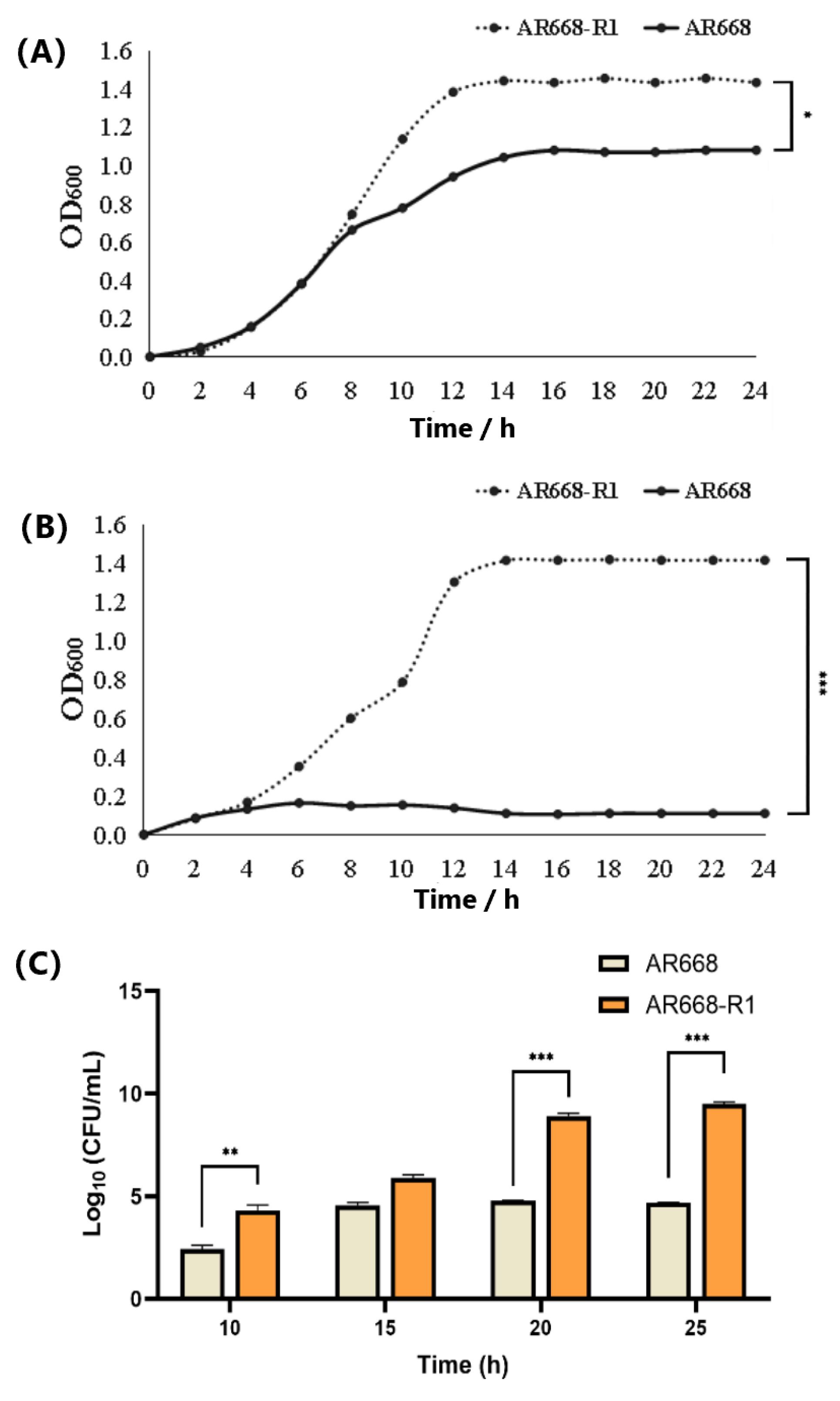
3.1. Hydrogen Peroxide Tolerance and Cell Growth of AR668-R1 and AR668

The maximum lethal concentration of H2O2 for AR668 and AR668-R1 was determined using the plate counting method (Table S3). It was found that AR668 completely died at 6.0 mM H2O2, while AR668-R1 retained viability (9.7% survival) even at 8.5 mM H2O2. Under anaerobic conditions, both AR668 and AR668-R1 grew normally (Figure 1A). AR668-R1 reached a maximum OD600 of 1.46, which was significantly higher than that of AR668 (1.08, p < 0.05). The anaerobic growth improvement observed in AR668-R1 may result in ALE-driven metabolic reorganization, which has been demonstrated to provide oxidative stress tolerance in S. cerevisiae [19]. Under aerobic conditions, the maximum growth OD600 of AR668-R1 reached 1.44, corresponding to 3.09 × 10⁹ CFU/mL (Figure 1B,C). This indicates that AR668-R1 could grow normally in the aerobic environment. In contrast, the growth of AR668 was severely inhibited under aerobic conditions. Therefore, the oxygen tolerance of AR668-R1 showed a significant improvement through ALE adaptation compared to AR668.

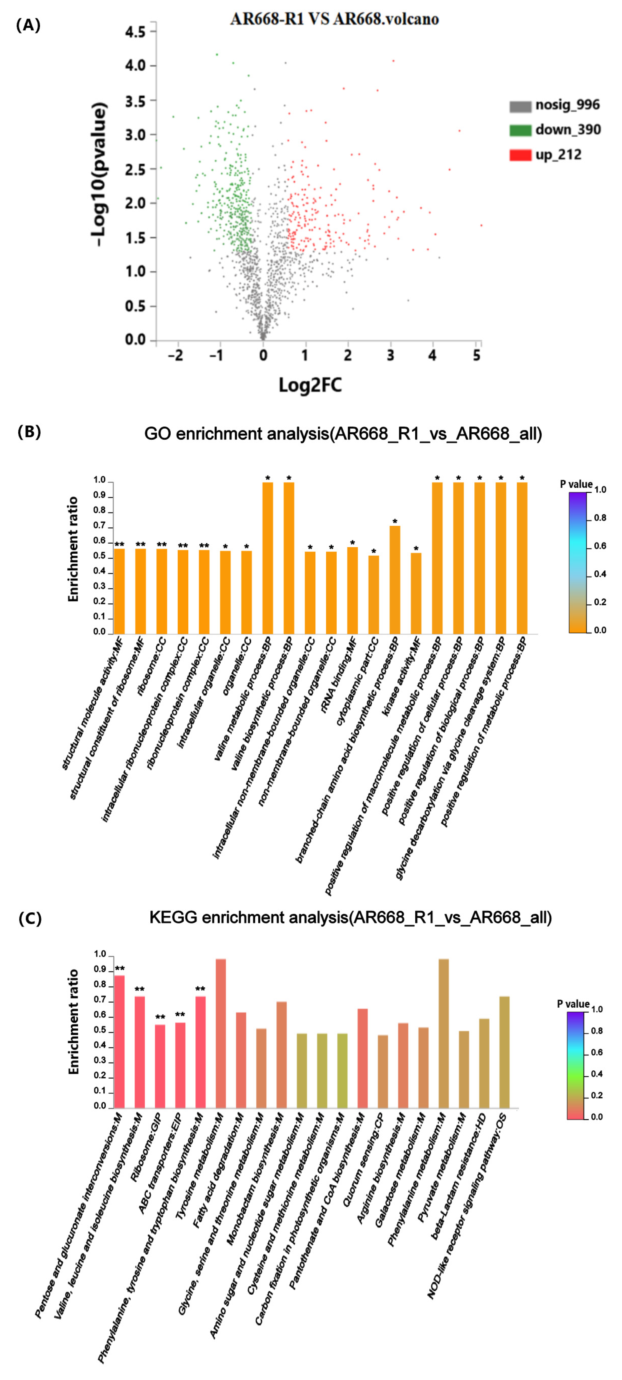
3.2. Functional Enrichment Significance Analysis

To understand the oxygen tolerance mechanism of AR668-R1, the protein expression levels of AR668-R1 and AR668 under aerobic conditions were determined by proteomic analysis, followed by functional annotation and pathway analysis using GO and KEGG. A total of 1598 proteins were detected in AR668 and AR668-R1, with 212 upregulated proteins and 390 downregulated proteins in AR668-R1 using AR668 as the control (Figure 2A). The top five significant (p < 0.01) GO functional enrichment results were all related to ribosomes, which function to synthesize polypeptide chains from amino acids according to mRNA instructions (Figure 2B). It indicated that the cultivation of B. animalis under an aerobic environment primarily affects the translation process, including protein synthesis and amino acid metabolism. In addition, the GO analysis revealed significant enrichment (p < 0.05) in valine metabolism and synthesis, rRNA binding, branched-chain amino acid synthesis, kinase activity, glycine cleavage, and the positive regulation of metabolic processes, which were potentially associated with oxygen tolerance in B. animalis. Similarly, the KEGG pathway analysis showed significant (p < 0.01) enriched pathways including pentose and glucuronate interconversions; valine, leucine, and isoleucine biosynthesis; ribosomes; and ABC transport systems (Figure 2C). It suggested that in addition to protein synthesis, the pathways related to carbohydrate metabolism and transmembrane transport systems are also important to oxygen tolerance.

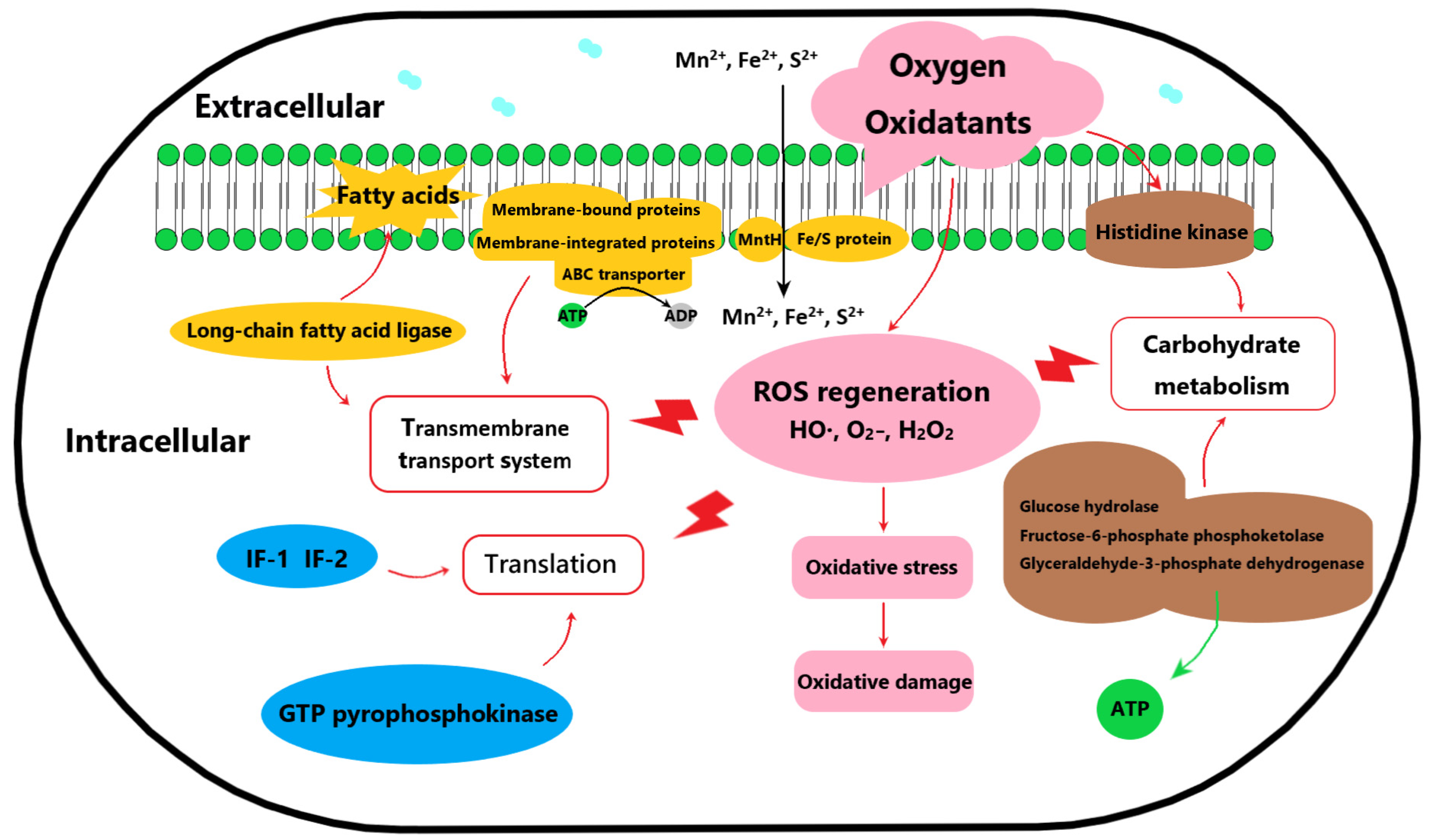
3.3. Analysis of Differential Expressed Proteins

Protein expression with significant differences (p < 0.01) were analyzed in the proteomic data (Table 2). Compared to AR668, 37 upregulated (FC > 2.0) and 38 downregulated (FC < 0.5) proteins were identified in AR668-R1. Based on the function of the differential proteins, a summary of the involved pathways associated with oxygen tolerance was found in AR668-R1 (Figure 3). For example, the ribosome serves as the exclusive site for protein biosynthesis, and proteins associated with ribosome modification and translation, including small subunit ribosomal RNA methyltransferase G (24.48-fold), 30 S ribosomal protein S6 (8.34-fold), and 50 S ribosomal proteins L24 and L33 (3.23-fold and 3.17-fold, respectively), were significantly upregulated in AR668-R1. Translation initiation factors IF-1 and IF-2 were also upregulated, suggesting the enhanced translation efficiency in AR668-R1. During the translation process, the elongation factor (EF) promotes peptide chain elongation, while the RelE factor cleaves messenger RNA at specific ribosomal sites to terminate translation. Therefore, the upregulation of RelE inhibits global protein synthesis, which leads to persistence and enhances aerobic survival capacity [20]. Notably, the GTP pyrophosphokinase protein enhances RelE activity, thereby significantly reducing translation efficiency [21]. Collectively, EF, IF−1, and IF-2 were upregulated 2.31- to 5.52-fold, while the GTP pyrophosphate kinase protein was downregulated by 4.34-fold. These differential expressions demonstrated faster translation speed and higher precision in protein synthesis under aerobic culture, which were directly associated with oxygen tolerance in B. animalis.
The cell membrane fatty acid composition is critical for stress resistance in Bifidobacterium [22]. In AR668-R1, long-chain fatty acid ligase and membrane-bound proteins were obviously downregulated, while ABC transporter proteins, implicated in substrate transport and oxygen tolerance [23], were upregulated (5.52-fold, 4.77-fold, and 3.03-fold for putative transporter protein, conserved membrane protein, and ABC transporter permease, respectively). Additionally, AR668-R1 exhibited enhanced carbohydrate metabolism, with a 2.8-fold upregulation of glyceraldehyde-3-phosphate dehydrogenase, a key enzyme in glycolysis [24], leading to elevated NADH/NADPH generation and ATP production under oxygen stress [25]. The two-component system in AR668-R1 was also more active, evidenced by a 2.44-fold upregulation of histidine kinase, enabling rapid environmental signal response and stress adaptation. As revealed by the proteomic analysis, the potential oxygen tolerance mechanism of AR668-R1 involves enhanced translation efficiency, improved transmembrane transport for membrane repair, and accelerated carbohydrate metabolism, which constitute intrinsic defense systems against oxidative stress (Figure 3). The coordinated extracellular protection and intracellular metabolic and translational adaptations enable AR668-R1 to maintain cellular integrity and function under aerobic conditions.

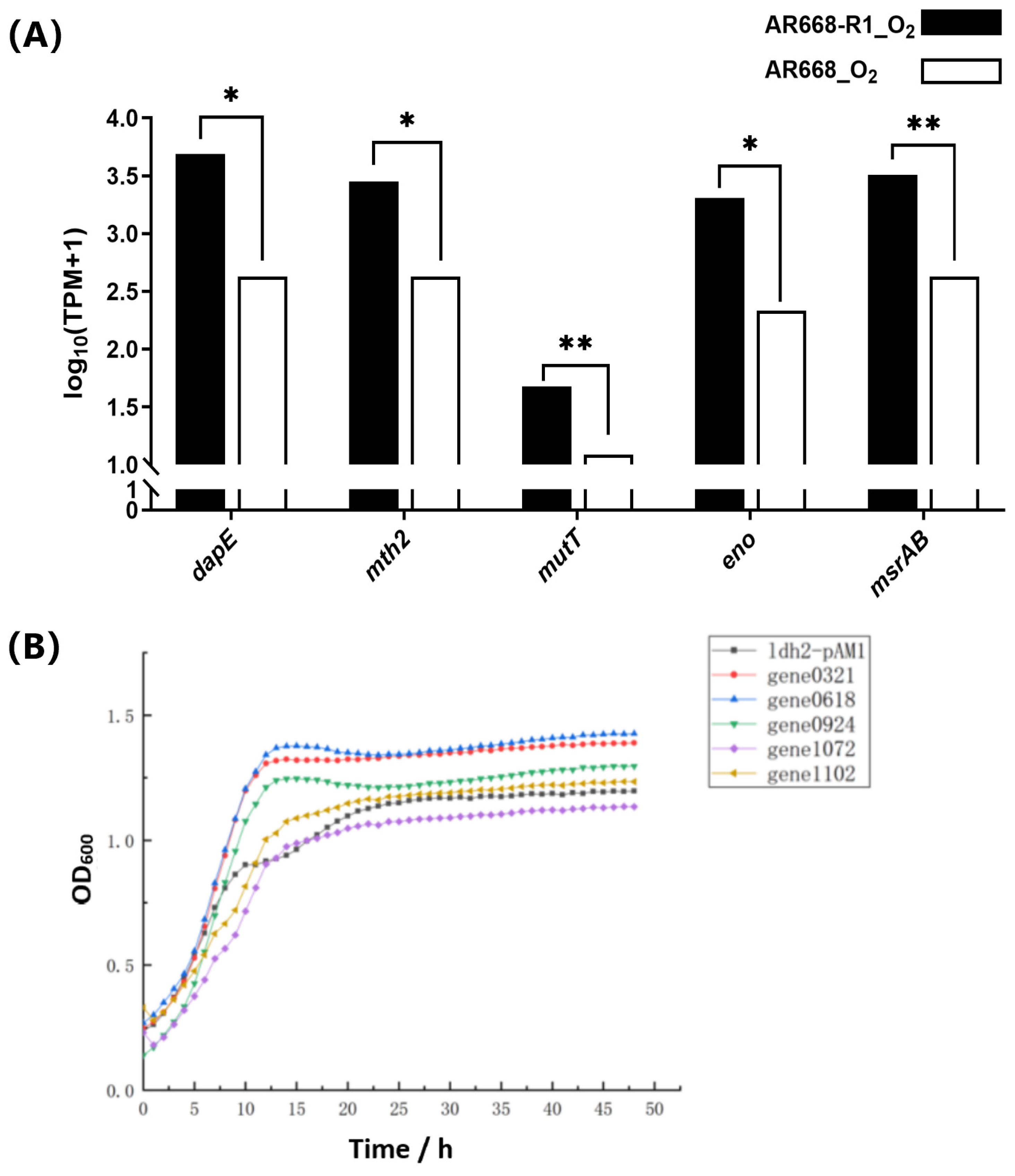
3.4. Validation of Potential Oxygen-Tolerance Proteins of B. animalis

According to the above proteomic analysis of differentially expressed proteins and our previous genomic analysis [15], we excluded certain proteins based on their functional annotation and literature evidence in metabolic pathways, including ribosome biogenesis-related proteins, non-oxygen-tolerant related proteins (e.g., cold-shock protein), and uncharacterized proteins. Notably, the upregulation of ribosomal proteins and translation factors in AR668-R1 indicated an enhanced translational capacity, which may contribute to oxygen tolerance through the rapid synthesis of stress-response proteins during oxidative challenge. This observation was supported by a previous report on E. coli, in which oxidative stress-resistant mutants increased the expression of translation machinery components to maintain proteostasis under ROS exposure [26]. The remaining candidates were evaluated through their functional contributions to essential oxygen-tolerance mechanisms, including membrane integrity maintenance demonstrated by membrane-bound transporters, oxidative DNA damage repair involving 8-oxo-dGTPase, energy metabolism through glycolytic enzymes, and redox homeostasis maintained by methionine sulfoxide reductases (Table 3). We further selected the following five proteins for experimental validation to confirm their roles that may be involved in the oxygen tolerance of AR668-R1. DapE (membrane protein) and Mth2 (7,8-dihydro-8-oxoguanine-triphosphatase) were selected based on the function in maintaining membrane stability under oxidative stress, as demonstrated in studies of oxygen stress responses in lactic acid bacteria [27]. MutT (DNA mismatch repair protein) was prioritized for its function as a crucial regulator in mitigating DNA damage during oxidative stress, reducing stress-induced oxidative damage by enhancing gene expression levels [11]. Eno (enolase) was included due to its regulatory function in diverse stress responses [28], while MsrAB (peptide methionine sulfoxide reductase) was chosen as a key component of the ModRS-regulated oxidative stress defense system [29].
The gene expression of these five potential oxygen-tolerance proteins was analyzed using RT-qPCR in B. animalis AR668 and AR668-R1 under aerobic conditions (Figure 4A). The expression levels of gene0321 (dapE), gene0618 (mth2), gene0924 (mutT), gene1072 (eno), and gene1102 (msrAB) were significantly (p  <  0.05) upregulated, which indirectly indicated the correctness of our proteome data. To further validate these five potential oxygen-tolerant proteins, we overexpressed their corresponding genes in B. animalis AR668-R1 using the ldh2 promoter. The RT-qPCR analysis showed that all the target mRNAs were upregulated, with transcript levels increasing >3-fold compared to the control (Figure S3). Under aerobic conditions, compared to the control strain, the overexpression of gene0321, gene0618, and gene0924 resulted in better growth (Figure 4B). Specifically, the OD600 of the strain overexpressing gene0321 and gene0618 were 17.6% and 19.8% higher than the control during the stationary phase, respectively. It confirmed that the DapE and Mth2 proteins are associated with the oxygen tolerance of B. animalis, providing valuable insights into the oxygen-tolerant mechanisms of B. animalis at the protein level. By analyzing the oxygen tolerance mechanism of the aerotolerant B. animalis AR668-R1 strain, this study provides a theoretical foundation for developing oxygen-resistant Bifidobacterium. Additionally, the inherent oxygen sensitivity of B. animalis has previously limited its applications in food products. Its wider application in fermented foods will be made possible by clarifying its oxygen tolerance mechanism. Furthermore, this insight can serve as a valuable reference for the genetic engineering of other oxygen-resistant strains, expanding the use of probiotics in a broader range of applications.

4. Conclusions

Bifidobacterium animalis AR668-R1, developed by ALE, exhibited high oxygen tolerance in comparison to the parental strain AR668. Comparative proteomic analysis identified 212 upregulated and 390 downregulated proteins in AR668-R1, revealing that the potential oxygen tolerance mechanism involves extracellular membrane components and intracellular transport systems to mitigate oxidative damage. Five potential oxygen-tolerance proteins (DapE, Mth2, MutT, Eno, and MsrAB) were validated, with Mth2 overexpression significantly improving growth by 19.8%. These findings provide valuable insights into the molecular basis of oxygen tolerance in B. animalis.

Supplementary Materials

The following supporting information can be downloaded at: https://www.mdpi.com/article/10.3390/foods14101766/s1, Table S1. The DNA sequences of potential oxygen tolerance genes; Table S2. Primers used in this study; Table S3. Hydrogen peroxide tolerance of Bifidobacterium; Figure S1. Oxygen-tolerant B. animalis AR668-R1 obtained through ALE; Figure S2. Electrophoresis analysis for validation of plasmid construction. The PCR validation results of colonies 1–5 are gene0321, gene0618, gene0924, gene1072, and gene1102, respectively. M: DL2000 DNA Marker; Figure S3. Transcriptional analysis of oxygen-tolerance related genes in engineered B. animalis AR668-R1.

Author Contributions

Y.L.: investigation, software, formal analysis, data curation, and writing—original draft. X.Z.: investigation and data curation. M.Y.: data curation and formal analysis. X.S.: investigation. G.W.: investigation. Y.X.: investigation. L.Z.: investigation. L.A.: writing—review and editing. Z.X.: conceptualization, methodology, supervision, and writing—review and editing. All authors have read and agreed to the published version of the manuscript.

Funding

This work was supported by Key Special Project of Synthetic Biology in Shanghai “Science and Technology Innovation Action Plan” (grant No. 23HC1400900), National Science Fund for Distinguished Young Scholars (grant No. 32025029), Natural Science Foundation of Shanghai (grants No. 22ZR1444000), Shanghai Education Committee Scientific Research Innovation Project (grant No. 2101070007800120), Yunnan Key Laboratory of Application Technology for Specialty Forest Fruits (No. 202305AG070001), Key Technology Research and Demonstration Application for Improving Quality and Efficiency of Tamarind Industry (No. 202304BP090008), and Shanghai Engineering Research Center of Food Microbiology (grant No. 19DZ2281100).

Institutional Review Board Statement

Not applicable.

Informed Consent Statement

Not applicable.

Data Availability Statement

The original contributions presented in this study are included in the article/Supplementary Materials. Further inquiries can be directed to the corresponding author.

Conflicts of Interest

The authors declare that they have no known competing financial interests or personal relationships that could have appeared to influence the work reported in this paper.

References

  1. Evdokimova, S.A.; Karetkin, B.A.; Guseva, E.V.; Gordienko, M.G.; Khabibulina, N.V.; Panfilov, V.I.; Menshutina, N.V.; Gradova, N.B. A study and modeling of Bifidobacterium and Bacillus coculture continuous fermentation under distal intestine simulated conditions. Microorganisms 2022, 10, 929. [Google Scholar] [CrossRef] [PubMed]
  2. Ji, J.; Lin, W.; Liu, S.J.; Jiao, Z.; Li, X. Probiotics, prebiotics, and postbiotics in health and disease. MedComm 2023, 4, e420. [Google Scholar] [CrossRef] [PubMed]
  3. Sun, Y.; Guo, S.; Kwok, L.Y.; Sun, Z.; Wang, J.; Zhang, H. Probiotic Bifidobacterium animalis ssp. lactis Probio-M8 improves the fermentation and probiotic properties of fermented milk. J. Dairy Sci. 2024, 107, 6643–6657. [Google Scholar] [CrossRef] [PubMed]
  4. Zhang, Y.; Hou, Y.; Zhang, S.; Jing, N.; Zhang, H.; Xie, Y.; Liu, H.; Yan, J.; Ren, J.; Jin, J. Bifidobacterium animalis A12, a probiotic strain that promotes glucose and lipid metabolism, improved the texture and aroma of the fermented sausage. Foods 2023, 12, 336. [Google Scholar] [CrossRef]
  5. Pimentel, T.C.; de Assis, B.B.T.; dos Santos Rocha, C.; Marcolino, V.A.; Rosset, M.; Magnani, M. Prebiotics in non-dairy products: Technological and physiological functionality, challenges, and perspectives. Food Biosci. 2022, 46, 101585. [Google Scholar] [CrossRef]
  6. Baussier, C.; Oriol, C.; Durand, S.; Py, B.; Mandin, P. Small RNA OxyS induces resistance to aminoglycosides during oxidative stress by controlling Fe–S cluster biogenesis in Escherichia coli. Proc. Natl. Acad. Sci. USA 2024, 121, e2317858121. [Google Scholar] [CrossRef]
  7. Cronin, M.; Zomer, A.; Fitzgerald, G.; van Sinderen, D. Identification of iron-regulated genes of Bifidobacterium breve UCC2003 as a basis for controlled gene expression. Bioengineered 2012, 3, 159–169. [Google Scholar] [CrossRef]
  8. Schöpping, M.; Vesth, T.; Jensen, K.; Franzén, C.J.; Zeidan, A.A. Genome-wide assessment of stress-associated genes in bifidobacteria. Appl. Environ. Microbiol. 2022, 88, e02251-21. [Google Scholar] [CrossRef]
  9. Zomer, A.; van Sinderen, D. Intertwinement of stress response regulons in Bifidobacterium breve UCC2003. Gut Microbes 2010, 1, 100–102. [Google Scholar] [CrossRef]
  10. Satoh, T.; Todoroki, M.; Kobayashi, K.; Niimura, Y.; Kawasaki, S. Purified thioredoxin reductase from O2-sensitive Bifidobacterium bifidum degrades H2O2 by interacting with alkyl hydroperoxide reductase. Anaerobe 2019, 57, 45–54. [Google Scholar] [CrossRef]
  11. Schöpping, M.; Zeidan, A.A.; Franzén, C.J. Stress response in bifidobacteria. Microbiol. Mol. Biol. Rev. 2022, 86, e00170-21. [Google Scholar] [CrossRef] [PubMed]
  12. Huang, G.; Pan, H.; Zhu, Z.; Li, Q. The complete genome sequence of Bifidobacterium longum LTBL16, a potential probiotic strain from healthy centenarians with strong antioxidant activity. Genomics 2020, 112, 769–773. [Google Scholar] [CrossRef] [PubMed]
  13. Nguyen, A.V.; Yaghoobi, M.; Zhang, S.; Li, P.; Li, Q.; Dogan, B.; Ahnrud, G.P.; Flock, G.; Marek, P.; Simpson, K.W.; et al. Adaptive Laboratory Evolution of Probiotics toward Oxidative Stress Using a Microfluidic-Based Platform. Small 2024, 20, 2306974. [Google Scholar] [CrossRef] [PubMed]
  14. Liu, Y.; Zhong, W.; Feng, S.; Tang, Z.; Zhang, Y.; Ai, L.; Xiong, Z. Identification of new reference genes for colony counting by reverse-transcription quantitative PCR in Bifidobacterium animalis. J. Dairy Sci. 2023, 106, 7477–7485. [Google Scholar] [CrossRef]
  15. Zhang, D.; Yang, M.; Song, X.; Xia, Y.; Ai, L.; Xiong, Z. Oxygen tolerance mechanism of Bifidobacterium animalis AR668-R1 based on genomic and phenotypic analyses. LWT 2025, 215, 117207. [Google Scholar] [CrossRef]
  16. Sahoo, S.; Mahapatra, S.R.; Misra, N.; Suar, M. Application of genomics, transcriptomics, and proteomics in probiotic research. In Probiotic Beverages; Panda, S.K., Kellershohn, J., Russell, I., Eds.; Academic Press: Newton, MA, USA, 2021; pp. 235–256. [Google Scholar] [CrossRef]
  17. Li, J.; Xiong, Z.; Yang, M.; Song, X.; Wang, G.; Xia, Y.; Ai, L. Improvement of electroporation-mediated transformation efficiency for Bifidobacterium animalis AR668. Food Biosci. 2024, 58, 103638. [Google Scholar] [CrossRef]
  18. Villa-Rodríguez, E.; Ibarra-Gámez, C.; de Los Santos-Villalobos, S. Extraction of high-quality RNA from Bacillus subtilis with a lysozyme pre-treatment followed by the Trizol method. J. Microbiol. Methods 2018, 147, 14–16. [Google Scholar] [CrossRef]
  19. Kocaefe-Özşen, N.; Yilmaz, B.; Alkım, C.; Arslan, M.; Topaloğlu, A.; Kısakesen, H.L.; Gülsev, E.; Çakar, Z.P. Physiological and molecular characterization of an oxidative stress-resistant Saccharomyces cerevisiae strain obtained by evolutionary engineering. Front. Microbiol. 2022, 13, 822864. [Google Scholar] [CrossRef]
  20. Starosta, A.L.; Lassak, J.; Jung, K.; Wilson, D.N. The bacterial translation stress response. FEMS Microbiol. Rev. 2014, 38, 1172–1201. [Google Scholar] [CrossRef]
  21. Gao, X.; Kong, J.; Zhu, H.; Mao, B.; Cui, S.; Zhao, J. Lactobacillus, Bifidobacterium and Lactococcus response to environmental stress: Mechanisms and application of cross-protection to improve resistance against freeze-drying. J. Appl. Microbiol. 2022, 132, 802–821. [Google Scholar] [CrossRef]
  22. Zafar, H.; Saier, M.H., Jr. Comparative analyses of the transport proteins encoded within the genomes of nine Bifidobacterium species. Microb. Physiol. 2022, 32, 30–44. [Google Scholar] [CrossRef] [PubMed]
  23. Duboux, S.; Muller, J.A.; De Franceschi, F.; Mercenier, A.; Kleerebezem, M. Using fluorescent promoter-reporters to study sugar utilization control in Bifidobacterium longum NCC 2705. Sci. Rep. 2022, 12, 10477. [Google Scholar] [CrossRef] [PubMed]
  24. Schöpping, M.; Goel, A.; Jensen, K.; Faria, R.A.; Franzén, C.J.; Zeidan, A.A. Novel Insights into the molecular mechanisms underlying robustness and stability in probiotic bifidobacteria. Appl. Environ. Microbiol. 2023, 89, e00082-23. [Google Scholar] [CrossRef] [PubMed]
  25. Zuo, F.; Yu, R.; Xiao, M.; Khaskheli, G.; Sun, X.; Ma, H.; Ren, F.; Zhang, B.; Chen, S. Transcriptomic analysis of Bifidobacterium longum subsp. longum BBMN68 in response to oxidative shock. Sci. Rep. 2018, 8, 17085. [Google Scholar] [CrossRef]
  26. Zhu, M.; Dai, X. Maintenance of translational elongation rate underlies the survival of Escherichia coli during oxidative stress. Nucleic Acids Res. 2019, 47, 7592–7604. [Google Scholar] [CrossRef]
  27. Ha, K.P.; Edwards, A.M. DNA repair in Staphylococcus aureus. Microbiol. Mol. Biol. Rev. 2021, 85, e00091-21. [Google Scholar] [CrossRef]
  28. Zhang, B.; Xu, J.; Sun, M.; Yu, P.; Ma, Y.; Xie, L.; Chen, L. Comparative secretomic and proteomic analysis reveal multiple defensive strategies developed by Vibrio cholerae against the heavy metal (Cd2+, Ni2+, Pb2+, and Zn2+) stresses. Front. Microbiol. 2023, 14, 1294177. [Google Scholar] [CrossRef]
  29. Scheible, M.; Nguyen, C.T.; Luong, T.T.; Lee, J.H.; Chen, Y.W.; Chang, C.; Wittchen, M.; Camacho, M.I.; Tiner, B.L.; Wu, C.; et al. The fused methionine sulfoxide reductase MsrAB promotes oxidative stress defense and bacterial virulence in Fusobacterium nucleatum. MBio 2022, 13, e03022-21. [Google Scholar] [CrossRef]
Figure 1. The growth curves of B. animalis AR668 and AR668-R1 under anaerobic (A) and aerobic (B) conditions. Determination of the viable cell counts of AR668 and AR668-R1 cultured under an aerobic environment (C). * p < 0.05, ** p < 0.01 and *** p < 0.001 by one-way analysis of variance.
Foods 14 01766 g001
Figure 2. Functional enrichment significance analysis. Volcano plot of differentially expressed genes between B. animalis AR668 and AR668-R1 under anaerobic conditions. Each dot represents an individual gene. The red dots indicate significantly upregulated genes, the green dots represent significantly downregulated genes, and the gray dots denote genes without significant differences (A). GO (B) and KEGG (C) functional enrichment significance analysis of differentially expressed proteins in B. animalis AR668 and AR668-R1 under aerobic conditions. The color scale indicates enrichment significance, with increasingly orange/red hues representing more significant enrichment (* p < 0.05 and ** p < 0.01).
Foods 14 01766 g002
Figure 3. A summary of the involved pathways of oxygen tolerance in B. animalis AR668-R1.
Foods 14 01766 g003
Figure 4. Validation of key proteins. The mRNA expression levels of potential oxygen-tolerance proteins by RT-qCPR using rplD as the control in B. animalis AR668 and AR668-R1 under aerobic conditions. * p < 0.05 and ** p < 0.01 by one-way analysis of variance (A). Aerobic growth curve of recombinant B. animalis AR668-R1 (B).
Foods 14 01766 g004
Table 1. Strains and plasmids.
Strains/PlasmidsDescriptionsReferences
Strains
Escherichia coli Top10Host for cloningOur laboratory
B. animalis AR668Wild-type, isolated from infant fecesOur laboratory
B. animalis AR668-R1High oxygen-tolerant, obtained through ALE using AR668 as the parental strainOur laboratory
Plasmids
pAM1-ldh2Emr (erythromycin resistance), Ampr (ampicillin resistance); pAM1 derivative for promoter Pldh2 replacement[17]
pLYP0321Emr, Ampr; pAM1-ldh2 derivative containing the gene0321This work
pLYP0618Emr, Ampr; pAM1-ldh2 derivative containing the gene0618This work
pLYP0924Emr, Ampr; pAM1-ldh2 derivative containing the gene0924This work
pLYP1072Emr, Ampr; pAM1-ldh2 derivative containing the gene1072This work
pLYP1102Emr, Ampr; pAM1-ldh2 derivative containing the gene1102This work
Table 2. Differentially expressed proteins in B. animalis AR668 and AR668-R1 under aerobic conditions. Fold change (FC) represented the ratio of protein expression between the AR668-R1 and AR668 strains.
The ID in
Database
SymbolFunctionDifferential
Regulation
FC (AR668-R1/AR668)
A0A2M9HL62rsmGRibosomal RNA small subunit methyltransferase Gup24.48
A0A0L7CYV1cspACold-shock proteinup20.85
D3R4Y3msrABpeptide methionine sulfoxide reductase MsrA/MsrBup8.82
A0A087BER2rpsF30S ribosomal protein S6up8.34
A0A315RYH0TMEM175DUF1211 domain-containing proteinup7.78
D3R6P3-Capsular polysaccharide synthesis proteinup6.63
D3R6Y8enoenolase 1/2/3up6.45
A0A087B4R0tktABTransketolaseup6.19
A0A0L0LVN5gatCAspartyl/glutamyl-tRNA (Asn/Gln) amidotransferase subunit Cup6.04
A0A068Z383MutTDNA mismatch repair proteinup6.01
A0A1C7FV15gadCAmino acid permeaseup6.01
A0A087AF24IF2Translation initiation factor IF-2up5.52
D3R5K0dapEConserved membrane protein (succinyl-diaminopimelate desuccinylase)up4.77
K4ILB4talBTransaldolaseup4.23
A0A086ZT91-Gp18up3.71
A0A315RUP5MTH28-oxo-dGTP diphosphataseup3.50
D3R6Z0-Collagen adhesion proteinup3.28
A0A087CFX6rplX50 S ribosomal protein L24up3.23
A0A0L7BVT2rpmG50 S ribosomal protein L33up3.17
D3R5P9livMABC transporter permease proteinup3.03
A0A2M9H761gapAType I glyceraldehyde-3-phosphate dehydrogenaseup2.80
A0A315S119nrdHNrdH-redoxinup2.77
A0A0L7BLN2-DeoR family transcriptional regulatorup2.55
A0A0L7BTZ0-Uncharacterized proteinup2.51
A0A087DR62infATranslation initiation factor IF-1up2.44
A0A0L7BN57priAPhosphoribosylanthranilate isomerase PriAup2.34
A0A261G534fadDAMP-binding proteinup2.33
A0A0L7BLU9efpElongation factor Pup2.31
B8DWS1atpCATP synthase epsilon chainup2.28
A0A315RWG9ctpACopper-translocating P-type ATPaseup2.26
A0A2N3QGE9ffhSignal recognition particle proteinup2.18
A0A2N3QFA6rplR50 S ribosomal protein L18up2.10
B8DV28-Zn-ribbon proteinup2.07
A0A2N5J2G0-Argininosuccinate synthaseup2.05
A0A315S096fadDLong-chain fatty acid-CoA ligaseup2.03
D3R3Q3-Hypothetical transporter (Sulfate transport family)up2.02
A0A315S6K7purCPhosphoribosylaminoimidazole-succinocarboxamide synthaseup2.01
A0A086ZTE6talATransaldolasedown0.50
A0A315RY65sufCFe-S cluster assembly ATPase SufCdown0.49
D3R515yajCYajCdown0.49
A0A1C7FUB9ftsHATP-dependent zinc metalloprotease FtsHdown0.49
D3R4Q0malLoligo-1,6-glucosidasedown0.49
D3R3A3ppk1Polyphosphate kinasedown0.49
A0A1V8PLA3ahpCAlkyl hydroperoxide reductasedown0.49
D3R6J3MalTMalT regulatory proteindown0.49
D3R4I6-DedA family proteindown0.48
B8DSM1musEPossible solute binding protein of ABC transporterdown0.48
A0A261G6F6ACSLAMP-binding proteindown0.48
A0A0L7BT82-ABC transporter permeasedown0.47
A0A0L7CYV9hupBIntegration host factordown0.46
A0A386K0D5musEABC transporter substrate-binding proteindown0.46
A0A133L2E1xfpD-xylulose 5-phosphate/D-fructose 6-phosphate phosphoketolasedown0.46
D3R7L4srtAPutative integral membrane proteindown0.46
B8DW33secYProtein translocase subunit SecYdown0.45
A0A0L7BMA7aqpZMajor intrinsic proteindown0.45
A0A315T437K06890BAX inhibitor (BI)-1/YccA family proteindown0.44
A0A087DDX0srtASortasedown0.44
D3R4S6butAC(S,S)-butane-2,3-diol dehydrogenasedown0.43
D3R7N6-Transporterdown0.43
A0A0L7BSH9-ABC transporter substrate-binding proteindown0.42
A0A087CM59rpmA50 S ribosomal protein L27down0.41
D3R7X1htsTMembrane-bound proteindown0.39
D3R462xynBBeta-xylosidasedown0.37
A0A087D495mrpIron-sulfur cluster carrier proteindown0.36
D3R5S2pflACEPyruvate formate-lyase activating enzymedown0.36
A0A133L2D5pflDFormate C-acetyltransferasedown0.35
A0A0A0UBM4trxAThioredoxindown0.35
A0A261FCR3ftsHATP-dependent zinc metalloprotease FtsHdown0.34
A0A2T3G7W9livKABC transporter substrate-binding proteindown0.34
Q564C5galRLacI family DNA-binding transcriptional regulatordown0.33
D3R5W5amdHDihydrodipicolinate reductasedown0.27
D3R5W3-GTP pyrophosphokinasedown0.23
A0A315T5L1mntHDivalent metal cation transporter MntHdown0.19
A0A174A5I1pflDFormate acetyltransferasedown0.18
A0A126SV27hupBIntegration host factordown0.17
Table 3. The potential oxygen-resistant genes identified in B. animalis AR668-R1.
The ID in
Database
Gene IDGenePredict Functions
D3R5K0gene0321dapEmembrane protein
A0A315RUP5gene0618mth27,8-dihydro-8-oxoguanine-triphosphatase
A0A068Z383gene0924mutTDNA mismatch repair protein MutT
D3R6Y8gene1072enoenolase
D3R4Y3gene1102msrABpeptide methionine sulfoxide reductase MsrA/MsrB
Disclaimer/Publisher’s Note: The statements, opinions and data contained in all publications are solely those of the individual author(s) and contributor(s) and not of MDPI and/or the editor(s). MDPI and/or the editor(s) disclaim responsibility for any injury to people or property resulting from any ideas, methods, instructions or products referred to in the content.

