Article

Prevalence and Antibiotic Resistance Phenotypes of Pseudomonas spp. in Fresh Fish Fillets

by
Nesrine Ben Mhenni
1,
Giulia Alberghini
2,
Valerio Giaccone
2,*,
Alessandro Truant
2 and
Paolo Catellani
2
1
Bioresources, Integrative Biology and Valorization Lab (BIOLIVAL), Higher Institute of Biotechnology of Monastir, University of Monastir, Monastir 5000, Tunisia
2
Department of Animal Medicine, Production and Health (MAPS), School of Agricultural Sciences and Veterinary Medicine, University of Padua, 35020 Legnaro, Italy
*
Author to whom correspondence should be addressed.
Foods 2023, 12(5), 950; https://doi.org/10.3390/foods12050950
Submission received: 18 January 2023 / Revised: 15 February 2023 / Accepted: 21 February 2023 / Published: 23 February 2023
(This article belongs to the Section Food Microbiology)

Abstract

:
In fresh fish products, excessive loads of Pseudomonas can lead to their rapid spoilage. It is wise for Food Business Operators (FBOs) to consider its presence both in whole and prepared fish products. With the current study, we aimed to quantify Pseudomonas spp. in fresh fillets of Salmo salar, Gadus morhua and Pleuronectes platessa. For all three fish species, we detected loads of presumptive Pseudomonas no lower than 104–105 cfu/g in more than 50% of the samples. We isolated 55 strains of presumptive Pseudomonas and carried out their biochemical identification; 67.27% of the isolates were actually Pseudomonas. These data confirm that fresh fish fillets are normally contaminated with Pseudomonas spp. and the FBOs should add it as a “process hygiene criterion” according to EC Regulation n.2073/2005. Furthermore, in food hygiene, it is worth evaluating the prevalence of antimicrobial resistance. A total of 37 Pseudomonas strains were tested against 15 antimicrobials, and they all were identified as being resistant to at least one antimicrobial, mainly penicillin G, ampicillin, amoxicillin, tetracycline, erythromycin, vancomycin, clindamycin and trimethoprim. As many as 76.47% of Pseudomonas fluorescens isolates were multi-drug resistant. Our results confirm that Pseudomonas is becoming increasingly resistant to antimicrobials and thus should be continuously monitored in foods.

1. Introduction

The microbiota of fishery products is made up of a set of microbial populations (bacteria, yeasts and/or molds), and the bacteria belonging to the Pseudomonadaceae family are one microbial population, alongside enterobacteria, coliforms or lactic acid bacteria. The genus Pseudomonas counts over 200 species divided into 11 subspecies. These bacteria are widely spread in the terrestrial and aquatic environment, both in fresh water and salt water. Based on currently available references, the species of Pseudomonas (hereafter sometimes only “P.”) most commonly isolated in fish are P. fluorescens, P. lundensis, P. fragi, P. anguilliseptica and P. putida [1,2,3]. The species of Pseudomonas that is mainly considered a human pathogen is P. aeruginosa, but it is not so common in food products [4].
Pseudomonads are one of the most relevant specific spoiling microorganisms (SSOs) because with their excessive proliferation, they enhance the splitting of nitrogenous compounds leading to product deterioration [5]. It is widely demonstrated that Gram-negative bacteria belonging to the Pseudomonadaceae family are one of the most active microbial populations in the production of proteolytic, lipolytic and saccharolytic enzymes (also extracellular ones). Furthermore, many species of Pseudomonas spp. are able to synthesize yellow, green fluorescent, blue and red pigments that can lead to the development of anomalous colors [5,6,7,8]. The discoloration and the development of unpleasant off-odors and slime coats characterize the spoilage of fresh fish, whole or portioned if they are stored in the air and even if stored at refrigeration temperatures [5,9]. In fact, Pseudomonadaceae are aerobic microorganisms and grow only in the presence of molecular oxygen. In vacuum- and CO2-packed stored fish, the number of Pseudomonas spp. is reduced [5].
Grooming and filleting operations bring additional loads of microbial community to the product being processed, with the risk of shortening the commercial life of the product itself [10].
Pseudomonas spp. can produce tough microbial biofilms on all surfaces they come into contact with, and these biofilms can offer shelter and protection to foodborne disease bacteria. The presence of high loads of Pseudomonas spp. in food or in any other substrate can favor the survival of Listeria monocytogenes, Staphylococcus aureus and enteropathogenic strains of E. coli (STEC) [11,12,13,14]. Due to the biofilm produced by Pseudomonas spp., it is not easy to eliminate the bacteria that are hosted there and, on the contrary, they are able to resist various environmental stresses, including chlorine disinfectants. Nikel and co-authors reported that P. putida possesses metabolic mechanisms that allow it to resist oxidative stress [15]. It is also known that Pseudomonadaceae are able to withstand a certain osmotic pressure (salinity of the medium) thanks to specific molecules present in their cytoplasm that can hook osmoprotective compounds such as choline, betaine and carnitine from the environment in which the bacteria are found, favoring the survival of species such as P. syringae and P. aeruginosa [16].
In general, it is accepted that a low number of Pseudomonas spp. does not pose a health problem for the consumer. Instead, Pseudomonadaceae can produce histamine and become a risk to human health if they proliferate excessively in fish that are inherently high in free histidine [17]. For this and for the reasons considered above, it is therefore wise that FBOs take into due consideration the presence of Pseudomonas spp. in whole fresh fish as well as in fresh fillets, formally giving to it the qualification of “process hygiene criterion” pursuant to EC Regulation n.2073/2005 [18] that establishes some “microbiological criteria for foodstuffs”.
The aforementioned Regulation is addressed to the FBOs who use it as a reference to check and validate their self-monitoring plan as well as the safety level of their products as required by European laws. The process hygiene criteria currently provided by the Regulation n.2073/2005 for fishery products are the loads of E. coli and coagulase-positive staphylococci; in addition, only shelled and shucked products of cooked crustaceans and shellfish are the fishery products considered.
The consumption of fishery products is regularly increasing, especially in low- and middle-income countries. Between 1961 and 2016, the demand for fish products increased by about 3.2% per year, while the need for meat from terrestrial animals increased by only 2.8% [19,20]. According to the FAO Report 2022 on “The State of World Fisheries and Aquaculture”, in recent years, there has been a sharp increase in the consumption of fresh, chilled and frozen fish products, while the consumption of salted, smoked or canned products remains stable. At the same time, aquatic food production is increasing; China, Norway, Vietnam, Chile and India are the major producers and exporters in the world [19]. In 2020, the global production of fishery products reached 178 million tons (with aquaculture representing 49.2% of the total), and it is forecasted to grow another 15% by 2030 [19].
Aquatic food systems are under growing pressure, and many reports document that the use of antimicrobials in fish farming is consequently growing. Researchers estimate that in 2030, about 13,495 tons of antimicrobials will be used in aquaculture, which, therefore, represents 5.7% of the total amount used in the world (by adding those used in humans and in all other animals) [21]. This also draws much attention to the fact that most classes of antimicrobials are used for the treatment of bacterial infections in both veterinary and human medicine [22]. This increases the speed of selection of drug-resistant bacteria [23], and antimicrobial-resistant bacteria can reduce the effectiveness of treatments, especially in Gram-negative bacterial infections [24]. Thus, the careless use of antibiotics in aquaculture is a contributing factor to the rise in antimicrobial resistance (AMR), leading to potential animal, human and ecosystem consequences [21].
The relationship between the detection of antibiotic-resistant bacteria in humans and antibiotic use in food animals continues to be a topic of discussion among researchers. Various studies have suggested that the use of antibiotics in animals can directly affect human health through direct contact with antibiotic-resistant bacteria from food animals [23]. Other authors argue indirect effects that result from contact with resistant organisms that have spread to various components of the ecosystem (e.g., water, soil, etc.), and so they can indirectly pass from animals to humans and vice versa. This indirect effect can be greatly increased by the horizontal transfer of mobile genetic elements such as conjugative plasmids, phages and transposons [25].
From a holistic view of AMR in different sectors, there are few studies that directly consider foods and not clinical isolates. Moreover, current AMR research, surveillance programs and dietary risk assessment studies (as the potential transmission of AMR), focus mainly on food-producing terrestrial animals and a few indicator bacteria. So, considering that world seafood production is increasing every year, our study aims to provide additional data to implement those already known regarding Pseudomonas spp. which is considered a dominant bacterial genus in food processing facilities and in the microbial community of many fish products [4].
Many strains of Pseudomonas have been shown to possess a high level of intrinsic resistance to most antibiotics [26]. This intrinsic resistance is mainly conferred by concurrent mechanisms, such as low outer membrane permeability, efflux systems that pump antibiotics out of the cell and the production of antibiotic-inactivating enzymes such as β-lactamases [17,27]. For now, various resistance genes have been described in the literature, and as it is a possible source of new resistance genes, Pseudomonas spp. should be better monitored [28] in their evolution.
Considering only the strains of Pseudomonas spp. isolated from food, previous studies reported in the literature confirm that various strains are able to resist some antimicrobial agents of different classes, especially β-lactams such as penicillins, cephalosporins, carbapenems, and monobactams [29,30]. For example, the study reported by Kačániová and co-authors [31] reveals a high proportion of resistant strains among the Pseudomonas spp. isolated originated from fish. Moreover, all Pseudomonas were resistant to meropenem. According to Fazeli and Momtaz [32], bacterial strains exhibited the highest level of resistance to penicillin (100%) followed by tetracycline (90.19%), streptomycin (64.70%) and erythromycin (43.13%). Shabana and co-authors [33] showed that P. fluorescens strains were resistant to more than two classes of antibiotics and, therefore, they are considered multidrug-resistant (MDR) strains and accounted for 29.7%.
The aim of our study was to determine and characterize a Pseudomonas spp. population in fresh fish fillets at the beginning of their shelf life and evaluate the degree of antimicrobial resistance of the isolated strains in order to provide useful data for monitoring its development from the point of view of food safety.

2. Materials and Methods

2.1. Samples Collection

A total of 75 fish fillets intended for large retailers were collected from a fish industry located in the Venetian area (Italy) in 5 different sampling times from March to May 2022. Each time, 15 samples were analyzed, namely:
  • Five salmon (Salmo salar) fillets reared in Norway;
  • Five plaice (Pleuronectes platessa) fillets caught using trawling in the Northeast Atlantic Ocean;
  • Five northern cod (Gadus morhua) fillets caught using longlining in the Northeast Atlantic Ocean.
The fish arrived in Italy by ship and under melting ice or frozen, as established by the EC Regulation n.853/2004. The samples of salmon and plaice were fresh, while the cod fillets were defrosted at the processing site. The fillets were collected from the fish industry early in the morning after packaging and immediately transferred to the laboratory. During transport (less than one hour), they were preserved at refrigeration temperature in expanded polystyrene containers with eutectic plates.
In the laboratory, the fillets were immediately subjected to microbiological analysis for Total Viable Count and Pseudomonas spp. count using the following methods: UNI EN ISO 6887-1: 2017, UNI EN ISO 7218: 2013, UNI EN ISO 4833-1: 2013, UNI EN ISO 11133: 2014, UNI EN ISO 13720:2010.

2.2. Samples Processing

According to the guidelines provided by UNI EN ISO 6887-1:2017, 10 g of each sample was moved into a sterilized container with 90 mL of Buffered Peptone Water (BPW) under strict hygienic measures, and homogenization was performed using a Stomacher 400 Lab blender.
From this mixture, 1 mL was transferred to a sterilized test tube containing 9 mL of BPW, from which ten-fold serial dilutions were processed up to 10−5. The prepared samples were subjected to the determination of Pseudomonas counts using Pseudomonas Agar Base (PAB) supplemented with CFC Supplement (Liofilchem®) and Total Viable Count (TVC) using Plate Count Agar (Merck®). The Pseudomonas Agar Base inoculated plates were incubated in a fridge-thermostat at 25 °C for 48 h, while the Plate Count Agar inoculated plates were incubated at 31 °C for 48–72 h.

2.3. Plate Counting and Statistical Analysis

The colonies were counted using the UNI EN ISO 7218:2013 standard as a reference. The results were expressed in the colony-forming unit (cfu)/g and then subjected to the determination of arithmetic mean and statistical analysis.
Log-transformed data (Total Viable Count and Pseudomonas spp.) were analyzed using a linear ANOVA model that included the fixed effects of fish type and sample ID (nested within the fish type). Least squares means (ls-means) were calculated and post hoc pairwise comparisons were performed using the Bonferroni correction. Data expressed as an index [formula: number of figures-1; 10^ (order of magnitude with powers of 10)] were submitted to non-parametric analysis (Kruskall–Wallis test) to assess the effect of the type of fish.
Post hoc pairwise comparisons were estimated using the Steel–Dwass–Critchlow–Flinger correction. p < 0.05 was considered significant. All the analyses were carried out with SAS (SAS Institute Inc, Cary, NC, USA 2017) and XLStat (XLSTAT statistical and data analysis solution. New York, NY, USA).

2.4. Isolation of Pseudomonas spp. and Species Identification

At least 5 colonies grown on PAB were selected from each plate and their oxidase and catalase activities were tested. The oxidase test was performed by smearing a fresh colony of each isolate onto a sterile oxidase filter paper disc soaked in distilled water. The appearance of purple color indicates a positive result of oxidase activity. The catalase test was carried out using hydrogen peroxide and the positive reaction was indicated by immediate bubbling.
At this stage, if the colonies gave positive results to the two tests mentioned, the non-fermentation activity was evaluated using the Kligler Iron test. In the Kligler Iron Agar infixion test, bacterial growth was evaluated after 24 h of incubation at 37 °C (time and temperature are those written in the technical data sheet of the media). Presumptive Pseudomonas spp. developed only on the surface because they are strictly aerobic bacteria. Furthermore, this bacterium does not ferment glucose or lactose but can split peptones, so the indicator turned purple (alkaline pH) on the surface.
Finally, the counts were corrected proportionally to the results of the tests provided by the ISO mentioned above.
Some of the colonies which proved positive for the 3 tests were identified by a unique alphanumeric code, purified on Brain Heart Infusion agar (Thermo Scientific™ Oxoid™), inoculated into Plate Count Agar tubes and stored in the refrigerator at 4 °C. Then, the isolated colonies (n = 55) were subjected to biochemical identification with the BIOLOG® system. Biolog’s carbon source utilization technology identifies microorganisms depending on their characteristic metabolic pattern produced as a consequence of some pre-selected test. The reading is performed by comparing the results to an extensive database.

2.5. Determination of Pseudomonas spp. Phenotypic Antibiotic Resistance

The analyses were performed following the recommendation of EUCAST 2022 for the method of execution [34]. Antimicrobial resistance was tested using the standard disc diffusion method.
Suspensions of Pseudomonas spp. were cultivated on Mueller Hinton agar (Merck®) with modifications regarding incubation temperature for the psychrotrophy of the bacterium itself. Then, antimicrobial discs (Liofilchem®) were placed on the agar surface taking care to preserve as much sterility as possible. All the strains of Pseudomonas spp. were tested against 15 different commercial antimicrobial agents chosen among the most used and important in human medicine and aquaculture worldwide [21,35,36]; the concentrations were instead chosen on the basis of preliminary studies. The antimicrobials were ciprofloxacin (5 μg/disk), enrofloxacin (5 μg/disk), tetracycline (30 μg/disk), rifampicin (30 μg/disk), erythromycin (15 μg/disk), vancomycin (30 μg/disk), clindamycin (10 μg/disk), meropenem (10 μg/disk), trimethoprim (5 μg/disk), penicillin G (10 IU/disk), ampicillin (10 μg/disk), amoxicillin (30 μg/disk), flumequine (30 μg/disk), florfenicol (30 μg/disk) and sulfadiazine (300 μg/disk). Inoculated plates were incubated at 31 °C for 24 h. Then, the diameters were measured in millimeters with a caliper.
For the interpretation of the results, neither EUCAST nor CLSI (Clinical and Laboratory Standards Institute) provides criteria and reference values for Pseudomonas spp., so, it was decided to consider as “highly resistant” those cases in which the diameter of the inhibition zone was ≤10 mm.

3. Results and Discussion

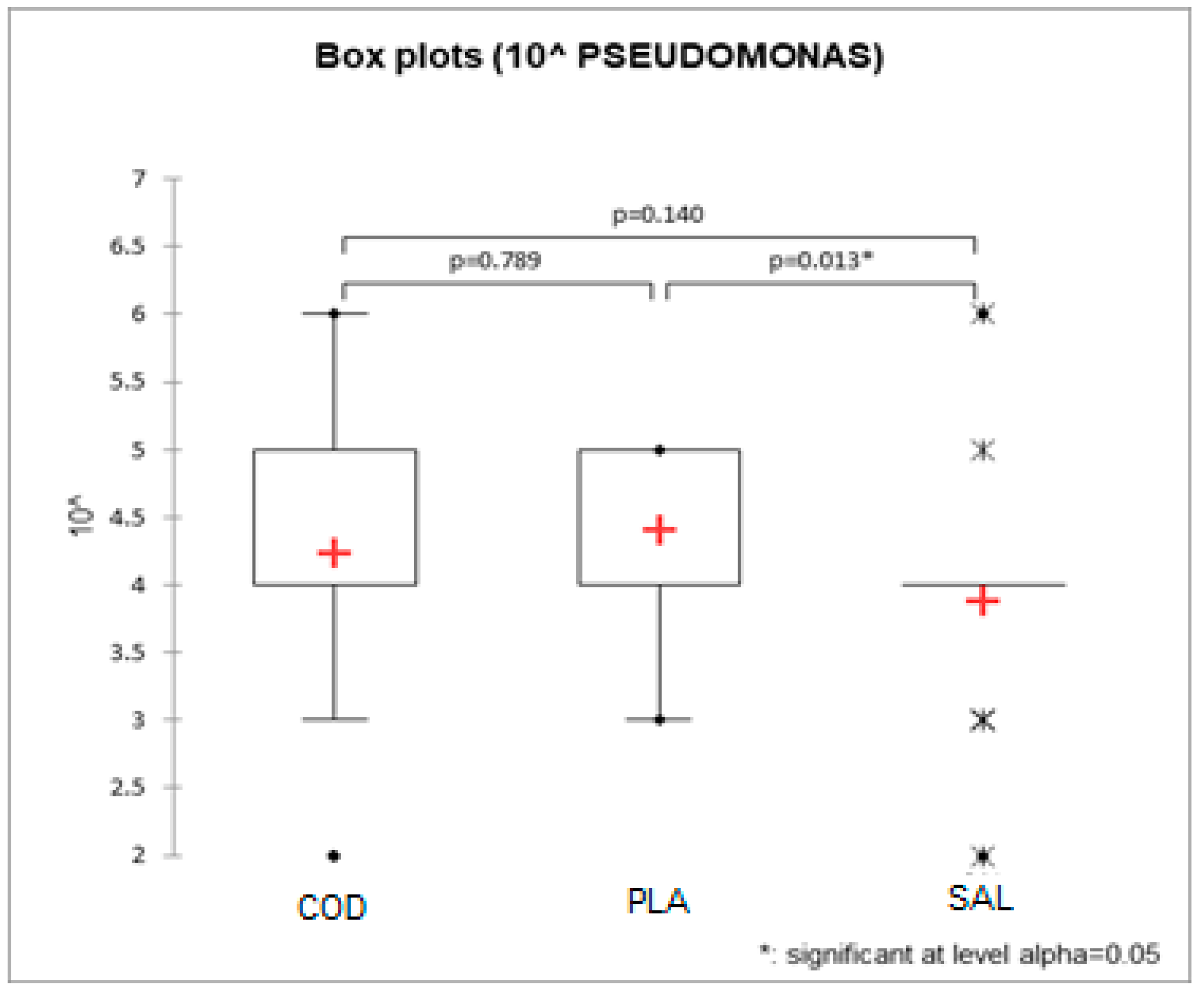
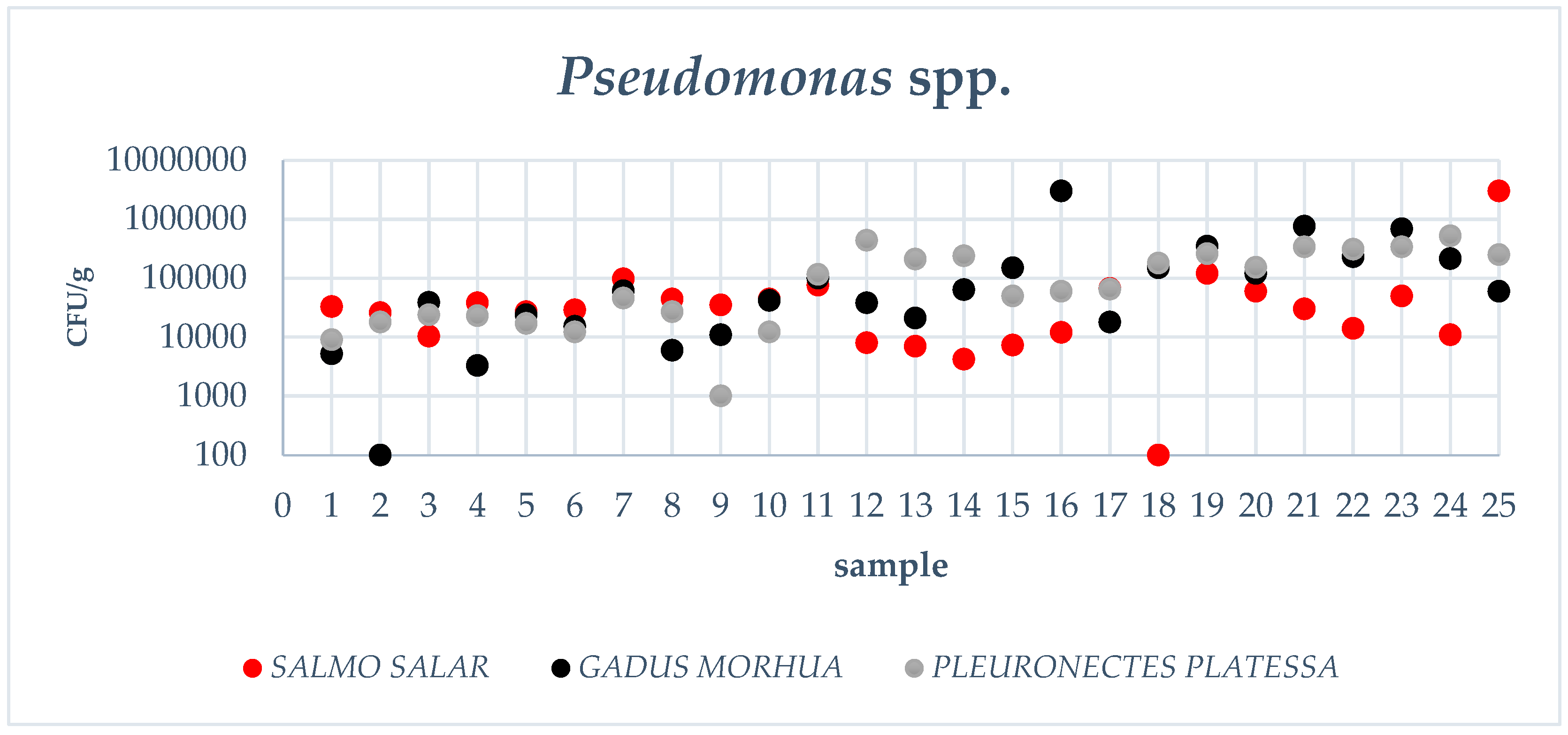
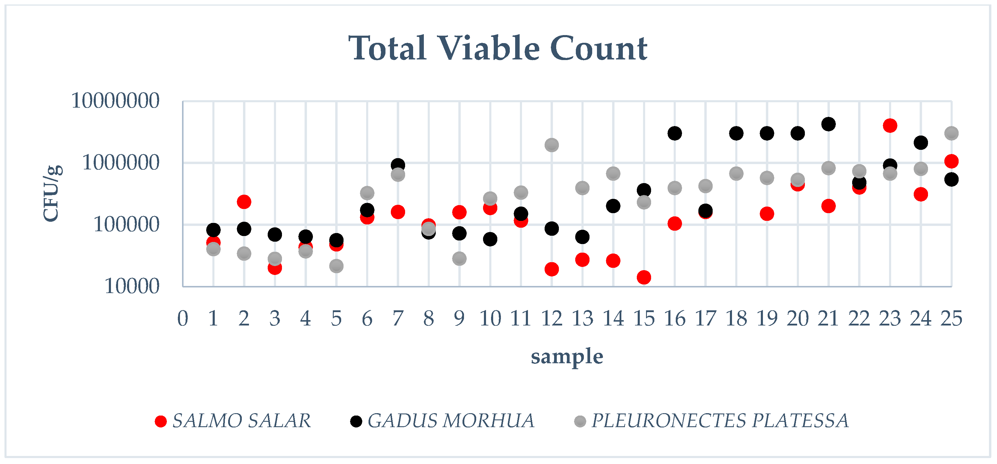
3.1. Pseudomonas spp. Count, Total Viable Count, Data Elaboration

The load of Pseudomonas spp. in the samples analyzed has been reported in Figure A1. Indeed, in the salmon fillets, in 19 samples out of 25 (76%), the initial loads were between 104 and 105 cfu/g, a rather significant load considering that these are fresh fish products. In the remaining samples, the charges of Pseudomonas spp. were between 103 and 104 cfu/g. In only one sample, 106 cfu/g has been detected.
In the plaice fillets, 13 out of 25 (52%) of the samples analyzed showed a load of Pseudomonas spp. between 104 and 105 cfu/g, and the remaining 12 samples showed loads between 105 and 106 cfu/g since the beginning of their shelf life.
According to Silbande and co-authors [37], Pseudomonas loads equal to or greater than 106 cfu/g may already be sufficient to compromise the organoleptic qualities of fresh fish products.
Therefore, in the salmon and plaice fillets, the loads of Pseudomonas spp. were, on average, between 104 and 105 cfu/g; nevertheless, some of the samples presented an initial load between 105 and 106 cfu/g of the fillet. Higher loads in some fillets could be due to transport or to the processing environment and also to the moment of processing (beginning vs. end) of that specific product compared to the other products belonging to the same batch.
In the defrosted northern cod fillets, the number of samples that contained 104–105 cfu/g was 14 out of 25 (56%), while the number of samples that showed amounts of Pseudomonas spp. between 105 and 106 cfu/g was 10. In only 1 sample of northern cod fillet, an exceptionally low load of Pseudomonas spp. (i.e., 102 cfu/g) was detected. In addition to the reasons described above for salmon and plaice, the remarkable loads of cod fillets could be also explained by the fact that cod fillets were a defrosted product, so the load of Pseudomonas spp. could have been affected by the specific pre-existing microbial population in the fish prior to freezing, although it is admitted that freezing may have inactivated part of this specific microbial population [38].
It is also interesting to note that loads of Pseudomonas spp. in plaice and cod fillets have progressively increased with the progress of the season and, therefore, going towards higher environmental temperatures. Indeed, the loads of Pseudomonas spp. were relatively low in the samples analyzed in March and April, while they increased in the April/May period [39]. However, this was not observed in salmon fillets that presented a rather “homogeneous” microbial distribution and, in any case, further and larger studies should be performed to confirm this hypothesis.
Generally, the average for the counts of pseudomonads detected in all three species analyzed is around 104 cfu/g (see Table 1) with some occasional increases towards higher loads (105–106 cfu/g) rather than lower levels.
Regarding the Total Viable Count (TVC) in the three species of fillets analyzed (see Figure A2), the values were in most cases comprised between 104 and 105 cfu/g or between 105 and 106 cfu/g. In 10 samples out of 75, the Total Viable Count was higher, between 106 and 107 cfu/g. The arithmetic mean values were around 105 cfu/g (see Table 1).
These values lead to the conclusion that a large part of the microbiota found in the analyses was made up of Pseudomonadaceae with other minor microbial populations. This confirms that the loads of Pseudomonas spp. have an important influence on the shelf-life of the fish fillets considered.
The ANOVA model using log-transformed Total Viable Count data showed a significant effect of the fish type (p < 0.001). Back transformed ls-means pointed out that the loads of Pseudomonas spp. in salmon fillets were significantly lower than those in cod and, respectively, in plaice fillets (1.07 × 10⁵ vs. 2.57 × 10⁵ vs. 2.56 × 10⁵ cfu/g, p < 0.01, see Table 1).
The effect of fish type was also significant for Pseudomonas spp. loads, with salmon being significantly lower than plaice (2.33 × 10⁴ vs. 6.71 × 10⁴ cfu/g, p = 0.028, Table 1) and cod being between the two above (4.67 × 10⁴ cfu/g). The Kruskall–Wallis test showed a significant effect of fish type only for Pseudomonas spp. (p = 0.021) with the same results as the previous linear ANOVA model (Figure 1).

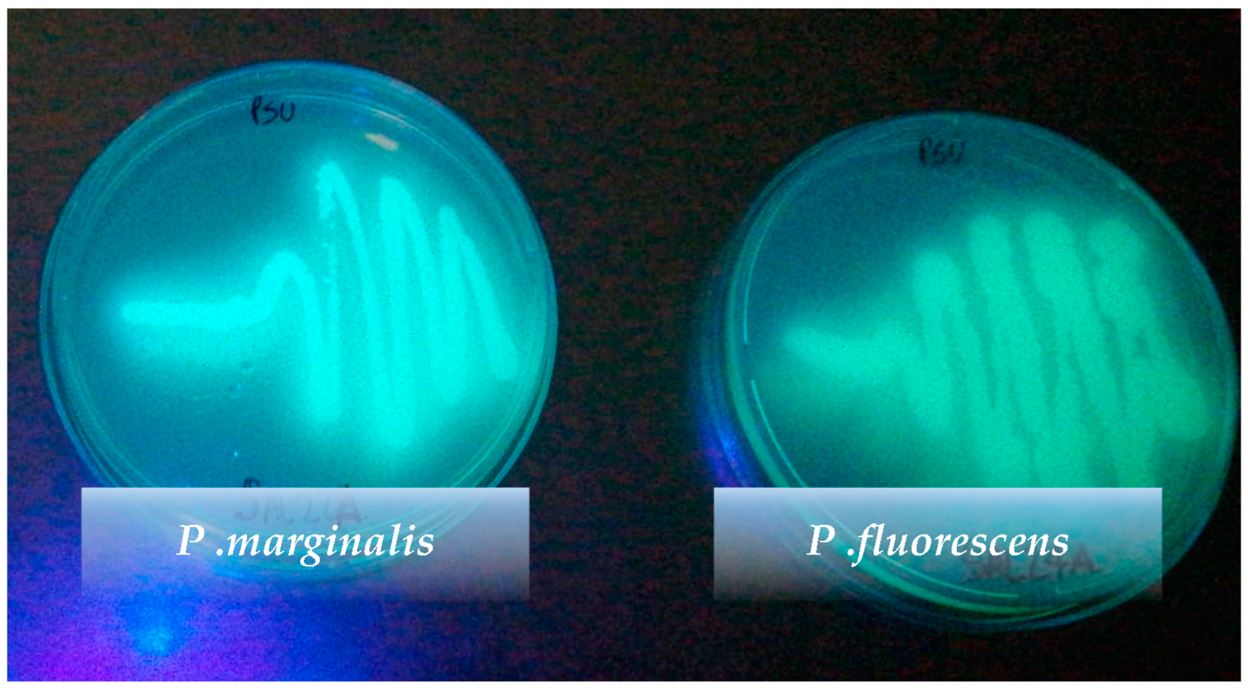
3.2. Phenotypic Identification of Pseudomonas Species

During our study, from the analyzed fish fillets, we selected 55 strains of presumptive Pseudomonas spp. that have been biochemically identified with the BIOLOG® system. Overall, 67.27% (37 up to 55) of the strains were definitively confirmed as Pseudomonas spp., while the other genera were all Gram-negative bacteria, such as Kluyvera, Proteus, Citrobacter, Klebsiella, Serratia, Roseomonas and Providencia. This finding is very similar to the one of another recent study carried out on the salmon processing environment where, out of 222 presumptive Pseudomonas isolates, 68% were confirmed as Pseudomonas [40]. This means that the identification of species on a certain number of bacterial colonies isolated is necessary to establish a higher degree of precision in the loads recorded. The bias of this methodological approach is represented by the economic costs and waiting times to obtain the final results.
Pseudomonas isolates originating from salmon, cod and plaice fillets are diverse with many species represented. Our isolated Pseudomonas species were P. fluorescens, P. fragi, P. lundensis, P. marginalis, P. syringae, P. taetrolens, P. chlororaphis, P. tolaasii and P. viridilivida. Indeed, 45.95% of the strains are represented by P. fluorescens, which largely dominates the Pseudomonadaceae population in the analyzed fillets, followed by P. fragi (21.62%), P. marginalis (10.81%), P. taetrolens (8.11%), P. lundensis, P. syringae, P. tolaasii, P. viridilivida and P. chlororaphis (2.70% each one).
It should be noted that this study has some limitations, mainly the lack of molecular identification using 16S rRNA and some housekeeping genes. It is, however, true that the genus Pseudomonas is very large and includes several hundred unclassified strains. So, for this complex, genus sequencing of the 16S rRNA gene can often only identify the three main lineages (P. aeruginosa, P. pertucinogena and P. fluorescens) but cannot give information about the species [40].
These data confirm what has already been described in the literature, which indicates that among the major components of Pseudomonadaceae in fishery products, there are P. fluorescens and P. fragi [1,2,3]. It should be noted that P. aeruginosa was never found among the strains isolated, and this is in line with what is reported by other studies [4] which highlight a low prevalence of the P. aeruginosa species in foods, including in drinking water.
As it is known, P. fluorescens and P. marginalis were able to produce fluorescent pigments on culture medium when illuminated with a Wood’s lamp light (Figure A3). Additionally, some strains of P. chlororaphis also produced fluorescent pigments.

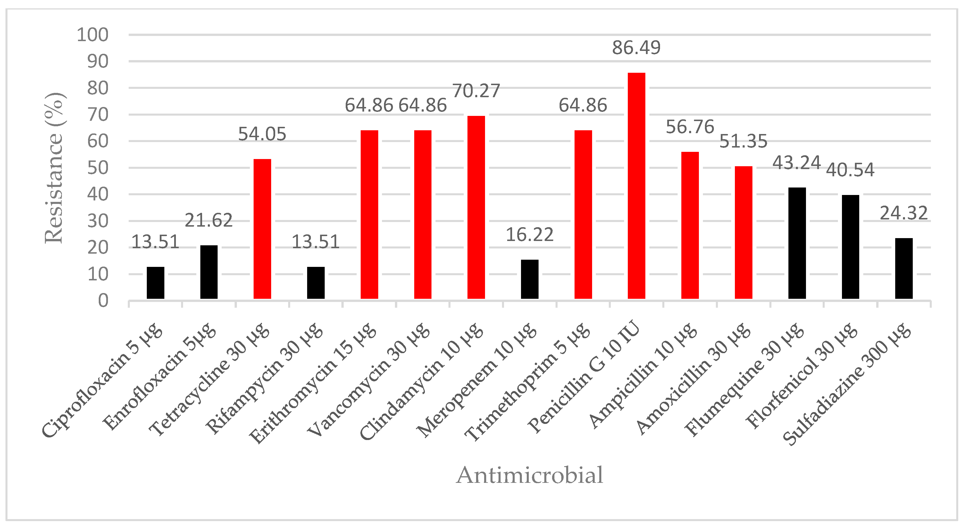
3.3. Antimicrobial Resistance

In order to ensure the best growing conditions, in our study we changed the temperature of incubation of the Mueller–Hinton plates as the tested isolates were psychrophiles and could not grow well at high temperatures. The assay for these was conducted at 31 °C for 24 h. It was decided to consider as “highly resistant” those cases in which the diameter of the inhibition zone was ≤10 mm as it was impossible to find guidelines that provided identification criteria. The reasons were that, on the one hand, the incubation temperature was different and, on the other hand, neither EUCAST nor CLSI give enough information about the Pseudomonas species.
Resistance was found with a prevalence greater than 50% (see Figure A4) in the following cases: penicillin G (penicillins), ampicillin (penicillins), amoxicillin (penicillins), tetracycline (tetracyclines), erythromycin (macrolides), vancomycin (glycopeptides), clindamycin (lincosamides) and trimethoprim (diaminopyrimidines).
It should be noted that, according to the WHO classification (2019) [36], macrolides and glycopeptides are CIAs (Critically Important Antimicrobials), while lincosamides and diaminopyrimidines are HIAs (Highly Important Antimicrobials).
Unlike Kačániová and co-authors [31], who found resistance of all Pseudomonas strains to meropenem, in our study, only 16.22% of the isolates were resistant to this antibiotic. Instead, according to Fazeli and Momtaz [32], the bacterial strains showed a high level of resistance to penicillin (86.49%), tetracycline and erythromycin but with greater resistance to erythromycin (64.86%) rather than to tetracycline (54.05%). In our case, these two were surpassed (or equaled) by clindamycin (70.27%), vancomycin and trimethoprim (both 64.86%).
In the literature, various resistance mechanisms are described that could explain some of the cases detected in our study, such as β-lactamases against penicillins, efflux pump against erythromycin and dihydrofolate reductase against trimethoprim [24,27,28,41].
Countless acquired resistance genes have also been discovered [28]; however, few studies on the possible mechanisms of the resistance owned by Pseudomonas spp. against clindamycin and vancomycin are reported in the scientific literature.
In our research, we noticed that 76.47% of the strains of P. fluorescens were resistant to more than two antibiotic classes and so we considered them as multi-drug resistant (MDR) according to Shabana and co-authors’ definition of “MDR strains” [33].

4. Conclusions

Based on the data obtained and the statistical analyses mentioned above, we can conclude that, under the basic hygienic conditions of production established by the EC Regulations n.852/2004 and 853/2004, fresh fish fillets (salmon, plaice, cod) intended for large-scale retailers have a load of Pseudomonas spp. of about, on average, 104 cfu/g. In salmon fillets, the counts of Pseudomonas spp. recorded have sporadically reached even 105 and 106 cfu/g. In contrast, in plaice and cod fillets, the upward swing of Pseudomonas loads to between 105 and 106 cfu/g were more frequent.
Moreover, in our study, we noted a significant effect of the fish type on the Pseudomonas population of the fillets. The loads of Pseudomonas spp. in salmon fillets were significantly lower than in plaice fillets (p = 0.028), while in cod fillets, they ranged between the two above mentioned species.
Considering the Total Viable Count of around 105 cfu/g (mean value), we can also conclude that a large part of the microbial community found on the analyzed fish fillets was made up of Pseudomonadaceae with other minor microbial populations. The most frequently isolated Pseudomonas species was P. fluorescens.
As Pseudomonas is the main SSO of fresh fish fillets, it is certainly worth keeping an eye on the colony-forming units per gram of fish muscle. In our opinion, fresh fish fillets should contain low loads of Pseudomonas spp. during production in order to avoid rapid degradation of the product during its shelf life. It would be advisable that in the fresh fish fillets, the initial Pseudomonas load should be lower than 104 or 105 cfu/g to not exceed the concretely spoiling loads of these bacteria. In general, in the literature [37,42], it is admitted that up to 106 cfu/g of SSO, the organoleptic quality is fine. Furthermore, considering the normal growth potential of Pseudomonas spp. in refrigerated fresh fish products, it is possible to act both on conservation techniques (for example vacuum packing) and on greater hygiene in the fish processing environment to limit their proliferation. It would be advisable that, at the refrigeration temperature, fresh fish fillets have a shelf life of no more than six days; five days would be even better.
Moreover, our antimicrobial resistance study on the 37 isolated strains of Pseudomonas spp. showed that these bacteria have a high level of phenotypic resistance to various antimicrobial classes. More specifically, 8 out of 15 antimicrobials tested proved ineffective in more than 50% of the strains. These eight antimicrobials are penicillin G 10 IU, ampicillin 10 μg, amoxicillin 30 μg, tetracycline 30 μg, erythromycin 15 μg, vancomycin 30 μg, clindamycin 10 μg and trimethoprim 5 μg. This highlights that 76.47% of P. fluorescens strains were multi-drug resistant.
In the literature, various resistance mechanisms are described (both intrinsic and acquired) such as low outer membrane permeability, β-lactamases synthesis and efflux pump systems. However, it is worth noting that few studies on the possible mechanisms of the resistance of Pseudomonas spp. against clindamycin and vancomycin are available. In order to deepen our research and obtain more relevant results, our recommendation is to complete this study with a genomic analysis by performing a whole genome sequencing of the isolated strains of Pseudomonas (WGS).
Therefore, our results suggest that Pseudomonas spp. should be monitored as possible source of other resistance genes. It would be preferable to complete this not only on strains isolated from clinical specimens but also on the strains isolated from food samples.

Author Contributions

Conceptualization, V.G., N.B.M. and P.C.; methodology, V.G. and G.A.; validation N.B.M. and A.T.; formal analysis, P.C. and N.B.M.; investigation, G.A. and N.B.M.; resources, V.G. and P.C.; data curation, G.A., N.B.M. and P.C.; writing—original draft preparation, V.G., N.B.M. and G.A.; writing—review and editing, G.A., A.T. and P.C.; visualization, N.B.M.; project administration, V.G. and P.C. All authors have read and agreed to the published version of the manuscript.

Funding

This work was supported by the University of Padova (project SID 2021 funded Dep. MAPS).

Institutional Review Board Statement

Not applicable.

Informed Consent Statement

Not applicable.

Data Availability Statement

Not applicable.

Conflicts of Interest

The authors declare no conflict of interest.

Appendix A

Figure A1. Representation of Pseudomonas spp. loads detected in the analyzed samples of the three studied fish species.
Foods 12 00950 g0a1
Figure A2. Representation of TVC in the analyzed samples of the three studied species.
Foods 12 00950 g0a2
Figure A3. Fluorescence of two selected strains under Wood’s lamp light.
Foods 12 00950 g0a3
Figure A4. Percentage of Pseudomonas spp. resistance against different tested antimicrobials (the antimicrobial concentration is expressed as dose/disk). Having no criteria in the literature, cases where the diameter of the inhibition zone was ≤10 mm were considered “highly resistant”. The red color highlights resistance in more than 50% of the strains. The black color, on the other hand, indicates resistance in less than 50% of the strains.
Foods 12 00950 g0a4

References

  1. Tryfinopoulou, P.; Tsakalidou, E.; Nychas, G.J.E. Characterization of Pseudomonas spp. associated with spoilage of gilt-head sea bream stored under various conditions. Appl. Environ. Microbiol. 2002, 68, 65–72. [Google Scholar] [CrossRef] [Green Version]
  2. Eissa, N.M.E.; Abou El-Ghiet, E.N.; Shaheen, A.A.; Abbass, A. Characterization of Pseudomonas species isolated from tilapia Oreochromis niloticus in Qaroun and Wadi-El-Rayan Lakes, Egypt. Glob. Vet. 2010, 5, 116–121. [Google Scholar] [CrossRef]
  3. Abd El Tawab, A.; Maarouf, A.; Ahmed, N. Detection of Virulence factors of Pseudomonas species isolated from fresh water fish by PCR. Benha Vet. Med. J. 2016, 30, 199–207. [Google Scholar] [CrossRef] [Green Version]
  4. Heir, E.; Moen, B.; Åsli, A.W.; Sunde, M.; Langsrud, S. Antibiotic resistance and phylogeny of Pseudomonas spp. Isolated over three decades from chicken meat in the norwegian food chain. Microorganisms 2021, 9, 207. [Google Scholar] [CrossRef]
  5. Gram, L.; Huss, H.H. Microbiological spoilage of fish and fish products. Int. J. Food Microbiol. 1996, 33, 121–137. [Google Scholar] [CrossRef]
  6. De Jonghe, V.; Coorevits, A.; Van Hoorde, K.; Messens, W.; Van Landschoot, A.; De Vos, P.; Heyndrickx, M. Influence of storage conditions on the growth of Pseudomonas species in refrigerated raw milk. Appl. Environ. Microbiol. 2011, 77, 460–470. [Google Scholar] [CrossRef] [Green Version]
  7. Doulgeraki, A.I.; Nychas, G.J.E. Monitoring the succession of the biota grown on a selective medium for pseudomonads during storage of minced beef with molecular-based methods. Food Microbiol. 2013, 34, 62–69. [Google Scholar] [CrossRef] [PubMed]
  8. Mohareb, F.; Iriondo, M.; Doulgeraki, A.I.; Van Hoek, A.; Aarts, H.; Cauchi, M.; Nychas, G.J.E. Identification of meat spoilage gen (Eissa et al., 2010) e biomarkers in Pseudomonas putida using gene profiling. Food Control 2015, 57, 152–160. [Google Scholar] [CrossRef] [Green Version]
  9. Caldera, L.; Franzetti, L.; Van Coillie, E.; De Vos, P.; Stragier, P.; De Block, J.; Heyndrickx, M. Identification, enzymatic spoilage characterization and proteolytic activity quantification of Pseudomonas spp. isolated from different foods. Food Microbiol. 2016, 54, 142–153. [Google Scholar] [CrossRef]
  10. Taliadourou, D.; Papadopoulos, V.; Domvridou, E.; Savvaidis, I.N.; Kontominas, M.G. Microbiological, chemical and sensory changes of whole and filleted Mediterranean aquacultured sea bass (Dicentrarchus labrax) stored in ice. J. Sci. Food Agric. 2003, 83, 1373–1379. [Google Scholar] [CrossRef]
  11. Carpentier, B.; Chassaing, D. Interactions in biofilms between Listeria monocytogenes and resident microorganisms from food industry premises. Int. J. Food Microbiol. 2004, 97, 111–122. [Google Scholar] [CrossRef]
  12. Mann, E.E.; Wozniak, D.J. Pseudomonas biofilm matrix composition and niche biology. FEMS Microbiol. Rev. 2012, 36, 893–916. [Google Scholar] [CrossRef] [Green Version]
  13. Giaouris, E.; Heir, E.; Desvaux, M.; Hébraud, M.; Møretrø, T.; Langsrud, S.; Doulgeraki, A.; Nychas, G.J.; Kacániová, M.; Czaczyk, K.; et al. Intra- and inter-species interactions within biofilms of important foodborne bacterial pathogens. Front. Microbiol. 2015, 6, 841. [Google Scholar] [CrossRef] [Green Version]
  14. Sterniša, M.; Klančnik, A.; Smole Možina, S. Spoilage Pseudomonas biofilm with Escherichia coli protection in fish meat at 5 °C. J. Sci. Food Agric. 2019, 99, 4635–4641. [Google Scholar] [CrossRef] [PubMed]
  15. Nikel, P.I.; Fuhrer, T.; Chavarría, M.; Sánchez-Pascuala, A.; Sauer, U.; de Lorenzo, V. Reconfiguration of metabolic fluxes in Pseudomonas putida as a response to sub-lethal oxidative stress. ISME J. 2021, 15, 1751–1766. [Google Scholar] [CrossRef]
  16. Chen, C.; Malek, A.A.; Wargo, M.J.; Hogan, D.A.; Beattie, G.A. The ATP-binding cassette transporter Cbc (choline/betaine/carnitine) recruits multiple substrate-binding proteins with strong specificity for distinct quaternary ammonium compounds. Mol. Microbiol. 2010, 75, 29–45. [Google Scholar] [CrossRef] [Green Version]
  17. Sheng, L.; Wang, L. The microbial safety of fish and fish products: Recent advances in understanding its significance, contamination sources, and control strategies. Compr. Rev. Food Sci. Food Saf. 2021, 20, 738–786. [Google Scholar] [CrossRef] [PubMed]
  18. Commission Regulation (EC) No 2073/2005 of 15 November 2005 on Microbiological Criteria for Foodstuffs. Official Journal of the European Union L338 22 December 2005. Available online: https://eur-lex.europa.eu/legal-content/EN/TXT/PDF/?uri=CELEX:32005R2073from=EN (accessed on 12 January 2023).
  19. FAO. The State of World Fisheries and Aquaculture 2022. In Towards Blue Transformation; FAO: Rome, Italy, 2022. [Google Scholar] [CrossRef]
  20. The European Market Observatory for Fisheries and Aquaculture. Available online: https://www.eumofa.eu/it/data (accessed on 12 January 2023).
  21. Schar, D.; Klein, E.Y.; Laxminarayan, R.; Gilbert, M.; Van Boeckel, T.P. Global trends in antimicrobial use in aquaculture. Sci. Rep. 2020, 10, 918. [Google Scholar] [CrossRef] [PubMed]
  22. Scott, A.; McEwen, P.J.C. Antimicrobial resistance: A One Health perspective. Trans. R. Soc. Trop. Med. Hyg. 2018, 111, 255–260. [Google Scholar] [CrossRef]
  23. Marshall, B.M.; Levy, S.B. Food animals and antimicrobials: Impacts on human health. Clin. Microbiol. Rev. 2011, 24, 718–733. [Google Scholar] [CrossRef] [PubMed] [Green Version]
  24. Delannoy, S.; Hoffer, C.; Youf, R.; Dauvergne, E.; Webb, H.E.; Brauge, T.; Tran, M.-L.; Midelet, G.; Granier, S.A.; Haenni, M.; et al. High Throughput Screening of Antimicrobial Resistance Genes in Gram-Negative Seafood Bacteria. Microorganisms 2022, 10, 1225. [Google Scholar] [CrossRef]
  25. Levy, S.B.; Bonnie, M. Antibacterial resistance worldwide: Causes, challenges and responses. Nat. Med. 2004, 10, S122–S129. [Google Scholar] [CrossRef] [PubMed]
  26. Kačániová, M.; Terentjeva, M.; Vukovic, N.; Puchalski, C.; Roychoudhury, S.; Kunová, S.; Klūga, A.; Tokár, M.; Kluz, M.; Ivanišová, E. The antioxidant and antimicrobial activity of essential oils against Pseudomonas spp. isolated from fish. Saudi Pharm. J. 2017, 65, 102–115. [Google Scholar] [CrossRef] [PubMed]
  27. Breidenstein, E.B.; de la Fuente-Núñez, C.; Hancock, R.E. Pseudomonas aeruginosa: All roads lead to resistance. Trends Microbiol. 2011, 19, 419–426. [Google Scholar] [CrossRef]
  28. Luczkiewicz, A.; Kotlarska, E.; Artichowicz, W.; Tarasewicz, K.; Fudala-Ksiazek, S. Antimicrobial resistance of Pseudomonas spp. isolated from wastewater and wastewater-impacted marine coastal zone. Environ. Sci. Pollut. Res. 2015, 22, 19823–19834. [Google Scholar] [CrossRef] [Green Version]
  29. King, D.T.; Sobhanifar, S.; Strynadka, N.C.J. Handbook of Antimicrobial Resistance. In Handbook of Antimicrobial Resistance; Springer: Berlin/Heidelberg, Germany, 2014; pp. 1–22. [Google Scholar] [CrossRef]
  30. Elbehiry, A.; Marzouk, E.; Aldubaib, M.; Moussa, I.; Abalkhail, A.; Ibrahem, M.; Hamada, M.; Sindi, W.; Alzaben, F.; Almuzaini, A.M.; et al. Pseudomonas species prevalence, protein analysis, and antibiotic resistance: An evolving public health challenge. AMB Express 2022, 12, 53. [Google Scholar] [CrossRef]
  31. Kačániová, M.; Klūga, A.; Kántor, A.; Medo, J.; Žiarovská, J.; Puchalski, C.; Terentjeva, M. Comparison of MALDI-TOF MS Biotyper and 16S rDNA sequencing for the identification of Pseudomonas species isolated from fish. Microb. Pathog. 2019, 132, 313–318. [Google Scholar] [CrossRef]
  32. Fazeli, N.; Momtaz, H. Virulence gene profiles of multidrug-resistant Pseudomonas aeruginosa isolated from Iranian hospital infections. Iran. Red Crescent Med. J. 2014, 16, e15722. [Google Scholar] [CrossRef] [Green Version]
  33. Shabana, B.M.; Elkenany, R.M.; Younis, G. Sequencing and multiple antimicrobial resistance of Pseudomonas fluorescens isolated from Nile tilapia fish in Egypt. Braz. J. Biol. 2022, 84, 1–9. [Google Scholar] [CrossRef]
  34. EUCAST Disk Diffusion Test Methodology. Available online: https://www.eucast.org/ast_of_bacteria/disk_diffusion_methodology (accessed on 12 January 2023).
  35. Simonsen, G.S.; Blix, H.S.; Grave, K.; Urdahl, A.M. NORM/NORM-VET 2020: Usage of Antimicrobial Agents and Occurrence of Antimicrobial Resistance in Norway; Norwegian Institute of Public Health: Tromsø/Oslo, Norway, 2021. [Google Scholar]
  36. WHO. Critically Important Antimicrobials for Human Medicine (6th revision). Available online: https://www.who.int/publications/i/item/9789241515528 (accessed on 12 January 2023).
  37. Silbande, A.; Adenet, S.; Smith-Ravin, J.; Joffraud, J.J.; Rochefort, K.; Leroi, F. Quality assessment of ice-stored tropical yellowfin tuna (Thunnus albacares) and influence of vacuum and modified atmosphere packaging. Food Microbiol. 2016, 60, 62–72. [Google Scholar] [CrossRef] [PubMed] [Green Version]
  38. Speck, M.L.; Ray, B. Effects of Freezing and Storage on Microorganisms in Frozen Foods: A Review. J. Food Prot. 1997, 40, 333–336. [Google Scholar] [CrossRef] [Green Version]
  39. Baña, Z.; Abad, N.; Uranga, A.; Azúa, I.; Artolozaga, I.; Unanue, M.; Iriberri, J.; Arrieta, J.M.; Ayo, B. Recurrent seasonal changes in bacterial growth efficiency, metabolism and community composition in coastal waters. Environ Microbiol. 2020, 22, 369–380. [Google Scholar] [CrossRef]
  40. Thomassen, G.M.B.; Reiche, T.; Tennfjord, C.E.; Mehli, L. Antibiotic Resistance Properties among Pseudomonas spp. Associated with Salmon Processing Environments. Microorganisms 2022, 10, 1420. [Google Scholar] [CrossRef] [PubMed]
  41. Livermore, D.M. Multiple mechanisms of antimicrobial resistance in Pseudomonas aeruginosa: Our worst nightmare? Clin. Infect. Dis. 2002, 34, 634–640. [Google Scholar] [CrossRef] [PubMed] [Green Version]
  42. Du, W.X.; Kim, J.; Cornell, J.A.; Huang, T.S.; Marshall, M.R.; Wei, C.I. Microbiological 2002, sensory, and electronic nose evaluation of yellowfin tuna under various storage conditions. J. Food Prot. 2001, 64, 2027–2036. [Google Scholar] [CrossRef] [PubMed]
Figure 1. Box plot of the 10^ index (order of magnitude with powers of 10) for Pseudomonas spp. The boxes contain 50% of the data distribution, the red crosses represent the average values, the whiskers are proportional to the interquartile range and the points and stars are the outliers as a function of the different interquartile ranges.“COD” refers to northern cod, “PLA” to plaice and “SAL” to salmon.
Foods 12 00950 g001
Table 1. TVC and Pseudomonas spp. back-transformed ls-means with 95% CI in the three species of fish fillets.
Fish FilletsTVC Mean Value (cfu/g)Pseudomonas spp. Mean Value (cfu/g)
Gadus morhua2.57 × 105 (1.83–3.61 × 10⁵) a4.67 × 104 (2.68–8.16 × 10⁴) a,b
Pleuronectes platessa2.56 × 105 (1.82–3.60 × 10⁵) a6.71 × 104 (3.85–1.17 × 10⁴) a
Salmo salar1.07 × 105 (7.65–1.51 × 10⁵) b2.33 × 104 (1.34–4.06 × 10⁴) b
a,b Different letters along columns mean significantly different values (p < 0.05).
Disclaimer/Publisher’s Note: The statements, opinions and data contained in all publications are solely those of the individual author(s) and contributor(s) and not of MDPI and/or the editor(s). MDPI and/or the editor(s) disclaim responsibility for any injury to people or property resulting from any ideas, methods, instructions or products referred to in the content.

