
Oxalate in Foods: Extraction Conditions, Analytical Methods, Occurrence, and Health Implications

 , ,
, ,  and
and 
Abstract
1. Introduction
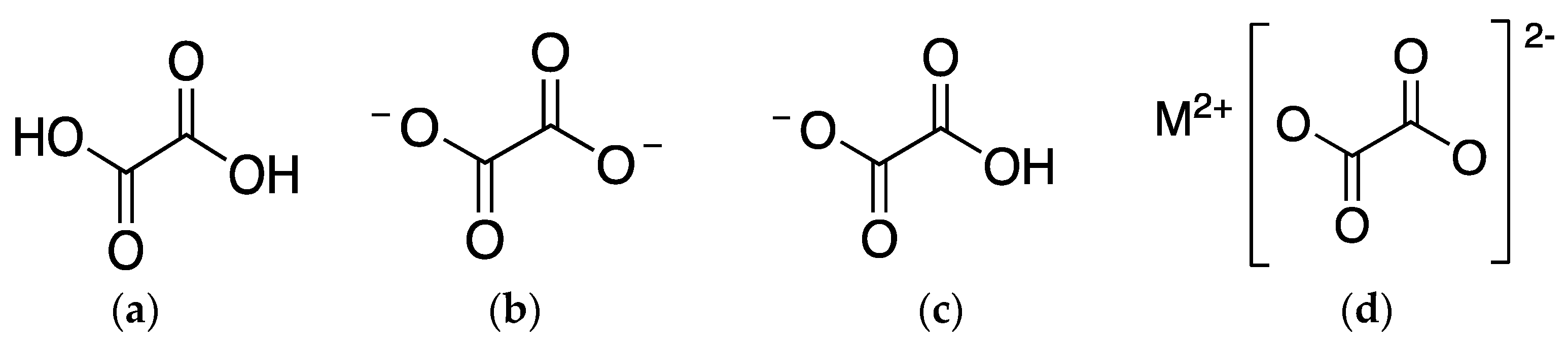
2. Oxalates
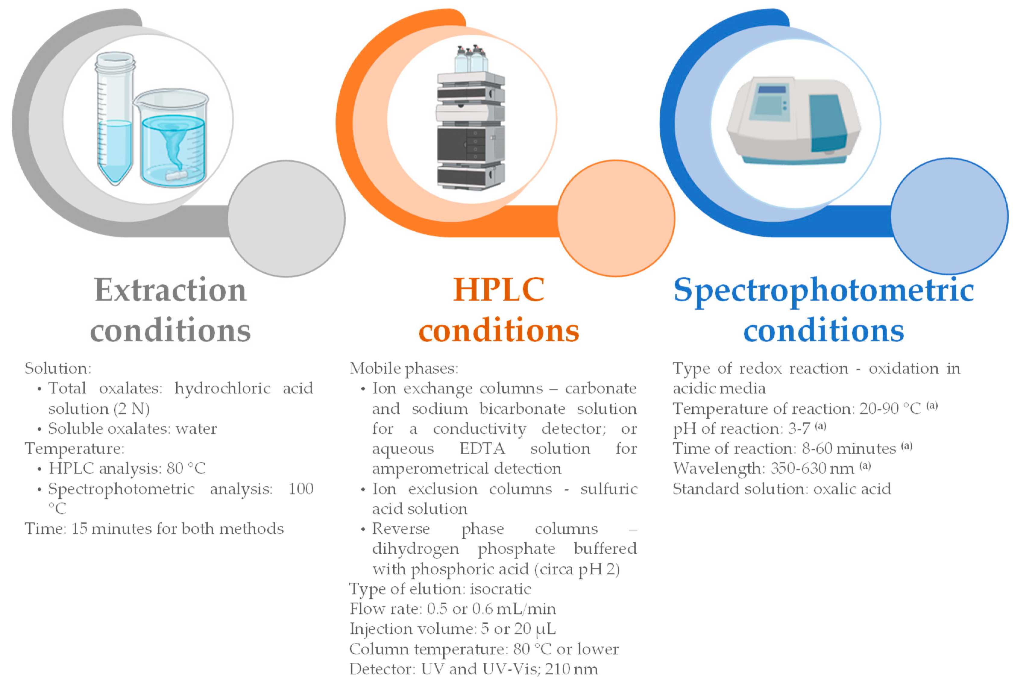
3. Analysis of Oxalates in Foods
3.1. Extraction Conditions
3.2. Methods’ Conditions
3.2.1. HPLC Conditions
3.2.2. Spectrophotometric Conditions
4. Oxalate Occurrence in Foods
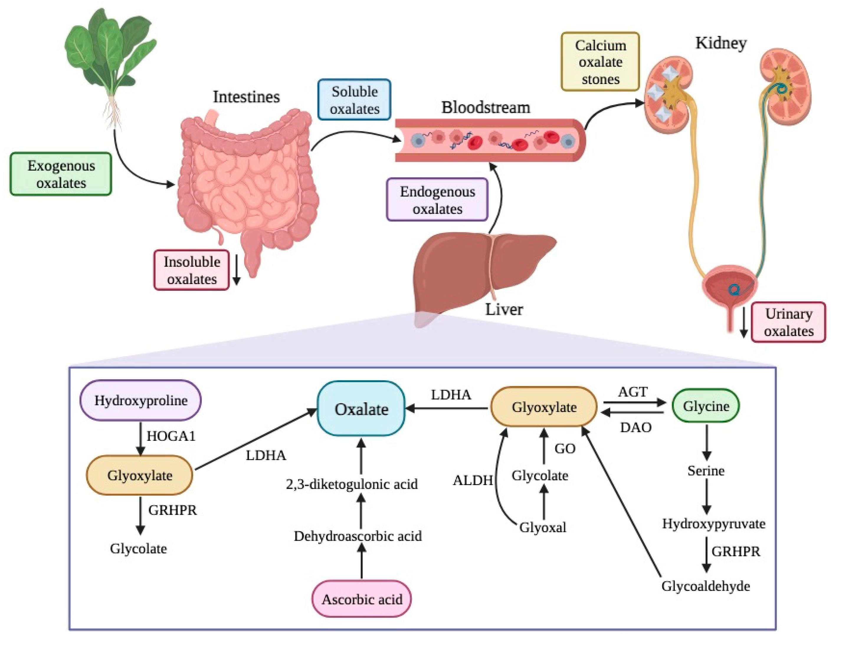
5. Health Implications of Oxalates
6. Conclusions
Author Contributions
Funding
Data Availability Statement
Conflicts of Interest
References
- Hönow, R.; Hesse, A. Comparison of Extraction Methods for the Determination of Soluble and Total Oxalate in Foods by HPLC-Enzyme-Reactor. Food Chem. 2002, 78, 511–521. [Google Scholar] [CrossRef]
- Li, P.; Liu, C.; Luo, Y.; Shi, H.; Li, Q.; Pinchu, C.; Li, X.; Yang, J.; Fan, W. Oxalate in Plants: Metabolism, Function, Regulation, and Application. J. Agric. Food Chem. 2022, 70, 16037–16049. [Google Scholar] [CrossRef] [PubMed]
- Petroski, W.; Minich, D.M. Is There Such a Thing as “Anti-Nutrients”? A Narrative Review of Perceived Problematic Plant Compounds. Nutrients 2020, 12, 2929. [Google Scholar] [CrossRef] [PubMed]
- Okombo, J.; Liebman, M. Oxalate Content of Selected Breads and Crackers. J. Food Compos. Anal. 2010, 23, 118–121. [Google Scholar] [CrossRef]
- Al-Wahsh, I.A.; Wu, Y.; Liebman, M. A Comparison of Two Extraction Methods for Food Oxalate Assessment. J. Food Res. 2012, 1, 233. [Google Scholar] [CrossRef]
- Ruan, Q.-Y.; Zheng, X.-Q.; Chen, B.-L.; Xiao, Y.; Peng, X.-X.; Leung, D.W.M.; Liu, E.-E. Determination of Total Oxalate Contents of a Great Variety of Foods Commonly Available in Southern China Using an Oxalate Oxidase Prepared from Wheat Bran. J. Food Compos. Anal. 2013, 32, 6–11. [Google Scholar] [CrossRef]
- Mou, B. Evaluation of Oxalate Concentration in the U.S. Spinach Germplasm Collection. HortScience 2008, 43, 1690–1693. [Google Scholar] [CrossRef]
- Kasidas, G.P.; Rose, G.A. Oxalate Content of Some Commond Foods: Determination by an Enzymatic Method. J. Hum. Nutr. 1980, 34, 255–266. [Google Scholar] [CrossRef]
- Milardović, S.; Grabarić, Z.; Rumenjak, V.; Jukić, M. Rapid Determination of Oxalate by an Amperometric Oxalate Oxidase-Based Electrode. Electroanalysis 2000, 12, 1051–1058. [Google Scholar] [CrossRef]
- Bohn, T.; Davidsson, L.; Walczyk, T.; Hurrell, R.F. Fractional Magnesium Absorption Is Significantly Lower in Human Subjects from a Meal Served with an Oxalate-Rich Vegetable, Spinach, as Compared with a Meal Served with Kale, a Vegetable with a Low Oxalate Content. Br. J. Nutr. 2004, 91, 601–606. [Google Scholar] [CrossRef]
- Chai, W.; Liebman, M. Effect of Different Cooking Methods on Vegetable Oxalate Content. J. Agric. Food Chem. 2005, 53, 3027–3030. [Google Scholar] [CrossRef]
- Pérez-Ruiz, T.; Martínez-Lozano, C.; Tomás, V.; Casajús, R. FIow Injection SpectrofIuorimetric Determination of Oxalate Based on Its Enhancing Effect on the Oxidation of Rhodamine B by Dichromate. Analyst 1995, 120, 2111–2114. [Google Scholar] [CrossRef]
- Shaidarova, L.G.; Leksina, Y.A.; Chelnokova, I.A.; Gedmina, A.V.; Budnikov, H.C. Flow Injection Amperometric Determination of Oxalic Acid on Graphite Electrode Covered by Nafion Film with Included Palladium Nanoparticles. Turk. Online J. Des. Art Commun. 2017, 7, 1879–1886. [Google Scholar]
- Pundir, C.S.; Chauhan, N.; Rajneesh; Verma, M.; Ravi, A. Novel Amperometric Biosensor for Oxalate Determination Using Multi-Walled Carbon Nanotube-Gold Nanoparticle Composite. Sens. Actuators B Chem. 2011, 155, 796–803. [Google Scholar] [CrossRef]
- Šljukić, B.; Baron, R.; Compton, R.G. Electrochemical Determination of Oxalate at Pyrolytic Graphite Electrodes. Electroanalysis 2007, 19, 918–922. [Google Scholar] [CrossRef]
- Fakhari, A.R.; Rafiee, B.; Ahmar, H.; Bagheri, A. Electrocatalytic Determination of Oxalic Acid by TiO2 Nanoparticles/Multiwalled Carbon Nanotubes Modified Electrode. Anal. Methods 2012, 4, 3314–3319. [Google Scholar] [CrossRef]
- Holmes, R.P.; Kennedy, M. Estimation of the Oxalate Content of Foods and Daily Oxalate Intake. Kidney Int. 2000, 57, 1662–1667. [Google Scholar] [CrossRef]
- Merusi, C.; Corradini, C.; Cavazza, A.; Borromei, C.; Salvadeo, P. Determination of Nitrates, Nitrites and Oxalates in Food Products by Capillary Electrophoresis with PH-Dependent Electroosmotic Flow Reversal. Food Chem. 2010, 120, 615–620. [Google Scholar] [CrossRef]
- Sotomayor, M.D.P.T.; Raimundo, I.M., Jr.; Oliveira Neto, G.O.; Kubota, L.T. Bi-Enzymatic Optode Detection System for Oxalate Determination Based on a Natural Source of Enzyme. Anal. Chim. Acta 2001, 447, 33–40. [Google Scholar] [CrossRef]
- Ojinnaka, M.C.; Ebinyasi, C.S.; Ihemeje, A.; Okorie, S.U. Nutritional Evaluation of Complementary Food Gruels Formulated from Blends of Soybean Flour and Ginger Modified Cocoyam Starch. Adv. J. Food Sci. Technol. 2013, 5, 1325–1330. [Google Scholar] [CrossRef]
- Adeniyi, S.A.; Orjiekwe, C.L.; Ehiagbonare, J.E. Determination of Alkaloids and Oxalates in Some Selected Food Samples in Nigeria. Afr. J. Biotechnol. 2009, 8, 110–112. [Google Scholar]
- Gouveia, C.S.S.; Ganança, J.F.T.; Lebot, V.; de Carvalho, M.Â.A.P. Quantitation of Oxalates in Corms and Shoots of Colocasia Esculenta (L.) Schott under Drought Conditions. Acta Physiol. Plant 2018, 40, 214. [Google Scholar] [CrossRef]
- Ikese, O.; Ubwa, S.; Sunday, A.; Lenka, J.; Inalegwu, J.; Ubwa, S.; Ocheje, M.; Inegedu, A. Proximate Composition, Antinutrients and Some Functional Properties of a Potential Infant Food Made from Wheat and Groundnut. Int. J. Food Sci. Nutr. 2016, 1, 59–63. [Google Scholar]
- Iwuozor, K.O. Qualitative and Quantitative Determination of Anti-Nutritional Factors of Five Wine Samples. Adv. J. Chem. 2019, 2, 136–146. [Google Scholar] [CrossRef]
- Baker, C.J.L. The Determination of Oxalates in Fresh Plant MateriaI. Analyst 1952, 77, 340–344. [Google Scholar] [CrossRef]
- Zhang, Y.; Li, Y.; Wei, J.; Sun, M.; Tian, Y.; Li, Z. Effects of Nitrogen and Calcium Nutrition on Oxalate Contents, Forms, and Distribution in Spinach. J. Plant Nutr. 2009, 32, 2123–2139. [Google Scholar] [CrossRef]
- Zhang, Y.; Lin, X.; Zhang, Y.; Shao, J.Z.; Du, S. Effects of Nitrogen Levels and Nitrate/Ammonium Ratios on Oxalate Concentrations of Different Forms in Edible Parts of Spinach. J. Plant Nutr. 2005, 28, 2011–2025. [Google Scholar] [CrossRef]
- Ohkawa, H. Gas Chromatographic Determination of Oxalic Acid in Foods. J. Assoc. Off. Anal. Chem. 1985, 68, 108–111. [Google Scholar] [CrossRef]
- Dong, M.W. Modern HPLC for Practicing Scientists; Wiley-Interscience: Hoboken, NJ, USA, 2006; ISBN 9780471727897. [Google Scholar]
- Altunay, N.; Gürkan, R. A Simple, Low-Cost, and Useful Preconcentration Method for Quantification of Soluble, Insoluble, and Total Oxalate in Selected Vegetables Through Spectrophotometry. Food Anal. Methods 2016, 9, 950–965. [Google Scholar] [CrossRef]
- Ensafi, A.A.; Abbasi, S.; Rezaei, B. Kinetic Spectrophotometric Method for the Determination of Oxalic Acid by Its Catalytic Effect on the Oxidation of Safranine by Dichromate. Spectrochim. Acta Part A 2001, 57, 1833–1838. [Google Scholar] [CrossRef]
- Savage, G.P.; Vanhanen, L.; Mason, S.M.; Ross, A.B. Effect of Cooking on the Soluble and Insoluble Oxalate Content of Some New Zealand Foods. J. Food Compos. Anal. 2000, 13, 201–206. [Google Scholar] [CrossRef]
- Radek, M.; Savage, G.P. Oxalates in Some Indian Green Leafy Vegetables. Int. J. Food Sci. Nutr. 2008, 59, 246–260. [Google Scholar] [CrossRef] [PubMed]
- Savage, G.P.; Mårtensson, L. Comparison of the Estimates of the Oxalate Content of Taro Leaves and Corms and a Selection of Indian Vegetables Following Hot Water, Hot Acid and in Vitro Extraction Methods. J. Food Compos. Anal. 2010, 23, 113–117. [Google Scholar] [CrossRef]
- Wilson, C.W.; Shaw, P.E.; Knight, R.J. Analysis of Oxalic Acid in Carambola (Averrhoa Carambola L.) and Spinach by High-Performance Liquid Chromatography. J. Agric. Food Chem. 1982, 30, 1106–1108. [Google Scholar] [CrossRef]
- Akhtar, M.S.; Israr, B.; Bhatty, N.; Ali, A. Effect of Cooking on Soluble and Insoluble Oxalate Contents in Selected Pakistani Vegetables and Beans. Int. J. Food Prop. 2011, 14, 241–249. [Google Scholar] [CrossRef]
- Ensafi, A.A.; Kazemzadeh, A. Flow Injection Spectrophotometric Determination of Ultra Trace Amounts of Oxalic Acid. Fresenius J. Anal. Chem. 2000, 367, 590–592. [Google Scholar] [CrossRef] [PubMed]
- Jiang, Z.-L.; Zhao, M.-X.; Liao, L.-X. Catalytic Spectrophotometric Methods for the Determination of Oxalic Acid. Anal. Chim. Acta 1996, 320, 139–143. [Google Scholar] [CrossRef]
- Liu, X.X.; Zhou, K.; Hu, Y.; Jin, R.; Lu, L.L.; Jin, C.W.; Lin, X.Y. Oxalate Synthesis in Leaves Is Associated with Root Uptake of Nitrate and Its Assimilation in Spinach (Spinacia oleracea L.) Plants. J. Sci. Food Agric. 2014, 95, 2105–2116. [Google Scholar] [CrossRef]
- Kim, D.-J.; Kim, H.; Kim, M.; Lee, J. Analysis of Oxalic Acid of Various Vegetables Consumed in Korea. Food Sci. Biotechnol. 2007, 16, 650–654. [Google Scholar]
- Morosanova, M.A.; Samodelov, Z.V.; Morosanova, E.I. Determination of Food Oxalates Using Silica–Titania Xerogel Modified with Eriochrome Cyanine R. Sensors 2018, 18, 864. [Google Scholar] [CrossRef]
- Fatoki, O.S. Determination of Oxalic Acid in Vegetables. In Modern Methods of Plant Analysis: Vegetables and Vegetable Products; Springer Berlin: Heidelberg, Germany, 1994; Volume 16, pp. 161–167. [Google Scholar]
- Singh, P.P.; Kothari, L.K.; Sharma, D.C.; Saxena, S.N. Nutritional Value of Foods in Relation to Their Oxalic Acid Content. Am. J. Clin. Nutr. 1972, 25, 1147–1152. [Google Scholar] [CrossRef] [PubMed]
- Natesh, H.N.; Abbey, L.; Asiedu, S.K. An Overview of Nutritional and Anti Nutritional Factors in Green Leafy Vegetables. Hortic. Int. J. 2017, 1, 58–65. [Google Scholar] [CrossRef]
- Huynh, N.K.; Nguyen, D.H.M.; Nguyen, H.V.H. Effects of Processing on Oxalate Contents in Plant Foods: A Review. J. Food Compos. Anal. 2022, 112, 104685. [Google Scholar] [CrossRef]
- Wang, Z.; Zhang, Y.; Zhang, J.; Deng, Q.; Liang, H. Recent Advances on the Mechanisms of Kidney Stone Formation (Review). Int. J. Mol. Med. 2021, 48, 149. [Google Scholar] [CrossRef] [PubMed]
- Massey, L.K. Food Oxalate: Factors Affecting Measurement, Biological Variation, and Bioavailability. J. Am. Diet. Assoc. 2007, 107, 1191–1194. [Google Scholar] [CrossRef]
- Witting, C.; Langman, C.B.; Assimos, D.; Baum, M.A.; Kausz, A.; Milliner, D.; Tasian, G.; Worcester, E.; Allain, M.; West, M.; et al. Pathophysiology and Treatment of Enteric Hyperoxaluria. Clin. J. Am. Soc. Nephrol. 2021, 16, 487–495. [Google Scholar] [CrossRef]
- Sharma, S.; Rao, R.N.; Pani, K.C.; Paul, P. Bone Marrow Oxalosis: An Unusual Cause of Cytopenia in End-Stage Renal Disease; Report of Two Cases. Indian J. Pathol. Microbiol. 2018, 61, 268–270. [Google Scholar] [CrossRef]
- Fogo, A.B.; Lusco, M.A.; Najafian, B.; Alpers, C.E. AJKD Atlas of Renal Pathology: Oxalosis. Am. J. Kidney Dis. 2017, 69, e13–e14. [Google Scholar] [CrossRef]
- Noonan, S.C.; Savage, G.P. Oxalate Content of Foods and Its Effect on Humans. Asia Pac. J. Clin. Nutr. 1999, 8, 64–74. [Google Scholar] [CrossRef]
- López-Moreno, M.; Garcés-Rimón, M.; Miguel, M. Antinutrients: Lectins, Goitrogens, Phytates and Oxalates, Friends or Foe? J. Funct. Foods 2022, 89, 104938. [Google Scholar] [CrossRef]
- Karamad, D.; Khosravi-Darani, K.; Hosseini, H.; Tavasoli, S. Analytical Procedures and Methods Validation for Oxalate Content Estimation. Biointerface Res. Appl. Chem. 2019, 9, 4305–4310. [Google Scholar] [CrossRef] [PubMed]
- Chamjangali, M.A.; Keley, V.; Bagherian, G. Kinetic Spectrophotometric Method for the Determination of Trace Amounts of Oxalate by an Activation Effect. Anal. Sci. 2006, 22, 333–336. [Google Scholar] [CrossRef] [PubMed]
- Libert, B.O. Rapid Determination of Oxalic Acid by Reversed-Phase High-Performance Liquid Chromatography. J. Chromatogr. 1981, 210, 540–543. [Google Scholar] [CrossRef]
- Arias-Rico, J.; Macías-León, F.J.; Alanís-García, E.; Cruz-Cansino, N.d.S.; Jaramillo-Morales, O.A.; Barrera-Gálvez, R.; Ramírez-Moreno, E. Study of Edible Plants: Effects of Boiling on Nutritional, Antioxidant, and Physicochemical Properties. Foods 2020, 9, 599. [Google Scholar] [CrossRef]
- Nemzer, B.; Al-Taher, F.; Abshiru, N. Extraction and Natural Bioactive Molecules Characterization in Spinach, Kale and Purslane: A Comparative Study. Molecules 2021, 26, 2515. [Google Scholar] [CrossRef]
- Kassie, W.; Washe, A.P.; Etsay, H. Spectrophotometric Determination of Oxalic Acid in Dietary Sources Through Catalytic Titration with Hexavalent Chromium. Food Sci. Qual. Manag. 2019, 83, 30–38. [Google Scholar] [CrossRef]
- Santamaria, P.; Elia, A.; Serio, F.; Todaro, E. A Survey of Nitrate and Oxalate Content in Fresh Vegetables. J. Sci. Food Agric. 1999, 79, 1882–1888. [Google Scholar] [CrossRef]
- Elia, A.; Santamaria, P.; Serio, F. Nitrogen Nutrition, Yield and Quality of Spinach. J. Sci. Food Agric. 1998, 76, 341–346. [Google Scholar] [CrossRef]
- Judprasong, K.; Charoenkiatkul, S.; Sungpuag, P.; Vasanachitt, K.; Nakjamanong, Y. Total and Soluble Oxalate Contents in Thai Vegetables, Cereal Grains and Legume Seeds and Their Changes after Cooking. J. Food Compos. Anal. 2006, 19, 340–347. [Google Scholar] [CrossRef]
- Ombódi, A.; Kosuge, S.; Saigusa, M. Effects of Polyolefin-Coated Fertilizer on Nutritional Quality of Spinach Plants. J. Plant Nutr. 2000, 23, 1495–1504. [Google Scholar] [CrossRef]
- Siener, R.; Seidler, A.; Hönow, R. Oxalate-Rich Foods. Food Sci. Technol. 2021, 41, 169–173. [Google Scholar] [CrossRef]
- Siener, R.; Hönow, R.; Voss, S.; Seidler, A.; Hesse, A. Oxalate Content of Cereals and Cereal Products. J. Agric. Food Chem. 2006, 54, 3008–3011. [Google Scholar] [CrossRef] [PubMed]
- Kusuma, D.S.; Vanhanen, L.P.; Savage, G.P. Evaluation of Extraction Parameters for Total Oxalate Determination in Spinach Using Design of Experiment Analysis. J. Food Compos. Anal. 2016, 51, 9–14. [Google Scholar] [CrossRef]
- Ghosh Das, S.; Savage, G.P. Oxalate Content of Indian Spinach Dishes Cooked in a Wok. J. Food Compos. Anal. 2013, 30, 125–129. [Google Scholar] [CrossRef]
- Vanhanen, L.; Savage, G. Comparison of Oxalate Contents and Recovery from Two Green Juices Prepared Using a Masticating Juicer or a High Speed Blender. NFS J. 2015, 1, 20–23. [Google Scholar] [CrossRef]
- Zulkhairi, A.M.; Razali, M.; Umikalsum, M.B.; Norfaizal, G.M.; Athirah, A.A.; Aisyah, M.N.S. Determination of Oxalates in Corms of Selected Taro (Colocasia esculenta) Varieties in Malaysia Using Ultra High-Performance Liquid Chromatography. Asian J. Chem. Sci. 2020, 7, 28–37. [Google Scholar] [CrossRef]
- Jaworska, G. Nitrates, Nitrites, and Oxalates in Products of Spinach and New Zealand Spinach: Effect of Technological Measures and Storage Time on the Level of Nitrates, Nitrites, and Oxalates in Frozen and Canned Products of Spinach and New Zealand Spinach. Food Chem. 2005, 93, 395–401. [Google Scholar] [CrossRef]
- Charrier, M.J.S.; Savage, G.P.; Vanhanen, L. Oxalate Content and Calcium Binding Capacity of Tea and Herbal Teas. Asia Pac. J. Clin. Nutr. 2002, 11, 298–301. [Google Scholar] [CrossRef]
- Yusenko, E.; Polyntseva, E.; Lyzhova, A.; Kalyakina, O. Determination of Oxalate and Some Inorganic Anions in Green and Black Tea. Proc. Latv. Acad. Sci. Sect. B 2013, 67, 429–432. [Google Scholar] [CrossRef]
- Siener, R.; Seidler, A.; Voss, S.; Hesse, A. Oxalate Content of Beverages. J. Food Compos. Anal. 2017, 63, 184–188. [Google Scholar] [CrossRef]
- Kaminishi, A.; Kita, N. Seasonal Change of Nitrate and Oxalate Concentration in Relation to the Growth Rate of Spinach Cultivars. HortScience 2006, 41, 1589–1595. [Google Scholar] [CrossRef]
- Ensafi, A.A.; Emadi, M. Spectrophotometric Reaction Rate Method for Determination of Oxalic Acid in Food Based on Its Enhancing Effect on the Oxidation of Pyrocathecol Violet by Dichromate. Anal. Lett. 2004, 37, 321–332. [Google Scholar] [CrossRef]
- Tabe, M.; Fujimoto, T.; Nakahara, R.; Yamaguchi, T.; Fujita, Y. Spectrophotometric Determination of Oxalate Ion with N,N′-Diethyl-N,N′-[[4,4′-Dihydroxy-1,1′-Binaphthalene]-3,3’-Diyl]Bisbenzamide and Copper(II). Anal. Sci. 2007, 23, 601–604. [Google Scholar] [CrossRef] [PubMed][Green Version]
- Tavallali, H.; Deilamy-Rad, G.; Mosallanejad, N. Development of a New Colorimetric Chemosensor for Selective Determination of Urinary and Vegetable Oxalate Concentration through an Indicator Displacement Assay (IDA) in Aqueous Media. Food Technol. Biotechnol. 2018, 56, 329–336. [Google Scholar] [CrossRef]
- Matsubara, C.; Yokoi, Y.; Tsuji, M.; Takamura, K. Flow Injection Analysis of Oxalate in Foods Using Titanium(IV)-Porphyrin Reagent. Anal. Sci. 1995, 11, 245–249. [Google Scholar] [CrossRef]
- Chamjangali, M.A.; Sharif-Razavian, L.; Yousefi, M.; Amin, A.H. Determination of Trace Amounts of Oxalate in Vegetable and Water Samples Using a New Kinetic-Catalytic Reaction System. Spectrochim. Acta A Mol. Biomol. Spectrosc. 2009, 73, 112–116. [Google Scholar] [CrossRef]
- Xu, X.-Q.; Zhang, Z.-Q. Kinetic Spectrophotometric Determination of Oxalic Acid Based on the Catalytic Oxidation of Bromophenol Blue by Dichromate. Mikrochim. Acta 2000, 135, 169–172. [Google Scholar] [CrossRef]
- Hassouna, M.E.M.; Elsuccary, S.A.A. Determination of Oxalate Based on Its Enhancing Effect on the Oxidation of Mn(II) by Periodate. Talanta 2002, 56, 193–202. [Google Scholar] [CrossRef]
- Yan, Z.-Y.; Xing, G.-M.; Li, Z.-X. Quantitative Determination of Oxalic Acid Using Victoria Blue B Based on a Catalytic Kinetic Spectrophotometric Method. Microchim. Acta 2004, 144, 199–205. [Google Scholar] [CrossRef]
- Safavi, A.; Banazadeh, A.R. Catalytic Determination of Traces of Oxalic Acid in Vegetables and Water Samples Using a Novel Optode. Food Chem. 2007, 105, 1106–1111. [Google Scholar] [CrossRef]
- Holloway, W.D.; Argall, M.E.; Jealous, W.T.; Lee, J.A.; Bradbury, J.H. Organic Acids and Calcium Oxalate in Tropical Root Crops. J. Agric. Food Chem. 1989, 37, 337–341. [Google Scholar] [CrossRef]
- Buslig, B.S.; Wilson, C.W.; Shaw, P.E. High-Performance Liquid Chromatographic Separation of Carboxylic Acids with Anion-Exchange and Reverse-Phase Columns. J. Agric. Food Chem. 1982, 30, 342–345. [Google Scholar] [CrossRef]
- Glod, B.K. Ion Exclusion Chromatography: Parameters Influencing Retention. Neurochem. Res. 1997, 22, 1237–1248. [Google Scholar] [CrossRef] [PubMed]
- Swartz, M. HPLC Detectors: A Brief Review. J. Liq. Chromatogr. Relat. Technol. 2010, 33, 1130–1150. [Google Scholar] [CrossRef]
- Hönow, R.; Bongartz, D.; Hesse, A. An Improved HPLC-Enzyme-Reactor Method for the Determination of Oxalic Acid in Complex Matrices. Clin. Chim. Acta 1997, 261, 131–139. [Google Scholar] [CrossRef] [PubMed]
- Farré, M.; Xirgu, J.; Salgado, A.; Peracaula, R.; Reig, R.; Sanz, P. Fatal Oxalic Acid Poisoning from Sorrel Soup. Lancet. 1989, 334, 1524. [Google Scholar] [CrossRef]
- Bargagli, M.; Tio, M.C.; Waikar, S.S.; Ferraro, P.M. Dietary Oxalate Intake and Kidney Outcomes. Nutrients 2020, 12, 2673. [Google Scholar] [CrossRef]
- Taylor, E.N.; Curhan, G.C. Oxalate Intake and the Risk for Nephrolithiasis. J. Am. Soc. Nephrol. 2007, 18, 2198–2204. [Google Scholar] [CrossRef]
- Mitchell, T.; Kumar, P.; Reddy, T.; Wood, K.D.; Knight, J.; Assimos, D.G.; Holmes, R.P. Dietary Oxalate and Kidney Stone Formation. Am. J. Physiol.-Ren. Physiol. 2019, 316, 409–413. [Google Scholar] [CrossRef]
- Huang, Y.; Zhang, Y.H.; Chi, Z.P.; Huang, R.; Huang, H.; Liu, G.Y.; Zhang, Y.F.; Yang, H.S.; Lin, J.H.; Yang, T.H.; et al. The Handling of Oxalate in the Body and the Origin of Oxalate in Calcium Oxalate Stones. Urol. Int. 2020, 104, 167–176. [Google Scholar] [CrossRef]
- Karamad, D.; Khosravi-Darani, K.; Khaneghah, A.M.; Miller, A.W. Probiotic Oxalate-Degrading Bacteria: New Insight of Environmental Variables and Expression of the Oxc and Frc Genes on Oxalate Degradation Activity. Foods 2022, 11, 2876. [Google Scholar] [CrossRef] [PubMed]
- Wang, B.; Wu, B.; Liu, J.; Yao, W.; Xia, D.; Li, L.; Chen, Z.; Ye, Z.; Yu, X. Analysis of Altered MicroRNA Expression Profiles in Proximal Renal Tubular Cells in Response to Calcium Oxalate Monohydrate Crystal Adhesion: Implications for Kidney Stone Disease. PLoS ONE 2014, 9, e0101306. [Google Scholar] [CrossRef] [PubMed]
- Ryall, R.L.; Fleming, D.E.; Doyle, I.R.; Evans, N.A.; Dean, C.J.; Marshall, V.R. Intracrystalline Proteins and the Hidden Ultrastructure of Calcium Oxalate Urinary Crystals: Implications for Kidney Stone Formation. J. Struct. Biol. 2001, 134, 5–14. [Google Scholar] [CrossRef] [PubMed]
- Panis, V.; Tosios, K.I.; Gagari, E.; Griffin, T.J.; Damoulis, P.D. Severe Periodontitis in a Patient With Hyperoxaluria and Oxalosis: A Case Report and Review of the Literature. J. Periodontol. 2010, 81, 1497–1504. [Google Scholar] [CrossRef] [PubMed]
- Bacchetta, J.; Farlay, D.; Abelin-Genevois, K.; Lebourg, L.; Cochat, P.; Boivin, G. Bone Impairment in Oxalosis: An Ultrastructural Bone Analysis. Bone 2015, 81, 161–167. [Google Scholar] [CrossRef]
- D’Costa, M.R.; Winkler, N.S.; Milliner, D.S.; Norby, S.M.; Hickson, L.T.J.; Lieske, J.C. Oxalosis Associated With High-Dose Vitamin C Ingestion in a Peritoneal Dialysis Patient. Am. J. Kidney Dis. 2019, 74, 417–420. [Google Scholar] [CrossRef]
- Blackmon, J.A.; Jeffy, B.G.; Malone, J.C.; Knable, A.L., Jr. Oxalosis Involving the Skin: Case Report and Literature Review. Arch. Dermatol. 2011, 147, 1302–1305. [Google Scholar] [CrossRef]
- Lorenz, E.C.; Michet, C.J.; Milliner, D.S.; Lieske, J.C. Update on Oxalate Crystal Disease. Curr. Rheumatol. Rep. 2013, 15, 340. [Google Scholar] [CrossRef]
- Bacchetta, J.; Boivin, G.; Cochat, P. Bone Impairment in Primary Hyperoxaluria: A Review. Pediatr. Nephrol. 2016, 31, 1–6. [Google Scholar] [CrossRef]
- Bhasin, B. Primary and Secondary Hyperoxaluria: Understanding the Enigma. World J. Nephrol. 2015, 4, 235–244. [Google Scholar] [CrossRef]
- Soliman, N.R.; Effat, B.A.M.; Mehanna, N.S.; Tawfik, N.F.; Ibrahim, M.K. Activity of Probiotics from Food Origin for Oxalate Degradation. Arch. Microbiol. 2021, 203, 5017–5028. [Google Scholar] [CrossRef]
- Ermer, T.; Nazzal, L.; Tio, M.C.; Waikar, S.; Aronson, P.S.; Knauf, F. Oxalate Homeostasis. Nat. Rev. Nephrol. 2023, 19, 123–138. [Google Scholar] [CrossRef]
- Food and Agriculture Organization of the United Nations; World Health Organization. Probiotics in Food: Health and Nutritional Properties and Guidelines for Evaluation; Food and Agriculture Organization of the United Nations: Rome, Italy, 2006. [Google Scholar]
- Gomathi, S.; Sasikumar, P.; Anbazhagan, K.; Sasikumar, S.; Kavitha, M.; Selvi, M.S.; Selvam, G.S. Screening of Indigenous Oxalate Degrading Lactic Acid Bacteria from Human Faeces and South Indian Fermented Foods: Assessment of Probiotic Potential. Sci. World J. 2014, 2014, 648059. [Google Scholar] [CrossRef]


| Oxalates | Matrix | Sample Amount (g) | Extraction Conditions | Further Procedures before Injection | References | ||||
|---|---|---|---|---|---|---|---|---|---|
| Solution | Volume (mL) | pH | Temp. (°C) | Time (min) | |||||
| Total | Rhubarb petioles | 25 | HCl (1 N) | 200 | - | 100 | 15 | Filtration and SPE (Sep-Pak cartridges) regenerated with 4 mL of methanol and 2 mL of water. The first 2 mL was discarded and the rest used for HPLC analysis. | [55] |
| Carambola and spinach | 100 | HCl (3 N) | 100 | - | - | 1 | Filtration with Buchner funnel and two extractions by stirring each time with 50 mL of water or 3 N HCl. Concentration to 100 mL at 30 °C under reduced pressure (20 mmHg). SPE (C-18 Sep-Pak cartridge) pretreated with 2 mL of acetonitrile and 5 mL of water. The first 2 mL of eluate was discarded and the rest was filtered through a 0.45 μm Millipore filter. Purification of some samples with ion exchange prior to HPLC analysis. | [35] | |
| Vegetables and spices | 1 | HCl (2 N) | 50 | 0.09 | 80 | 15 | Centrifugation at 3000 rpm and filtration of 10 mL of the supernatant through a 0.45 mm cellulose acetate membrane. | [32,33] | |
| Vegetables, cereal grains, and legume seeds | 1 | [61] | |||||||
| Spinach | 1 | HCl (0.05 N) | - | - | - | - | - | [62] | |
| HCl (0.5 N) | 5 (+5 deionized water) | - | 100 | 20 | Centrifugation at 12,000× g for 10 min. Supernatant removed and the volume increased to 20 mL with deionized water. Filtration with a filter with pore size 0.22 μm. | [39] | |||
| Fruits, vegetables, beverages, spices, herbs, nuts, cereals, algae, and mushrooms | 2 | HCl (25 %) | 4 | - | 80 | 30 and 180 | Filtration of 1 mL of the solutions. | [1] | |
| 100 | |||||||||
| HCl (2 N) | 100 | ||||||||
| 21 | 15 | ||||||||
| 30 | |||||||||
| 80 | 15 | ||||||||
| 30 | |||||||||
| Cereal, cereal products, and plants of the Fabaceae, Convolvulaceae, and Malvaceae families | 2 | HCl (2 N) | 4 | - | 21 | 15 | Filtration. | [63,64] | |
| Spinach | HCl | 40 | 0.93–5.81 | 25–95 | 15–120 | Cooled by standing in running cold water (11.5 °C) for 5 min. Volume made up to 100 mL with the extraction solution. Filtration of the sample solutions using a 0.45 μm syringe filter into 1 mL glass vial. | [65] | ||
| Taro leaves and corms and Indian vegetables | 3 or 0.5 | HCl (2 N) | 50 | 0.7 | 80 | 15 | Centrifugation at 3000 rpm and filtration of 10 mL of the supernatant through a 0.45 mm cellulose acetate membrane. | [34] | |
| Spinach | 0.3 | HCl (0.2 N) | 40 | - | 80 | 15 | Centrifugation at 2889× g for 15 min. Filtration of the supernatant through a 0.45 mm cellulose nitrate filter. | [66] | |
| Green juices with spinach and other vegetables and fruits | 5 | 20 | The extracts were allowed to cool and then made up to 100 mL, in a volumetric flask, with HCl (0.2 N). | [67] | |||||
| Korean vegetables | HCl (2 N) | 40 (+20 after homogenization) | - | 80 | 15 | Filtration with filter paper. Dilution with deionized water (90 mL). Filtration with 0.45 μm regenerated cellulose microfilters. | [40] | ||
| Pakistani vegetables and beans | 5 (mixed with 5 mL of distilled water) | HCl (2 N) | 50 | - | - | - | Centrifugation at 5000 rpm for 20 min and the supernatants were transferred to 100 mL volumetric flasks and made up to final volume with distilled water. | [36] | |
| Taro corms | 0.5 | HCl (2 N) | 20 | - | 80 | 15 | The extracts were cooled at room temperature and made up to 50 mL with HCl (2 N). Centrifugation at 2889 rpm for 15 min. Separation and filtration of the supernatant through a 0.45 mm cellulose nitrate membrane. | [68] | |
| Mexican vegetables | HPO3 (45 g/L) | 25 | - | - | 15 | Centrifugation (30 min, 8960× g) and the supernatant made up to 25 mL with metaphosphoric acid. | [56] | ||
| Ethiopian collard greens and mango | 4 | HCl (2 N) | 50 | - | - | 50 (at 250 rpm) | The solution was removed from the shaker and 50 mL HPLC grade H2O was added. Filtration through 0.45 μm syringe filter. The filtrate was transferred into a 2 mL vial. | [58] | |
| Various foods | 2 to 3 | HCl (0.2 N) | - | - | 60 | 60 | Centrifugation (15,000× g; 5 min) and filtration through 25 mm diameter 0.2 µm PTFE filter. | [17] | |
| Spinach and kale | - | Potassium phosphate buffer (0.2 M) | - | 2.4 | - | - | - | [57] | |
| Soluble | Carambola, spinach, spinach products, and New Zealand spinach | 100 | Water | 100 | - | - | 1 | Filtration with Buchner funnel and two extractions by stirring each time with 50 mL of water or 3 N HCl. Concentration to 100 mL at 30 °C under reduced pressure (20 mmHg). SPE (C-18 Sep-Pak cartridge) pretreated with 2 mL of acetonitrile and 5 mL of water. The first 2 mL of eluate was discarded and the rest was filtered through a 0.45 μm Millipore filter. Purification of some samples with ion exchange prior to HPLC analysis. | [35,69] |
| Spinach | 0.5 | Carbonate (1.8 mM) and sodium bicarbonate (1.7 mM) solution | 50 | - | - | - | - | [60] | |
| Fresh vegetables | 25 | 30 | [59] | ||||||
| Taro corms | Deionized water | 20 | - | 80 | 15 | The extracts were cooled at room temperature and made up to 50 mL with deionized water. Centrifugation at 2889 rpm for 15 min. Separation and filtration of the supernatant through a 0.45 mm cellulose nitrate membrane. | [68] | ||
| Mexican vegetables | Distilled water | 25 | - | - | 15 | Centrifugation (30 min, 8960× g) and the supernatant was made up to 25 mL with metaphosphoric acid. | [56] | ||
| Vegetables and spices | 1 | Distilled water | 50 | 6.90 | 80 | 15 | Centrifugation (3000 rpm) and filtration of 10 mL of the supernatant through a 0.45 mm cellulose acetate membrane. | [32,33] | |
| Vegetables, cereal grains, and legume seeds | 5.50 | [61] | |||||||
| Samples of herbal, green, oolong, and black teas | - | Distilled water | 50 | 6.90 | 80 | 15 | Each sample of tea was filtered through a 0.45 mm cellulose acetate membrane syringe filter. | [70] | |
| Taro leaves and corms and Indian vegetables | 3 or 0.5 | Distilled water | 50 | 6.50 | 80 | 15 | Centrifugation at 3000 rpm and filtration of 10 mL of the supernatant through a 0.45 mm cellulose acetate membrane. | [34] | |
| Fruits, vegetables, beverages, spices, herbs, nuts, cereals, algae, and mushrooms | 2 | Distilled water | 4 | - | 21 | 15 | Filtration and acidification by adding HCl (50 μL/mL, 2 N) to stabilize ascorbic acid. | [1] | |
| 30 | |||||||||
| 80 | 15 | ||||||||
| 30 | |||||||||
| 21 | 15 | ||||||||
| Green and black tea | 2 | Water | - | - | 100 | 6 | Filtration of the obtained extract through a disposable 0.45 μm filter. Dilution 10 times. | [71] | |
| Cereal, cereal products, and plants of the Fabaceae, Convolvulaceae, and Malvaceae families | Distilled water | 4 | - | 21 | 15 | Filtration and acidification by adding HCl (50 μL/mL, 2 N) to stabilize ascorbic acid. | [63,64] | ||
| Korean vegetables | 5 | Water | 40 (+20 after homogenization) | - | 80 | 15 | Filtration with filter paper. Dilution with deionized water (90 mL). Filtration with 0.45 μm regenerated cellulose microfilters. | [40] | |
| Green juices with spinach and other vegetables and fruits | Nanopure II water | 40 | - | 80 | 20 | The extracts were allowed to cool and then made up to 100 mL, in a volumetric flask, with Nanopure II water. | [67] | ||
| Pakistani vegetables and beans | 5 (mixed with 5 mL of distilled water) | Distilled water | 50 | - | - | - | Centrifugation at 5000 rpm for 20 min and the supernatants were transferred to 100 mL volumetric flasks and made up to final volume with distilled water. | [36] | |
| Spinach | 0.3 | Nanopure water | 40 | - | 80 | 15 | The extracts were allowed to cool and then transferred into 100 mL volumetric flasks and made up to final volume. Centrifugation at 2889× g for 15 min. The supernatant was filtered through a 0.45 mm cellulose nitrate filter. | [66] | |
| Alcoholic and non-alcoholic beverages | 1.75 to 7 | Water | 200 or 150 | - | 70 | 5 | Filtration of 4 mL of samples and acidification with 50 μL HCl (2 N). | [72] | |
| Spinach | - | Deionized water | 10 times the volume of fresh weight | - | RT | 5 | Filtration through filtrate paper (ADVANTEC no. 3; Advantec, Tokyo) on ice. Further filtration using the spin column Ultra-free-MC 0.45 μm PTFE membrane (Millipore, Billerica, Mass.) and centrifugation at 5000× g for 60 min at 4 °C. Storage of samples at –20 °C. Frozen filtrates were thawed on ice, diluted to 10× with deionized water, and transferred to a disposable vial (S/T micro vial; Tomsic, Tokyo). | [73] | |
| Spinach | 1 | Deionized water | 5 (+5 after cooling) | - | 100 | 20 | Centrifugation at 12,000× g for 10 min. The supernatant was removed and the volume made up to 20 mL with deionized water. Filtration with a filter with pore size 0.22 μm. | [39] | |
| Oxalates | Matrix | Sample Amount (g) | Extraction Conditions | Further Procedures before Analysis | References | |||
|---|---|---|---|---|---|---|---|---|
| Solution | Volume (mL) | Temp. (°C) | Time (min) | |||||
| Total | Cress, dill, parsley, cauliflower, broccoli, celery, black cabbage, red radish, lettuce, and leek | 3 | HCl (0.2 N) | 30 | 60 | 15 | The mixture of the sample with HCl was degassed and digested under ultrasonic power (300 W, 50 Hz). It was allowed to cool and then filtered by using a membrane filter of 0.45 μm into a 100 mL volumetric flask. The final volume was diluted to 100 mL with ultrapure water before analysis. | [30] |
| Ethiopian collard greens, cabbage, lettuce, beetroot, pineapple, and mango | 1 | (a) Water, (b) HCl (6 N), (c) octanol | (a) 150, (b) 27.5, (c) 2 drops | 100 | 25 | The mixture was cooled, transferred to a 250 mL volumetric flask, and the volume completed. Filtration through Whatman 541 filter paper. Evaporation of 10 mL of this filtrate at 40–45 °C in a vacuum oven and redissolution in 10 mL of 0.01 M H2SO4. | [58] | |
| Spinach | 0.50 | HCl (2 N) | 20 | - | - | Centrifugation at 2500 rpm for 6 min and the supernatant was filtered through Whatman No. 1 paper. The residue retained by the filter was treated twice with 10 mL of HCl (2 N) and the combined filtrates were diluted to 250 mL with water. | [37] | |
| 5 | HCl (2 N) | 20 | - | - | [38] | |||
| Spinach and mushroom | 5 | HCl (2 N) | 20 | - | - | Centrifugation of the suspension at 2500 rpm for 6 min. Filtration of the supernatant through filter paper (Whatman No. 1). The residue retained by the filter was treated twice with 10 mL of HCl (2 N) and then filtered. The combined filtrates were mixed and diluted to 100 mL with water. | [74] | |
| Soluble | Spinach and mushroom | 2.5 or 25 | Water | - | 100 | 20 | The suspension, previously cooled, was centrifuged and then filtered through filter paper (Whatman No. 1). Dilution of the filtrate to 1000 cm3 and 2.0 cm3 of the sample solution was used in the proposed method. | [54] |
| Cress, dill, parsley, cauliflower, broccoli, celery, black cabbage, red radish, lettuce, and leek | 3 | Ultrapure water | 30 | 60 | 15 | The mixture of the sample with water was degassed and digested under ultrasonic power (300 W, 50 Hz). It was allowed to cool and then filtered by using a membrane filter of 0.45 μm into a 100 mL volumetric flask. The final volume was diluted to 100 mL with ultrapure water before analysis. | [30] | |
| Sorrel, spinach, parsley, ginger, and black pepper | 10 | Distilled water | 100 | 100 | 15 | Filtration after cooling. The solution pH was adjusted to 3.0 and the final solution was diluted to 100 mL. Dilution five times. | [41] | |
| Tap water | 50 | - | - | 100 | 20 | - | [75] | |
| Spinach and mushroom | 3 or 15 | Water | Necessary to dilute to 100 mL in a calibrated flask | 100 | 45 | After cooling, the filtration of the suspension was carried out through Whatman No. 1 filter paper. Dilution to 250 mL. Adjustment of the pH to about 10 by dropwise addition of NaOH (0.1 N). The solution was centrifuged (MS-3400 centrifuge; Cole-Parmer, St. Neots, UK) at 1492× g for 5 min. After the neutralization of the solution with HCl (0.1 N), it was diluted in a 100 mL volumetric flask. | [76] | |
| Vegetables, beverages, and fruits | 20 | Distilled water | 60 (+HCl to adjust to pH 2–3) | 50 | 15 | After cooling to room temperature and adjusting to pH 3.0 with potassium hydroxide, the mixtures were transferred to 100 mL volumetric flasks and they were made up with distilled water. Filtration through membrane filters (0.45 μm). | [77] | |
| Spinach and mushrooms | 3 or 15 | Water | - | 100 | 45 | The suspension was filtered twice through filter paper (Whatman No. 1) and the filtrate was diluted to 250 mL. Adjustment of the pH to about 10 by dropwise addition of 0.10 N sodium hydroxide solution. Centrifugation at 2000 rpm for 5 min. The resulting solution was decanted, neutralized with HCl (0.10 N), and diluted in a 100 mL volumetric flask. Then, 1.0 mL of the solution obtained was used for the proposed method. | [78] | |
| Spinach, mushroom, and kidney bean | 5 to 10 | Water | 250 | - | 10 | Centrifugation at 2500 rpm for 5 min. Filtration of the supernatant dryly through Whatman No. 1 paper. | [79] | |
| Spinach | 5 | Water | - | 100 | 30 | Filtration through a filter paper, after being cooled. The filtrate was diluted to 250 mL and 5 mL of this solution was used to determine oxalate. | [80] | |
| Spinach | 5 | Water | Necessary to dilute to 50 mL in a volumetric flask | - | 1200 | Centrifugation of the suspension at 2500 rpm for 10 min. Filtration of the supernatant through Whatman No. 1 paper. | [81] | |
| Spinach | 5 | Water | Necessary to dilute to 50 mL in a volumetric flask | - | - | Centrifugation of the suspension at 2500 rpm for 5 min. Filtration of the supernatant through Whatman No. 1 paper. | [31] | |
| Beetroot, spinach, and mushroom | 50 | Water | - | 100 | 50 | The mixture was cooled and filtered through a membrane filter. | [82] | |
| Matrix | Analytes | Chromatographic Conditions | Validation Data | References |
|---|---|---|---|---|
| Rhubarb petioles | Total oxalates | Column: LiChrosorb RP-8 (250 × 4.6 mm; 10 μm particle size) Guard column: LC C-18 guard column Detector (λ, nm): UV (220) Mobile phase: 0.5% KH2PO4 and 0.005 M TBA buffered at pH 2.00 with orthophosphoric acid Type of elution: Isocratic Injection volume (µL): 5 Flow rate (mL/min): 2 Column temp: (°C): - Run time (min): 7 | Linearity range: 0–0.40 mg/mL Determination coefficient (r2): - LOD: < 0.001 mg/mL LOQ: - | [55] |
| Carambola and spinach | Total oxalates | Column: Zorbax amine (250 × 4.6 mm; 7 μm particle size) Guard column: Amino guard column (10 μm particle size) Detector (λ, nm): UV (206) Mobile phase: Buffer solution of aqueous NaH2PO4 (0.15 M) at pH 2.4 Type of elution: Isocratic Injection volume (µL): 20 Flow rate (mL/min): 1.1 Column temp: (°C): - Run time (min): - | Linearity range: - Determination coefficient (r2): - LOD: - LOQ: - | [35] |
| - | Organic acids (e.g., oxalic acid) | Columns: (1) Radial compression column with C-18 or C-8 functionality (100 mm; 10 μm particle size); (2) C-8 analytical columns of two manufacturers (250 × 4.6 mm, 6 and 10 μm particle size); (3) propylamine anion exchange column (25 cm × 4.6 mm; 6 μm particle size) (4) diethylaminoethyl (DEAE) anion exchange column (250 × 4.6 mm; 5 μm particle size); and (5) Hamilton HA-X8.00 column with strong anion exchange resin in the sulfate form (255 × 5 mm; 7–10-μm particle size) Guard column: - Detector (λ, nm): RI and UV (254 and 206) Mobile phase: (1–2) 2% NH4H2PO4 adjusted to pH 2.4 with phosphoric acid; (3) 0.15 M NaH2PO4 (pH = 4.2); (4) 0.30 M NH4H2PO4 adjusted to pH 6.5 with concentrated ammonium hydroxide; (5) 0.5 M (NH4)2SO4 adjusted to pH 7.25 with ammonium hydroxide; 0.3 M (NH4)2SO4 containing 10% methanol and 0.1–1.5 M MgSO4 solutions (for the gradient analysis) Type of elution: (1–5) Isocratic and (5) linear gradient Injection volume (µL): 20 Flow rate (mL/min): 1; 1.5 and 2 Column temp: (°C): (columns 1 to 4) RT and (column 5) 80 Run time (min): - | Linearity range: - Determination coefficient (r2): - LOD: - LOQ: - | [84] |
| Spinach | Soluble oxalates | Column: IonPac AS4A (250 × 4 mm; 15 µm particle size) Guard column: IonPac AG4A Detector (λ, nm): Conductivity Mobile phase: Na2CO3 (1.8 mM) and NaHCO3 (1.7 mM) solution Type of elution: Isocratic Injection volume (µL): - Flow rate (mL/min): - Column temp: (°C): - Run time (min): - | Linearity range: - Determination coefficient (r2): - LOD: - LOQ: - | [60] |
| Vegetables | Soluble oxalates | Column: IonPac AS4A (250 × 4 mm; 15 µm particle size) Guard column: IonPac AG4A Detector (λ, nm): Conductivity Mobile phase: Na2CO3 (1.8 mM) and NaHCO3 (1.7 mM) solution Type of elution: Isocratic Injection volume (µL): - Flow rate (mL/min): 2 Column temp: (°C): - Run time (min): - | Linearity range: - Determination coefficient (r2): - LOD: - LOQ: - | [59] |
| Spinach | Total oxalates | Column: Dionex IonPac AS4A-SC (250 × 4 mm; 13 µm particle size) Guard column: - Detector (λ, nm): - Mobile phase: - Type of elution: - Injection volume (µL): - Flow rate (mL/min): - Column temp: (°C): - Run time (min): - | Linearity range: - Determination coefficient (r2): - LOD: - LOQ: - | [62] |
| Spinach, swiss chard, broccoli, carrot, parsnip, rhubarb stalks, and beetroot | Total and soluble oxalates | Column: Bio-Rad Aminex ion exclusion HPX-87H (300 × 7.8 mm; 9 µm particle size) Guard column: Aminex Cation-H guard column Detector (λ, nm): UV–Vis (210) Mobile phase: H2SO4 (0.0125 M) filtered through a 0.45 µm membrane and degassed using vacuum Type of elution: Isocratic Injection volume (µL): 5 Flow rate (mL/min): 0.5 (0.1 prior to use and in between sample sets) Column temp: (°C): RT Run time (min): - | Linearity range: 0.01–0.2 mg/mL Determination coefficient (r2): 0.999 (total) and 0.986 (soluble) LOD: - LOQ: - | [32] |
| Various foods | Soluble oxalates | Column: Alltech All-Sep anion exchange column (100 × 4.6 mm; 7 µm particle size) Guard column: - Detector (λ, nm): Conductivity Mobile phase: Na2CO3 (0.9 mM) and NaHCO3 (0.85 mM) Type of elution: Isocratic Injection volume (µL): - Flow rate (mL/min): 1.2 Column temp: (°C): - Run time (min): - | Linearity range: 0.5 × 10⁻⁹–1 × 10⁻⁸ mg/mL Determination coefficient (r2): 0.990 LOD: - LOQ: 0.2 mg/100 g | [17] |
| Samples of herbal, green, oolong, and black teas | Soluble oxalates | Column: Bio-Rad Aminex ion exclusion HPX-87H (300 × 7.8 mm; 9 µm particle size) Guard column: - Detector (λ, nm): UV (210) Mobile phase: H2SO4 (0.0125 M) Type of elution: Isocratic Injection volume (µL): 5 Flow rate (mL/min): 0.5 Column temp: (°C): 50 Run time (min): - | Linearity range: - Determination coefficient (r2): - LOD: - LOQ: - | [70] |
| Fruits, vegetables, beverages, spices, herbs, nuts, cereals, algae, and mushrooms | Total and soluble oxalates | Column: Dionex IonPac AS4A anion exchange column (250 × 4 mm; 15 µm particle size) Guard column: - Detector (λ, nm): Amperometric Mobile phase: 2.0 g EDTA/L distilled water adjusted to pH 5.0 by adding 15 mL of 0.3% NaOH suprapur Type of elution: Isocratic Injection volume (µL): - Flow rate (mL/min): - Column temp: (°C): - Run time (min): - | Linearity range: - Determination coefficient (r2): - LOD: 0.68 µM LOQ: - | [1] |
| Spinach | Soluble oxalates | Column: Mightysil RP-C18 Aqua column (250 × 4.6 mm; 5 μm particle size) Guard column: - Detector (λ, nm): UV (210) Mobile phase: Tetrabutylammonium chloride (5 mM) in phosphate ammonium buffer (pH 6.8) Type of elution: Isocratic Injection volume (µL): - Flow rate (mL/min): 1 Column temp: (°C): 30 Run time (min): - | Linearity range: - Determination coefficient (r2): - LOD: - LOQ: - | [73] |
| Cereal and cereal products | Total and soluble oxalates | Column: Dionex IonPac AS4A anion exchange column (250 × 4 mm; 15 µm particle size) Guard column: - Detector (λ, nm): Amperometric Mobile phase: 2.0 g of EDTA/L of distilled water adjusted to pH 5.0 by adding 15 μL of 0.3 N NaOH Type of elution: Isocratic Injection volume (µL): - Flow rate (mL/min): - Column temp: (°C): - Run time (min): - | Linearity range: - Determination coefficient (r2): - LOD: - LOQ: - | [64] |
| Thai vegetables, cereal grains, and legume seeds | Total and soluble oxalates | Column: Bio-Rad Aminex ion exclusion HPX-87H (300 × 7.8 mm; 9 µm particle size) Guard column: - Detector (λ, nm): UV (210) Mobile phase: H2SO4 (0.0125 M) Type of elution: Isocratic Injection volume (µL): - Flow rate (mL/min): - Column temp: (°C): - Run time (min): - | Linearity range: 0.1–0.5 mg/mL Determination coefficient (r2): 0.997 LOD: - LOQ: 3 mg/100 g | [61] |
| Korean vegetables | Total and soluble oxalates | Column: Bio-Rad Aminex ion exclusion HPX-87H (300 × 7.8 mm; 9 µm particle size) Guard-column: Aminex Cation-H guard column Detector (λ, nm): UV (215) Mobile phase: H2SO4 (0.008 N) Type of elution: Isocratic Injection volume (µL): 20 Flow rate (mL/min): 0.6 Column temp: (°C): - Run time (min): - | Linearity range: 0.0168–1.3131 mg/mL Determination coefficient (r2): 0.9995 LOD: - LOQ: - | [40] |
| Taro leaves and corms and Indian vegetables | Total and soluble oxalates | Column: Bio-Rad Aminex ion exclusion HPX-87H (300 × 7.8 mm; 9 µm particle size) Guard column: Aminex Cation-H guard column Detector (λ, nm): UV–Vis (210) Mobile phase: H2SO4 (0.0125 M) filtered through a 0.45 µm membrane and degassed using vacuum Type of elution: Isocratic Injection volume (µL): 5 Flow rate (mL/min): 0.5 (0.1 prior to use and in between sample sets) Column temp: (°C): RT Run time (min): - | Linearity range: - Determination coefficient (r2): - LOD: 5 mg/100 g DM LOQ: - | [34] |
| Pakistani vegetables and beans | Total and soluble oxalates | Column: Supelco reversed-phase column (250 × 4.6 mm; 5 µm particle size) Guard column: - Detector (λ, nm): UV (210) Mobile phase: 0.25% dihydrogenate phosphate and 0.0025 M tetrabutylammonium hydrogen sulfate buffered at pH 2.0 with ortho-phosphoric acid Type of elution: Isocratic Injection volume (µL): 5 Flow rate (mL/min): 1 Column temp: (°C): - Run time (min): - | Linearity range: 1 × 10⁻³–0.04 mg/mL Determination coefficient (r2): 0.9773 LOD: - LOQ: - | [36] |
| Spinach | Total and soluble oxalates | Column: Rezex ROA ion exclusion organic acid column (300 × 7.8 mm; 8 µm particle size) Guard column: Bio-Rad cation-H guard column Detector (λ, nm): UV–Vis (210) Mobile phase: H2SO4 (25 mM) Type of elution: Isocratic Injection volume (µL): 20 Flow rate (mL/min): 0.6 Column temp: (°C): 25 Run time (min): - | Linearity range: - Determination coefficient (r2): - LOD: - LOQ: - | [66] |
| Green and black tea | Soluble oxalates | Column: Shodex IC SI-90 anion exchange column (250 × 4 mm; 9 μm particle size) filled with KanK-ASt (120 × 5 mm, 14 μm particle size) Guard column: - Detector (λ, nm): Conductivity Mobile phase: Na2CO3 (1.9 mM) and NaHCO3 (2.4 mM) solution Type of elution: Isocratic Injection volume (µL): 20 Flow rate (mL/min): 1.5 Column temp: (°C): 33 Run time (min): - | Linearity range: 1 × 10⁻⁴–0.02 mg/mL Determination coefficient (r2): 0.9998 LOD: 3 × 10⁻⁵ mg/mL LOQ: 1 × 10⁻⁴ mg/mL | [71] |
| Spinach | Total and soluble oxalates | Column: Hypersil C18 column (250 × 4.6 mm; 5 μm particle size) Guard column: - Detector (λ, nm): UV (220) Mobile phase: Aqueous solution containing 0.5% KH2PO4 and 0.5 mM tetra-n-butyl ammonium hydrogen sulfate (pH 2.0) degassed with an ultrasonic generator for 20 min Type of elution: Isocratic Injection volume (µL): 5 Flow rate (mL/min): 0.5 Column temp: (°C): 40 Run time (min): - | Linearity range: - Determination coefficient (r2): - LOD: - LOQ: - | [39] |
| Green juices with spinach and other vegetables and fruits | Total and soluble oxalates | Column: Rezex ROA ion exclusion organic acid column (300 × 7.8 mm; 8 µm particle size) Guard column: Bio-Rad cation-H guard column Detector (λ, nm): UV–Vis (210) Mobile phase: H2SO4 (25 mM) Type of elution: Isocratic Injection volume (µL): 20 Flow rate (mL/min): 0.6 Column temp: (°C): - Run time (min): - | Linearity range: 0.01–0.25 mg/mL Determination coefficient (r2): - LOD: - LOQ: - | [67] |
| Spinach | Total oxalates | Column: Rezex ROA ion exclusion organic acid column (300 × 7.8 mm; 8 µm particle size) Guard column: Bio-Rad cation-H guard column Detector (λ, nm): UV–Vis (210) Mobile phase: H2SO4 (25 mM) Type of elution: Isocratic Injection volume (µL): 20 Flow rate (mL/min): 0.6 Column temp: (°C): 25 Run time (min): - | Linearity range: 0.01–0.25 mg/mL Determination coefficient (r2): - LOD: - LOQ: - | [65] |
| Alcoholic and non-alcoholic beverages | Soluble oxalates | Column: Dionex IonPac AS4A anion exchange column (250 × 4 mm; 15 µm particle size) Guard column: - Detector (λ, nm): Amperometric Mobile phase: Aqueous EDTA solution (2.0 g/L) adjusted to pH 5.0 with 0.3 M NaOH Type of elution: Isocratic Injection volume (µL): - Flow rate (mL/min): 0.6 Column temp: (°C): - Run time (min): - | Linearity range: - Determination coefficient (r2): - LOD: - LOQ: - | [72] |
| Vegetables and fruits | Total oxalates | Column: Agilent Poroshell-C18 (250 × 4.6 mm; 2.7 μm particle size) Guard column: - Detector (λ, nm): UV (210) Mobile phase: 50 mM KH2PO4, H3PO4 (pH 2.8) Type of elution: Isocratic Injection volume (µL): 5 Flow rate (mL/min): 1 Column temp: (°C): 50 Run time (min): 25 | Linearity range: 0.1–1.0 mg/mL Determination coefficient (r2): - LOD: - LOQ: - | [58] |
| Column: Agilent Poroshell-C18 (100 × 2.1 mm; 2.7 μm particle size) Guard column: - Detector (λ, nm): UV (210) Mobile phase: 50 mM KH2PO4, H3PO4 (pH 2.8) Type of elution: Isocratic Injection volume (µL): 5 Flow rate (mL/min): 0.6 Column temp: (°C): 20 Run time (min): 25 | ||||
| Taro corms | Total and soluble oxalates | Column: Rezex ROA ion exclusion organic acid column (300 × 7.8 mm; 8 µm particle size) Guard column: Cation H-guard column Detector (λ, nm): DAD (210) Mobile phase: 60% H2SO4 (0.005 N) and 40% CH3CN Type of elution: Isocratic Injection volume (µL): 5 Flow rate (mL/min): 0.5 Column temp: (°C): 50 Run time (min): 20 | Linearity range: 6.25 × 10⁻³–0.2 mg/mL Determination coefficient (r2): 0.999 LOD: - LOQ: - | [68] |
| Mexican vegetables | Total and soluble oxalates | Column: Sphereclone ODS-column (250 × 4.6 mm; 5 µm particle size) Guard column: - Detector (λ, nm): UV–Vis (215) Mobile phase: H2SO4 (1.8 μM) in distilled water (pH 2.6) Type of elution: Isocratic Injection volume (µL): 20 Flow rate (mL/min): 0.4 Column temp: (°C): - Run time (min): - | Linearity range: - Determination coefficient (r2): - LOD: - LOQ: - | [56] |
| Beans, lentils, sweet potato, and others | Total and soluble oxalates | Column: Dionex IonPac AS4A anion exchange column (250 × 4 mm; 15 µm particle size) Guard column: - Detector (λ, nm): Amperometric Mobile phase: Aqueous EDTA solution (2.0 g/L) adjusted to pH 5.0 with 0.3 M NaOH Type of elution: Isocratic Injection volume (µL): - Flow rate (mL/min): - Column temp: (°C): - Run time (min): - | Linearity range: - Determination coefficient (r2): - LOD: - LOQ: - | [63] |
| Matrix | Analytes | Reaction (a) | Spectrophotometric Conditions | Standard Solution | Validation Data | References |
|---|---|---|---|---|---|---|
| Fruits, beverages, and vegetables | Soluble oxalates | Oxidation of oxalate by oxygen in the presence of oxalate oxidase, forming hydrogen peroxide, which will form a monoperoxo complex: (COOH)2 + O2 → 2CO2 + H2O2 TiO(tpypH4)4+ + H2O2 → TiO2(tpypH4)4+ + H2O. | Temperature of reaction (°C): 75 pH of reaction: 3 Time of reaction (min): - Oxalate effect: - Wavelength (nm): 450 | Sodium oxalate | Linearity range (μg/mL): 0.067–33.5 Determination coefficient (r2): 0.998 LOD (μg/mL): - LOQ (μg/mL): - | [77] |
| Spinach | Total oxalates | Oxidation of rhodamine B by potassium dichromate in sulfuric acid. | Temperature of reaction (°C): 90 pH of reaction: - Time of reaction (min): 8 Oxalate effect: Catalyst Wavelength (nm): 555 | Oxalic acid | Linearity range (μg/mL): 0.06–40 Determination coefficient (r2): - LOD (μg/mL): 0.02 LOQ (μg/mL): - | [38] |
| Vegetables | Soluble oxalates | Oxidation of bromophenol blue by potassium dichromate in dilute sulfuric acid media. | Temperature of reaction (°C): 60 pH of reaction: - Time of reaction (min): 10 Oxalate effect: Catalyst Wavelength (nm): 600 | Oxalic acid | Linearity range (μg/mL): 0.1–8.0 Determination coefficient (r2): 0.998 LOD (μg/mL): 0.04 LOQ (μg/mL): - | [79] |
| Spinach | Total oxalates | Oxidation of brilliant cresyl blue by potassium dichromate in acidic media. | Temperature of reaction (°C): 80 pH of reaction: - Time of reaction (min): - Oxalate effect: Catalyst Wavelength (nm): 625 | Oxalic acid | Linearity range (μg/mL): 0.020–4.70 Determination coefficient (r2): 0.996 LOD (μg/mL): 0.005 LOQ (μg/mL): - | [37] |
| Spinach | Soluble oxalates | Oxidation of safranine by potassium dichromate in dilute sulfuric acid media. | Temperature of reaction (°C): 60 pH of reaction: - Time of reaction (min): - Oxalate effect: Catalyst Wavelength (nm): 530 | Oxalic acid | Linearity range (μg/mL): 0.10–10.0 Determination coefficient (r2): 0.998 LOD (μg/mL): 0.08 LOQ (μg/mL): - | [31] |
| Spinach | Soluble oxalates | Oxidation of Mn(II) to MnO4− by potassium periodate: MnSO4 + KIO4 → MnO4− + 2IO3− | Temperature of reaction (°C): 35 pH of reaction: - Time of reaction (min): 18 Oxalate effect: Catalyst Wavelength (nm): 525 | Sodium oxalate | Linearity range (μg/mL): 0.05–1.25 and 0.05–1.75 Determination coefficient (r2): 0.998 (for the range of 0.05–1.25 μg/mL) LOD (μg/mL): 0.027 and 0.005 LOQ (μg/mL): - | [80] |
| Spinach and mushrooms | Total oxalates | Oxidation of pyrocathecol violet with potassium dichromate in acidic media. | Temperature of reaction (°C): 30 pH of reaction: - Time of reaction (min): - Oxalate effect: Catalyst Wavelength (nm): 450 | Oxalic acid | Linearity range (μg/mL): 0.08–1.30 Determination coefficient (r2): 0.9993 LOD (μg/mL): 0.07 LOQ (μg/mL): - | [74] |
| Spinach | Soluble oxalates | Oxidation of Victoria blue B by potassium dichromate in dilute sulfuric acid media. | Temperature of reaction (°C): 60 pH of reaction: - Time of reaction (min): 9 Oxalate effect: Catalyst Wavelength (nm): 610 | Oxalic acid | Linearity range (μg/mL): 0.06–9.0 Determination coefficient (r2): ≥ 0.996 LOD (μg/mL): 0.12 LOQ (μg/mL): - | [81] |
| Spinach and mushrooms | Soluble oxalates | Oxidation of iodide by bromate in acidic media catalyzed by iron(II) in the presence of oxalate ion as activator: BrO3− + 9I− + 6H+ → 3I3− + 3H2O + Br−. | Temperature of reaction (°C): 20 pH of reaction: 5 Time of reaction (min): - Oxalate effect: Activator Wavelength (nm): 352 | Sodium oxalate | Linearity range (μg/mL): 0.10–7.0 Determination coefficient (r2): 0.998 LOD (μg/mL): 0.08 LOQ (μg/mL): - | [54] |
| Spinach, beetroot, and mushrooms | Soluble oxalates | Oxidation of Victoria blue 4R by dichromate in acidic media. | Temperature of reaction (°C): 25 pH of reaction: 4 Time of reaction (min): - Oxalate effect: Catalyst Wavelength (nm): 615 | Oxalic acid | Linearity range (μg/mL): 2.0–180 Determination coefficient (r2): 0.995 LOD (μg/mL): 0.7 LOQ (μg/mL): - | [82] |
| Tap water | Soluble oxalates | Reduction of copper(II) complex to copper (I) by oxalate ion. | Temperature of reaction (°C): RT pH of reaction: - Time of reaction (min): 10 Oxalate effect: - Wavelength (nm): 533 | Sodium oxalate | Linearity range (μg/mL): 0.1–2.0 Determination coefficient (r2): - LOD (μg/mL): - LOQ (μg/mL): - | [75] |
| Spinach and mushrooms | Soluble oxalates | Oxidation of crystal violet by potassium dichromate in sulfuric acid media. | Temperature of reaction (°C): 20 pH of reaction: - Time of reaction (min): - Oxalate effect: Catalyst Wavelength (nm): 630 | Sodium oxalate | Linearity range (μg/mL): 0.2–1.8 and 1.8–5.5 Determination coefficient (r2): 0.993 and 0.994 LOD (μg/mL): 0.05 LOQ (μg/mL): - | [78] |
| Vegetables | Total and soluble oxalates | Ion association of stable anionic complex, which is produced by the reaction of oxalate with Mo(VI) and with Toluidine blue (TBH2+): (MoO4)2− + Ox2− ↔ [MoO3(Ox)]2− + H2O [MoO3(Ox)]2− + TBH2+ ↔ TBH2+[MoO3(Ox)] or [MoO3(Ox)]2− + 2TB+ ↔ (TB)2[MoO3(Ox)]. | Temperature of reaction (°C): - pH of reaction: 6 Time of reaction (min): - Oxalate effect: - Wavelength (nm): 627 | Sodium oxalate | Linearity range (μg/mL): 0.0012- 0.012 and 0.012–0.240 Determination coefficient (r2): 0.9974 and 0.9915 LOD (μg/mL): 0.00036 LOQ (μg/mL): 0.0012 | [30] |
| Vegetables and aromatic plants | Soluble oxalates | Interaction of oxalates with a sensor material of Si-Ti/ECR, silica–titania xerogel with eriochrome cyanine R. | Temperature of contact (°C): - pH of contact: 3 Time of contact (min): 15 Oxalate effect: - Wavelength (nm): 570 | Oxalic acid | Linearity range (μg/mL): 35–900 Determination coefficient (r2): 0.9982 LOD (μg/mL): 10.5 LOQ (μg/mL): 35 | [41] |
| Spinach and mushrooms | Soluble oxalates | Reaction of Reactive blue 4-Cu2+ with oxalate: RB4-Cu2+ + (C2O4)2− → CuC2O4 + RB4. | Temperature of reaction (°C): - pH of reaction: 5–7 Time of reaction (min): - Oxalate effect: - Wavelength (nm): 607 | Potassium oxalate | Linearity range (μg/mL): 0.29–8.21 Determination coefficient (r2): 0.9983 LOD (μg/mL): 0.10 LOQ (μg/mL): 0.34 | [76] |
| Vegetables and fruits | Total oxalates | Reduction of hexavalent chromium by oxalic acid in presence of Mn(II) as a catalyst. | Temperature of reaction (°C): 25 pH of reaction: 3 Time of reaction (min): 60 Oxalate effect: - Wavelength (nm): 350 | Potassium oxalate | Linearity range (μg/mL): 1.660–332.4 Determination coefficient (r2): 0.997 LOD (μg/mL): 0.20 LOQ (μg/mL): 0.66 | [58] |
| Food | n | Type of Sample | Total Oxalates (mg/100 g FW) | Soluble Oxalates (mg/100 g FW) | Insoluble Oxalates (mg/100 g FW) | References |
|---|---|---|---|---|---|---|
| Amaranth Amaranthus mangostamus | - | Raw | 1510.8 | 835.1 | 675.7 | [40] |
| Apple Malus domestica | 1 | Granny Smith, raw | 3.5 | 1.8 | 1.7 | [1] |
| Apple Malus sylvestris | 26 | Cox Kent, raw | - | 0.4 | 0.6 | |
| 28 | 1 | - | ||||
| Apricot Prunus armeniaca | 2 | Raw | 6.8 | 1.9 | 4.9 | |
| Artichoke Cynara scolymus | 1 | Boiled | 6.8 | 6.8 | 0 | |
| Arugula Eruca vesicaria | 10 | Raw | - | ND | - | [59] |
| Asparagus Asparagus officinalis | 3 | Boiled | 2.6 | 0.9 | 1.7 | [1] |
| Asparagus chicory Cichorium intybus | 10 | Raw | - | 1 | - | [59] |
| Aubergine Solanum melogena (or eggplant) | 3 | Green, long, and raw; boiled | 55; 38 | 45; 19 | 10; 19 | [61] |
| - | Raw | 54.4 | 53.7 | 0.7 | [40] | |
| 1 | Boiled | 12.8 | 4.8 | 8 | [1] | |
| 2 | Raw | 16.2 | 15.7 | 0.5 | ||
| Avocado Persea gratissima | 2 | Raw | 1.3 | 1.3 | 0 | |
| Bamboo shoot Bambusa spp. | 3 | Cultivated and raw; boiled | 222; 93 | 163; 51 | 60; 42 | [61] |
| 3 | Pickled and raw; boiled | 71; 51 | 23; 10 | 47; 41 | ||
| Banana Musa paradisiaca | 7 | Raw | 6.8 | 0.7 | 6.1 | [1] |
| Bean Phaseolus vulgaris | 2 | Preserved white | 54.2 | 1.9 | 52.3 | |
| 2 | White, seeds, dry | 547.9 | 38.8 | 509.1 | [63] | |
| 3 | White, raw; boiled | 158; 47 | 52; 12 | 106; 34 | [36] | |
| 3 | Red, raw; boiled | 113; 72 | 37; 22 | 76; 50 | ||
| 1 | Quail, seeds, dry | 176.7 | 16.9 | 159.8 | [63] | |
| 3 | Red kidney, seeds, and raw; boiled | 91; 32 | 26; 10 | 65; 27 | [61] | |
| 1 | Kidney | - | 32 | - | [79] | |
| 1 | Red kidney | 13.9 | 1.5 | 12.4 | [1] | |
| 1 | Red kidney, seeds, dry | 74.6 | 4.8 | 69.8 | [63] | |
| 1 | Green, raw | 65.2 | 8.4 | 56.8 | ||
| Beetroot Beta vulgaris | 3 | Raw; boiled | 67; 52 | 45; 38 | 22; 15 | [36] |
| 5 | Raw | - | 1431 | - | [82] | |
| 2 | Boiled | 36.9 | 16.3 | 20.6 | [1] | |
| 6 | Raw | - | 74,9 | - | [59] | |
| - | Raw; boiled | 45.6; 76.0 | 38.6; 72.3 | 7; 3.7 | [32] | |
| Bellflower root Platycodon grandiflorum | - | Raw | ND | ND | - | [40] |
| Bilberry Vaccinium myrtillus | 3 | Raw | 1.5 | 0.1 | 1.4 | [1] |
| Blackcurrent Ribes nigrum | 1 | Raw | 19 | 3 | 16 | |
| Bramble (blackberry) Rubus fruticosus | 4 | Raw | 29.2 | 0.9 | 28.3 | |
| Broccoli Brassica oleracea | - | Raw | ND | ND | - | [40] |
| 2 | Boiled | 1.4 | 1.1 | 0.3 | [1] | |
| 5 | Raw | - | 0,5 | - | [59] | |
| - | Raw; boiled | 16.1; 10.1 | 11.6; 6.6 | 4.5; 3.5 | [32] | |
| Broccoli raab Brassica rapa | 10 | Raw | - | 0,1 | - | [59] |
| Brussel sprout Brassica oleracea | 3 | Boiled | 1.2 | 0.8 | 0.4 | [1] |
| Burdock Arctium lappa | 1 | Raw | 64.8 | 62.7 | 2.1 | [40] |
| Cabbage Brassica oleracea | - | Raw | ND | ND | - | |
| 3 | Raw; boiled | 7; 5 | ND; 4 | 7; <DL | [61] | |
| 3 | Raw | - | ND | - | [59] | |
| Carrot Daucus carota | 3 | Raw; boiled | 29; 12 | 24; 7 | 5; 5 | [61] |
| 3 | Raw; boiled | 49; 26 | 28; 14 | 21; 12 | [36] | |
| - | Raw | 16.4 | 16.2 | 0.2 | [40] | |
| 24 | Raw | 17.8 | 9.0 | 8.8 | [1] | |
| 1 | Boiled | 4.9 | 2.3 | 2.6 | ||
| 7 | Raw | - | 12 | - | [59] | |
| - | Raw; boiled | 35.6; 32.3 | 22.6; 19.3 | 13.0; 13.0 | [32] | |
| Cauliflower Brassica oleracea | 1 | Raw | - | 0.3 | 0.1 | [1] |
| 2 | 0.4 | - | ||||
| 4 | Raw | - | ND | - | [59] | |
| 3 | Raw; boiled | 27; 8 | ND; ND | 27; 6 | [61] | |
| Celery Apium graveolens | - | Raw | 23.2 | ND | 23.2 | [40] |
| 1 | Canned | 6.7 | 3.5 | 3.2 | [1] | |
| 6 | Raw | - | 0,5 | - | [59] | |
| Chanterelles Cantharellus cibarius | 2 | Canned | 0.5 | 0.5 | 0 | [1] |
| Cherry Prunus avium | 5 | Sweet, raw | 2.4 | 1.3 | 1.1 | |
| Chickpeas Cicer arietinum | 2 | Seeds, dry | 14.3 | 13.7 | 0.6 | [63] |
| Chinese convolvulus Lpomoea reptans | 3 | Raw; boiled | 156; 135 | 21; 7 | 135; 128 | [61] |
| Chinese kale Brassica oleracea | 3 | Raw; boiled | 23; 7 | ND; ND | 22; 7 | |
| Chinese cucumber Momordica charantia | 3 | Raw; boiled | 71; 56 | 57; 22 | 14; 34 | |
| Coriander Coriandrum sativum | - | Dried | 40.6 | ND | 40.6 | [33] |
| 4 | Raw, air dried | 40.5 | ND | 40.5 | [34] | |
| Cowpea Vigna unguiculata | 3 | Seeds and raw; boiled | ND; 5 | ND; ND | ND; 5 | [61] |
| Cress Nasturtium officinale | 1 | - | ND | ND | - | [1] |
| Crown daisy Chrysanthemum coronarium | - | Raw | 96.0 | 58.8 | 37.2 | [40] |
| Cucumber Cucumis sativus | - | Raw | ND | ND | - | |
| 1 | Raw | 0.4 | 0.3 | 0.1 | [1] | |
| Elderberry Sambucus nigra | 4 | Black, raw | 72.1 | 7.1 | 65 | |
| Endive Cichorium endivia | 5 | Raw | - | 0.2 | - | [59] |
| Fava beans Vicia faba | 1 | Seeds, raw | 1.3 | 0.9 | 0.4 | [63] |
| Fennel Foeniculum vulgare | 2 | Boiled | 5.3 | 3.3 | 2 | [1] |
| 1 | Raw | 19.7 | 17.2 | 2.5 | ||
| 11 | Raw | - | 12.4 | - | [59] | |
| Fig Ficus carica | 3 | Raw | 20.5 | 3.3 | 17.2 | [1] |
| 2 | Dried | - | 5.5 | 89.6 | ||
| 1 | 95.1 | - | ||||
| Garlic Allium sativum | - | Raw | ND | ND | - | [40] |
| 4 | Raw | - | ND | - | [59] | |
| Green amaranth Amaranthus viridis | - | Leaves, dried | 1939.9 | 901.7 | 1038.1 | [33] |
| 4 | Raw, air dried | 1940.8 | 902.3 | 1038.7 | [34] | |
| Green peas Pisum sativum | 2 | Seeds, dry | 3.3 | 2.7 | 0.6 | [63] |
| Green onion Allium cepa | 3 | Raw | - | ND | - | [59] |
| Green pepper Capsicum annuum | - | Raw | 31.0 | 27.5 | 3.5 | [40] |
| Gooseberry Grossularia uva crispa | 1 | Red | 21.6 | 3.2 | 18.4 | [1] |
| 1 | Green, raw | 27 | 3.1 | 23.9 | ||
| Granadilla Passiflora edulis | 1 | Raw | 1 | 0.6 | 0.4 | |
| Grape Vitis vinifera | 9 | Green, raw | 1.7 | 0.6 | 1.1 | |
| Huauzontle Chenopodium Nuttalliae | 3 | Raw; boiled | 162.0; 97.7 | 135.0; 97.7 | 27.1; 0 | [56] |
| Ivy gourd Coccinia grandis | 3 | Raw; boiled | 36; 24 | 10; 5 | 29; 19 | [61] |
| Kale Brassica oleracea | - | Raw | ND | ND | - | [40] |
| Kiwi fruit Actinida chinesis | - | - | - | 4.50 | - | [77] |
| 6 | Raw | 23 | 2.4 | 20.6 | [1] | |
| Kohlrabi Brassica oleracea | 1 | Boiled | 0.7 | 0.7 | 0 | |
| 3 | Raw | - | ND | - | [59] | |
| Leaf chicory Cichorium intybus | - | Raw | 47.6 | 42.3 | 5.3 | [40] |
| 8 | Raw | - | ND | - | [59] | |
| Leek Allium porrum | 2 | Raw | 17.0 | 9.4 | 7.6 | [1] |
| Leek Allium tuberosum | - | Raw | 48.6 | 45.1 | 3.5 | [40] |
| Lemon Citrus medica | 2 | Raw | 3.1 | 0.5 | 2.6 | [1] |
| Lentil Lens culinaris | 2 | Brown, seeds, dry | 24.0 | 14.3 | 9.7 | [63] |
| 2 | Red, seeds, dry | 13.8 | 7.8 | 6 | ||
| Lentil Lens esculenta | 1 | Dried | 13.3 | 1.9 | 11.4 | [1] |
| Lettuce Lactuca sativa | - | Raw | 40 | ND | 40 | [40] |
| 2 | Raw | 0.3 | 0.3 | 0 | [1] | |
| 16 | Raw | - | ND | - | [59] | |
| Lettuce Valeriana locusta | 1 | Raw | 1.3 | 0.9 | 0.4 | [1] |
| Licorice Glycyrrhiza glabra | 2 | Root | 3569.3 | 165 | 3404.3 | [63] |
| Lime Citrus auranttifolia | 1 | Raw | 7.5 | 0.5 | 7 | [1] |
| Lotus root Nelumbo nucifera | - | Raw | ND | ND | - | [40] |
| Mandarin Citrus nobilis | 2 | Raw | 8.5 | 0.3 | 8.2 | [1] |
| Mango Magnifera indica | 4 | Raw | - | 0.5 | 1.1 | |
| 5 | 1.6 | - | ||||
| Mirabelle Prunus domestica syriaca | 4 | Raw | 8.1 | 0.2 | 7.9 | |
| Mung bean Vigna radiata | 3 | Seeds and raw; boiled | 24; 5 | 12; ND | 12; 4 | [61] |
| Mushroom Agaricus bisporus | 5 | Raw | - | 320 | - | [78] |
| 1 | Raw | - | 41 | - | [79] | |
| 5 | Raw | - | 36 | - | [82] | |
| 1 | Raw | - | - | [74] | ||
| 5 | Raw | - | 326 | - | [54] | |
| 3 | Canned | 0.7 | 0.4 | 0.3 | [1] | |
| 1 | Boiled | 0.5 | 0.1 | 0.4 | ||
| - | Raw | - | 482 | - | [76] | |
| Muskmelon Cucumis melo | 6 | Raw | - | 0.9 | 0.1 | [1] |
| 7 | 1 | - | ||||
| New Zealand Spinach Tetragonia tetragonioides | - | Raw; boiled | 1764.7; 1322.6 | 364.6; 129.3 | 1400.1; 1193.3 | [32] |
| Okra Abdelmoschus esculentus | 1 | Raw | 317.2 | 56.3 | 260.9 | [63] |
| Olive Olea europaea | 2 | Green, canned | 45.7 | 1.2 | 44.5 | [1] |
| 3 | Black, canned | 13.9 | 1.6 | 12.3 | ||
| Onion Allium cepa | - | Raw | ND | ND | - | [40] |
| 2 | Raw | 1.7 | 1.6 | 0.1 | [1] | |
| 6 | Raw | - | ND | - | [59] | |
| Onion stalks Allium cepa | - | Dried | 29.1 | ND | 29.1 | [33] |
| 4 | Raw, air dried | 29.3 | ND | 29.3 | [34] | |
| Orange Citrus sinsensis | 1 | Raw | 1.8 | 0.2 | 1.6 | [1] |
| Oval kumquat Fortunella margarita | 1 | Raw | 3.5 | 0.8 | 2.7 | |
| Papaloquelite Porophyllum ruderale | 3 | Raw | 15.8 | 15.8 | ND | [56] |
| Papaya Carica papaya | 3 | Raw; boiled | 5; 11 | ND; ND | ND; 8 | [61] |
| 3 | Raw | 1.3 | 0.5 | 0.8 | [1] | |
| Parsley | 3 | Raw | - | 782 | - | [41] |
| Parsley Petroselinum sativum | 1 | - | - | 76 | 60 | [1] |
| 2 | 136 | - | ||||
| Parsley Petroselinum crispum | - | Raw | 270.7 | 72.0 | 198.8 | [40] |
| 8 | Raw | - | 0.5 | - | [59] | |
| Pea Pisum sativum | 1 | Canned | 6.2 | 6.2 | 0 | [1] |
| 1 | Boiled | 0.2 | 0.2 | 0 | ||
| 2 | Green, dried | ND | ND | - | ||
| Peach Prunus persica | 3 | Raw | 2.5 | 0.2 | 2.3 | |
| Pear Pyrus communis | 10 | Raw | - | 0.9 | 1.8 | |
| 11 | 2.7 | - | ||||
| 2 | Peeled | - | 3.8 | - | ||
| 3 | 3.7 | |||||
| Persimmon Diospyros kaki | - | - | - | 2.61 | - | [77] |
| Pineapple Ananas comosus | 2 | Preserved without sugar | - | 0.9 | 4 | [1] |
| 3 | 4.9 | - | ||||
| Plum Prunus domestica | 8 | Raw | 1.7 | 0.5 | 1.2 | |
| Potato Solanum tuberosum | 3 | Boiled | 24.3 | 12.8 | 11.5 | |
| 1 | Baked | 13.0 | 11.7 | 1.3 | ||
| 1 | Deep fried | 26.9 | 17.0 | 9.9 | ||
| 1 | Chips | 47.0 | 45.8 | 1.2 | ||
| 2 | Raw | 17.1 | 13.0 | 4.1 | ||
| 3 | Raw | - | 0,4 | - | [59] | |
| Pumpkin Cucurbita pepo | - | Raw | ND | ND | - | [40] |
| 1 | Raw | ND | ND | - | [1] | |
| Purple amaranth Amaranthus cruentus | - | Leaves, dried | 1355.3 | 594.9 | 760.4 | [33] |
| 4 | Raw, air dried | 1353.7 | 594.2 | 759.5 | [34] | |
| Quelite Chenopodium album | 3 | Raw; boiled | 110.0; 59.8 | 72.4; 31.0 | 37.6; 28.9 | [56] |
| Radicchio Cichorium intybus | 3 | Raw | - | ND | - | [59] |
| Radish Raphanus sativus | - | White | - | 1.46 | - | [77] |
| 1 | Raw, red | 1.7 | 1.4 | 0.3 | [1] | |
| 1 | Raw, white | ND | ND | - | ||
| Radish leaves | - | Dried | 12.3 | ND | 12.3 | [33] |
| 4 | Raw, air dried | 12.3 | ND | 12.3 | [34] | |
| 6 | Raw | - | ND | - | [59] | |
| Radish roots | - | Raw | ND | ND | - | [40] |
| 6 | Raw | - | ND | - | [59] | |
| Raspberry Rubus idaeus | 4 | Raw | 18.9 | 3.4 | - | [1] |
| Redcurrant Ribes rubrum | 4 | Raw | 19.8 | 4.9 | - | |
| Rhubarb Rheum rhabarbarum | 1 | Raw | - | 380 | 855 | |
| 2 | 1235 | - | ||||
| Rhubarb petioles | 4 | Raw | 1080 | - | - | [55] |
| Rhubarb stalks | - | Raw; boiled | 986.7; 756.3 | 287.3; 80.7 | 699.4; 675.6 | [32] |
| Romerito Suaeda torreyana | 3 | Raw; boiled | 94.2; 24.0 | 67.0; 11.2 | 27.2; 12.8 | [56] |
| Sauerkraut Brassica oleracea | 1 | Raw | 7.1 | 7.1 | 0 | [1] |
| Savoy cabbage Brassica oleracea | 1 | Boiled | 3.5 | 1.3 | 2.2 | |
| 2 | Raw | - | 0.1 | - | [59] | |
| Sorrel Rumex acetosa | 3 | Raw | - | 1079 | - | [41] |
| Soybean Glycine max | 3 | Seeds, raw | 204 | 58 | 145 | [61] |
| 3 | Raw; boiled | 497; 224 | 155; 64 | 343; 162 | [36] | |
| 1 | Flakes | 218.4 | 32.6 | 185.8 | [63] | |
| 1 | Flour | 124 | 29.9 | 94,1 | ||
| 2 | Seeds, dry | 276.8 | 37.9 | 238.9 | ||
| Soybean sprout | 1 | Raw | 26.5 | 12.7 | 13.8 | [40] |
| Spinach Spinacia oleracea | - | Raw | 2350 | 2000 | 350 | [39] |
| - | Raw | 460 | - | - | [38] | |
| 10 | Raw | 390 | - | - | [37] | |
| 2 | Raw | - | 731 | - | [80] | |
| 5 | Raw | - | 762 | - | [54] | |
| 3 | Raw | - | 2.61 | - | [81] | |
| 5 | Raw | - | 415 | - | [82] | |
| 2 | Raw | - | - | [74] | ||
| 3 | Raw | - | - | [79] | ||
| - | Raw | - | 421 | - | [31] | |
| 5 | Raw | - | 1330 | - | [78] | |
| - | - | - | 515 | - | [77] | |
| 182 | Frozen, cultivated in winter | - | 1092.9 | - | [73] | |
| Frozen, cultivated in spring | - | 890.3 | - | |||
| Frozen, cultivated in summer | - | 752.5 | - | |||
| Frozen, cultivated in fall | - | 614.9 | - | |||
| 3 | Raw; boiled | 978; 477 | 543; 184 | 435; 293 | [36] | |
| 2 | Boiled | 364 | 101 | 263 | [1] | |
| 1 | Boiled with cream | 412 | 123 | 289 | ||
| - | Raw | 1272.2 | 1176.1 | 96.0 | [40] | |
| 12 | Raw | - | 542.6 | - | [59] | |
| - | Raw; boiled | 329.6; 154.8 | 266.2; 90.9 | 63.4; 63.9 | [32] | |
| 1 | Raw | 1370 | 90 | 1280 | [35] | |
| - | Dried | 768.4 | 727.1 | 41.3 | [33] | |
| 4 | Raw, air dried | 691.7 | 643.5 | 48.2 | [34] | |
| - | Raw | - | 631 | - | [76] | |
| Spring onion Allium fistulosum | - | Raw | 33.3 | 28.2 | 5.1 | [40] |
| Star fruit Averrhoa carambola | 4 | Raw | 295.4 | 138.9 | 156.5 | [1] |
| - | Raw | 160 | 90 | 70 | [35] | |
| Strawberry Fragaria | 8 | Raw | 2.9 | 0.9 | 2 | [1] |
| Sultana Vitis vinifera | 1 | Dried | 8.5 | 3.2 | 5.3 | |
| Swamp morning glory Ipomoea aquatica | 3 | White stems and raw; boiled | 79; 56 | 58; 40 | 21; 16 | [61] |
| 3 | Red stems and raw; boiled | 94; 59 | 61; 23 | 33; 36 | ||
| Sweet potato Ipomea batatas | 2 | Raw | 495.6 | 76.7 | 418.9 | [63] |
| 3 | Raw | - | ND | - | [59] | |
| Swiss chard Beta vulgaris | 6 | Raw | 874 | 327 | 547 | [1] |
| - | Raw | 1458.1 | 1082.7 | 375.4 | [40] | |
| 12 | Raw | - | 207,7 | - | [59] | |
| Swiss chard stems | - | Raw; boiled | 127.4; 148.4 | 24.0; 19.4 | 103.4; 129.0 | [32] |
| Swiss chard leaves | - | Raw; boiled | 525.5; 291.1 | 252.3; 117.7 | 273.2; 173.4 | [32] |
| Taro leaves Colocasia esculenta | 4 | Maori cultivar, raw | 443.7 | 204.1 | 239.5 | [34] |
| 4 | Maori cultivar, baked | 721.9 | 367.2 | 354.7 | ||
| 4 | Maori cultivar, baked with milk | 397.5 | 173.3 | 224.2 | ||
| 4 | Japanese cultivar, raw | 424.7 | 267.0 | 157.7 | ||
| 4 | Japanese cultivar, baked | 533.9 | 352.6 | 181.3 | ||
| 4 | Japanese cultivar, baked with milk | 300.2 | 144.6 | 155.6 | ||
| Tassel hyacinth Leopoldia comosa | 3 | Raw | - | ND | - | [59] |
| Tamarillo Tamarindus indica | 1 | Raw | 19.9 | 3.7 | 16.2 | [1] |
| Tomato Lycopersicum esculentum | 3 | Raw | 11 | 7 | 4 | [61] |
| 3 | Raw | 8.5 | 3.6 | 4.9 | [1] | |
| 1 | Canned, peeled | 12.7 | 3.1 | 9.6 | ||
| Verdolaga Portulaca oleracea | 3 | Raw; boiled | 91.5; 119.8 | 62.7; 50.1 | 28.7; 69.7 | [56] |
| Viper’s grass Scorzonera hispanica | 1 | Canned | 9.1 | 6.5 | 2.6 | [1] |
| Water dropwort Oenanthe javanica | - | Raw | 93.0 | 64.7 | 28.3 | [40] |
| Watermelon Citrullus lanatus | 1 | Raw | 0.3 | 0.3 | 0 | [1] |
| Winged bean Psophocarpus tetragonolobus | 3 | Pods and raw; boiled | 7; 5 | ND; ND | 4; 5 | [61] |
| Yard long bean Vigna sesquipedalis | 3 | Green and raw; boiled | 38; 29 | 9; 3 | 29; 23 | |
| Yellow plum Prunus domestica ssp syriaca | 2 | Raw | 1.4 | 0.4 | 1 | [1] |
Disclaimer/Publisher’s Note: The statements, opinions and data contained in all publications are solely those of the individual author(s) and contributor(s) and not of MDPI and/or the editor(s). MDPI and/or the editor(s) disclaim responsibility for any injury to people or property resulting from any ideas, methods, instructions or products referred to in the content. |
© 2023 by the authors. Licensee MDPI, Basel, Switzerland. This article is an open access article distributed under the terms and conditions of the Creative Commons Attribution (CC BY) license (https://creativecommons.org/licenses/by/4.0/).
Share and Cite
Salgado, N.; Silva, M.A.; Figueira, M.E.; Costa, H.S.; Albuquerque, T.G. Oxalate in Foods: Extraction Conditions, Analytical Methods, Occurrence, and Health Implications. Foods 2023, 12, 3201. https://doi.org/10.3390/foods12173201
Salgado N, Silva MA, Figueira ME, Costa HS, Albuquerque TG. Oxalate in Foods: Extraction Conditions, Analytical Methods, Occurrence, and Health Implications. Foods. 2023; 12(17):3201. https://doi.org/10.3390/foods12173201
Chicago/Turabian StyleSalgado, Neuza, Mafalda Alexandra Silva, Maria Eduardo Figueira, Helena S. Costa, and Tânia Gonçalves Albuquerque. 2023. "Oxalate in Foods: Extraction Conditions, Analytical Methods, Occurrence, and Health Implications" Foods 12, no. 17: 3201. https://doi.org/10.3390/foods12173201
APA StyleSalgado, N., Silva, M. A., Figueira, M. E., Costa, H. S., & Albuquerque, T. G. (2023). Oxalate in Foods: Extraction Conditions, Analytical Methods, Occurrence, and Health Implications. Foods, 12(17), 3201. https://doi.org/10.3390/foods12173201