Article

The GRAS Salts of Na2SiO3 and EDTA-Na2 Control Citrus Postharvest Pathogens by Disrupting the Cell Membrane

1
National Key Laboratory for Germplasm Innovation & Utilization of Horticultural Crops, National R&D Center for Citrus Preservation, National Centre of Citrus Breeding, Huazhong Agricultural University, Wuhan 430070, China
2
Shenzhen Institute of Nutrition and Health, Huazhong Agricultural University, Wuhan 430070, China
3
Shenzhen Branch, Guangdong Laboratory for Lingnan Modern Agriculture, Genome Analysis Laboratory of the Ministry of Agriculture, Agricultural Genomics Institute at Shenzhen, Chinese Academy of Agricultural Sciences, Shenzhen 518120, China
*
Author to whom correspondence should be addressed.
Foods 2023, 12(12), 2368; https://doi.org/10.3390/foods12122368
Submission received: 28 March 2023 / Revised: 28 May 2023 / Accepted: 29 May 2023 / Published: 15 June 2023
(This article belongs to the Section Food Packaging and Preservation)

Abstract

:
Sodium silicate (Na2SiO3) and ethylenediaminetetraacetic acid disodium salt (EDTA-Na2) are inorganic salts classified as ‘Generally Recognized as Safe’ (GRAS) compounds with great advantages in controlling various pathogens of postharvest fruits and vegetables. Here, we determined the median effective concentration (EC50) of Na2SiO3 (0.06%, 0.05%, 0.07% and 0.08%) and EDTA-Na2 (0.11%, 0.08%, 0.5%, and 0.07%) against common pathogens affecting postharvest citrus fruit, including Penicillium digitatum, Penicillium italicum, Geotrichum citri-aurantii, and Colletotrichum gloeosporioides. Na2SiO3 and EDTA-Na2 treatments at the EC50 decreased the spore germination rate, visibly disrupted the spore cell membrane integrity, and significantly increased the lipid droplets (LDs) of the four postharvest pathogens. Moreover, both treatments at EC50 significantly reduced the disease incidence of P. italicum (by 60% and 93.335, respectively) and G. citri-aurantii (by 50% and 76.67%, respectively) relative to the control. Furthermore, Na2SiO3 and EDTA-Na2 treatment resulted in dramatically lower disease severity of the four pathogens, while also demonstrating no significant change in citrus fruit quality compared with the control. Therefore, Na2SiO3 and EDTA-Na2 present a promising approach to control the postharvest diseases of citrus fruit.

1. Introduction

Citrus fruits such as orange, lemon, lime, mandarin orange, and pomelo, which are cultivated in more than 130 countries, are in high demand worldwide [1]. Citrus fruits are a rich source of vitamin C, dietary fiber, phenolic acids, and other bioactive compounds [2,3]. According to FAO, global citrus production exceeded 147 million tons in 2022, playing an essential role in the development of the global agricultural economy (http://www.fao.org/faostat/en/?#data/QC, accessed on 12 December 2020). However, various citrus diseases cause huge economic losses and seriously affect the development of the citrus industry [4]. Penicillium digitatum, Penicillium italicum, and Geotrichum citri-aurantii are three pathogens responsible for causing the greatest post-harvest loss of citrus fruit. They can infect the fruit through wound during transport, packing, and post-harvest storage [5]. Anthracnose is another serious citrus disease caused by Colletotrichum spp., and can infect citrus fruit not only in the field but also during post-harvest storage, which has serious negative impacts on the yield and quality of citrus fruit [6,7]. Overall, these post-harvest losses were estimated to reach 20–50% in developing countries and 10–20% in developed countries in the fresh and juice citrus industries [8].
Chemical fungicides, including thiabendazole, imazalil, fludioxonil, pyrimethanil, and sodium o-phenylphenate, have been used for continuous control of citrus post-harvest diseases for many years [9]. However, no chemical fungicide has shown satisfactory performance in controlling G. citri-aurantii. It has been reported that the ergosterol demethylation inhibitor (DMI) triazole propiconazole (PCZ) is the only highly effective fungicide for post-harvest sour rot after the prohibition of guazatine in the European Union (EU) [10]. Furthermore, due to its potential harmful effects on human health, this active substance was prohibited by the EU on 28 November 2018. Consequently, there is a lack of effective and safe fungicides to control sour rot in Europe [11]. Currently, there are increasing concerns about the side effects of fungicide residues, environmental pollution, and pathogen fungicide resistance [1].
To solve these problems, some natural alternatives have been proposed to control citrus post-harvest diseases, such as certain microorganisms as biocontrol agents [12,13,14,15], essential oils [16,17,18], and plant extracts [19,20]. However, the commercial use of these alternatives has been seriously hindered by the low efficacy or instability of the formulated products [21]. Therefore, there is an urgent need for safe and commercially viable strategies for the control of citrus post-harvest diseases.
Organic and inorganic salts classified as ‘Generally Recognized as Safe’ (GRAS) compounds exhibit great advantages in post-harvest disease control, such as high solubility in water, easy synthesis, and wide availability at relative low costs. More importantly, the United States Food and Drug Administration (US FDA) has exempted the residue detection of these compounds on all agricultural commodities [22,23]. Furthermore, they are also approved to be widely used in food without safety concerns by international regulators. Therefore, GRAS salts may be a promising and safe alternative to fungicides for controlling post-harvest diseases in fresh citrus fruit [24].
In previous studies, several GRAS salts have been demonstrated to effectively control citrus post-harvest diseases. For example, alkaline (alEW) and acidic (acEW) electrolyzed water of sodium metabisulfite (SM), potassium sorbate (PS), potassium carbonate (PC), and sodium chloride (SC) have direct impacts on the radial growth, conidial germination, germ tube elongation, and morphological changes of green and blue molds, without negative effects on the quality properties of ‘Valencia’ late orange [25]. Potassium silicate (PSi), sodium benzoate (SB), sodium methylparaben (SMP), and sodium ethylparaben (SEP) can effectively reduce the incidence and severity of both green and blue molds [26,27,28]. In addition, the curative activity of PS, SMP (200 mM), SEP (200 mM), or SB (3% w/v) was found to reduce the sour rot incidence and severity by up to 90% [11,29]. Moreover, coatings containing 2% PS, 2% SB, and 2% PSi are the most effective to reduce anthracnose severity by up to 70% on mandarins [7]. With great progress in the relevant research fields, more and more GRAS salts have been commercially applied to citrus industries in Western countries [5]. However, there are fewer studies on the application of GRAS salts in China, where citrus post-harvest diseases are still mostly controlled by traditional chemical fungicides. Moreover, only a small proportion of GRAS salts have been used and there is a serious lack of broad-spectrum antifungal GRAS salts.
By referring to the relevant literature, this study used the Oxford cup method to evaluate the inhibitory activity of GRAS salts on different pathogens by determining the diameter of the inhibition zone (IZ), and evaluated the activity of the screened GRAS salts against Penicillium digitatum, Penicillium italicum, Geotrichum citri-aurantii, and Colletotrichum gloeosporioides in vitro, as well as explored the possible inhibition mechanisms of the selected GRAS salts. Furthermore, the ability of the selected GRAS salts to control the four citrus post-harvest diseases was assessed by in vivo experiments with artificial pathogen inoculation on mandarins and oranges. The findings will provide an important reference for developing asepsis and high-efficiency antiseptic and freshening agents for citrus fruit.

2. Materials and Methods

2.1. Fungal Strain, Citrus Fruit and GRAS Salts

Penicillium digitatum (referred to as P44) and Penicillium italicum (referred to as B3) were obtained from the University of Bari Aldo Moro (donated by Professor Antonio Ippolito). Geotrichum citri-aurantii (referred to as AY-1) and Colletotrichum gloeosporioides (referred to as NF28) were isolated from Newhall navel orange in Anyuan city, Jiangxi Province of China in 2012. They were activated on PDA medium (2% glucose and 1.5% agar in an infusion from potatoes) at 25 °C for 3–5 days, and identified and purified three times as described in our previous study [30]. All pathogens were identified with ITS primers again.
Valencia orange fruits were harvested from the orchard (Huazhong Agricultural University). The GRAS salts were of food grade and purchased from Zhejiang Yinuo Biological Technology Co., Ltd. (Lanxi, China)
The experimental scheme was designed as shown in Figure 1.

2.2. High-Throughput Screening of GRAS Salts for Citrus Postharvest Diseases

There are hundreds of GRAS salts that can be used for controlling citrus post-harvest diseases. Here, we first collected the data of all GRAS salts according to the United States Food and Drug Administration (US FDA), and then screened the most appropriate GRAS salts of high solubility in water, steady chemical property, no unpleasant smell, no stimulation of the skin, no harm, no toxicity, and wide availability at relatively low costs.

2.3. Evaluation of Antifungal Activity In Vitro

The antifungal activity of GRAS salts against P. digitatum, P. italicum, G. citri-aurantii, and C. gloeosporioides was evaluated using the Oxford cup technique with some modifications [31]. Briefly, 100 μL spore suspension (1.0 × 10⁶ CFU mL−1) of these pathogens were added into PDA medium at 40 °C to avoid scalding to death. Then, sterilized Oxford cups were inserted gently in the center of the PDA medium, and 250 μL GRAS salt solution (1%) was dropped. The diameter of the inhibition zone (IZ) was measured after 5 d of incubation at 25 °C.
After selection of GRAS salts with obvious IZ, the inhibition rate of every GRAS salt at 1% was determined and calculated. Then, the EC50 of GRAS salts with a broad spectrum of antifungal function to the four citrus post-harvest pathogens was detected, and the inhibitory effect of these salts on the growth of hyphae was determined with the agar dilution method as described in a previous study [32].
The PDA medium contained different concentrations (0, 0.02%, 0.04%, 0.06%, 0.08%, and 0.16%) of sodium silicate (Na2SiO3, SS), and ethylenediaminetetraacetic acid disodium salt (EDTA-Na2, EA) was prepared into final concentrations of 0, 0.03%, 0.06%, 0.12%, 0.24%, and 0.48%. Then, 2.5 μL of fresh spore suspension (106 CFU mL−1) was deposited in the center of the culture dish and incubated at 25 °C in a constant-temperature incubator (Bo er Si, BES600SH). Finally, each measurement consisted of three 90-mm Petri dishes. The logarithm of the concentration of 10 was used as the abscissa and the probability value corresponding to the inhibition rate was used as the ordinate to obtain the regression equation and the correlation coefficient R2 for each test concentration. The EC50 value and its 95% confidence interval were calculated based on the probability value corresponding to 50% inhibition [33].

2.4. Effect of Na2SiO3 and EDTA-Na2 on Spore and Hyphal Morphology

First, 50 μL of 106 CFU mL−1 fresh spore suspension (P. Digitatum, P. italicum, G. citri-aurantii, and C. gloeosporioides) was prepared and added into 50 mL PDB. After shaking for 12 h at 25 °C and 150 r min−1, Na2SiO3 and EDTA-Na2 were added into the PDB medium to the final EC50 concentration, respectively. The PDB without the addition of any pathogen was used as the negative control. All 12 samples continued to be incubated for 12 h. Finally, the morphology of spores and hyphae was observed via light microscopy (Nikon Eclipse E100, Nikon corporation, Tokyo, Japan).

2.5. Effect of Na2SiO3 and EDTA-Na2 on Spore Germination

The spore suspension of every sample was prepared using the same method as in the previous section. The germination of spores was recorded via light microscopy. The rate of spore germination (RSG) = (the number of germination spore/the number of total spores) × 100%.

2.6. Cell Wall Integrity, Cell Membrane Integrity, and Lipid Droplet Accumulation Assays

The spore suspension and the PDB medium with Na2SiO3 and EDTA-Na2 were prepared as described in Section 2.4 above. Then, 100 μL spore suspensions (1 × 10⁶ CFU mL−1) of four citrus post-harvest pathogens were added in the prepared PDB medium, and these suspensions were shaken for 8 h at 25 °C, centrifuged at 8000 r min−1 for 10 min [34], and the supernatant was removed.
Cell wall integrity assay: Calcofluor white (CFW, Coolaber, Beijing, China) was used to show the cell wall because of the special etch reactive. The cell wall integrity of spores and hyphae was determined with a modified protocol [34]. Spores were dyed with 10 μL CFW containing 10% KOH [32], and then observed using a fluorescence microscope (Nikon Eclipse 90i).
Cell membrane integrity assay: The cell membrane integrity of spores and hyphae was determined with a modified method [32]. Spores and hyphae were collected and dyed with 10 μg mL−1 propidium iodide (PI, (Coolaber Technology Co., Ltd., Beijing, China)). They were stained at 37 °C for 30 min [35] and the floating dye was washed off with PBS for three times. All samples were observed using a fluorescence microscope (Nikon Eclipse 90i).
Lipid droplet (LD) accumulation assay: LDs of all samples were observed according to the modified protocol developed in a previous study [32]. Fresh spores and hyphae were dyed with the Nile Red solution (Coolaber, Beijing, China) for 3–6 min, then washed twice with 0.1 × PBS buffer. LDs were observed using a fluorescence microscope.

2.7. Release of Cell Components

Detection of the cell components for these citrus post-harvest pathogens after EC50 Na2SiO3 and EDTA-Na2 treatment was performed with a modified method [34]. The spore suspensions were prepared and treated as described in Section 2.4 above; however, all 12 samples only continued to be incubated for 2 h after adding Na2SiO3 and EDTA-Na2 at the EC50. Finally, the fungal suspensions were centrifuged at 8000 r min−1 for 10 min, and then detected at 260 nm using a UV-spectrophotometer (UV-1500, AOE INSTRUMENTS (Shanghai, China) Co., Ltd.).

2.8. Fruit Decay Test

Healthy unwounded citrus fruit were soaked in 2% (v/v) sodium hypochlorite for 2 min and air-dried after washing twice with distilled water. Then, three wounds (4 mm deep and 4 mm wide) were created at equal spaces in the equator with an inoculating needle. The optimal concentrations for in vivo experiments were determined according to the in vitro results. Each wound of the fruit was inoculated with 20 μL spore suspension (107 CFU mL−1) of the four citrus post-harvest pathogens, respectively [35]. After air-drying of the wounds, the fruits were dipped in 0 (control) and EC95 Na2SiO3 and EDTA-Na2 solution for 2 min and then stored in a plastic crisper with wet tissue at room temperature. The disease incidence and lesion size were determined after 7 d. The disease incidence (DI) = (decaying wounds number/total wounds number) × 100%. Every experimental group included five fruits, and was repeated three times.

2.9. Fruit Quality Evaluation In Vivo

To verify the practical value of Na2SiO3 and EDTA-Na2 in the citrus industry, long-term storage experiments were performed in a storehouse in Zhijiang city, Hubei province in China. The fruits were soaked in EC95 Na2SiO3 and EDTA-Na2 solution for 2 min, with tap-water as the control. Treated fruits were stored for three months under natural conditions, during which the fruit quality (fruit weight-loss rate, soluble solid, titration acid contents, and VC) was constantly monitored. Each treatment contained 300 fruit in three replicates.

2.10. Statistical Analysis

All experiments were conducted with a completely randomized design and repeated three times. SPSS 26.0 statistical software was used to analyze the data. The results were reported as the average value of the three replicates. Moreover, the standard error and significant differences were calculated with one-way ANOVA followed by Duncan’s Multiple Range test (p < 0.05).

3. Results

3.1. Inhibition Rates of 17 GRAS Salts (1%)

After screening of 42 GRAS salts with the potential to control citrus post-harvest diseases according to the above-mentioned criteria, 17 GRAS salts were found to exhibit different degrees of inhibitory effect on the growth of P. Digitatum, P. italicum, G. citri-aurantii and C. gloeosporioides (Table 1). The inhibition rate of Sodium silicate (Na2SiO3, 1%) and Ethylenediaminetetraacetic acid disodium salt (EDTA-Na2, 1%) was 100% on P. italicum, G. citri-aurantii, and C. gloeosporioides, and 100% and 87.90% on P. digitatum, respectively.

3.2. Digital Photography of Na2SiO3 and EDTA-Na2 against Four Postharvest Pathogens

As shown in Figure 2, Na2SiO3 and EDTA-Na2 inhibited the hyphal growth of four pathogens in a concentration-dependent manner (Figure 2). Table 2 shows that the EC50 of Na2SiO3 was 0.06%, 0.05%, 0.07%, and 0.08%, and that of EDTA-Na2 was 0.11%, 0.08%, 0.5%, and 0.07% for G. citri-aurantii, P. digitatum, P. italicum, and C. gloeosporioides, respectively.

3.3. Effect of Na2SiO3 and EDTA-Na2 on the Spore and Hyphal Morphology

As shown in Figure 3, the number of spores decreased after Na2SiO3 and EDTA-Na2 treatment (red arrow); however, non-treated hyphae of the four pathogens showed a normal morphology with clear boundaries and loose arrangement. The hyphae were tightly aggregated and adhered to each other under Na2SiO3 and EDTA-Na2 treatment. Moreover, Na2SiO3 and EDTA-Na2 treatment caused abnormal bulges and swelling on the fungal hyphae (blue arrow and blue numbers of 1, 2, 3, 4, 5, 6, 7 and 8). In general, Na2SiO3 and EDTA-Na2 treatment obviously disrupted the fungal spores and hyphae of P. digitatum, P. italicum, G. citri-aurantii, and C. gloeosporioides.

3.4. Effect of Na2SiO3 and EDTA-Na2 on Spore Germination

As shown in Figure 4, Na2SiO3 and EDTA-Na2 remarkably inhibited the spore germination of four pathogens (Figure 4A). The germination rate of P. digitatum, P. italicum, G. citri-aurantii, and C. gloeosporioides was 3.46%, 0.94%, 0.23%, and 0 under the EC50 Na2SiO3 treatment, and 10.82%, 8.97%, 7.16%, and 14.47% under EC50 EDTA-Na2 treatment, respectively which were significantly decreased compared with that of the control (80.71%, 80.48%, 64.87%, and 97.11%) (p < 0.05) (Figure 4B). Obviously, Na2SiO3 treatment resulted in a lower germination rate than the EDTA-Na2 treatment. Moreover, Na2SiO3 and EDTA-Na2 also showed certain inhibitory effects on the germ tube elongation (Figure 4A).

3.5. Effect of Na2SiO3 and EDTA-Na2 on Cell Wall Integrity and Lipid Droplet Accumulation

As shown in Figure 5A, the blue fluorescence brightness of spore cell walls of the four pathogens generally remained unchanged after Na2SiO3 and EDTA-Na2 treatment compared with that of the control in CFW-staining assay, suggesting that Na2SiO3 and EDTA-Na2 treatments caused no obvious damage to the cell wall integrity.
The accumulation of LD in spores and hyphae was observed using LD staining with Nile red solution (Figure 5B). The fluorescence intensity of the experiment group was obviously higher than that of the control, because of a significant increase in LD biogenesis under Na2SiO3 and EDTA-Na2 treatments.

3.6. Effect of Na2SiO3 and EDTA-Na2 on Cell Membrane Integrity

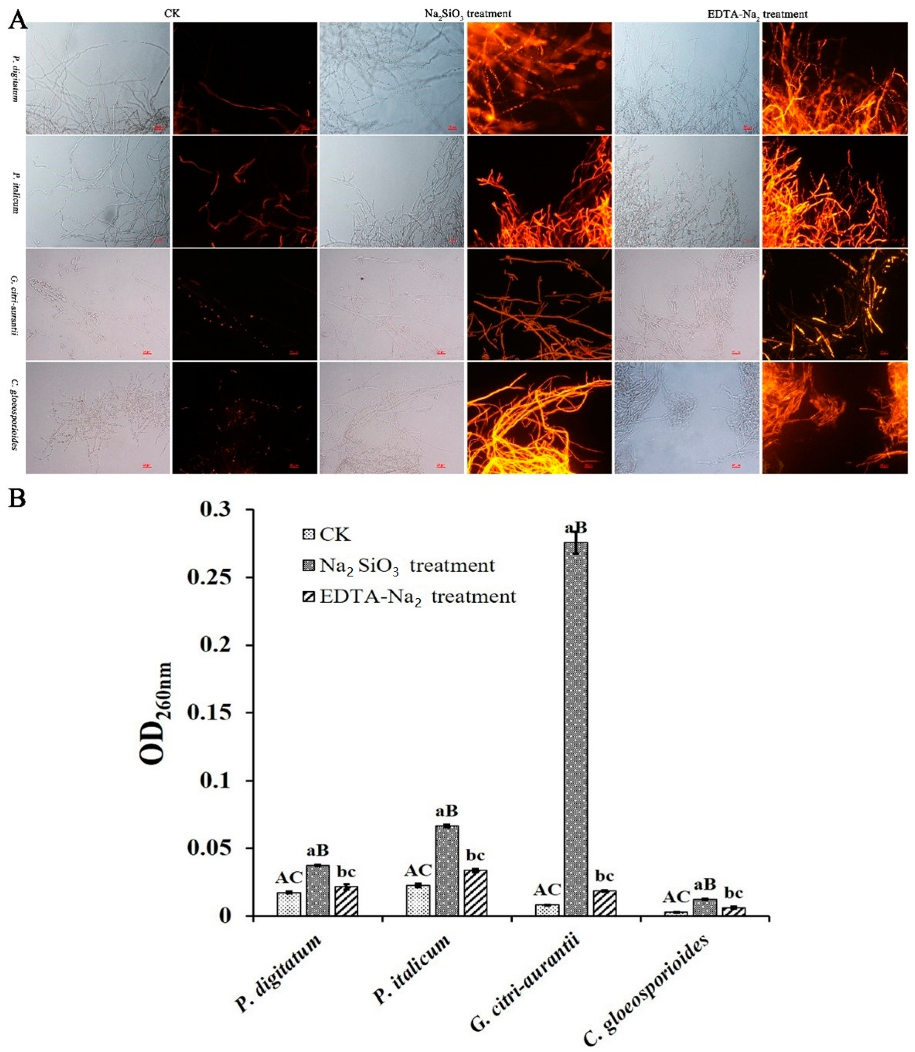
As shown in Figure 6A, Na2SiO3 and EDTA-Na2 treatments led to clear red fluorescence compared with the control, indicating that the two treatments disrupt the hyphal cell membrane of pathogens.

3.7. Effect of Na2SiO3 and EDTA-Na2 on the Nucleic Acid Leakage

The nucleic acid concentration was determined to further confirm the disruption of cell membrane integrity. The nucleic acid concentration increased under Na2SiO3 and EDTA-Na2 treatments. As shown in Figure 6B, the OD260 values of P. digitatum, P. italicum, G. citri-aurantii, and C. gloeosporioides were 0.037, 0.067, 0.28, and 0.01 after Na2SiO3 treatment, and 0.022, 0.034, 0.019, and 0.006 after EDTA-Na2 treatment, respectively, which were all apparently higher than those of the control (0.017, 0.023, 0.008, and 0.003, respectively). These results indicated that Na2SiO3 and EDTA-Na2 disrupted the cell membrane, resulting in a massive leakage of nucleic acid.

3.8. Pathogen Inhibition Ability of Na2SiO3 and EDTA-Na2 In Vivo

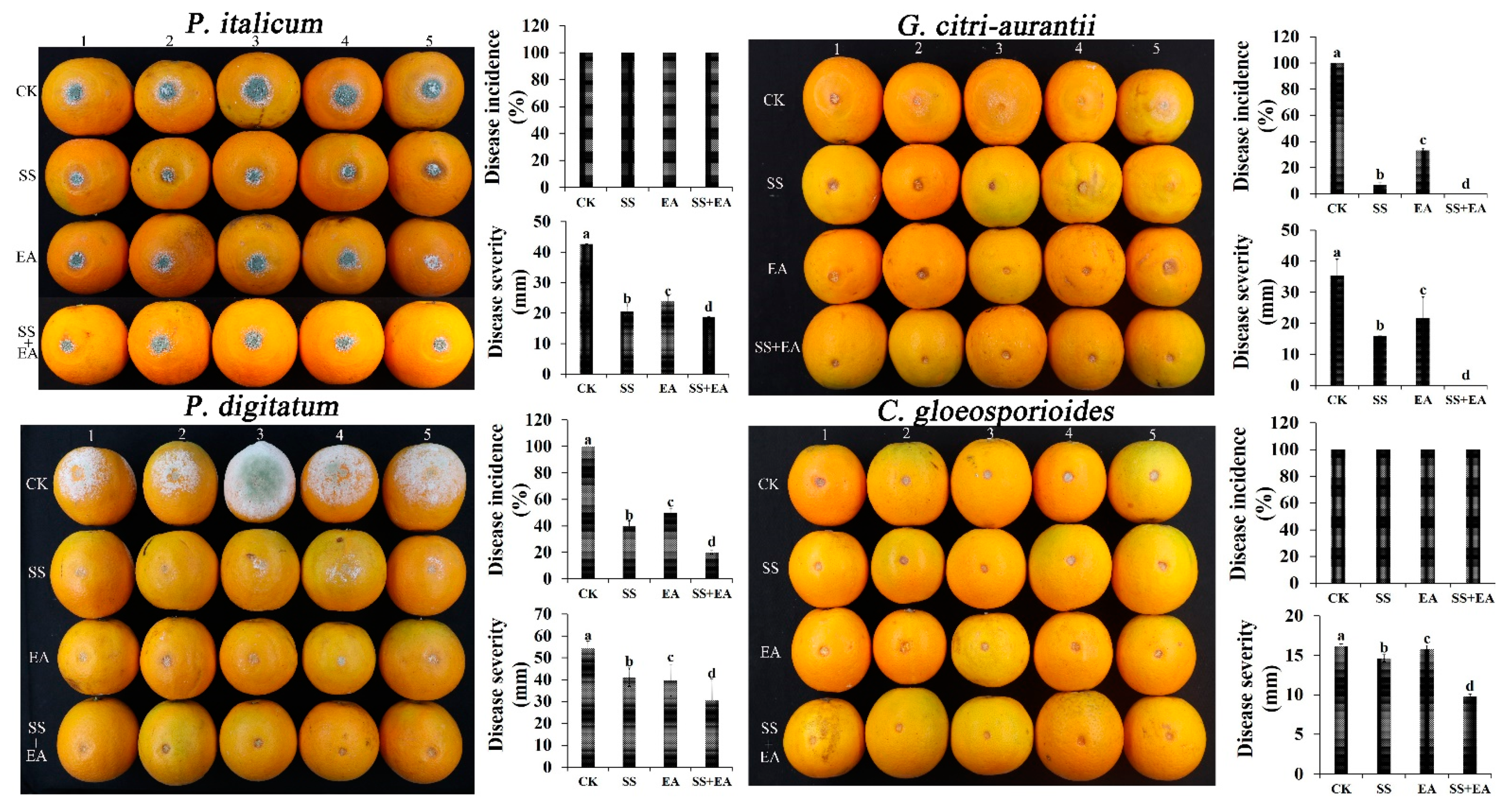
The inhibition ability of Na2SiO3 and EDTA-Na2 on citrus fruit post-harvest pathogens is shown in Figure 7. After treatment with Na2SiO3 and EDTA-Na2, the decay rates caused by P. italicum (40 ± 3.64% and 50 ± 2.83%) and G. citri-aurantii (6.67 ± 2.46% and 33.33 ± 1.63%) were lower than that in the control (100% and 100%). Furthermore, the disease severity of P. digitatum (20.67 ± 3.64 mm and 24.02 ± 2.83 mm), P. italicum (41.08 ± 4.31 mm and 39.76 ± 7.23 mm), G. citri-aurantii (16.00 ± 0.02 mm and 21.82 ± 6.67 mm), and C. gloeosporioides (14.64 ± 0.45 mm and 15.79 ± 0.47 mm) was also dramatically reduced compared with that in the control (42.68 ± 0.64 mm, 54.50 ± 3.10 mm, 35.34 ± 5.45 mm, and 16.19 ± 0.27 mm). Moreover, a mixture of Na2SiO3 and EDTA-Na2 solution reduced the risk of the incidence of P. italicum (20 ± 1.32%) and the disease severity of P. digitatum (18.76 ± 1.32 mm), P. italicum (30.56 ± 9.59 mm), and C. gloeosporioides (9.78 ± 0.30 mm). No fruit decay was observed under treatment of the mixture. These results suggest that Na2SiO3 and EDTA-Na2 treatment can effectively control the four citrus post-harvest diseases.

3.9. Fruit Quality

As shown in Table 3, EC50 Na2SiO3 and EDTA-Na2 treatments resulted in lower weight-loss rates of fruit than the control treatment, and significantly better fruit quality in terms of soluble solid, titratable acid, and VC, suggesting that EC50 Na2SiO3 and EDTA-Na2 treatments had no negative impact on the fruit quality and could be used as preservatives to replace traditional chemicals.

4. Discussion

In previous studies, some GRAS salts were found to be capable of effectively controlling various post-harvest diseases of fruits and vegetables [36]. For example, 2% PS, 2% SB, and 2% PSi could control anthracnose severity [7]; sodium dehydroacetate [37] and polyhexamethylene biguanide (PHMB) [38] could effectively reduce citrus sour rot; and cinnamic acid (CA) could decrease the incidence of blue mold caused by Penicillium italicum in “Orah” mandarin during storage [39]. However, there have been no reports on GRAS salts with broad-spectrum inhibitory effects and their inhibition mechanism on the growth of citrus post-harvest pathogens. The Na2SiO3 and EDTA-Na2 reported in this study may provide more options for the prevention and control of citrus post-harvest diseases.
In fact, Na2SiO3, an inexpensive GRAS salt, has been widely used to control the postharvest diseases of muskmelon and grape [40]. However, there has been no study and application of Na2SiO3 in controlling citrus post-harvest diseases. The in vitro experiments in this study showed that the hyphal growth and spore germination of four pathogens could be almost completely inhibited by Na2SiO3 at a concentration of 1%, which is lower than that of PS, SB, and Psi (2%) used for controlling C. gloeosporioides [7]. Na2SiO3 treatment significantly reduced the disease incidence of blue mold and sour rot in citrus fruit compared with the control, as well as decreased the disease severity of P. italicum, P. digitatum, G. citri-aurantii, and C. gloeosporioides. It has been reported that Na2SiO3 increases the resistance of muskmelon and grape fruit against post-harvest diseases by activating reactive oxygen species metabolism and phenylpropanoid pathway, thereby maintaining the post-harvest quality of fruit [40]. This study demonstrated that Na2SiO3 decreased the incidence and disease severity of citrus postharvest diseases by inhibiting the hyphal growth of pathogens, providing a theoretical foundation for the application of Na2SiO3 to control citrus postharvest diseases as an important alternative.
EDTA-Na2 is a stable food additive used in the food industry, but there are no reports about its application in the control of post-harvest diseases in fruits and vegetables. In this study, we demonstrated for the first time the antifungal ability of EDTA-Na2 to control citrus post-harvest pathogens in vitro. Notably, the combination of EDTA-Na2 and Na2SiO3 decreased the sour rot incidence to 0 in vivo, as well as significantly reduced the lesion diameter of green mold, blue mold, and anthracnose, indicating that the combination has a better inhibitory effect on the pathogen than EDTA-Na2 and Na2SiO3 alone.
The cell wall plays an important role in maintaining the morphology and integrity of the cells [35,36,41]. In this study, the CFW and PI staining results revealed that some spores had no blue fluorescence (Figure 5A) and all hyphae had more visible red fluorescence (Figure 6A) after EDTA-Na2 and Na2SiO3 treatments compared with the control, suggesting that the treatments have nearly no effect on the cell wall, but disrupt the cell membrane integrity of the post-harvest pathogens. In addition, EC50 Na2SiO3 and EDTA-Na2 treatments increased the OD600nm, suggesting that the treatment might severely disrupt the cell membrane integrity and increase the membrane permeability, leading to nucleic acid leakage, which is consistent with the results obtained from PI staining. Moreover, Na2SiO3 treatment resulted in much higher OD600nm values of P. digitatum, P. italicum, G. citri-aurantii, and C. gloeosporioides than EDTA-Na2 treatment, indicating that Na2SiO3 causes further damage to the cell membrane. Notably, Na2SiO3 caused the most significant damage to the cell membrane of G. citri-aurantii (Figure 6B).
LDs are involved in regulating the balance of lipid metabolism by changing the LD size and number [42,43]. Furthermore, in a previous study, a significant increase in LD biogenesis was observed under rapamycin treatment in Magnaporthe grisea, Botrytis cinerea, Fusarium oxysporum, Fusarium annularis, Alternaria alternaria, and Fusarium graminearum [32]. In this study, clearly visible green fluorescence was observed under Na2SiO3 and EDTA-Na2 treatments, indicating that Na2SiO3 and EDTA-Na2 induce the accumulation of LDs of P. digitatum, P. italicum, G. citri-aurantii, and C. gloeosporioides. These results indicate a potential future research direction to study the inhibition mechanism of GRAS salts against citrus post-harvest pathogens.
During citrus post-harvest storage, fruit quality traits, including cumulative weight-loss rate, soluble solid, and titratable acid, are usually determined before and after treatment to measure the applicability of antifungal agents [44]. So far, studies have confirmed that no GRAS salt would impair the fruit quality when used to control post-harvest decay in fruit [35,39]. In our previous study, KCl (K+) was found to be capable of controlling sour rot with decreasing weight-loss rate during a 90-day storage period [30]. Moreover, US FDA and the European Food Safety Authority (EFSA) have exempted the residue detection of GRAS salts in all agricultural commodities [5,24]. Therefore, considering our in vitro and in vivo results, EDTA-Na2 + Na2SiO3 treatment might present an effective approach to control the post-harvest diseases of citrus fruit.

5. Conclusions

The two GRAS salts, Na2SiO3 and EDTA-Na2, can alter the microstructure of spores and hyphal cell membrane and increase the cell membrane permeability, resulting in the leakage of nucleic acid and synergistic inhibition on the hyphal growth and spore germination of P. digitatum, P. italicum, G. citri-aurantii, and C. gloeosporioides. Moreover, the EC50 Na2SiO3 and EDTA-Na2 treatment conspicuously induced LD accumulation and reduced both disease incidence and disease severity without posing a negative impact on the fruit quality. These findings indicate Na2SiO3 + EDTA-Na2 treatment as a promising approach in controlling the post-harvest diseases of citrus fruit.

Author Contributions

J.Z.: Conceptualization, Methodology, Data analysis, Writing—original draft. Y.W.: Experiment. Q.L.: Experiment. S.L.: Experiment. H.P.: Experiment. Y.C.: Project administration. C.L.: Project administration, Supervision. All authors have read and agreed to the published version of the manuscript.

Funding

This study was financially supported by the National Natural Science Foundation of China (Grant No. 32172255 & 31972122), HZAU-AGIS Cooperation Fund (No. SZYJY2023001) and China Agriculture Research System of MOF and MARA and the Key R&D Program of Hubei Province (NO. 2021BBA095).

Data Availability Statement

Data is contained within the article.

Conflicts of Interest

The authors declare that they have no known competing financial interests.

References

  1. Sun, L.; Nasrullah, K.F.; Nie, Z.; Wang, P.; Xu, J. Citrus genetic engineering for disease resistance: Past, present and future. Int. J. Mol. Sci. 2019, 20, 5256. [Google Scholar] [CrossRef] [PubMed] [Green Version]
  2. Wang, S.P.; Ruan, C.Q.; Yi, L.H.; Deng, L.L.; Yao, S.X.; Zeng, K.F. Biocontrol ability and action mechanism of Metschnikowia citriensis against Geotrichum citri-aurantii causing sour rot of postharvest citrus fruit. Food Microbiol. 2020, 87, 103375. [Google Scholar] [CrossRef] [PubMed]
  3. Xue, Y.H.; Li, H.; Li, H.; Liu, C.; Luo, H.J.; Liu, C.X.; Chen, J.F.; Zou, K.; Liu, S.H. Metabolite profiling reveals comprehensive effects of Chaetomium globosum on citrus preservation. Food Chem. 2022, 369, 130959. [Google Scholar] [CrossRef] [PubMed]
  4. Chen, O.; Deng, L.; Ruan, C.; Yi, L.; Zeng, K. Pichia galeiformis induces resistance in postharvest citrus by activating the phenylpropanoid biosynthesis pathway. J. Agric. Food Chem. 2021, 69, 2619–2631. [Google Scholar] [CrossRef]
  5. Palou, L. Postharvest treatments with gras salts to control fresh fruit decay. Horticulturae 2018, 4, 46. [Google Scholar] [CrossRef] [Green Version]
  6. Wang, W.; Silva, D.; Moslemi, A.; Edwards, J.; Taylor, P. Colletotrichum species causing anthracnose of citrus in australia. J. Fungi 2021, 7, 47. [Google Scholar] [CrossRef]
  7. Martínez-Blay, V.; Pérez-Gago, M.B.; Fuente, B.; Carbó, R.; Palou, L. Edible coatings formulated with antifungal gras salts to control citrus anthracnose caused by colletotrichum gloeosporioides and preserve postharvest fruit quality. Coatings 2020, 10, 730. [Google Scholar] [CrossRef]
  8. Palou, L. Penicillium digitatum, Penicillium italicum (green mold, blue mold). In Postharvest Decay; Academic Press: Cambridge, MA, USA, 2014; pp. 25–102. [Google Scholar] [CrossRef]
  9. Liu, S.; Wang, W.; Deng, L.; Ming, J.; Yao, S.; Zeng, K. Control of sour rot in citrus fruit by three insect antimicrobial peptides. Postharvest Biol. Technol. 2019, 149, 200–208. [Google Scholar] [CrossRef]
  10. McKay, A.; Förster, H.; Adaskaveg, J. Efficacy and application strategies for propiconazole as a new postharvest fungicide for managing sour rot and green mold of citrus fruit. Plant Dis. 2012, 96, 235–242. [Google Scholar] [CrossRef] [Green Version]
  11. Soto-Muoz, L.; Taberner, V.; Fuente, B.; Jerbi, N.; Palou, L. Curative activity of postharvest GRAS salt treatments to control citrus sour rot caused by Geotrichum citri-aurantii. Int. J. Food Microbiol. 2020, 335, 108860. [Google Scholar] [CrossRef]
  12. Pereyra, M.M.; Diaz, M.A.; Soliz-Santander, F.F.; Poehlein, A.; Meinhardt, F.; Daniel, R.; Dib, J.R. Screening methods for isolation of biocontrol epiphytic yeasts against Penicillium digitatum in lemons. J. Fungi 2021, 7, 166. [Google Scholar] [CrossRef] [PubMed]
  13. Chen, O.; Hong, Y.; Ma, J.; Deng, L.; Zeng, K. Screening lactic acid bacteria from pickle and cured meat as biocontrol agents of Penicillium digitatum on citrus fruit. Biol. Control 2021, 158, 104606. [Google Scholar] [CrossRef]
  14. Klein, M.N.; Kupper, K.C. Biofilm production by aureobasidium pullulans improves biocontrol against sour rot in citrus. Food Microbiol. 2018, 69, 1–10. [Google Scholar] [CrossRef] [PubMed] [Green Version]
  15. Chen, O.; Yi, L.; Deng, L.; Ruan, C.; Zeng, K. Screening antagonistic yeasts against citrus green mold and the possible biocontrol mechanisms of Pichia galeiformis (BAF03). J. Sci. Food Agric. 2020, 100, 3812–3821. [Google Scholar] [CrossRef] [PubMed]
  16. Wang, Y.; Yin, C.; Cheng, X.; Li, G.; Zhu, X. β-cyclodextrin inclusion complex containing litsea cubeba essential oil: Preparation, optimization, physicochemical, and antifungal characterization. Coatings 2020, 10, 850. [Google Scholar] [CrossRef]
  17. Gan, Z.; Huang, J.; Chen, J.; Nisar, M.F.; Qi, W. Synthesis and antifungal activities of cinnamaldehyde derivatives against penicillium digitatum causing citrus green mold. J. Food Qual. 2020, 2020, 8898692. [Google Scholar] [CrossRef]
  18. Serna-Escolano, V.; Serrano, M.; Valero, D.; Rodríguez-López, M.I.; Gabaldón, J.A.; Castillo, S.; Guillén, F.; Zapata, P.J.; Martínez-Romero, D. Effect of thymol and carvacrol encapsulated in hp-Β-cyclodextrin by two inclusion methods against Geotrichum citri-aurantii. J. Food Sci. 2019, 84, 1513–1521. [Google Scholar] [CrossRef]
  19. Lin, Y.; Fan, L.; He, J.; Wang, Z.; Li, Z. Anthocyanins contribute to fruit defense against postharvest green mold. Postharvest Biol. Technol. 2021, 181, 111661. [Google Scholar] [CrossRef]
  20. Karim, H.; Boubaker, H.; Askarne, L.; Talibi, I.; Msanda, F.; Boudyach, E.H.; Saadi, B.; Ait, B.A.A. Antifungal properties of organic extracts of eight cistus l. species against postharvest citrus sour rot. Lett. Appl. Microbiol. 2016, 62, 16–22. [Google Scholar] [CrossRef] [Green Version]
  21. Bazioli, J.M.; Belinato, J.R.; Costa, J.H.; Akiyama, D.Y.; Fill, T.P. Biological control of citrus postharvest phytopathogens. Toxins 2019, 11, 460. [Google Scholar] [CrossRef] [Green Version]
  22. Palou, L.; Smilanick, J.L.; Droby, S. Alternatives to conventional fungicides for the control of citrus postharvest green and blue molds. Stewart Postharvest Rev. 2008, 4, 1–16. [Google Scholar] [CrossRef]
  23. Palou, L.; Ali, A.; Fallik, E.; Romanazzi, G. Gras, plant- and animal-derived compounds as alternatives to conventional fungicides for the control of postharvest diseases of fresh horticultural produce. Postharvest Biol. Technol. 2016, 122, 41–52. [Google Scholar] [CrossRef]
  24. D’Aquino, S.; Palma, A. Reducing or Replacing Conventional Postharvest Fungicides with Low Toxicity Acids and Salts. In Postharvest Pathology of Fresh Horticultural Produce; CRC Press, Taylor and Francis Group: Boca Raton, FL, USA, 2020; pp. 595–632. [Google Scholar] [CrossRef]
  25. Khamis, Y.; Hussien, A. Electrolysed water and salt solutions can reduce green and blue molds while maintain the quality properties of ‘valencia’ late oranges. Postharvest Biol. Technol. 2019, 159, 111025. [Google Scholar] [CrossRef]
  26. Moscoso-Ramírez, P.A.; Palou, L. Preventive and curative activity of postharvest potassium silicate treatments to control green and blue molds on orange fruit. Eur. J. Plant Pathol. 2014, 138, 721–732. [Google Scholar] [CrossRef]
  27. Montesinos-Herrero, C.; Moscoso-Ramírez, P.A.; Palou, L. Evaluation of sodium benzoate and other food additives for the control of citrus postharvest green and blue molds. Postharvest Biol. Technol. 2016, 115, 72–80. [Google Scholar] [CrossRef]
  28. Moscoso-Ramírez, P.A.; Montesinos-Herrero, C.; Palou, L. Characterization of postharvest treatments with sodium methylparaben to control citrus green and blue molds. Postharvest Biol. Technol. 2013, 77, 128–137. [Google Scholar] [CrossRef]
  29. Smilanick, J.L.; Mansour, M.F.; Gabler, F.M.; Sorenson, D. Control of citrus postharvest green mold and sour rot by potassium sorbate combined with heat and fungicides. Postharvest Biol. Technol. 2008, 47, 226–238. [Google Scholar] [CrossRef]
  30. Zhao, J.; Liu, Q.Y.; Wang, Y.Q.; Lu, X.J.; Pan, H.; Long, C.A. Curative activity of KCl treatments to control citrus sour rot. LWT-Food Sci. Technol. 2022, 168, 113853. [Google Scholar] [CrossRef]
  31. Zhang, H.; Huangfu, H.P.; Wang, X.; Zhao, S.S.; Tan, Z. Antibacterial activity of lactic acid producing leuconostoc mesenteroides qz1178 against pathogenic Gallibacterium anatis. Front. Vet. Sci. 2021, 8, 630294. [Google Scholar] [CrossRef]
  32. Liu, N.; Yun, Y.Z.; Yin, Y.N.; Matthias, H.; Ma, Z.H. Lipid droplet biogenesis regulated by the fgnem1/spo7-fgpah1 phosphatase cascade plays critical roles in fungal development and virulence in Fusarium graminearum. New Phytol. 2019, 223, 412–429. [Google Scholar] [CrossRef]
  33. Jurick, W.M.; Macarisin, O.; Gaskins, V.L.; Janisiewicz, W.J.; Peter, K.A.; Cox, K.D. Baseline sensitivity of Penicillium spp. to difenoconazole. Plant Dis. 2019, 103, 331–337. [Google Scholar] [CrossRef] [Green Version]
  34. Cheng, X.; Yang, Y.; Zhu, X.; Yuan, P.; Shan, Y. Inhibitory mechanisms of cinnamic acid on the growth of Geotrichum citri-aurantii. Food Control 2021, 131, 108459. [Google Scholar] [CrossRef]
  35. Liu, S.; Du, Y.; Zhang, D.; Yang, F.; He, X.; Long, C.A. Aluminum sulfate inhibits green mold by inducing chitinase activity of Penicillium digitatum and enzyme activity of citrus fruit. Food Control 2022, 136, 108854. [Google Scholar] [CrossRef]
  36. Li, X.; Wang, W.; Liu, S.; Ruan, C.; Zeng, K. Effects of the peptide h-ooww-nh2 and its derived lipopeptide c12-ooww-nh2 on controlling of citrus postharvest green mold. Postharvest Biol. Technol. 2019, 158, 110979. [Google Scholar] [CrossRef]
  37. Li, L.; Tang, X.; Ouyang, Q.; Tao, N.G. Combination of sodium dehydroacetate and sodium silicate reduces sour rot of citrus fruit. Postharvest Biol. Technol. 2019, 151, 19–25. [Google Scholar] [CrossRef]
  38. Wu, Y.; Duan, X.; Jing, G.; OuYang, Q.; Tao, N.G. Cinnamaldehyde inhibits the mycelial growth of Geotrichum citri-aurantii and induces defense responses against sour rot in citrus fruit. Postharvest Biol. Technol. 2017, 129, 23–28. [Google Scholar] [CrossRef]
  39. Li, J.; Li, H.; Ji, S.; Chen, T.; Tian, S.; Qin, G.Z. Enhancement of biocontrol efficacy of Cryptococcus laurentii by cinnamic acid against Penicillium italicum in citrus fruit. Postharvest Biol. Technol. 2019, 149, 42–49. [Google Scholar] [CrossRef]
  40. Wang, Y.F.; Bi, Y.; Ren, Y.L.; Wang, Y.; Fan, C.F.; Li, D.Q.; Yang, Z.M. Control of postharvest diseases and potentiation of reactive oxygen species metabolism in muskmelon (Cucumis melo L.) fruits treated by sodium silicate. Sci. Agric. Sin. 2012, 45, 2242–2248. Available online: https://211.155.251.135:81/jwk_zgnykx/en/y2012/v45/i11/2242 (accessed on 1 May 2023).
  41. Ouyang, Q.; Duan, X.; Li, L.; Tao, N. Cinnamaldehyde exerts its antifungal activity by disrupting the cell wall integrity of Geotrichum citri-aurantii. Front. Microbiol. 2019, 10, 55. [Google Scholar] [CrossRef] [Green Version]
  42. Xu, C.; Fan, J. Links between autophagy and lipid droplet dynamics. J. Exp. Bot. 2022, 73, 2848–2858. [Google Scholar] [CrossRef]
  43. Robenek, H.; Hofnagel, O.; Buers, I.; Robenek, M.J.; Severs, N.J. Adipophilin-enriched domains in the er membrane are sites of lipid droplet biogenesis. J. Cell Sci. 2006, 119, 4215–4224. [Google Scholar] [CrossRef] [PubMed] [Green Version]
  44. Duan, X.; Ouyang, Q.; Jing, G.; Tao, N. Effect of sodium dehydroacetate on the development of sour rot on satsuma mandarin. Food Control 2016, 65, 8–13. [Google Scholar] [CrossRef]
Figure 1. Technical route of this study. SS: Na2SiO3, EA: EDTA-Na2, LDs: lipid droplets; EC50: the median effective concentration.
Foods 12 02368 g001
Figure 2. Effects of different concentrations of Na2SiO3 ((A); 0, 0.02%, 0.04%, 0.06%, 0.08%, and 0.16%) and EDTA-Na2 ((B); 0, 0.03%, 0.06%, 0.12%, 0.24%, and 0.48%) on the hyphal growth of P. digitatum, P. italicum, G. citri-aurantii, and C. gloeosporioides.
Foods 12 02368 g002
Figure 3. Number of P. digitatum, P. italicum, G. citri-aurantii, and C. gloeosporioides spores under Na2SiO3 and EDTA-Na2 treatment. The scale bar in all photos is 50 µm. (Spores (red arrow), mycelium (blue arrow)).
Foods 12 02368 g003
Figure 4. Effects of EC50 Na2SiO3 and EDTA-Na2 treatments on the spore germination of P. digitatum, P. italicum, G. citri-aurantii, and C. gloeosporioides. (A) Inhibition efficacy of EC50 Na2SiO3 and EDTA-Na2 on the spore germination of P. digitatum, P. italicum, G. citri-aurantii, and C. gloeosporioides; (B) Percentage of spore germination. The scale bar of B is 10 µm, and that of other photos is 100 µm. Error bar means the standard error. Different lower-case letters are significantly different (p < 0.05), different upper-case letters are significantly different (p < 0.01).
Foods 12 02368 g004
Figure 5. Fluorescence microscopy images of EC50 Na2SiO3 and EDTA-Na2 on spores of P. digitatum, P. italicum, G. citri-aurantii, and C. gloeosporioides. (A) Spores under bright field and propidium iodide (CFW); (B) LDs under bright field and propidium iodide (Nile Red). The scale bar in the photos is 20 µm.
Foods 12 02368 g005
Figure 6. Effect of EC50 Na2SiO3 and EDTA-Na2 on hyphal cell membrane integrity of P. digitatum, P. italicum, G. citri-aurantii, and C. gloeosporioides. (A) Fluorescence microscopy image of hyphae under bright field and propidium iodide (PI), the scale bar in the photos is 25 µm; (B) Effect of cinnamic acid on nucleic acid leakage. The results were reported by the average value. Error bar means the standard error. Different lower-case letters are significantly different (p < 0.05), different upper-case letters are significantly different (p < 0.01).
Foods 12 02368 g006
Figure 7. Disease incidence and disease severity of P. digitatum, P. italicum, G. citri-aurantii and C. gloeosporioides under EC50 Na2SiO3 and EDTA-Na2 treatments. SS: Na2SiO3, EA: EDTA-Na2, SS + SA: Na2SiO3 + EDTA-Na2. Each group contained five fruit. The results are reported by the average value. Error bar means the standard error. Different lower-case letters are significantly different (p < 0.05).
Foods 12 02368 g007
Table 1. Inhibition rates of 17 GRAS salts (1%) on P. digitatum, P. italicum, G. citri-aurantii, and C. gloeosporioides.
NumberGRAS Salts
(1%)
P. digitatum (B3, %)P. italicum (P44, %)G.citri-aurantii (AY-1, %)C. gloeosporioides (NF28, %)
1Sodium silicate100100100100
2Ethylenediaminetetraacetic acid disodium salt87.90 ± 0.19100100100
3Sodium benzoate10086.04 ± 0.45100100
4Sodium diacetate10088.09 ± 1.010100
5Succinic acid079.08 ± 0.5289.47 ± 0.4785.09 ± 0.19
6Maleic acid079.66 ± 0.9090.36 ± 0.4284.61 ± 1.03
7Sodium carbonate10010000
8Sodium sesquicarbonate0010089.58 ± 0.69
9Sodium stearyl lactate027.18 ± 1.6618.63 ± 2.490
10Adipic acid0086.34 ± 0.38100
11Aconitic acid0089.21 ± 0.6683.46 ± 1.09
12Fumaric acid001000
13Calcium glycerophosphate0007.01 ± 1.78
14Aspartame020.33 ± 3.7000
15DL-Malic acid0089.69 ± 0.330
16Citric acid038.38 ± 1.7800
17Ferrous gluconate hydrate091.26 ± 0.2800
Note: The results are reported as the average value ± standard error.
Table 2. EC50 of Na2SiO3 and EDTA-Na2 on the hyphal growth of G.citri-aurantii, P. digitatum, P. italicum, and C. gloeosporioides.
GRAS SaltsCitrus Postharvest DiseaseVirulence Equation (Y = ax + b)EC50 (%)EC95 (%)
Sodium silicateG. citri-aurantiiy = 3.4498x + 16.2160.060.17
P. digitatumy = 3.6899x + 17.21080.050.14
P. italicumy = 4.1557x + 18.05280.070.14
C. gloeosporioidesy = 1.0819x + 7.26170.080.19
Ethylenediaminetetraacetic acid disodium saltG.citri-aurantiiy = 2.9653x + 13.79190.110.39
P. digitatumy = 1.7099x + 10.27810.080.75
P. italicumy = 1.9901x + 9.58040.53.35
C. gloeosporioidesy = 2.0019x + 11.33640.070.45
Table 3. Quality of fruit treated with Na2SiO3 and EDTA-Na2 during the storage of 30, 60, 80, and 90 d.
TimeSamplesWeight Loss Rate (%)Soluble SolidTitratable AcidVC (mg/100 g)
30 dCK0.64 ± 0.06 AC14.00 ± 0.00 AC 0.83 ± 0.00 AC0.66 ± 0.01 AC
Na2SiO30.56 ± 0.04 aB13.50 ± 0.00 aB0.98 ± 0.02 aB0.64 ± 0.04 AB
EDTA-Na20.52 ± 0.03 bc 13.43 ± 0.06 Bc 0.76 ± 0.01 bc0.65 ± 0.02 Bc
60 dCK2.41 ± 0.20 AC13.67 ± 0.06 AC0.84 ± 0.06 AC0.62 ± 0.01 AC
Na2SiO32.29 ± 0.17 aB15.20 ± 0.10 aB 1.06 ± 0.18 AB0.74 ± 0.05 aB
EDTA-Na22.13 ± 0.15 bc13.90 ± 0.00 bc0.79 ± 0.04 bC0.67 ± 0.01 bc
80 dCK4.99 ± 0.42 AC13.45 ± 0.25 AC0.75 ± 0.07 AC0.60 ± 0.02 AC
Na2SiO34.88 ± 0.39 aB14.67 ± 0.06 aB0.85 ± 0.03 aB0.70 ± 0.01 aB
EDTA-Na24.49 ± 0.33 bc13.57 ± 0.21 bC0.67 ± 0.01 bC0.64 ± 0.01 bc
90 dCK6.19 ± 0.53 AC14.07 ± 0.06 AC0.77 ± 0.03 AC0.58 ± 0.01 AC
Na2SiO36.10 ± 0.19 AB14.57 ± 0.06 aB0.89 ± 0.01 aB0.69 ± 0.01 aB
EDTA-Na25.65 ± 0.43 bc14.17 ± 0.15 bC0.75 ± 0.01 bc0.62 ± 0.01 bc
Note: CK: control group; Na2SiO3: Na2SiO3 treatment group; EDTA-Na2: EDTA-Na2 treatment group. Values represent the mean ± standard deviations of three replicates. Different lower-case letters are significantly different (p < 0.05), different upper-case letters are significantly different (p < 0.01).
Disclaimer/Publisher’s Note: The statements, opinions and data contained in all publications are solely those of the individual author(s) and contributor(s) and not of MDPI and/or the editor(s). MDPI and/or the editor(s) disclaim responsibility for any injury to people or property resulting from any ideas, methods, instructions or products referred to in the content.