Article

The Quantification of IgG Specific to α-Gal Could Be Used as a Risk Marker for Suffering Mammalian Meat Allergy

by
Alejandro Joral
1,
Nahikari Azketa
2,
Patricia Sanchez
2,
Ainara Vélez-del-Burgo
2,
María-Ascensión Aranzabal-Soto
3,
Susana Lizarza
1,
Jorge Martínez
2 and
Idoia Postigo
2,*
1
Servicio de Alergología, Hospital Universitario Donostia, 20014 San Sebastián, Spain
2
Parasitology and Allergy Research Group, Lascaray Research Institute, Department of Immunology, Microbiology, and Parasitology, Faculty of Pharmacy, The University of Basque Country, 01006 Vitoria, Spain
3
OSI Goierri Alto Urola, 20700 Zumárraga, Spain
*
Author to whom correspondence should be addressed.
Foods 2022, 11(3), 466; https://doi.org/10.3390/foods11030466
Submission received: 8 December 2021 / Revised: 1 February 2022 / Accepted: 2 February 2022 / Published: 4 February 2022

Abstract

:
The alpha-Gal Syndrome is a delayed meat allergy characterized by the presence of sIgE against α-Gal epitope. It is known that the α-Gal present in tick saliva induces the sensitization to this epitope ending in the production of sIgG and sIgE to α-Gal. It could be considered that the more times a person is bitten by tick species, the higher the probability of making the switch from sIgG to sIgE to α-Gal and developing allergy, but it is no clear when the switch occurs. To determine the likelihood that a subject bitten by ticks but without AGS be at risk of developing this allergy, we quantified the levels of sIgG to α-Gal by an automated system (ImmunoCap). To stablish a cut-off value for sIgG to α-Gal, a receiving operating curve (ROC) was constructed. The statistical analysis demonstrated that the risk of suffering AGS in individuals bitten by ticks was 35% when the sIgG to α-Gal was greater than or equal to 40 µg/mL. Our data indicate that the sIgG values against α-Gal could be used as a prognostic marker for developing mammalian meat allergy.

1. Introduction

Mammalian meat allergy, also referred to as alpha-Gal syndrome (AGS), is a special kind of delayed food allergy to the carbohydrate moiety galactosyl-α-1,3-galactose (α-Gal) [1,2]. Clinically, α-Gal allergy is characterized by reactions to mammalian meat and innards, including beef, pork, and lamb, which occur 3–6 h following meat consumption [3,4,5,6]. The symptoms of this allergy are variable ranging from abdominal pain and diarrhea to urticaria episodes and anaphylaxis [7]. The α-Gal carbohydrate is present in tissues from several mammalians except humans [8,9] and it has been demonstrated in the salivary glands of several tick species [10,11,12]. It is assumed that the main cause of sensitization in AGS is the recurrent tick bite [13,14] and, the most prevalent tick species in each continent could be responsible for this allergy [15,16].
In Europe, the frequency of positivity of sIgE to α-Gal has been reported to be 5.5% in Denmark [17], 15.7% in a representative Spanish cohort [18] and 24.7% in a rural area in northeast Italy [19]. In the United States (U.S.), more than 5000 cases have been described [20]. In addition, new cases that are not directly related to meat intake are being described, e.g., immediate allergic reactions in α-Gal positive patients to common vaccines, which contain mammalian-derived gelatin [21].
The knowledge about this syndrome began in the southeastern of the U.S. with the use of cetuximab, a mouse–human chimeric antibody (Ab) for the treatment of cancer [22]. Although the clinical trials demonstrated a low risk of allergy to the drug, these patients from a specific U.S. geographic area presented severe drug allergic reactions, higher than the expected [23]. Further investigations demonstrated that the patients who experienced hypersensitivity reactions had pre-existing IgE antibodies (Abs) that bound to the α-Gal carbohydrate moiety found in the murine portion of the chimeric Ab [23]. Therefore, they should have been sensitized in some way. At the same time and, in the same southern region of the U.S., physicians perceived an increase in cases of healthy individuals experiencing urticaria, angioedema or anaphylaxis several hours after consuming red meat [24]. In many cases, the individuals who experienced these hypersensitivity responses had a history of consuming meat for decades with no adverse reaction [25]. Further work revealed that α-Gal-specific IgE contributed significantly to the allergic response to red meat in these individuals [26]. Later on, the research groups realized that both cetuximab-induced hypersensitivity and meat allergy cases were restricted to the same geographical area where the lone star tick was prevalent [26]. In Australia, the investigations revealed that a large number of patients with meat allergy also had a history of tick bites [27] and, in 2007, the first report of the capacity of ticks to induce mammalian red meat allergy was published [28]. Since then, this syndrome is becoming a global problem and an increasing number of cases are being reported from almost all continents [29,30,31,32,33].
Old World monkeys, apes, and humans do not express the α-Gal containing oligosaccharide because, in humans, the α-1,3-galactosyltransferase (α-1,3GT) is expressed in an enzymatically inactive form [34]. That is the reason why all immunocompetent humans can express in a “natural” way anti-α-Gal Abs [34] against the α-Gal epitopes. It is though that the antigenic source for developing the anti-α-Gal Abs is the continuous exposition to this moiety present in the outer membrane of bacteria from the intestinal microbiome [35]. Thus, when foreign α-Gal antigens enter the body, the anti-α-Gal B cells are stimulated and can produce large amounts of high-affinity anti-α-Gal Abs [36]. It is estimated that one percent of human-circulating B-lymphocytes are capable of producing the natural anti-α-Gal Abs, mainly IgM and IgG isotypes [37].
The AGS-patients are characterized by elevated values of specific IgE and IgG Abs against α-Gal epitope [38,39,40,41] and, probably, most AGS-patients who had tolerated red meat for many years could have sensitized to α-Gal through tick bites [40]. The α-Gal epitope has been identified in the salivary glands [42] and cement [43] of several tick species, including the most prevalent hard tick in Europe: Ixodes ricinus [10,12]. The mechanism by which tick bites sensitize individuals to this epitope and, as a result, induce the meat allergy is not clear yet. Given that α-Gal exposure alone does not induce an IgE response [44], there must be a sensitization process, maybe by means α-Gal antigen present on tick salivary proteins or through the presence of immunomodulatory factors in tick saliva, such as prostaglandin E2 (PGE2) [45]. Maybe, the continuous exposition to tick bites could induced in exposed individuals the switch of pre-existing anti-α-Gal-IgG Abs to sIgE to α-Gal helped by immunomodulatory agents, such as PGE2.
There are several works on the Ab titers of the different Ig isotypes and IgG subclasses expressed in both AGS patients and healthy donors, including their relationship to the blood group of the individuals [46,47] and, the published data on the concentration of IgG to α-Gal in normal human serum are controversial depending on the methodology used for quantification. [37,48,49,50]
The objective of our study was to quantify, by means of an automated and standardized method, the levels of sIgG to α-Gal in four different groups of adult subjects: clinical diagnosed AGS-patients, atopic subjects, healthy donors, and in a group of subjects bitten by ticks but who had not developed AGS. The aim was to stablish a sIgG to α-Gal cut-off value. Before, we analyze the likelihood that a subject bitten by ticks, with sIgG to α-Gal but without AGS, could be at risk of developing this allergy.

2. Materials and Methods

2.1. Study Design and Ethical Approval

The study protocol was approved by the Ethics Committee of the Hospital BioDonosti- Osakidetza, in accordance with The Code of Ethics of the World Medical Association, Declaration of Helsinki.
Two hundred people living in the Basque Country community (North of Spain) were contacted through social networks (Facebook) to participate in the study and, fifty subjects voluntarily accepted to participate. Forty-six people from the fifty volunteers were recruited through a survey for this cross-sectional study. The age range was 20–60 years. All selected individuals declared to be bitten by ticks, but none reported clinical AGS-related symptoms or had been clinically diagnosed with AGS. This group was labeled as the risk population group. The community health centers give each individual a medical flyer for blood collection and written informed consent was obtained from all of the individuals. The frozen serum samples were sent to the Parasitology and Allergy Laboratory-Lascaray Research Center-UPV/EHU for analysis. The basic epidemiological information, such as age, gender, habitat, presence of allergies and some items about the meat consumption behavior or the contact with animals (dog, cat) of each subject in the risk-population group, were recorded by survey.
Sera from 15 adult (20–60 years) patients clinically diagnosed with AGS were included in the study as positive control group. The AGS diagnostic was based on case history, positive skin prick tests, and the presence of α-Gal-specific IgE in the serum of these patients in concentrations higher than 0.35 kUA/L. All subjects mentioned being bitten by ticks. This group was labeled as AGS patients.
Sera from 108 healthy individuals who did not report any allergic symptoms and showed no allergen-specific IgE Ab (ImmunoCap ISAC. Thermo Fisher Scientific, Waltham, MA, USA) were selected from our serum collection (National Register of Biobank Serum Collections, code C.0002774; Instituto de Salud Carlos III, Ministry of Economy and Competitiveness/Lascaray Research Center, University of the Basque Country, Vitoria, Spain). All the samples belonged to adult people living in urban areas. None of them mentioned tick bites. This group was considered as the healthy population control group.
Finally, 64 sample sera from atopic adult subjects constituted the allergic control group. None of them mentioned being bitten by ticks and all of them came from urban areas.

2.2. Determination of sIgE Antibodies against Different Allergens

To assess the profile of sensitization in the risk and atopic populations, the sIgE against a panel of 112 allergens, including grass pollen, tree pollen, mites, fungi, food allergens, animal dander, insect venom and CCDs, was measured by Immuno Solid-phase Allergen Chip (ImmunoCAP ISAC 112. Phadia AB, Uppsala, Sweden) following the manufacturer’s instructions [51]. This technique is a multiplex assay based on component-resolved diagnosis [52]. The allergen components are spotted in triplets and covalently immobilized on a polymer-coated slide. Briefly, 30 μL of serum samples was added to each microarray and incubated at room temperature for 120 min. After washing, 30 μL fluorescence-labeled antihuman IgE Abs were added. Following incubation for 30 min, unbound labeled Abs were removed by washing, and fluorescence was measured with a laser scanner. The results were evaluated using Phadia Microarray Image Analysis (MIA) software. ImmunoCAP ISAC is a semi-quantitative test and results are reported in ISAC Standardized Units (ISU) giving indications of specific IgE Ab levels within a measuring range of 0.3–100 ISU-E. The ISU-E are standardized to ImmunoCAP Specific IgE units [51].
In this study, all samples in the range of 0.3–100 ISU-E were considered positives and the results were expressed as the percentage of positives to each allergen in the studied populations.

2.3. Quantification of sIgE and sIgG Specific Antibodies against the α-Gal Epitope

In all samples, the sIgE and sIgG Ab levels against α-Gal epitope were quantified by fluoro-enzyme-immunoassay (FEIA) using an automated system (ImmunoCap, Thermo Fisher Scientific), according to the manufacturer’s instructions. The cut-off value stablished by the manufacturer for the sIgE Ab was 0.35 kUA/L.
A receiver operating characteristic (ROC) curve was constructed to establish the sIgG cut-off value to α-Gal in order to differentiate the patients bitten by ticks who developed AGS from the control groups (atopic subjects and healthy individuals) not bitten by ticks [53].

2.4. Statistical Analysis

Data were entered in Graph Pad Prism 7.0 for statistical analysis. Statistical differences among populations were determined using the no parametric Kruskal–Wallis test and Dunn’s multiple comparisons. Differences were considered statistically significant for p < 0.05 (95% CI).
A ROC curve was plotted using the GraphPad Prism v 7.0 software and the area under the curve was calculated to quantify the accuracy of the test.
To assess the association between the likelihood of suffering AGS and the sIgG levels to α-Gal, the contingency analysis was performed using the Fisher exact test. A p ≤ 0.05 was considered statistically significant. The relative and attributable risk were calculated by Koopman asymptomatic score and odds ratio by the Baptista–Pike method with 95% CI.

3. Results

3.1. Clinical and Demographic Data of Participants

3.1.1. AGS Patients

The 15 AGS patients had experienced meat-induced symptoms occurring 3–7 h after ingestion that comprised anaphylaxis (53.3%), acute urticaria (13.3%), and recurrent urticaria (33.3%). The IgE specific to α-Gal ranged from 12.3 to >100 kUA/L, the average being 62.5 kUA/L (Table 1).

3.1.2. Risk-Population Group

The 78% of the risk population subjects declared to be daily meat consumers (mainly beef and/or pork) and the 9% declared suffering from some intolerance directly related with meat consumption. The basic epidemiological information of participants in risk-population group (n = 46) are given in Table 2. A total of 83% of the participants live in an urban environment and 85% declared animal contact (dogs and cats). A total of 30% declared suffering from chronic diseases, such as Lyme, asthma, arthritis, and fibromyalgia. A total of 9% declared other diseases, such as anxiety, psoriasis, hypothyroidism, hyperactivity, hearing loss, or acute pyelonephritis.

3.1.3. Profiling of sIgE Antibodies in Risk- and Atopic Population Groups

Data on the atopic condition of the risk and atopic populations are summarized in Table 3. A total of 54% of the risk population declared having some type of allergy. The data on IgE specific to the array of studied allergens (ImmunoCap ISAC. Thermo Fisher Scientific) demonstrated that Der 1 and Der 2 mite allergens (30%), grass pollen (Phl p1) (15%), cat uteroglobin (Fel d 1) (13%), and lipocalins from cat, dog and mouse (12.9%) were the allergens implicated in the risk-population atopic condition. Sensitization to food and molds allergens was not demonstrated. One individual showed IgE specific to cross-reactive carbohydrates determinants (Mux F3-bromelain) and two people to wasp venom (Pol d 5- antigen 5).
In the atopic group, the sensitization to grass pollen (53%) was the most prevalent followed by the sensitization to tree pollens (olive and cypress) and to Der 1 and Der 2 mites allergens (30 and 37%, respectively). Sensitization to food allergens, such as apricot (20%), hazel (7.8%), kiwifruit (7.8%), shrimp (4.6%) and egg (4.6%), was demonstrated and to fungal allergens, such as Alternaria alternata (12.5%) and Aspergillus fumigatus (7.8%). Finally, 32% of the atopic subjects showed sensitization to cat epithelium, with sensitization to cat uteroglobulin (Fel d 1) being the most prevalent (26%).

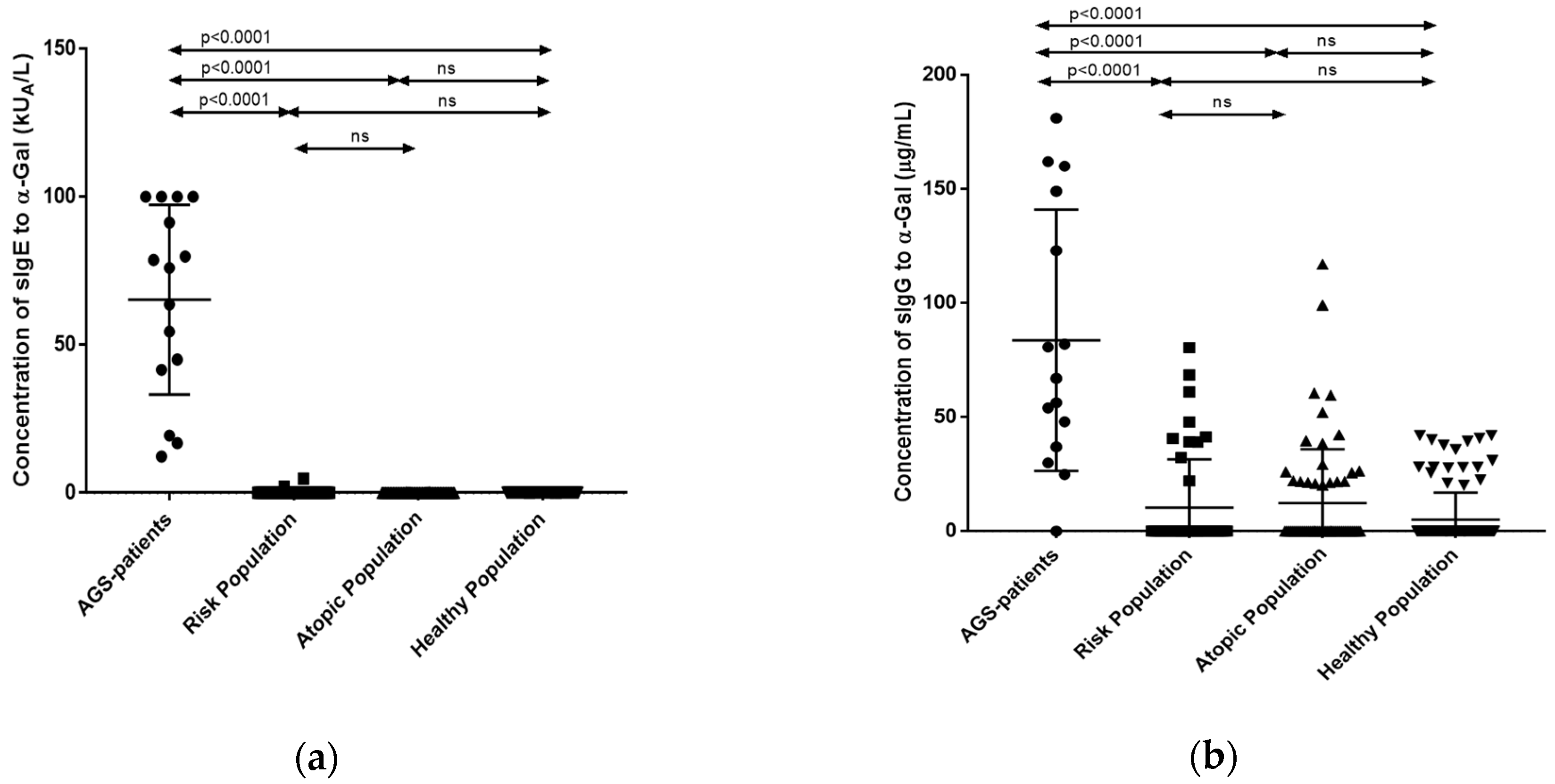
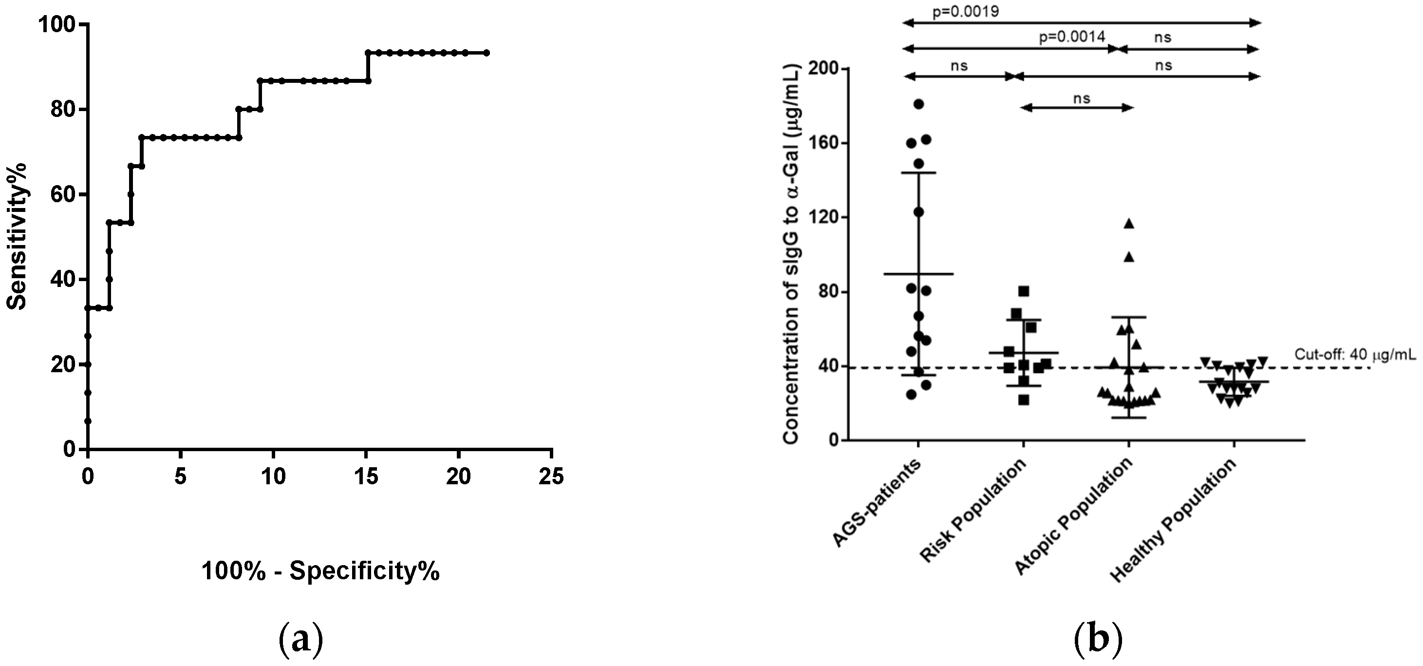
3.2. Quantification of sIgE and sIgG Antibodies against the α-Gal Epitope

The sIgE mean value against α-Gal in the AGS patients was 62.5 ± 8.3 kUA/L (Figure 1a). No positive values (≥0.35 kUA/L) were demonstrated in atopic and healthy populations (mean values 0.014 ± 0.003 kUA/L and 0.013 ± 0.001 kUA/L, respectively). Two participants (4.3%) in the risk-population group have the concentrations of sIgE to α-Gal 4.82 and 2.13 kUA/L, which was higher than the minimal values for the positive test >0.35 kUA/L). Statistically significate differences were only demonstrated among AGS patients and the risk-population group, the atopic population group, and the healthy population group (p < 0.001).
In 14 of 15 of AGS patients (93%), α-Gal-specific IgG Abs were found at concentrations between 25 and 190 µg/mL. The mean value obtained for this group was 83 ± 1.4 µg/mL) (Figure 1b).
A total of 21.7% of the subjects in the risk-population group (10/46), showed IgG specific to α-Gal (mean value: 10.3 ± 3.1 µg/mL). The 31.2% and the 15.7% of the atopic and healthy population, respectively, demonstrated sIgG against α-Gal epitope (mean values: 12.3 ± 2.9 µg/mL and 0.5 ± 1.1 µg/mL, respectively). Statistically significate differences were only demonstrated among AGS patients and risk-population group, atopic group, and the healthy population (p < 0.001).
A ROC was constructed to establish the sIgG Ab cut-off value against the α-Gal epitope (Figure 2a). The optimal cut-off point for specific IgG against α-Gal was obtained by ROC analysis (area under the curve: 0.931; standard error: 0.042; 95% confidence interval: 0.721 to 0.847; p < 0.001). The mean value plus SD (40 µg/mL) was chosen as the cut-off value (sensitivity, 86.7%; specificity, 92.6%). A new statistical analysis of the data was made according to the cut-off value (Figure 2b).
In this case, the statistical analysis showed no differences between the AGS patient group and the risk-population group included in this study (p > 0.999). Statistically significate differences were demonstrated among AGS patients and the atopic population and healthy population (p = 0.002 and p = 0.001, respectively). A contingency analysis was performed to assess the association between the likelihood of suffering AGS and the sIgG levels to α-Gal. The statistical analysis showed a likelihood of 35% of suffering AGS when the sIgG to α-Gal was greater than or equal to 40 µg/mL in people bitten by ticks. The risk for suffering AGS was increased by 17.5 times (CI 6.333–50.271) when sIgG values to α-Gal was greater than the cut-off value. Serum IgG specific to α-Gal was a significant predictor of AGS with an odds ratio of 27.2 (CI 8.153–81.190). The attributable risk was 33.5% (CI 0.175–0.526).

4. Discussion

Although the studies of the biochemical nature and function of α-Gal began more than two decades ago in xenotransplantation [54,55], it was in 2008 when Chung et al. published the anaphylaxis reaction to α-Gal epitope present in cetuximab, a chimeric monoclonal Ab used in the treatment of some cancer [18]. Commins et al., in 2008, demonstrated that patients with IgE specific to α-Gal epitope suffer from delayed anaphylaxis, angioedema, or urticaria after the consumption of red meat. In 2007, van Nunen et al. published the first report on the capacity of ticks to induce red meat allergy [28]. Since then, several authors have provided elegant proofs of the association between the injection of α-Gal present in tick saliva and cement with the development of mammalian meat allergy [56,57].
The association between tick bite reactions and red meat allergy in humans was described in Australia [57]. Before, different studies on red meat allergy have demonstrated that AGS culprit tick species are found in almost all continents [27]. Our study was carried out in people from the North of Spain (Basque Country), where the most prevalent ticks are I. ricinus and Hemaphysalis punctate [58]. All the clinically diagnosed AGS patients had been bitten by ticks, presented IgE specific to α-Gal and, almost all of them (14/15) presented IgG specific to α-Gal. In the same way, all the risk population subjects were bitten by ticks, but none of them was AGS diagnosed. However, two participants presented IgE specific to α-Gal positive values (4.82 kUA/L and 2.13 kUA/L) revealing the sensitization to the epitope without clinical symptoms. Mabelane et al. (2018) established that the α-Gal IgE value above which there was a 95% probability of meat allergy was 5.5 kUA/L [59]. These data explain, probably, that none one of these participants were referred for suffering red meat allergy, although other authors stablished the cut-off value at >0.54 kUA/L for sIgE [60]. No individuals in control groups (healthy and atopic population) showed IgE Abs specific to α-Gal. Nevertheless, all the studied groups demonstrated IgG Abs specific to this oligosaccharide moiety. Humans do not express the α-Gal carrying oligosaccharide because, in humans, α-1,3GT is expressed in an enzymatically inactive form [8,9]. The origin of this mutation occurred thousands of years ago and, it was probably an evolutionary step for humans in the defense against viruses, bacteria, and parasites carrying this oligosaccharide on its surfaces, such as Trypanosoma and Leishmania [61]. All immunocompetent humans can develop a strong immune response against the α-Gal epitope [33], which is considered as the only naturally abundantly expressed Abs in humans [8,9]. Hamanova et al. (2015) studied the kinetics for the formation of anti-α-Gal Abs (IgM, IgA, and IgG) in a group of infants along their first two years of life [62]. They demonstrated the transplacental transfer of the anti-α-Gal IgG Abs, which started to increase slowly with increasing age [63]. It is suggested that these Abs are produced at all human ages against the α-Gal epitopes present in the outer membrane of bacteria from the intestinal microbiome [34,35]. The continuous antigen stimulation by gut bacteria induces that as much as 1% of the human B cell population (memory B cells) in an individual is capable of producing anti-α-Gal Abs [36]. However, the data on the concentration of IgG to α-Gal in normal human serum are controversial depending on the methodology used for quantification. Galili et al. (1984) established that the level of anti-α-Gal Abs in healthy donors was 1% [8]; Yu et al. (1996) quantified by ELISA that the value was in the range of 0 to 15 µg/mL [27], and Tomlinson and Nussenzweig (1997) established this value between 0.25 and 0.5% of total Igs [48]; Obukhova et al. (2007) indicated that the level of Igs against α-Gal in healthy donors was about 10-fold less than the established by Galili et al. (1984) [49]. Recently, Zappe et al. (2021) published that the level of IgG anti-α-Gal in a commercial concentrate of human IgG is about 10% (IgG1 isotype) [50].
In our study, it was demonstrated that the AGS patients had higher statistically significant anti-α-Gal sIgG levels than the risk population and the control groups according to other authors [46,47]. These data were of interest, and we performed a ROC to establish an α-Gal IgG cut-off value to study the possibility of using anti-α-Gal IgG as an AGS prognostic marker. The statistical analysis of the re-arranged data demonstrated that the sIgG positive values at the risk population and AGS patients followed a similar distribution from the statistical point of view. Then, we calculated the probability that tick-bitten people with sIgG Ab levels to α-Gal greater than 40 µg/mL would develop AGS. The results indicated that the presence of sIgG Ab to α-Gal at levels greater than the cut-off value in serum was a risk factor for developing sIgE Ab to α-Gal greater than 0.35 kUA/L. This event was calculated to occur in a ratio of 27:1 with a likelihood of 93.9%. In the same way, people bitten by ticks with anti-α-Gal IgG values greater than the cut-off value had a likelihood of suffering AGS of 35%.
Nowadays, two hypotheses have been proposed for explaining the sensitization to α-Gal and posterior allergy development. One proposes that α-Gal antigen is present on salivary proteins [44]. After biting, the α-Gal glycoproteins are presented to antigen-presenting cells (APCs) and B-lymphocytes in the context of Th2 cell-mediated immunity [44]. The second hypothesis implies the presence of immunomodulatory factors in tick saliva, such as PGE2 that triggers immunoglobulin class switching to anti-α-Gal IgE-producing B cells from preexisting mature B-cell clones producing anti-α-Gal IgM and/or IgG [45]. Oliveira et al. (2011) demonstrated the presence of non-protein molecules in tick saliva with potent immunomodulatory properties. Among these molecules, PGE2 was found in several tick species from major genera, such as Ixodes [64]. Gao et al. (2016) demonstrated that PGE2 promotes IgE production in vivo contributing to asthma development [63]. Specifically, PGE2 induces a class switch recombination on mature B cells [65]. Cabezas-Cruz et al. (2019) postulated that tick salivary PGE2 triggers Ab class switching in mature B cells, increasing the levels of anti-α-Gal IgE Abs [20]. Given that α-Gal exposure alone does not induce an IgE response [43], our results may be supported by the second hypothesis because all the AGS patients and risk-population individuals bitten by ticks demonstrated sIgG values to α-Gal statistically different from the atopic and healthy populations. Our data demonstrated that tick-bitten subjects present IgG Abs to α-Gal greater than subjects who were not bitten by ticks. From a statistical point of view, there seems to be a direct relationship between the levels of IgG and the possibility of developing AGS.
However, our study has some limitations, such as the characteristics of the control selected populations and the relative low number of AGS patients included in the study. In following studies, it would be convenient to take into account the anti-α-Gal IgG subclass studied and the blood group of the individuals [41,46,47]. Finally, other characteristics of the individuals, such as age, the diet composition or the presence of intestinal parasites, could affect the composition of the microbiota and, therefore, the normal levels of anti-α-Gal Abs [65].

5. Conclusions

The relative levels of specific IgG against α-Gal can be quantified by an automated system using relative standardized calibrators defined for specific IgG. This quantification allowed us to establish a cut-off point for this parameter. According to the statistical results obtained, the quantification of IgG against α-Gal in subjects bitten by ticks could be used as a prognostic marker for developing mammalian meat allergy.

Author Contributions

Conceptualization, A.J., J.M. and I.P.; methodology, A.J., N.A., P.S., A.V.-d.-B., M.-A.A.-S., S.L., J.M. and I.P.; software, P.S. and A.V.-d.-B.; validation, A.J., J.M. and I.P.; formal analysis, I.P.; investigation, A.J., N.A., M.-A.A.-S., S.L., P.S. and A.V.-d.-B.; resources, A.J., M.-A.A.-S., S.L., J.M. and I.P.; data curation, A.J., P.S., A.V.-d.-B. and I.P.; writing—original draft preparation, A.J., N.A., P.S., A.V.-d.-B., M.-A.A.-S., S.L., J.M. and I.P.; writing—review and editing, I.P., A.J. and J.M.; supervision, I.P.; project administration, I.P. and J.M.; funding acquisition, A.J., J.M. and I.P. All authors have read and agreed to the published version of the manuscript.

Funding

This research was funded by the Sociedad Española de Alergología e Inmunología Clínica (SEAIC2014) and, by the Department of Education of the Basque Government Grants to support the activities of research groups in the Basque University System (Project IT-1043-16).

Institutional Review Board Statement

The study was approved by the Ethics Committee of the Hospital BioDonosti-Osakidetza on 19 March 2014 and conducted according to the guidelines of the Declaration of Helsinki (Law 14/2007).

Informed Consent Statement

Informed consent was obtained from all subjects involved in the study.

Data Availability Statement

The data presented in this study are available in the article.

Acknowledgments

We would like to thank the individuals who have volunteered to participate in this project.

Conflicts of Interest

The authors declare no conflict of interest. The funders had no role in the design of the study; in the collection, analyses, or interpretation of data; in the writing of the manuscript, or in the decision to publish the results.

References

  1. Commins, S.P. Diagnosis & management of alpha-gal syndrome: Lessons from 2500 patients. Expert Rev. Clin. Immunol. 2020, 16, 667–677. [Google Scholar] [CrossRef]
  2. Hilger, C.; Fischer, J.; Wolbing, F.; Biedermann, T. Role and mechanism of galactose-alpha-1,3-galactose in the elicitation of delayed anaphylactic reactions to red meat. Curr. Allergy Asthma Rep. 2019, 19, 3. [Google Scholar] [CrossRef] [Green Version]
  3. Commins, S.P.; James, H.R.; Stevens, W.; Pochan, S.L.; Land, M.H.; King, C.; Mozzicato, S.; Platts-Mills, T.A.E. Delayed clinical and ex vivo response to mammalian meat in patients with IgE to galactose-alpha-1,3- galactose. J. Allergy Clin. Immunol. 2014, 134, 108–115. [Google Scholar] [CrossRef] [Green Version]
  4. Fischer, J.; Hebsaker, J.; Caponetto, P.; Platts-Mills, T.A.E.; Biedermann, T. Galactose-alpha-1,3-galactose sensitization is a prerequisite for pork-kidney allergy and cofactor-related mammalian meat anaphylaxis. J. Allergy Clin. Immunol. 2014, 134, 755–759. [Google Scholar] [CrossRef] [PubMed]
  5. Platts-Mills, T.A.E.; Commins, S.P.; Biedermann, T.; van Hage, M.; Levin, M.; Beck, L.A.; Diuk-Wasser, M.; Jappe, U.; Apostolovic, D.; Minnicozzi, M.; et al. On the cause and consequences of IgE to galactose-α-1,3-galactose: A report from the national institute of allergy and infectious diseases workshop on understanding IgE-mediated mammalian meat allergy. J. Allergy Clin. Immunol. 2020, 145, 1061–1071. [Google Scholar] [CrossRef] [PubMed]
  6. Wilson, J.M.; Schuyler, A.J.; Workman, L.; Gupta, M.; James, H.R.; Posthumus, J.; McGowan, E.C.; Commins, S.P.; Platts-Mills, T.A.E. Investigation into the α-Gal syndrome: Characteristics of 261 children and adults reporting red meat allergy. J. Allergy Clin. Immunol. 2019, 7, 2348–2358. [Google Scholar] [CrossRef] [PubMed]
  7. Young, I.; Prematunge, C.; Pussegoda, K.; Corrin, T.; Waddell, L. Tick exposures and alpha-gal syndrome: A systematic review of the evidence. Ticks Tick-Borne Dis. 2021, 12, 101674. [Google Scholar] [CrossRef] [PubMed]
  8. Galili, U.; Shohet, S.B.; Kobrin, E.; Stults, C.L.; Macher, B.A. Man, apes, and Old World monkeys differ from other mammals in the expression of alpha-galactosyl epitopes on nucleated cells. J. Biol. Chem. 1988, 263, 17755–17762. [Google Scholar] [CrossRef]
  9. Galili, U. Human Natural Antibodies to Mammalian Carbohydrate Antigens as Unsung Heroes Protecting against Past, Present, and Future Viral Infections. Antibodies 2020, 9, 25. [Google Scholar] [CrossRef]
  10. Apostolovic, D.; Mihailovic, J.; Commins, S.P.; Wijnveld, M.; Kazimirova, M.; Starkhammar, M.; Stockinger, H.; Platts-Mills, T.A.E.; Cirkovic Velickovic, T.; Hamsten, C.; et al. Allergenomics of the tick Ixodes ricinus reveals important α-Gal-carrying IgE-binding proteins in red meat allergy. Allergy 2020, 75, 217–220. [Google Scholar] [CrossRef]
  11. Araujo, R.N.; Franco, P.F.; Rodrigues, H.; Santos, L.C.B.; McKayç, C.S.; Sanhueza, C.A.; Nascimento Brito, C.R.; Araújo Azevedo, M.; Venuto, A.P.; Cowan, P.J.; et al. Amblyomma sculptum tick saliva: α-Gal identification, antibody response and possible association with red meat allergy in Brazil. Int. J. Parasitol. 2016, 46, 213–220. [Google Scholar] [CrossRef] [Green Version]
  12. Hamsten, C.; Starkhammar, M.; Tran, T.A.; Johansson, M.; Bengtsson, U.; Ahlén, G.; Sällberg, M.; Grönlund, H.; van Hage, M. Identification of galactose-a-1,3-galactose in the gastrointestinal tract of the tick Ixodes ricinus; Possible relationship with red meat allergy. Allergy Eur. J. Allergy Clin. Immunol. 2013, 68, 549–552. [Google Scholar] [CrossRef] [PubMed]
  13. Commins, S.P.; James, H.R.; Kelly, L.A.; Pochan, S.L.; Workman, L.J.; Perzanowski, M.S.; Kocan, K.M.; Fahy, J.V.; Nganga, L.W.; Ronmark, E.; et al. The relevance of tick bites to the production of IgE antibodies to the mammalian oligosaccharide galactose-α-1,3-galactose. J. Allergy Clin. Immunol. 2011, 127, 1286–1293. [Google Scholar] [CrossRef] [PubMed] [Green Version]
  14. Mitchell, C.L.; Lin, F.C.; Vaughn, M.; Apperson, C.S.; Meshnick, S.R.; Commins, S.P. Association between lone star tick bites and increased alpha-gal sensitization: Evidence from a prospective cohort of outdoor workers. Parasites Vectors 2020, 13, 470. [Google Scholar] [CrossRef]
  15. Kwak, M.; Somerville, C.; van Nunen, S. A novel Australian tick Ixodes (Endopalpiger) australiensis inducing mammalian meat allergy after tick bite. Asia Pac. Allergy 2018, 8, 31. [Google Scholar] [CrossRef] [PubMed]
  16. Chinuki, Y.; Ishiwata, K.; Yamaji, K.; Takahashi, H.; Morita, E. Haemaphysalis longicornis tick bites are a possible cause of red meat allergy in Japan. Allergy 2016, 71, 421–425. [Google Scholar] [CrossRef] [PubMed]
  17. Gonzalez-Quintela, A.; Dam Laursen, A.S.; Vidal, C.; Skaaby, T.; Gude, F.; Linneberg, A. IgE antibodies to alpha-gal in the general adult population: Relationship with tick bites, atopy, and cat ownership. Clin. Exp. Allergy 2014, 44, 1061–1068. [Google Scholar] [CrossRef]
  18. Mateo-Borrega, M.B.; Garcia, B.; Larramendi, C.H.; Azofra, J.; González-Mancebo, E.; Alvarado, M.I.; Alonso-Díaz-de-Durana, M.D.; Núñez-Orjales, R.; Diéguez, M.C.; Guilarte, M.; et al. IgE-Mediated Sensitization to Galactose-α-1,3- Galactose (α-Gal) in Urticaria and Anaphylaxis in Spain: Geographical Variations and Risk Factors. J. Investig. Allergol. Clin. Immunol. 2019, 29, 436–443. [Google Scholar] [CrossRef] [Green Version]
  19. Villalta, D.; Cecchi, L.; Farsi, A.; Chiarini, F.; Minale, P.; Voltolini, S.; Scala, E.; Quercia, O.; Muratore, L.; Pravettoni, V.; et al. Galactose-α-1,3-galactose syndrome: An Italian survey. Eur. Ann. Allergy Clin. Immunol. 2017, 49, 263–269. [Google Scholar] [CrossRef] [Green Version]
  20. Cabezas-Cruz, A.; Hodžić, A.; Román-Carrasco, P.; Mateos-Hernández, L.; Duscher, G.G.; Sinha, D.K.; Hemmer, W.; Swoboda, I.; Estrada-Peña, A.; de la Fuente, J. Environmental and Molecular Drivers of the α-Gal Syndrome. Front. Immunol. 2019, 31, 1210. [Google Scholar] [CrossRef] [Green Version]
  21. Commins, S.P. Invited Commentary: Alpha-Gal Allergy: Tip of the Iceberg to a Pivotal Immune Response. Curr. Allergy Asthma Rep. 2016, 16, 61. [Google Scholar] [CrossRef]
  22. Cunningham, D.; Humblet, Y.; Siena, S.; Khayat, D.; Bleiberg, H.; Santoro, A.; Bets, D.; Mueser, M.; Harstrick, A.; Verslype, C.; et al. Cetuximab monotherapy and cetuximab plus irinotecan in irinotecan-refractory metastatic colorectal cancer. N. Engl. J. Med. 2004, 351, 337–345. [Google Scholar] [CrossRef] [Green Version]
  23. Chung, C.H.; Mirakhur, B.; Chan, E.; Le, Q.T.; Berlin, J.; Morse, M.; Murphy, B.A.; Satinover, S.M.; Hosen, J.; Mauro, D.; et al. Cetuximab-induced anaphylaxis and IgE specific for galactose-alpha-1,3-galactose. N. Engl. J. Med. 2008, 358, 1109–1117. [Google Scholar] [CrossRef] [PubMed] [Green Version]
  24. Commins, S.P.; Satinover, S.M.; Hosen, J.; Mozena, J.; Borish, L.; Lewis, B.D.; Woodfolk, J.A.; Platts-Mills, T.A. Delayed anaphylaxis, angioedema, or urticaria after consumption of red meat in patients with IgE antibodies specific for galactose-alpha-1,3-galactose. J. Allergy Clin. Immunol. 2009, 123, 426–433. [Google Scholar] [CrossRef] [Green Version]
  25. Commins, S.P.; Jerath, M.R.; Cox, K.; Erickson, L.D.; Platts-Mills, T. Delayed anaphylaxis to alpha-gal, an oligosaccharide in mammalian meat. Allergol. Int. 2016, 65, 16–20. [Google Scholar] [CrossRef] [PubMed] [Green Version]
  26. Steinke, J.W.; Platts-Mills, T.A.; Commins, S.P. The alpha-gal story: Lessons learned from connecting the dots. J. Allergy Clin. Immunol. 2015, 135, 589–596. [Google Scholar] [CrossRef] [Green Version]
  27. Van Nunen, S.A.; O’Connor, K.S.; Clarke, L.R.; Boyle, R.X.; Fernando, S.L. An association between ticks bite reactions and red meat allergy in humans. Med. J. Aust. 2009, 190, 510–514. [Google Scholar] [CrossRef]
  28. Van Nunen, S.A.; Fernando, S.L.; Clarke, L.R.; Boyle, R.X. The association between Ixodes holocyclus tick bite reactions and red meat allergy (abstract). Intern. Med. J. 2007, 37 (Suppl. 5), A132. [Google Scholar]
  29. Binder, A.M.; Commins, S.P.; Altrich, M.L.; Wachs, T.; Biggerstaff, B.J.; Beard, C.B.; Petersen, L.R.; Kersh, G.J.; Armstrong, P.A. Diagnostic testing for galactose-alpha-1,3-galactose. Ann. Allergy Asthma Immunol. 2021, 126, 411–416. [Google Scholar] [CrossRef]
  30. Hamsten, C.; Tran, T.A.T.; Starkhammar, M.; Brauner, A.; Commins, S.P.; Platts-Mills, T.A.E.; van Hage, M. Red meat allergy in Sweden: Association with tick sensitization and B-negative blood groups. J. Allergy Clin. Immunol. 2013, 132, 1431–1434. [Google Scholar] [CrossRef] [PubMed] [Green Version]
  31. Morisset, M.; Richard, C.; Astier, C.; Jacquenet, S.; Croizier, A.; Beaudouin, E.; Cordebar, V.; Morel-Codreanu, F.; Petit, N.; Moneret-Vautrin, D.A.; et al. Anaphylaxis to pork kidney is related to IgE antibodies specific for galactose-alpha-1,3-galactose. Allergy 2012, 67, 699–704. [Google Scholar] [CrossRef]
  32. Fernández-Soto, P.; Dávila, I.; Laffond, E.; Lorente, F.; Encinas-Grandes, A.; Pérz-Sánchez, R. Tick bite-induced anaphylaxis in Spain. Ann. Trop. Med. Parasitol. 2001, 95, 97–103. [Google Scholar] [CrossRef] [Green Version]
  33. Macher, B.A.; Galili, U. The Galα1,3Galβ1,4GlcNAc-R (α-Gal) epitope: A carbohydrate of unique evolution and clinical relevance. Biochim. Biophys. Acta 2008, 1780, 75–88. [Google Scholar] [CrossRef] [PubMed] [Green Version]
  34. Minanov, O.P.; Itescu, S.; Neethling, F.A.; Morgenthau, A.S.; Kwiatkowski, P.; Cooper, D.K.; Michler, R.E. Anti-gal IgG antibodies in sera of newborn humans and baboons and its significance in pig xenotransplantation. Transplantation 1997, 63, 182–186. [Google Scholar] [CrossRef] [PubMed]
  35. Montassier, E.; Al-Ghalith, G.A.; Mathe, C.; Le Bastard, Q.; Douillard, V.; Garnier, A.; Guimon, R.; Raimondeau, B.; Touchefeu, Y.; Duchalais, E.; et al. Distribution of bacterial alpha1,3-galactosyltransferase genes in the human gut microbiome. Front. Immunol. 2019, 10, 3000. [Google Scholar] [CrossRef] [PubMed] [Green Version]
  36. Galili, U.; Anaraki, F.; Thall, A.; Hill-Black, C.; Radic, M. One percent of human circulating B lymphocytes are capable of producing the natural anti-Gal antibody. Blood 1993, 82, 2485–2493. [Google Scholar] [CrossRef] [Green Version]
  37. Yu, P.B.; Holzknecht, Z.E.; Bruno, D.; Parker, W.; Platt, J.L. Modulation of natural IgM binding and complement activation by natural IgG antibodies: A role for IgG anti-Gal alpha1-3Gal antibodies. J. Immunol. 1996, 157, 5163–5168. [Google Scholar] [PubMed]
  38. Platts-Mills, T.A.E.; Li, R.C.; Keshavarz, B.; Smith, A.R.; Wilson, J.M. Diagnosis and Management of Patients with the α-Gal Syndrome. J. Allergy Clin. Immunol. Pract. 2020, 8, 15–23. [Google Scholar] [CrossRef]
  39. Fischer, J.; Yazdi, A.S.; Biedermann, T. Clinical spectrum of a-Gal syndrome: From immediate-type to delayed immediate-type reactions to mammalian innards and meat. Allergo J. Int. 2016, 25, 55–62. [Google Scholar] [CrossRef] [Green Version]
  40. Platts-Mills, T.A.E.; Schuyler, A.J.; Tripathi, A.; Commins, S.P. Anaphylaxis to the carbohy-drate side chain alpha-gal. Immunol. Allergy Clin. N. Am. 2015, 35, 247–260. [Google Scholar] [CrossRef] [Green Version]
  41. Kollmann, D.; Nagl, B.; Ebner, C.; Emminger, W.; Wöhrl, S.; Kitzmüller, C.; Vrtala, S.; Mangold, A.; Ankersmit, H.J.; Bohle, B. The quantity and quality of α-gal-specific antibodies differ in individuals with and without delayed red meat allergy. Allergy 2017, 72, 266–273. [Google Scholar] [CrossRef] [PubMed]
  42. Park, Y.; Kim, D.; Boorgula, G.D.; De Schutter, K.; Smagghe, G.; Šimo, L.; Archer-Hartmann, S.A.; Azadi, P. Alpha-Gal and Cross-Reactive Carbohydrate Determinants in the N-Glycans of Salivary Glands in the Lone Star Tick, Amblyomma americanum. Vaccines 2020, 8, 18. [Google Scholar] [CrossRef] [PubMed] [Green Version]
  43. Villar, M.; Pacheco, I.; Merino, O.; Contreras, M.; Mateos-Hernández, L.; Prado, E.; Barros-Picanço, D.K.; Lima-Barbero, J.F.; Artigas-Jerónimo, S.; Alberdi, P.; et al. Tick and Host Derived Compounds Detected in the Cement Complex Substance. Biomolecules 2020, 10, 555. [Google Scholar] [CrossRef] [Green Version]
  44. Chandrasekhar, J.L.; Cox, K.M.; Erickson, L.D. B Cell Responses in the Development of Mammalian Meat Allergy. Front. Immunol. 2020, 11, 1532. [Google Scholar] [CrossRef]
  45. Carvalho-Costa, T.M.; Mendes, M.T.; da Silva, M.V.; da Costa, T.A.; Tiburcio, M.G.; Anhê, A.C.; Rodrigues, V., Jr.; Oliveira, C.J.F. Immunosuppressive effects of Amblyomma cajennense tick saliva on murine bone marrow-derived dendritic cells. Parasites Vectors 2015, 8, 22. [Google Scholar] [CrossRef] [Green Version]
  46. Rispens, T.; Derksen, N.I.; Commins, S.P.; Platts-Mills, T.A.; Aalberse, R.C. IgE production to α-gal is accompanied by elevated levels of specific IgG1 antibodies and low amounts of IgE to blood group B. PLoS ONE 2013, 8, e55566. [Google Scholar] [CrossRef]
  47. Apostolovic, D.; Rodrigues, R.; Thomas, P.; Starkhammar, M.; Hamsten, C.; van Hage, M. Immunoprofile of α-Gal- and B-antigen-specific responses differentiates red meat-allergic patients from healthy individuals. Allergy 2018, 73, 1525–1531. [Google Scholar] [CrossRef]
  48. Tomlinson, S.; Nussenzweig, V. Human alternative complement pathway-mediated lysis of rabbit erythrocytes is enhanced by natural anti-Galalpha1-3Gal antibodies. J. Immunol. 1997, 159, 5606–5609. [Google Scholar]
  49. Obukhova, P.; Rieben, R.; Bovin, N. Normal human serum contains high levels of anti-Gala1-4GlcNAc antibodies. Xenotransplantation 2007, 14, 627–635. [Google Scholar] [CrossRef] [PubMed]
  50. Zappe, A.; Rosenlöcher, J.; Kohla, G.; Hinderlich, S.; Parr, M.K. Purification and Characterization of Antibodies Directed against the -Gal Epitope. BioChem 2021, 1, 81–97. [Google Scholar] [CrossRef]
  51. van Hage, M.; Schmid-Grendelmeier, P.; Skevaki, C.; Plebani, M.; Canonica, W.; Kleine-Tebbe, J.; Nystrand, M.; Jafari-Mamaghani, M.; Jakob, T. Performance evaluation of ImmunoCAP® ISAC 112: A multi-site study. Clin. Chem. Lab. Med. 2017, 55, 571–577. [Google Scholar] [CrossRef] [PubMed]
  52. Sastre, J. Molecular diagnosis in allergy. Clin. Exp. Allergy 2010, 40, 1442–1460. [Google Scholar] [CrossRef] [PubMed]
  53. Viñas, M.; Postigo, I.; Suñen, E.; Martınez, J. Urticaria and silent parasitism by Ascaridoidea: Component-resolved diagnosis reinforces the significance of this association. PLoS Negl. Trop. Dis. 2020, 14, e0008177. [Google Scholar] [CrossRef] [Green Version]
  54. Cooper, D.K. Depletion of natural antibodies in non-human primates-a step towards successful discordant xenografting in humans. Clin. Transpl. 1992, 6, 178–183. [Google Scholar]
  55. Kobayashi, T.; Cooper, D.K. Anti-Gal, alpha-Gal epitopes, and xenotransplantation. Subcell. Biochem. 1999, 32, 229–257. [Google Scholar] [CrossRef]
  56. Commins, S.P.; Platts-Mills, T.A. Tick bites and read meat allergy. Curr. Opin. Allergy Clin. Immunol. 2013, 13, 354–359. [Google Scholar] [CrossRef] [PubMed] [Green Version]
  57. Van Nunen, S.A. Tick-induced allergies: Mammalian meat allergy, tick anaphylaxis and their significance. Asia Pac. Allergy 2015, 5, 3–16. [Google Scholar] [CrossRef] [PubMed] [Green Version]
  58. Barral, M.; García-Pérez, A.L.; Juste, R.A.; de Luco, D.F.; Dehesa, V. Distribución y Actividad de los Ixódidos Presentes en la Vegetación de la Comunidad Autónoma Vasca; Informe Técnico N°54; Servicio Central de Publicaciones del Gobierno Vasco: Vitoria-Gasteiz, Spain, 1993. [Google Scholar]
  59. Mabelane, T.; Basera, W.; Botha, M.; Thomas, H.F.; Ramjith, J.; Levin, M.E. Predictive values of alpha-gal IgE levels and alpha-gal IgE: Total IgE ratio and oral food challenge-proven meat allergy in a population with a high prevalence of reported red meat allergy. Pediatr. Allergy Immunol. 2018, 29, 841–849. [Google Scholar] [CrossRef]
  60. Fischer, J.; Huynh, H.N.; Hebsaker, J.; Forchhammer, S.; Yazdi, A.S. Prevalence and Impact of Type I Sensitization to Alpha-Gal in Patients Consulting an Allergy Unit. Int. Arch. Allergy Immunol. 2020, 18, 119–127. [Google Scholar] [CrossRef]
  61. Avila, J.L.; Rojas, M.; Galili, U. Immunogenic Gal alpha 1—3Gal carbohydrate epitopes are present on pathogenic American Trypanosoma and Leishmania. J. Immunol. 1989, 142, 2828–2834. [Google Scholar]
  62. Hamanova, M.; Chmelikova, M.; Nentwich, I.; Thon, V.; Lokaj, J. Anti-Gal IgM, IgA and IgG natural antibodies in childhood. Immunol. Lett. 2015, 164, 40–43. [Google Scholar] [CrossRef] [PubMed]
  63. Gao, Y.; Zhao, C.; Wang, W.; Jin, R.; Li, Q.; Ge, Q.; Guan, Y.; Zhang, Y. Prostaglandins E2 signal mediated by receptor subtype EP2 promotes IgE production in vivo and contributes to asthma development. Sci. Rep. 2016, 6, 20505. [Google Scholar] [CrossRef] [PubMed] [Green Version]
  64. Oliveira, C.J.; Sá-Nunes, A.; Francischetti, I.M.; Carregaro, V.; Anatriello, E.; Silva, J.S.; Santos, I.K.; Ribeiro, J.M.; Ferreira, B.R. Deconstructing tick saliva: Non-protein molecules with potent immunomodulatory properties. J. Biol. Chem. 2011, 286, 10960–10969. [Google Scholar] [CrossRef] [PubMed] [Green Version]
  65. Wilson, J.M.; Keshavarz, B.; James, H.R.; Retterer, M.K.C.; Schuyler, A.J.; Knoedler, A.; Workman, L.J.; Nganga, L.; Chico, M.E.; Rönmark, E.; et al. α-Gal specific-IgE prevalence and levels in Ecuador and Kenya: Relation to diet, parasites, and IgG4. J. Allergy Clin. Immunol. 2021, 147, 1393–1401.e7. [Google Scholar] [CrossRef]
Figure 1. Concentrations of the sIgE (a) and sIgG (b) antibodies specific to α-Gal in the studied populations: AGS patients (circles) ; Risk Population (squares); Atopic Population (triangles up-ward); Healthy Population (downward).
Foods 11 00466 g001
Figure 2. ROC curve (a) and distribution of the studied populations considering 40 µg/mL as sIgG to α-Gal cut-off value (b). AGS patients (circles) ; Risk Population (squares); Atopic Population (triangles upward); Healthy Population (downward).
Foods 11 00466 g002
Table 1. Clinical data on the AGS-patients.
PatientGenderTotal IgE (kUA/L)IgE Specific to α-Gal [kUA/L]Clinical Symptoms
1m738>100Recurrent Urticaria
2m569>100Anaphylaxis
3m452>100Anaphylaxis
4f325>100Recurrent Urticaria
5m52391.3Recurrent Urticaria
6m23879.9Anaphylaxis
7m26778.7Anaphylaxis
8m46176.0Anaphylaxis
9m26963.7Anaphylaxis
10m23054.4Anaphylaxis
11m91.745.0Acute Urticaria
12m67141.6Recurrent Urticaria
13m10219.4Recurrent Urticaria
14m13916.8Acute Urticaria
15m31112.3Anaphylaxis
Table 2. Characteristics of the risk-population group (n = 46) recorded by survey. Results are expressed as percentage (%) of total number of participants.
Percentage (%)
Male34
Female66
Demographic dataAge (20–60 yrs.)90
Urban83
Rural17
Dog41
Animal contactCat24
Others20
Daily meat consumption 78
Meat-consumption related symptomsNone90
Intolerance9
Lyme10
Chronic diseasesAsthma10
Fibromyalgia4
Arthritis4
Others9
Table 3. Prevalence of sensitization in the risk and atopic population measured by ImmunoCAP ISAC. Results are expressed as percentage (%) of total number of participants.
Percentage (%)
AllergenMain Source of AllergenRisk Population (n = 46)Atopic Population (n = 64)
Phl p 1Grass pollen1553.1
Ole e 1Olive tree pollen8.637
Cup a 1Cypress tree pollen4.334
Cry j 1Japanese cedar pollen2.126
Bet v 2Birch pollen4.321.8
Der 1Mites Group 1 allergens3030
Der 2Mites Group 2 allergens3037.5
Der p 10Mites Tropomyosin04.6
Alt a 1Alternaria alternata012.5
Asp f 1Aspergillus fumigatus07.8
Pru p 3Apricot020
Cor a 8Hazel07.8
Act d 1Kiwifruit07.8
Pen m 1Shrimp04.6
Gad c 1Egg04.6
Fel d 1Cat (uteroglobin)1326
Fel d 4Cat (lipocalin)6.56.25
Can f 1Dog (lipocalin)4.34.6
Mus m 1Mouse (lipocalin)2.16.2
Equ c 1Horse (lipocalin)03.1
Api m 1Bee venom (phospholipase A2)01.5
Pol d 5 (array)Wasp venom (Antigen 5)4.30
Mux F3 (array)Carbohydrates (CCDs)3.70
Publisher’s Note: MDPI stays neutral with regard to jurisdictional claims in published maps and institutional affiliations.

