Article

Mutation of Signal Transducer and Activator of Transcription 5 (STAT5) Binding Sites Decreases Milk Allergen αS1-Casein Content in Goat Mammary Epithelial Cells

1
Shaanxi Key Laboratory of Molecular Biology for Agriculture, College of Animal Science and Technology, Northwest A&F University, Yangling 712100, China
2
College of Animal Science and Technology, Anhui Agricultural University, Hefei 230036, China
*
Author to whom correspondence should be addressed.
Foods 2022, 11(3), 346; https://doi.org/10.3390/foods11030346
Submission received: 4 January 2022 / Revised: 23 January 2022 / Accepted: 23 January 2022 / Published: 26 January 2022
(This article belongs to the Section Dairy)

Abstract

:
αS1-Casein (encoded by the CSN1S1 gene) is associated with food allergy more than other milk protein components. Milk allergy caused by αS1-casein is derived from cow milk, goat milk and other ruminant milk. However, little is known about the transcription regulation of αS1-casein synthesis in dairy goats. This study aimed to investigate the regulatory roles of signal transducer and activator of transcription 5 (STAT5) on αS1-casein in goat mammary epithelial cells (GMEC). Deletion analysis showed that the core promoter region of CSN1S1 was located at −110 to −18 bp upstream of transcription start site, which contained two putative STAT5 binding sites (gamma-interferon activation site, GAS). Overexpression of STAT5a gene upregulated the mRNA level and the promoter activity of the CSN1S1 gene, and STAT5 inhibitor decreased phosphorylated STAT5 in the nucleus and CSN1S1 transcription activity. Further, GAS site-directed mutagenesis and chromatin immunoprecipitation (ChIP) assays revealed that GAS1 and GAS2 sites in the CSN1S1 promoter core region were binding sites of STAT5. Taken together, STAT5 directly regulates CSN1S1 transcription by GAS1 and GAS2 sites in GMEC, and the mutation of STAT5 binding sites could downregulate CSN1S1 expression and decrease αS1-casein synthesis, which provide the novel strategy for reducing the allergic potential of goat milk and improving milk quality in ruminants.

1. Introduction

Dairy products are rich in nutrients such as essential amino acids and bioactive peptides needed by human body, which are important sources of dietary protein. Milk proteins are divided into caseins and whey proteins [1]. Caseins consist of αS1-, αS2-, β- and κ-casein, and whey proteins mainly include α-lactalbumin and β-lactoglobulin [2]. Despite the nutritiousness of milk proteins, they also carry the risk of a milk allergy for many people [3]. Milk protein contains antigen epitopes that can be recognized by the human immune system, and may cause skin itching, vomiting, diarrhea, allergic rhinitis and other adverse symptoms [4]. The prevalence of a food allergy caused by milk protein is as high as 2–7.5% in different countries, especially for infants and children [5,6]. Among various milk protein components, αS1-casein is highly susceptible to human allergies, which is sensitive for approximately 65% of patients suffering from a milk protein allergy [7,8]. Goat milk contains a considerable variation of αS1-casein content (4–26% of total protein) compared to cow milk (36–40%) [9,10]. Therefore, the means of manipulating αS1-casein gene transcription to decrease milk allergic reactions is necessary to be elucidated.
Transcription factors that play key roles in regulating the transcription of milk protein genes mainly include signal transducer and activator of transcription 5 (STAT5), glucocorticoid receptor (GR), activator protein 1 (AP-1), CCAAT enhancer binding protein (C/EBP) and YinYang1 (YY1) in mammals. Among these transcription factors, STAT5 plays a central role in the transcription of milk protein genes [11]. During the lactation period, hormones such as prolactin, glucocorticoids and insulin could bind to corresponding receptors on the mammary epithelial cell membranes to activate STAT5 [12]. Then phosphorylated STAT5 transfers to the nucleus and binds specifically to the GAS (gamma-interferon activation site) response element of the milk protein gene promoter regions to initiate the transcription process [13,14]. Breast specific STAT5-knockout mice had impaired development of the mammary gland and the decrease of whey protein content in milk [15]. In the mammary epithelial cell lines of cows [16] and dairy goats [17], STAT5 binds to the CSN2 gene promoter and promotes the transcription and synthesis of β-casein.
To address the situation of milk allergic reactions due to αS1-casein in ruminants, it is essential to clarify the transcriptional regulation of αS1-casein (CSN1S1). Although previous studies have shown that STAT5 plays crucial roles in the processes of milk protein synthesis; however, whether it could regulate αS1-casein transcription in ruminants remains obscure. Therefore, the objective of the present study was to dissect the transcriptional regulation mechanism of CSN1S1 by STAT5 in goat mammary epithelial cells (GMEC). Here, we found that STAT5 directly regulated the transcription of the CSN1S1 gene by GAS binding sites in GMEC, which provides the novel way to reduce milk allergic reactions through the mutation of these GAS sites in ruminants.

2. Materials and Methods

2.1. Ethics Statement

All experimental procedures were approved by the Institutional Animal Care and Use Committee of Northwest A&F University, YangLing, China (protocol number 15-516).

2.2. Goat CSN1S1 Promoter Region Cloning and Bioinformatics Analysis

Goat genome sequence of the CSN1S1 promoter region (GenBank no. NC_030813.1) was used for primer design. Genomic DNA extracted from goat blood samples by using TIANamp Genomic DNA Kit (DP304, Tiangen, Beijing, China). The 2201 bp fragment of the CSN1S1 gene promoter was amplified and ligated to pMD-19T vector (Takara, Shiga, Japan). The sequences of forward −2018 and reverse +183 primers were shown in Table 1. Then, JASPAR (http://jaspar.genereg.net/, accessed on 20 April 2020) and TRANSFAC databases (http://www.gene-regulation.com, accessed on 20 April 2020) were used to predict the putative binding sites of the transcription factor in the CSN1S1 promoter region.

2.3. Vector Construction

The deletion fragment primers of the CSN1S1 promoter for −1715, −1354, −1049, −598, −346, −110 and −18 bp were designed (Table 1), then amplified using −2018/+183 bp as a template. DNA fragments of the CSN1S1 promoter region were gel purified, digested with KpnI and HindIII (Takara) and cloned into a pGL3-Basic vector. The plasmids were confirmed by gel electrophoresis (Figure S1) and sequencing. The site-directed mutant primers of STAT5 binding sites for GAS1, GAS2 and GAS3 in the CSN1S1 promoter were showed in Table 1. Overlapping PCR technology was used to construct GAS site-directed mutagenesis vectors. All plasmids were verified by DNA sequencing.

2.4. Cell Culture

The GMEC were isolated from 3 healthy Xinong Saanen dairy goats at the peak of lactation, as previously described [18]. The protocol for the purification and identification procedure have been described in the previous study [19]. Cells were investigated by immunofluorescence with Cytokeratin 18 (66187-1-Ig, Proteintech, Rosemont, IL, USA, 1:200; Figure S2), which is specific for epithelial cells. Cells were cultured at 37 °C in 5% CO2. The culture medium contained basal DMEM/F12 (SH30023, Hyclone, Logan, UT, USA) medium, 5 mg/L insulin (16634, Sigma, St. Louis, MO, USA), 1 mg/L hydrocortisone (H0888, Sigma), 10 μg/L epidermal growth factor (PHG0311, Invitrogen, Carlsbad, CA, USA), 1 × 10⁵ U/L streptomycin/penicillin (080092569, Harbin Pharmaceutical Group, Harbin, China) and 10% FBS (10099141, Gibco, Gaithersburg, MD, USA). To promote lactogenesis, GMEC were switched to a lactogenic medium supplemented with 2 mg/L prolactin (L6520, Sigma) for 48 h before performing the experiments.

2.5. Cell Treatment

The GMEC were approximately 80% confluence before applying treatments. For luciferase assays, cells were cultured in 48-well plates at a density of 5 × 10⁴ cells per well and transfected with the CSN1S1 promoter vectors by X-treme GENE HP DNA Transfection Regent (06366236001, Roche, Mannheim, Germany) for 48 h. Similarly, cells were incubated with pcDNA3.1-STAT5a expression vector [20] or STAT5 inhibitor STAT5-IN-1 (CAS 285986-31-4, MedChemExpress, Monmouth Junction, NJ, USA; 200 μM, dissolved in DMSO) followed by CSN1S1 promoter plasmids transfection, respectively. The pRL-TK vector (renilla luciferase) was the internal control of pGL3-Basic vector (the mass ratio was 1:50). For quantitative real-time PCR (qRT-PCR) assays, cells were cultured in 12-well plates at a density of 2 × 10⁵ cells per well and treated with pcDNA3.1-STAT5a vector or STAT5-IN-1 (200 μM) for 48 h, respectively. For western blot analysis, cells were cultured in 60 mm dish and incubated with STAT5-IN-1 (200 μM) for 48 h, then cellular nuclear and cytoplasmic proteins were extracted, respectively. In addition, cells seeded in the culture medium were incubated with STAT5-IN-1 (200 μM, or DMSO) followed by prolactin (2 mg/L) treatment for 48 h, then total RNA or protein was extracted. Similarly, cells seeded in the culture medium were co-treated with pGL3-CSN1S1 vector and prolactin (2 mg/L) for luciferase assays.

2.6. Luciferase Assays

The GMEC cultured in 48-well plates were harvested and lysed for luciferase activity measurement. The luciferase activity was detected by the Dual-Luciferase Reporter System (E1910, Promega, Madison, WI, USA) on a Fluoroskan Ascent (ThermoFisher Scientific, Waltham, MA, USA). The relative luciferase activity was calculated as the ratio of firefly compared with renilla luciferase activity.

2.7. RNA Extraction and Quantitative Real-Time PCR

The total RNA of GMEC was extracted by Trizol reagent (9109, Takara). The concentration and purity of RNA were measured by NanoDrop2000 (ThermoFisher Scientific). The optical density 260/280 was 1.9–2.1, and the concentration of RNA was 400–500 ng/μL. The purified RNA was used to synthesize cDNA by PrimeScript RT kit (RR047A, Takara). qRT-PCR were detected by SYBR green (RR820A, Takara) in a CFX96 sequence detector (Bio-Rad, Hercules, CA, USA). The qRT-PCR reaction condition was performed at 95 °C for 30 s, followed by 40 cycles of 95 °C for 5 s and 60 °C for 30 s. Ribosomal protein S9 (RPS9) and ubiquitously expressed transcript (UXT) were used as internal control genes [21], and data were analyzed using 2−ΔΔCt method [22]. Primers for qRT-PCR are shown in Table 2.

2.8. Western Blot

Total protein of GMEC was harvested using RIPA lysis buffer (R0010, Solarbio) containing phosphatase and protease inhibitor cocktails (04906845001 and 04693132001, Roche). Nuclear and cytoplasmic proteins were extracted separately according to the protocol of protein extraction kit (DE201, TransGen, Beijing, China). Protein concentration was measured by BCA kit (23227, Thermo Scientific). The protein was separated using 10% SDS-PAGE and blotted onto a PVDF membrane (Roche) by a Bio-Rad Trans-Blot SD. Nonspecific binding sites were blocked for 2 h by 5% skim milk (232100, BD Biosciences, Franklin Lakes, NJ, USA), then probed overnight at 4 °C with primary antibodies: rabbit anti-αS1-casein (SAB1401093, Sigma-Aldrich; 1:1000), mouse anti-p-STAT5a (611964, BD Biosciences; 1:500), mouse anti-STAT5a (610191, BD Biosciences; 1:1000), mouse anti-Histone H3 (100005-MM01, Sino Biological, Beijing, China; 1:2000) and mouse anti-β-actin (CW0096, CW Biotech; 1:2000). Horseradish peroxidase-conjugated goat anti-mouse-IgG (CW0102, CW Biotech; Beijing, China; 1:5000) and goat anti-rabbit-IgG (CW0103, CW Biotech; 1:5000) secondary antibodies were performed. Signals were examined by the chemiluminescent kit (1705061, Bio-Rad). ImageJ (http://imagej.nih.gov, accessed on 16 November 2020) was used for measuring the intensities of bands. Relative expression of αS1-casein was normalized to β-actin, and p-STAT5a expression was normalized by comparison to STAT5a or Histone H3.

2.9. Chromatin Immunoprecipitation (ChIP) Assays

The ChIP assays were performed according to the protocol of a commercial kit (P2048, Beyotime, Shanghai, China). GMEC were cultured in 100 mm dish at a density of 2 × 10⁶ cells per well until 80% confluence before cell collection. Cells were crosslinked with a final concentration of 1% formaldehyde for 10 min at 37 °C, then washed in cold PBS containing phosphatase and protease inhibitors. Chromosomal DNA was sheared into lengths of 100 to 500 bp by Bioruptor UCD-200 (Diagenode, Seraing, Belgium). The chromatin–protein complexes were treated with protein A/G beads and rabbit p-STAT5 antibody (9351S, Cell Signaling Technology, Danvers, MA, USA; 1:50) or normal rabbit IgG antibody (A7016, Beyotime; 1:50) at 4 °C for 12 h. The primers used in the ChIP assays for amplification are shown in Table 2.

2.10. Statistical Analysis

All experiments were carried out with each goat as a biological replicate (n = 3). The data were processed using SPSS20.0 (IBM, Chicago, IL, USA). Statistical analysis was performed using Student’s t-test for two group comparison, and one-way ANOVA was performed with Tukey test for multiple comparisons. Data were shown as means ± SEM. Significant differences were declared at p < 0.05 (* p < 0.05, ** p < 0.01).

3. Results

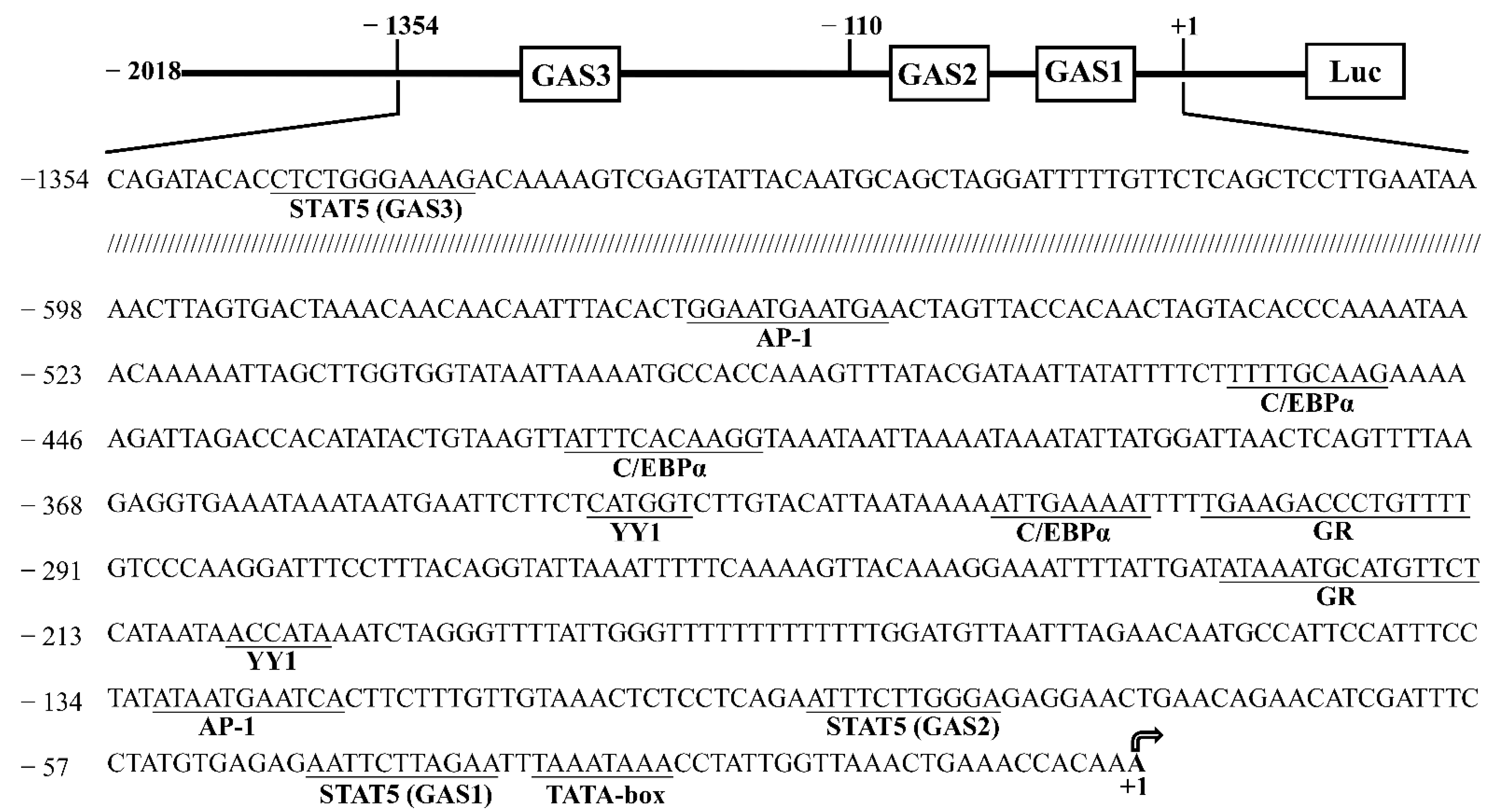
3.1. Cloning and Characterization of the Goat CSN1S1 Promoter

We obtained a 2201-bp 5′-flanking sequence of the CSN1S1 promoter containing 2018 bp upstream of the transcription start site (+1), the first exon (+1 to +107 bp) and part of the first intron. Using the JASPAR and TRANSFAC database analysis of the CSN1S1 promoter sequence, we revealed some consensus binding sites of transcription factors, such as STAT5 (−46, −92, −1345), AP-1 (−131, −566), C/EBP (−318, −420, −459), GR (−228, −306), YY1 (−206, −341) and TATA-box (−33, Figure 1).

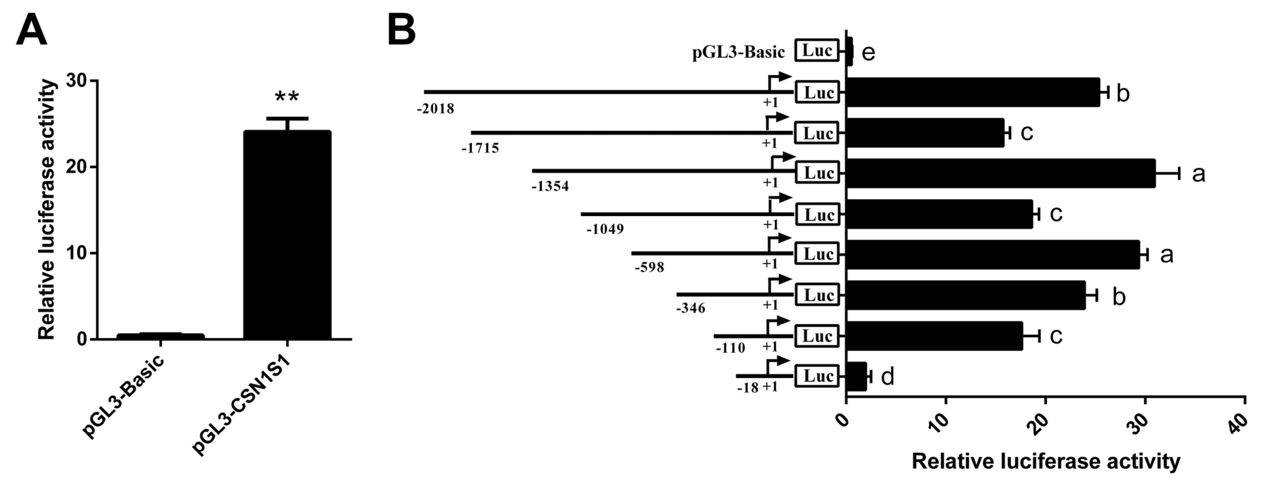
3.2. Core Promoter Region Identification of CSN1S1 Gene

The pGL3-CSN1S1 vector was transfected into GMEC, and luciferase activity was measured. The result shows that the CSN1S1 promoter activity is more active than the control vector (p < 0.01, Figure 2A). Eight promoter fragments of various lengths (−2018/+183, −1715/+183, −1354/+183, −1049/+183, −598/+183, −346/+183, −110/+183, −18/+183 bp) were cloned into pGL3-Basic vectors to confirm the core region of transcription activity in the CSN1S1 promoter. The luciferase assays showed that there are transcriptional activator elements in −2018~−1715 bp and −1354~−1049 bp regions, while transcriptional suppressor elements are present in −1715~−1354 bp and −1049~−598 bp regions. The promoter activity was progressively declined when the region between −598 and −110 bp slightly deleted (p < 0.05; Figure 2B). Further deletion from −110/+183 to −18/+183 bp reduced the activity by 89%, almost abolishing the promoter activity (p < 0.05; Figure 2B). These findings suggest that the region from −110 to −18 bp is required for maintaining basal promoter activity of the goat CSN1S1 gene, which contains two putative STAT5 binding sites (GAS sites).

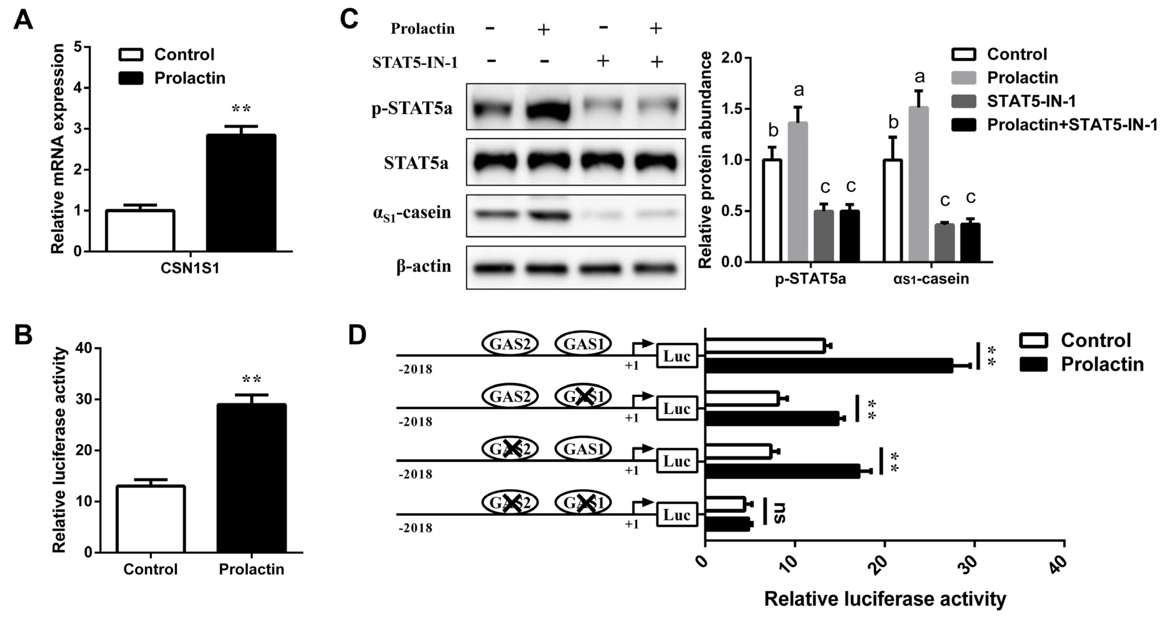
3.3. STAT5 Upregulates CSN1S1 Expression

Next, to define the regulator effect of STAT5 on αS1-casein transcription, GMEC were treated with pcDNA3.1-STAT5a or STAT5-IN-1. Compared with control, mRNA abundance of STAT5a and CSN1S1 was effectively increased in STAT5a overexpression group (p < 0.01, Figure 3A); moreover, the CSN1S1 promoter activity was also enhanced (p < 0.01, Figure 3B). Compared with the DMSO group, STAT5 inhibitor STAT5-IN-1 decreased the mRNA level and the promoter activity of CSN1S1 (p < 0.01, Figure 3C,D). In addition, STAT5a phosphorylation in nuclear was markedly reduced (p < 0.05), as well as αS1-casein synthesis in cytoplasm by STAT5 inhibition (p < 0.01, Figure 3E). Collectively, we indicate that STAT5 activates CSN1S1 transcription activity, and CSN1S1 may be a target gene of STAT5 in GMEC.

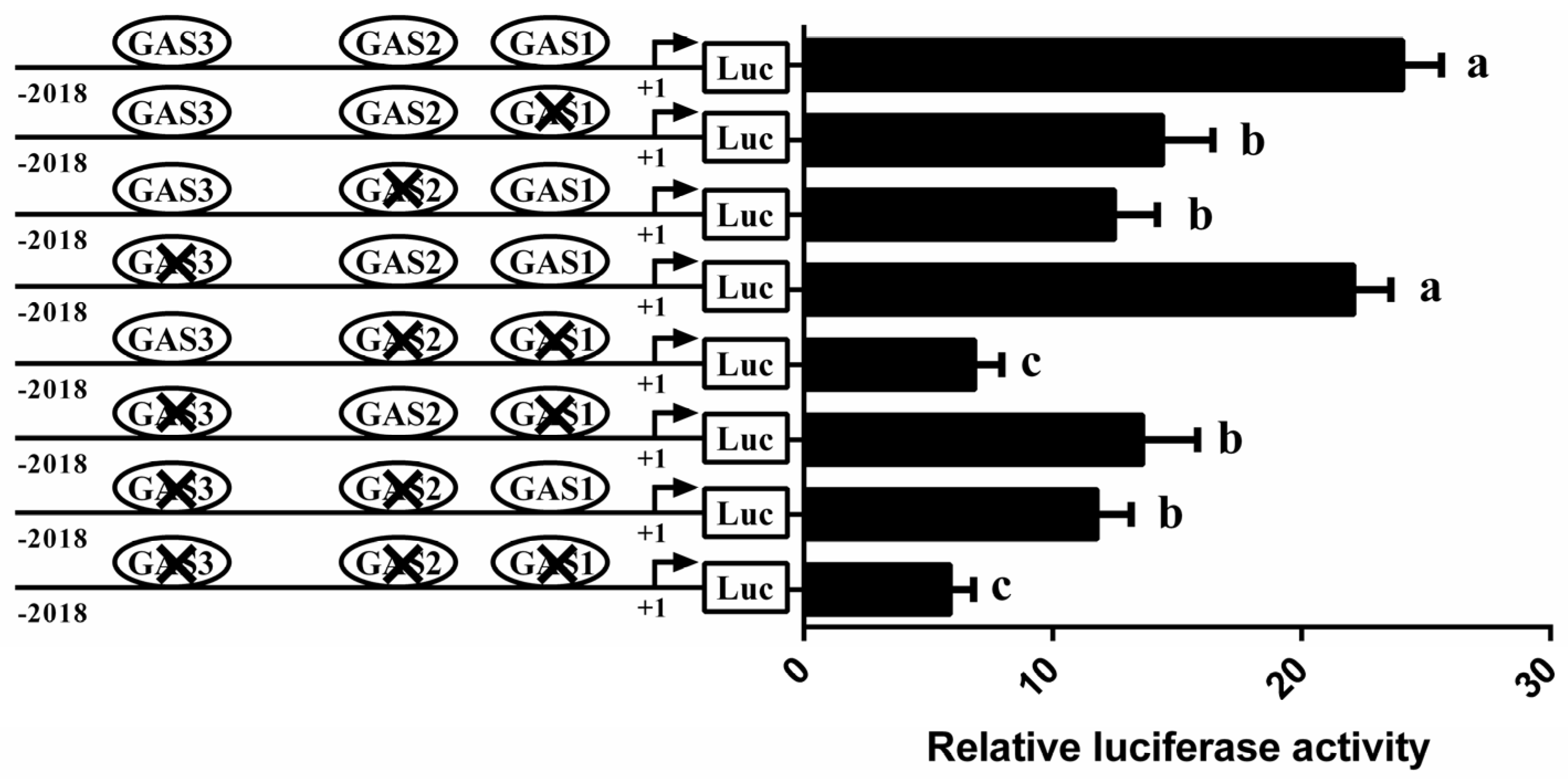
3.4. Involvement of STAT5 in Transcriptional Regulation of CSN1S1

Bioinformatics analysis revealed three conserved GAS binding sites in the CSN1S1 promoter region (Figure 1). To examine which elements are functional for STAT5-mediated CSN1S1 regulation, vectors containing GAS site-mutated (GAS1, GAS2 and GAS3) versions of the CSN1S1 promoter were transfected into GMEC for promoter activity assays. Compared with the wild-type promoter vector, GAS1 or GAS2 mutation decreased the CSN1S1 promoter activity by an average 40% and 48%, respectively (p < 0.05; Figure 4). In contrast, individual mutation of GAS3 could not affect the CSN1S1 promoter activity. Similarly, mutations of both GAS1 and GAS2 sites (or all 3 sites) resulted in very low promoter activity (Figure 4). These results show that the GAS1 and GAS2 binding sites of STAT5 are critical for CSN1S1 promoter activation.
Further, we overexpressed or inhibited STAT5 to identify the role of GAS1 and GAS2 elements in the regulation of CSN1S1 by STAT5. The GMEC were co-treated with pcDNA3.1-STAT5a (or STAT5-IN-1) and the site-mutated vectors. Results showed that STAT5a overexpression could also increase the CSN1S1 promoter activity during the individual GAS1 or GAS2 site mutation compared with the pcDNA3.1-NC control (p < 0.01, Figure 5A). However, double mutations of GAS1 and GAS2 sites could abolish the stimulatory effect of STAT5 on the CSN1S1 promoter activity (Figure 5A). Consistently, STAT5 inhibition markedly decreased the CSN1S1 promoter activity during the individual GAS1 or GAS2 mutation (p < 0.05; Figure 5B). While STAT5 inhibition could not affect the promoter activity during the double mutations of GAS1 and GAS2 binding sites (Figure 5B). Collectively, these results indicate that GAS1 and GAS2 are required for CSN1S1 promoter transcription in response to STAT5.

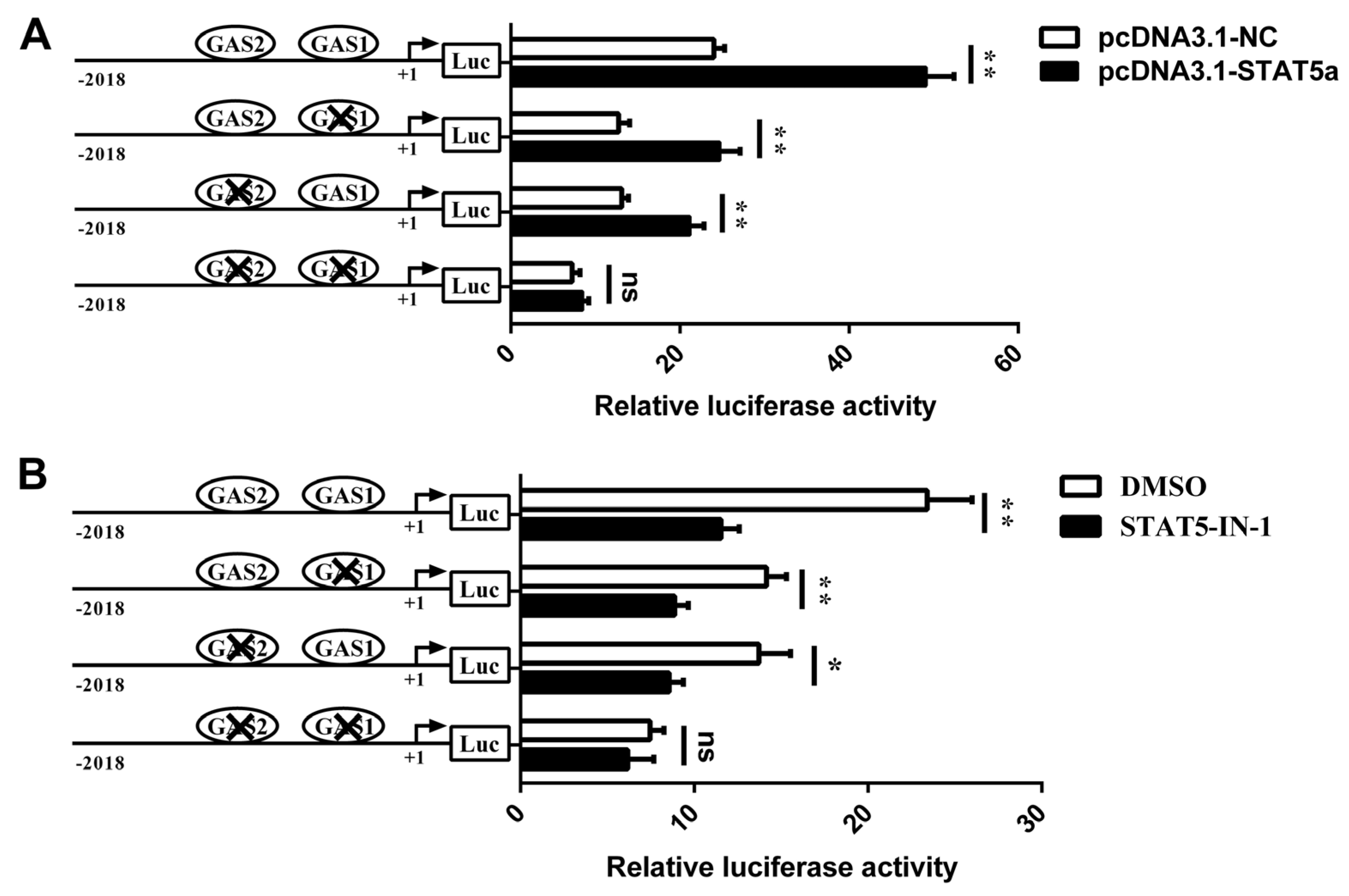
3.5. Prolactin Increases CSN1S1 Promoter Activity via STAT5

In mammary epithelial cells, prolactin could activate STAT5 phosphorylation. To explore whether prolactin affected CSN1S1 promoter activity and expression via STAT5 signaling, the GMEC seeded in the culture medium were treated with prolactin. Compared with the control, prolactin effectively increased the mRNA level and promoter activity of the CSN1S1 gene (p < 0.05, Figure 6A,B). Additionally, we found that STAT5a phosphorylation and αS1-casein synthesis were increased after incubation with prolactin (p < 0.05, Figure 6C), while STAT5-IN-1 abolished the stimulation of prolactin on p-STAT5a and αS1-casein expression (Figure 6C). These findings indicate that the prolactin-mediated increase of CSN1S1 expression might be involved with STAT5.
Further, we co-treated with GAS site-mutated vectors and prolactin in GMEC to identify the STAT5 binding sites involved in prolactin-induced CSN1S1 expression. Results showed that the prolactin-mediated increase of CSN1S1 promoter activity was reduced when a single mutation of GAS1 or GAS2 occurred (p < 0.01; Figure 6D). However, the stimulatory effect of prolactin on CSN1S1 promoter activity was completely abolished in GMEC transfected with the GAS1 and GAS2 simultaneously mutated CSN1S1 promoter (Figure 6D). Taken together, these results suggest that GAS1 and GAS2 sites are responsible for the activation of prolactin on the activity of the CSN1S1 promoter.

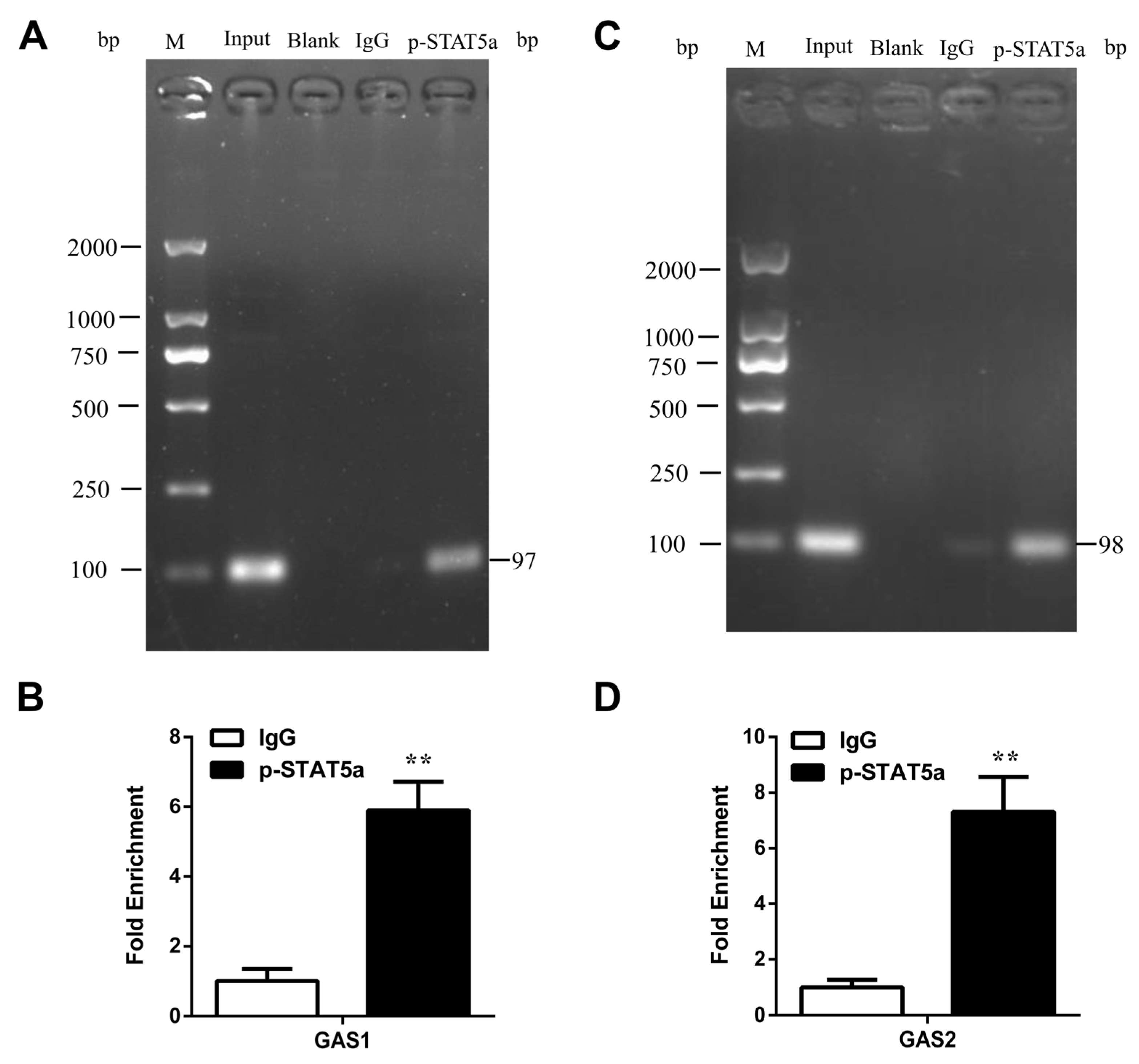
3.6. STAT5 Binds to the GAS Sites of CSN1S1 Promoter Region

To confirm whether the transcription factor STAT5 binds to the GAS1 and GAS2 sites on the CSN1S1 promoter region, ChIP assays were performed. We found that STAT5 interacted with the CSN1S1 promoter at the GAS1 and GAS2 sites (Figure 7A,C). qRT-PCR results showed that the fold enrichment levels of GAS1 and GAS2 sites were ~5.9 and 7.3, respectively (Figure 7B,D). Collectively, we conclude that STAT5 directly binds to GAS sites in the CSN1S1 promoter region, and that the GAS1 and GAS2 sites are necessary for STAT5 to regulate CSN1S1 transcription and αS1-casein synthesis.

4. Discussion

As one of the main components of milk protein, αS1-casein content is closely related to the milk allergy potential for humans [23]. The problem of αS1-casein allergy could be caused by cow milk, goat milk and other ruminant milk [4]. Reducing the αS1-casein content in goat milk and other ruminant milk is beneficial for the intake of dairy products for people with a milk protein allergy. The transcription of αS1-casein mainly depends on the activity of the CSN1S1 gene promoter in mammary epithelial cells [24]. Bioinformatics analysis revealed multiple polymorphism sites in the promoter region of the CSN1S1 gene [25,26]. According to the transcription factor binding sites in the CSN1S1 gene promoter, the locus mutation of the CSN1S1 promoter is associated with the transcription efficiency and the αS1-casein content in cow milk [27]. There are polymorphism and insertion–deletion sites in the promoter region of the CSN1S1 gene, which affect the protein content in mare milk [28]. Although the promoter sequence of the CSN1S1 gene has been well analyzed in silico, the research on its transcriptional regulation mechanism remains scarce [29]. In this study, we focus on the regulation mechanism of CSN1S1 promoter activity in goat mammary epithelial cells, which contributes to the improvement of milk quality for human consumption [30].
There are many transcription factor binding motifs in the CSN1S1 promoter region of dairy cows and goats, such as activating factor STAT5, GR, C/EBP, AP-1 and inhibitory factor YY1 [31,32]. The methylation of the milk protein gene promoter affects the binding of transcription factors to cis-acting elements and inhibits the expression of the milk protein [33]. During the lactation period of dairy cows, the methylation level of the specific site in the CSN1S1 promoter was negatively correlated with the CSN1S1 mRNA level in the mammary gland [34]. CpG island methylation of the STAT5 binding site in the CSN1S1 promoter led to the decrease in milk protein production in dairy cows [35]. In mammary epithelial cells, STAT5, GR and C/EBP could interact with acetyltransferase p300 and recruit RNA polymerase II, which affects β-casein gene transcription [36]. In the present study, we cloned 2201 bp sequence of the 5′-flanking promoter region of the goat CSN1S1 gene. In addition to the TATA-box near the transcription start site, the putative binding sites of transcription factors were comprehensively predicted by using JASPAR and TRANSFAC databases [37]. These transcription factors associated with milk protein synthesis [38], such as STAT5, may modulate the transcriptional activity of the goat CSN1S1 promoter.
In ruminants, the active center of gene promoter plays a vital role in transcription activity [39,40]. In order to determine the core promoter region of the CSN1S1 gene in dairy goats, we performed a segment-by-step deletion analysis starting from −2018 bp upstream of transcription start site [41]. When the CSN1S1 promoter fragment was deleted from −110 to −18 bp, the promoter activity decreased by 89%; therefore, −110~−18 bp upstream of transcription start site was identified as the active center of the goat CSN1S1 promoter [42]. The core promoter region contains two STAT5 binding sites (GAS1 and GAS2), thus we hypothesized that STAT5 may have a great effect on the transcriptional activity of the CSN1S1 gene. In addition, another potential STAT5 binding site (GAS3) may also exist in −1354~−1049 bp upstream. Sequence analysis of the CSN1S1 promoter region in bovine showed that there were three conserved STAT5 binding sites similar to the sequences of dairy goats [32]. Therefore, the investigation of CSN1S1 promoter activity in dairy goats could also provide the theoretical support for CSN1S1 transcriptional regulation in bovine, which contribute to the reduction of αS1-casein content in ruminant milk.
Previous studies have shown that STAT5 plays the crucial regulatory role in mammalian milk protein synthesis [43]. After the knockout of STAT5 in the mouse mammary gland, the content of α-lactalbumin and β-lactoglobulin in milk was reduced by 23% and 39%, respectively [15]. In addition to STAT5, mammalian target of rapamycin (mTOR) also regulates the synthesis of milk proteins such as α-lactalbumin and αS1-casein [44,45]. In bovine mammary epithelial cells, suppressor of cytokine signaling 3 inhibited STAT5a activity, and milk protein synthesis was significantly reduced after the phosphorylation of STAT5a was blocked [46,47]. In both mouse cell lines and immortalized GMEC, STAT5a binds to CSN2 promoter and promotes the transcription of β-casein [48]. In primary GMEC, STAT5a binds to the CSN2 promoter region, and STAT5 activity inhibition markedly reduced CSN2 promoter activity and β-casein synthesis [20]. Consistent with the previous studies, in the present study, the inhibition of STAT5 activity significantly reduced phosphorylated STAT5a in the nucleus, which functions as a transcription factor, and downregulated CSN1S1 mRNA level and αS1-casein synthesis. Site-directed mutagenesis of the three potential STAT5 binding sites in the CSN1S1 promoter region revealed that GAS1 and GAS2 sites in the core region could affect the promoter activity, while the distal GAS3 site had no significant effect on the promoter activity. Simultaneous mutation analysis of multiple sites showed that GAS1 and GAS2 sites could independently affect the promoter activity. The activity of the CSN1S1 promoter was severely weakened after simultaneous mutation of the two sites in the core region, and even the overexpression of STAT5a could not enhance its activity.
Prolactin is a key hormone regulating milk protein synthesis in mammals [49]. In bovine mammary epithelial cells, prolactin activates STAT5 phosphorylation via the prolactin receptor, promotes the expression of β-casein gene and maintains cell proliferation and milk protein synthesis [50,51]. Mouse mammary epithelial cells could not synthesize and secrete β-casein properly under the condition of prolactin deficiency, and prolactin induces β-casein synthesis by activating the proximal promoter and distal enhancer of the CSN2 gene [52]. Therefore, our study attempted to use prolactin to activate the phosphorylation of STAT5 in GMEC. Consistent with the results of direct inhibition of STAT5 activity, the promoter activity of the CSN1S1 gene was severely inhibited after the mutation of GAS1 and GAS2 sites. In this case, prolactin could also not activate its transcriptional activity, indicating that these two sites were the main binding sites of STAT5 in the CSN1S1 promoter. Compared with the site mutation analysis, the ChIP assay is the direct evidence to confirm the binding of transcription factors to the promoter region [40,42]. In the present study, ChIP assays showed that STAT5 could directly bind to GAS1 and GAS2 sites and promote the transcription of the CSN1S1 gene. These results indicate that STAT5 can directly regulate the transcription of the CSN1S1 gene and the synthesis of αS1-casein in GMEC. In addition, the complicated mechanism under which STAT5 binding site mutation at goat genome affects the synthesis of αS1-casein and other milk proteins requires further investigation.

5. Conclusions

The core promoter region of the goat CSN1S1 gene is located at −110~−18 bp upstream of transcription start site, which contains two STAT5 binding sites (GAS). STAT5 activates CSN1S1 gene promoter activity and αS1-casein synthesis. Furthermore, GAS1 and GAS2 sites in the core region of the CSN1S1 promoter are the binding sites of STAT5. Overall, this study reveals that STAT5 can directly regulate CSN1S1 transcription and αS1-casein synthesis through GAS1 and GAS2 sites. Our findings provide the novel basis for decreasing milk allergic reactions by mutating the STAT5 binding sites on αS1-casein gene promoter in ruminants.

Supplementary Materials

The following supporting information can be downloaded at: https://www.mdpi.com/article/10.3390/foods11030346/s1, Figure S1: Construction of deletion fragments pGL3-Basic vectors of the goat CSN1S1 gene promoter. Figure S2: Identification of GMEC by immunofluorescence with Cytokeratin 18.

Author Contributions

Conceptualization, N.S., J.L. and L.H.; methodology, N.S., S.Z. and Q.H.; software, formal analysis, data curation, and writing—original draft preparation, N.S.; validation, N.S. and L.H.; investigation, N.S., S.Z. and J.W.; resources, J.L.; writing—review and editing, L.H. and J.H.; supervision, J.L. All authors have read and agreed to the published version of the manuscript.

Funding

This research was funded by the National Natural Science Foundation of China (31772575), the Transgenic New Species Breeding Program of China (2018ZX0800802B) and the Innovation Support Plan of Shaanxi Province (2021TD-31).

Institutional Review Board Statement

The animal study protocol was approved by the Institutional Animal Care and Use Committee of Northwest A&F University, YangLing, China (protocol code 15–516, 13 September 2015).

Informed Consent Statement

Not applicable.

Data Availability Statement

Not applicable.

Conflicts of Interest

The authors declare no conflict of interest.

References

  1. Gillespie, A.L.; Green, B.D. The bioactive effects of casein proteins on enteroendocrine cell health, proliferation and incretin hormone secretion. Food Chem. 2016, 211, 148–159. [Google Scholar] [CrossRef] [PubMed] [Green Version]
  2. Park, Y.W.; Juarez, M.; Ramos, M.; Haenlein, G.F.W. Physico-chemical characteristics of goat and sheep milk. Small Rumin. Res. 2007, 68, 88–113. [Google Scholar] [CrossRef] [Green Version]
  3. Hochwallner, H.; Schulmeister, U.; Swoboda, I.; Focke-Tejkl, M.; Reininger, R.; Civaj, V.; Campana, R.; Thalhamer, J.; Scheiblhofer, S.; Balic, N. Infant milk formulas differ regarding their allergenic activity and induction of T cell and cytokine responses. Allergy 2016, 72, 416–424. [Google Scholar] [CrossRef] [PubMed] [Green Version]
  4. Clark, S.; Garcia, M.B.M. A 100-Year Review: Advances in goat milk research. J. Dairy Sci. 2017, 100, 10026–10044. [Google Scholar] [CrossRef]
  5. Fiocchi, A.; Schuenemann, H.J.; Brozek, J.; Restani, P.; Beyer, K.; Troncone, R.; Martelli, A.; Terracciano, L.; Bahna, S.L.; Rance, F.; et al. Diagnosis and Rationale for Action against Cow’s Milk Allergy (DRACMA): A summary report. J. Allergy Clin. Immun. 2010, 126, 1119–1128.e12. [Google Scholar] [CrossRef]
  6. Hochwallner, H.; Schulmeister, U.; Swoboda, I.; Spitzauer, S.; Valenta, R. Cow’s milk allergy: From allergens to new forms of diagnosis, therapy and prevention. Methods 2014, 66, 22–33. [Google Scholar] [CrossRef]
  7. Caubet, J.C.; Lin, J.; Ahrens, B.; Gimenez, G.; Bardina, L.; Niggemann, B.; Sampson, H.A.; Beyer, K. Natural tolerance development in cow’s milk allergic children: IgE and IgG4 epitope binding. Allergy 2017, 72, 1677–1685. [Google Scholar] [CrossRef]
  8. Schulmeister, U.; Hochwallner, H.; Swoboda, I.; Focketejkl, M.; Geller, B.; Nystrand, M.; Härlin, A.; Thalhamer, J.; Scheiblhofer, S.; Keller, W. Cloning, Expression, and Mapping of Allergenic Determinants of alpha S1-Casein, a Major Cow’s Milk Allergen. J. Immunol. 2009, 182, 7019–7029. [Google Scholar] [CrossRef]
  9. Clark, S.; Sherbon, J.W. Genetic variants of alpha(s1)-CN in goat milk: Breed distribution and associations with milk composition and coagulation properties. Small Rumin. Res. 2000, 38, 135–143. [Google Scholar] [CrossRef]
  10. Carillier-Jacquin, C.; Larroque, H.; Robert-Granie, C. Including a(s1) casein gene information in genomic evaluations of French dairy goats. Genet. Sel. Evol. 2016, 48, 54. [Google Scholar] [CrossRef] [Green Version]
  11. Shin, H.Y.; Hennighausen, L.; Yoo, K.H. STAT5-Driven Enhancers Tightly Control Temporal Expression of Mammary-Specific Genes. J. Mammary Gland Biol. 2019, 24, 61–71. [Google Scholar] [CrossRef] [PubMed]
  12. Radler, P.D.; Wehde, B.L.; Wagner, K. Crosstalk between STAT5 activation and PI3K/AKT functions in normal and transformed mammary epithelial cells. Mol. Cell. Endocrinol. 2017, 451, 31–39. [Google Scholar] [CrossRef] [PubMed]
  13. Maurer, B.; Kollmann, S.; Pickem, J.; Hoelbl-Kovacic, A.; Sexl, V. STAT5a and STAT5B-Twins with Different Personalities in Hematopoiesis and Leukemia. Cancers 2019, 11, 1726. [Google Scholar] [CrossRef] [PubMed] [Green Version]
  14. Li, S.; Huang, S.; Qiao, S.; Jiang, J.; Shi, D.; Li, X. Cloning and functional characterization of STAT5a and STAT5b genes in buffalo mammary epithelial cells. Anim. Biotechnol. 2020, 31, 59–66. [Google Scholar] [CrossRef]
  15. Reichenstein, M.; Rauner, G.; Barash, I. Conditional repression of STAT5 expression during lactation reveals its exclusive roles in mammary gland morphology, milk-protein gene expression, and neonate growth. Mol. Reprod. Dev. 2011, 78, 585–596. [Google Scholar] [CrossRef]
  16. Zhong, W.; Shen, J.; Liao, X.; Liu, X.; Zhang, J.; Zhou, C.; Jin, Y. Camellia (Camellia oleifera Abel.) seed oil promotes milk fat and protein synthesis-related gene expression in bovine mammary epithelial cells. Food Sci. Nutr. 2019, 8, 419–427. [Google Scholar] [CrossRef] [Green Version]
  17. Kung, M.H.; Lee, Y.J.; Hsu, J.T.; Huang, M.C.; Ju, Y.T. A functional study of proximal goat beta-casein promoter and intron 1 in immortalized goat mammary epithelial cells. J. Dairy Sci. 2015, 98, 3859–3875. [Google Scholar] [CrossRef]
  18. Song, N.; Luo, J.; Huang, L.; Tian, H.; Chen, Y.; He, Q. miR-204-5p and miR-211 synergistically downregulate the alpha(S1)-casein content and contribute to the lower allergy of goat milk. J. Agric. Food Chem. 2021, 69, 5353–5362. [Google Scholar] [CrossRef]
  19. Shi, H.; Shi, H.; Luo, J.; Wang, W.; Haile, A.B.; Xu, H.; Li, J. Establishment and characterization of a dairy goat mammary epithelial cell line with human telomerase (hT-MECs). Anim. Sci. J. 2014, 85, 735–743. [Google Scholar] [CrossRef]
  20. Song, N.; Chen, Y.; Luo, J.; Huang, L.; Tian, H.; Li, C.; Loor, J.J. Negative regulation of alpha(S1)-casein (CSN1S1) improves beta-casein content and reduces allergy potential in goat milk. J. Dairy Sci. 2020, 103, 9561–9572. [Google Scholar] [CrossRef]
  21. Yao, D.; Luo, J.; He, Q.; Shi, H.; Li, J.; Wang, H.; Xu, H.; Chen, Z.; Yi, Y.; Loor, J.J. SCD1 alters long-chain fatty acid (LCFA) composition and its expression is directly regulated by SREBP-1 and PPAR 1 in dairy goat mammary cells. J. Cell. Physiol. 2017, 232, 635–649. [Google Scholar] [CrossRef]
  22. Bionaz, M.; Loor, J.J. Identification of reference genes for quantitative real-time PCR in the bovine mammary gland during the lactation cycle. Physiol. Genom. 2007, 29, 312–319. [Google Scholar] [CrossRef]
  23. Archila, L.D.; Khan, F.S.; Bhatnagar, N.; Robinson, D.; Farrington, M.L.; Kwok, W.W. Alpha(S1)-Casein elucidate major T-cell responses in cow’s milk allergy. J. Allergy Clin. Immun. 2017, 140, 854–857. [Google Scholar] [CrossRef] [Green Version]
  24. Dong, X.; Zhou, Z.; Wang, L.; Saremi, B.; Helmbrecht, A.; Wang, Z.; Loor, J.J. Increasing the availability of threonine, isoleucine, valine, and leucine relative to lysine while maintaining an ideal ratio of lysine: Methionine alters mammary cellular metabolites, mammalian target of rapamycin signaling, and gene transcription. J. Dairy Sci. 2018, 101, 5502–5514. [Google Scholar] [CrossRef] [Green Version]
  25. Ahmed, A.; Rahmatalla, S.; Bortfeldt, R.; Arends, D.; Reissmann, M.; Brockmann, G. Milk protein polymorphisms and casein haplotypes in Butana cattle. J. Appl. Genet. 2017, 58, 261–271. [Google Scholar] [CrossRef]
  26. Noce, A.; Pazzola, M.; Dettori, M.L.; Amills, M.; Castello, A.; Cecchinato, A.; Bittante, G.; Vacca, G.M. Variations at regulatory regions of the milk protein genes are associated with milk traits and coagulation properties in the Sarda sheep. Anim. Genet. 2016, 47, 717–726. [Google Scholar] [CrossRef]
  27. Bonfatti, V.; Giantin, M.; Gervaso, M.; Coletta, A.; Dacasto, M.; Carnier, P. Effect of CSN1S1-CSN3 (alpha(s1)-kappa-casein) composite genotype on milk production traits and milk coagulation properties in Mediterranean water buffalo. J. Dairy Sci. 2012, 95, 3435–3443. [Google Scholar] [CrossRef]
  28. Cieslak, J.; Wodas, L.; Borowska, A.; Pawlak, P.; Czyzak-Runowska, G.; Wojtowski, J.; Puppel, K.; Kuczynska, B.; Mackowski, M. 5′-flanking variants of equine casein genes (CSN1S1, CSN1S2, CSN2, CSN3) and their relationship with gene expression and milk composition. J. Appl. Genet. 2019, 60, 71–78. [Google Scholar] [CrossRef] [Green Version]
  29. Ramos Matosinho, C.G.; Rosse, I.C.; Souza Fonseca, P.A.; de Oliveira, F.S.; Dos Santos, F.G.; Gomes Araujo, F.M.; de Matos Salim, A.C.; Lopes, B.C.; Arbex, W.A.; Machado, M.A.; et al. Identification and in silico characterization of structural and functional impacts of genetic variants in milk protein genes in the Zebu breeds Guzerat and Gyr. Trop. Anim. Health Prod. 2021, 53, 524. [Google Scholar] [CrossRef]
  30. Wal, J.M. Structure and function of milk allergens. Allergy 2001, 56, 35–58. [Google Scholar] [CrossRef]
  31. Bai, W.; Yin, R.; Dou, Q.; Yang, J.; Zhao, S.; Ma, Z.; Yin, R.; Luo, G.; Zhao, Z. A single nucleotide polymorphism and sequence analysis of CSN1S1 gene promoter region in Chinese BOS Grunniens (YAK). Anim. Biotechnol. 2010, 21, 36–41. [Google Scholar] [CrossRef]
  32. Ramunno, L.; Cosenza, G.; Rando, A.; Illario, R.; Gallo, D.; Berardino, D.D.; Masina, P. The goat alpha s1-casein gene: Gene structure and promoter analysis. Gene 2004, 334, 105–111. [Google Scholar] [CrossRef]
  33. Wu, Y.; Chen, J.; Sun, Y.; Dong, X.; Wang, Z.; Chen, J.; Dong, G. PGN and LTA from staphylococcus aureus induced inflammation and decreased lactation through regulating DNA methylation and histone H3 acetylation in bovine mammary epithelial cells. Toxins 2020, 12, 238. [Google Scholar] [CrossRef] [Green Version]
  34. Montazer-Torbati, M.B.; Hue-Beauvais, C.; Droineau, S.; Ballester, M.; Coant, N.; Aujean, E.; Petitbarat, M.; Rijnkels, M.; Devinoy, E. Epigenetic modifications and chromatin loop organization explain the different expression profiles of the Tbrg4, WAP and Ramp3 genes. Exp. Cell Res. 2008, 314, 975–987. [Google Scholar] [CrossRef]
  35. Nguyen, M.; Boutinaud, M.; Petridou, B.; Gabory, A.; Pannetier, M.; Chat, S.; Bouet, S.; Jouneau, L.; Jaffrezic, F.; Lalo, D. DNA methylation and transcription in a distal region upstream from the bovine alphaS1 casein gene after once or twice daily milking. PLoS ONE 2014, 9, e111556. [Google Scholar] [CrossRef]
  36. Buser, A.C.; Obr, A.E.; Kabotyanski, E.B.; Grimm, S.L.; Rosen, J.M.; Edwards, D.P. Progesterone receptor directly inhibits beta-casein gene transcription in mammary epithelial cells through promoting promoter and enhancer repressive chromatin modifications. Mol. Endocrinol. 2011, 25, 955–968. [Google Scholar] [CrossRef] [Green Version]
  37. Yao, D.; Luo, J.; He, Q.; Xu, H.; Li, J.; Shi, H.; Wang, H.; Chen, Z.; Loor, J.J. Liver X receptor alpha promotes the synthesis of monounsaturated fatty acids in goat mammary epithelial cells via the control of stearoyl-coenzyme A desaturase 1 in an SREBP-1-dependent manner. J. Dairy Sci. 2016, 99, 6391–6402. [Google Scholar] [CrossRef]
  38. Qian, X.; Zhao, F. Regulatory roles of Oct proteins in the mammary gland. Biochim. Biophys. Acta 2016, 1859, 812–819. [Google Scholar] [CrossRef] [Green Version]
  39. Li, S.; Khan, R.; Raza, S.H.A.; Jieyun, H.; Chugang, M.; Kaster, N.; Cheng, G.; Zhao, C.; Schreurs, N.M.; Zan, L. Function and characterization of the promoter region of perilipin 1 (PLIN1): Roles of E2F1, PLAG1, C/EBP beta, and SMAD3 in bovine adipocytes. Genomics 2020, 112, 2400–2409. [Google Scholar] [CrossRef]
  40. Xu, H.; Luo, J.; Ma, G.; Zhang, X.; Yao, D.; Li, M.; Loor, J.J. Acyl-CoA synthetase short-chain family member 2 (ACSS2) is regulated by SREBP-1 and plays a role in fatty acid synthesis in caprine mammary epithelial cells. J. Cell. Physiol. 2018, 233, 1005–1016. [Google Scholar] [CrossRef]
  41. Xu, H.F.; Luo, J.; Wang, H.P.; Wang, H.; Zhang, T.Y.; Tian, H.B.; Yao, D.W.; Loor, J.J. Sterol regulatory element binding protein-1 (SREBP-1)c promoter: Characterization and transcriptional regulation by mature SREBP-1 and liver X receptor a in goat mammary epithelial cells. J. Dairy Sci. 2016, 99, 1595–1604. [Google Scholar] [CrossRef] [Green Version]
  42. He, Q.; Luo, J.; Wu, J.; Li, Z.; Yao, W.; Zang, S.; Niu, H. ELOVL6 promoter binding sites directly targeted by sterol regulatory element binding protein 1 in fatty acid synthesis of goat mammary epithelial cells. J. Dairy Sci. 2021, 104, 6253–6266. [Google Scholar] [CrossRef]
  43. Geng, Z.; Shan, X.; Lian, S.; Wang, J.; Wu, R. LPS-induced SOCS3 antagonizes the JAK2-STAT5 pathway and inhibits beta-casein synthesis in bovine mammary epithelial cells. Life Sci. 2021, 278, 119547. [Google Scholar] [CrossRef]
  44. Cai, J.; Wang, D.; Zhao, F.; Liang, S.; Liu, J. AMPK-mTOR pathway is involved in glucose-modulated amino acid sensing and utilization in the mammary glands of lactating goats. J. Anim. Sci. Biotechnol. 2020, 11, 32. [Google Scholar] [CrossRef]
  45. Luo, C.; Zhao, S.; Zhang, M.; Gao, Y.; Wang, J.; Hanigan, M.D.; Zheng, N. SESN2 negatively regulates cell proliferation and casein synthesis by inhibition the amino acid-mediated mTORC1 pathway in cow mammary epithelial cells. Sci. Rep.-UK 2018, 8, 3912. [Google Scholar] [CrossRef]
  46. Khan, M.Z.; Khan, A.; Xiao, J.; Ma, Y.; Ma, J.; Gao, J.; Cao, Z. Role of the JAK-STAT pathway in bovine mastitis and milk production. Animals 2020, 10, 2107. [Google Scholar] [CrossRef]
  47. Huang, Y.; Zhao, F.; Luo, C.; Zhang, X.; Si, Y.; Sun, Z.; Zhang, L.; Li, Q.; Gao, X. SOCS3-mediated blockade reveals major contribution of JAK2/STAT5 signaling pathway to lactation and proliferation of dairy cow mammary epithelial cells in vitro. Molecules 2013, 18, 12987–13002. [Google Scholar] [CrossRef] [Green Version]
  48. Ghosh, A.; Radhakrishnan, R. Time-dependent antagonist-agonist switching in receptor tyrosine kinase-mediated signaling. BMC Bioinform. 2019, 20, 242. [Google Scholar] [CrossRef] [Green Version]
  49. Tian, M.; Qi, Y.; Zhang, X.; Wu, Z.; Chen, J.; Chen, F.; Guan, W.; Zhang, S. Regulation of the JAK2-STAT5 Pathway by Signaling Molecules in the Mammary Gland. Front. Cell Dev. Biol. 2020, 8, 604896. [Google Scholar] [CrossRef]
  50. Zhou, J.; Jiang, M.; Shi, Y.; Song, S.; Hou, X.; Lin, Y. Prolactin regulates LAT1 expression via STAT5 (signal transducer and activator of transcription 5) signaling in mammary epithelial cells of dairy cows. J. Dairy Sci. 2020, 103, 6627–6634. [Google Scholar] [CrossRef]
  51. Wang, B.; Men, J.; Wang, C.; Hou, X.; Zhao, F. Laminin-dependent integrin beta 1 signaling regulates milk protein synthesis via prolactin/STAT5 pathway in bovine mammary epithelial cells. Biochem. Bioph. Res. Commun. 2020, 524, 288–294. [Google Scholar] [CrossRef] [PubMed]
  52. Kobayashi, K.; Oyama, S.; Kuki, C.; Tsugami, Y.; Matsunaga, K.; Suzuki, T.; Nishimura, T. Distinct roles of prolactin, epidermal growth factor, and glucocorticoids in beta-casein secretion pathway in lactating mammary epithelial cells. Mol. Cell. Endocrinol. 2017, 440, 16–24. [Google Scholar] [CrossRef] [PubMed]
Figure 1. Schematic representation of the goat αS1-casein gene (CSN1S1) promoter region. The underlined sequence of bases indicates the putative binding sites, and names of transcription factors appear below the underline. +1 represents transcriptional start site and gamma-interferon activation site (GAS) represents the binding site of signal transducer and activator of transcription 5 (STAT5).
Foods 11 00346 g001
Figure 2. Deletion analysis of the goat CSN1S1 promoter. (A) Relative luciferase activity of full-length CSN1S1 promoter. (B) Relative luciferase activity of the CSN1S1 promoter in different lengths. Serial CSN1S1 promoter deletions (−2018/+183, −1715/+183, −1354/+183, −1049/+183, −598/+183, −346/+183, −110/+183, −18/+183 bp) were transfected into GMEC and incubated at 48 h for luciferase assays. Data are shown as means ± SEM for 3 biological replicates. ** p < 0.01. Differences between 2 groups are considered significant at lowercase letters, p < 0.05.
Foods 11 00346 g002
Figure 3. Effects of STAT5 on the expression and the promoter activity of the CSN1S1 gene in goat mammary epithelial cells (GMEC). (A) The mRNA expression levels of STAT5a and CSN1S1 and (B) the promoter activity of CSN1S1 in GMEC transfected with pcDNA3.1-STAT5a or pcDNA3.1-NC for 48 h. (C) The mRNA expression level and (D) the promoter activity of CSN1S1 in GMEC treated with STAT5 inhibitor STAT5-IN-1 (200 μM) or DMSO for 48 h. (E) GMEC were treated with STAT5-IN-1 (200 μM) or DMSO. After 48 h incubation, p-STAT5a and αS1-casein expression were examined. Relative expression of αS1-casein was normalized by comparison to β-actin. Relative expression of nuclear p-STAT5a was normalized by comparison to Histone H3. Data are shown as means ± SEM for 3 biological replicates. * p < 0.05, ** p < 0.01.
Foods 11 00346 g003
Figure 4. Relative luciferase activities of mutagenesis of STAT5 binding sites (GAS) in the CSN1S1 promoter region. Data are shown as means ± SEM for 3 biological replicates. Differences between 2 groups are considered significant at lowercase letters, p < 0.05.
Foods 11 00346 g004
Figure 5. STAT5 regulates the CSN1S1 promoter activity by directly binding to the GAS sites in the CSN1S1 promoter region. (A) Effects of STAT5a overexpression on site-mutations of CSN1S1 promoter activity. GMEC were transfected with the wild-type pGL3-CSN1S1 (or GAS site-mutated constructs) followed by pcDNA3.1-STAT5a (or pcDNA3.1-NC) transfection, respectively. At 48 h treatment, cells were measured for luciferase activity. (B) Effects of STAT5 inhibition on site-mutations of CSN1S1 promoter activity. GMEC were treated with STAT5-IN-1 (200 μM, or DMSO) before wild-type pGL3-CSN1S1 (or GAS site-mutated constructs) transfection, respectively. At 48 h treatment, cells were measured for luciferase activity. Data are shown as means ± SEM for 3 biological replicates. * p < 0.05, ** p < 0.01. ns, no significance.
Foods 11 00346 g005
Figure 6. Effects of prolactin on the expression and promoter activity of the CSN1S1 gene. (A) The mRNA expression level and (B) promoter activity of CSN1S1 in GMEC incubated with prolactin (2 mg/L) for 48 h in culture medium. (C) GMEC seeded in culture medium were treated with STAT5-IN-1 (200 μM, or DMSO) before prolactin (2 mg/L) treatment for 48 h, then total protein was extracted. Relative expression of αS1-casein was normalized by comparison to β-actin. Relative expression of p-STAT5a was normalized by comparison to STAT5a. (D) Relative luciferase activity of CSN1S1 promoter activity in GMEC co-treated with prolactin (2 mg/L) and the wild-type pGL3-CSN1S1 (or GAS site-mutated constructs) for 48 h in culture medium. Data are shown as means ± SEM for 3 biological replicates. ** p < 0.01. Differences between 2 groups are considered significant at lowercase letters, p < 0.05. ns, no significance.
Foods 11 00346 g006
Figure 7. Transcription of the CSN1S1 gene is regulated by direct STAT5 binding to GAS sites in GMEC. (A) GAS1 and (C) GAS2 binding sites in CSN1S1 promoter region were recognized by STAT5 directly using chromatin immunoprecipitation (ChIP)-PCR analysis. (B) GAS1 and (D) GAS2 binding sites in the CSN1S1 promoter region were recognized by STAT5 directly using ChIP-qPCR analysis. Data are shown as means ± SEM for 3 biological replicates. ** p < 0.01.
Foods 11 00346 g007
Table 1. Primers used for cloning, deletion and site-directed mutagenesis of the goat CSN1S1 gene.
PrimersPrimer Sequence (5′–3′)Binding Region
Cloning and 5′ deletion primers
Forward 1CGGGGTACCGGTGGCAAGAATAGTATTACAG−2018
Forward 2CGGGGTACCGACATCACTTTACTGATAAG−1715
Forward 3CGGGGTACCCAGATACACCTCTGGGAAAG−1354
Forward 4CGGGGTACCGATTCCTTTCTTATAAACAA−1049
Forward 5CGGGGTACCAACTTAGTGACTAAACAACA−598
Forward 6CGGGGTACCCTTCTCATGGTCTTGTACAT−346
Forward 7CGGGGTACCGTAAACTCTCCTCAGAATTTC−110
Forward 8CGGGGTACCGTTAAACTGAAACCACAAAAT−18
ReverseCCCAAGCTTCAACTGCGTATTAGTGAAGA+183
Site-directed mutagenesis primers
GAS1-mutCCTATGTGAGAGAAcgtTTAtgcTTTAAA−46
GAS1-anti-mutTAGGTTTATTTAAAgcaTAAacgTTCTCT−46
GAS2-mutACTCTCCTCAGAATcgtTTGtgcGAGGAA−92
GAS2-anti-mutCTGTTCAGTTCCTCgcaCAAacgATTCTG−92
GAS3-mutAAGTCCAGATACACgcaTGGacgAGACAAAA−1345
GAS3-anti-mutTACTCGACTTTTGTCTcgtCCAtgcGTGTAT−1345
Lowercase letters represent mutation sites, italics indicate restriction enzyme sites. Mut = mutagenesis forward primer; anti-mut = mutagenesis reverse primer.
Table 2. Primers used for quantitative real-time PCR.
GenBank IDGenePrimer Sequence (5′–3′)Length (bp)
XM_018049127.1CSN1S1F, TCCACTAGGCACACAATACACTGA61
R, GCCAATGGGATTAGGGATGTC
XM_018065112.1STAT5aF, CCATCGACCTGGACAATCCC96
R, CGACTTGGTGCTCTGCCTTCTT
XM_005700842.2UXTF, CAGCTGGCCAAATACCTTCAA125
R, GTGTCTGGGACCACTGTGTCAA
XM_005709411.1RPS9F, CCTCGACCAAGAGCTGAAG64
R, CCTCCAGACCTCACGTTTGTTC
ChIP primersGAS1F, GAGGAACTGAACAGAACATC97
R, GTAAAATGCTAATTTTGTGG
GAS2F, ACAATGCCATTCCATTTCCTAT98
R, AGGAAATCGATGTTCTGTTCAG
Publisher’s Note: MDPI stays neutral with regard to jurisdictional claims in published maps and institutional affiliations.

