Article

Isolation and Identification of Constituents Exhibiting Antioxidant, Antibacterial, and Antihyperuricemia Activities in Piper methysticum Root

1
Center for Research and Technology Transfer, Vietnam Academy of Science and Technology (VAST), Hanoi 122100, Vietnam
2
Transdisciplinary Science and Engineering Program, Graduate School of Advanced Science and Engineering, Hiroshima University, Hiroshima 739-8529, Japan
3
Agricultural Genetic Institute, Pham Van Dong Street, Hanoi 122000, Vietnam
4
Center for Agricultural Innovation, Vietnam National University of Agriculture, Hanoi 131000, Vietnam
5
Center for the Planetary Health and Innovation Science (PHIS), The IDEC Institute, Hiroshima University, Higashi-Hiroshima 739-8529, Japan
*
Author to whom correspondence should be addressed.
Foods 2022, 11(23), 3889; https://doi.org/10.3390/foods11233889
Submission received: 4 October 2022 / Revised: 26 November 2022 / Accepted: 28 November 2022 / Published: 1 December 2022

Abstract

:
The aqueous extract of kava (Piper methysticum) root is known as a traditional beverage for daily intake in the Western Pacific Islands, such as Fiji, Tonga, and Vanuatu, to induce relaxation and health-beneficial effects. In this study, the antioxidant, anti-hyperuricemia, and antibacterial properties of kava root were investigated through the isolation and purification of bioactive compounds in ten fractions separated by column chromatography (CC). They included six flavonoids, 5-hydroxy-4′,7-dimethoxyflavanone (C1), matteucinol (C2), isosakuranetin (C3), 5,7- dimethoxyflavanone (C4), 2′,4′-dihydroxy-6′-methoxydihydrochalcone (in MC5) and alpinetin (C10), and seven kavalactones, 5,6-dehydrokawain (DK) (in MC5 and C6), kavain (in MC7), yangonin (in MC7 and C8), dihydro-5,6-dehydrokavain (DDK) (in MC9), 7,8-dihydromethysticin (in MC9), dihydromethysticin (in MC9), methysticin (in MC9). The chemical structures of the compounds were illustrated by the analyses of gas chromatography–mass spectrometry (GC–MS), electrospray ionization–mass spectrometry (ESI–MS), nuclear magnetic resonance (1H and 13C-NMR), and X-ray diffraction. The evaluation of the free radical scavenging activity of the isolated substances via the DPPH and ABTS assays revealed that C3 (IC50: ABTS = 76.5; DPPH = 74.8 µg/mL) possessed the strongest antioxidant property. In terms of anti-hyperuricemia activity evaluated via the xanthine oxidase inhibitory in vitro assay, the compound C10 was the most promising inhibitor, revealing an IC50 of 134.52 µg/mL. The two kavalactone mixtures in MC5 and a pure compound C6 inhibited the growth of bacteria Listeria monocytogenes, while MC7 can constrain the development of Klebsiella pneumoniae. This is the first study to isolate, purify, and identify the flavonoids isosakuranetin, 2′,4′-dihydroxy-6′-methoxydihydrochalcone and alpinetin in kava root and report their pharmaceutical potential. The identified bioactive compounds showed potent antioxidant, anti-hyperuricemia, and antibacterial activity and thus can enhance the value of beverages and foods derived from kava root.

1. Introduction

Free radicals are described as highly reactive and unstable molecules, which is caused by an unpaired electron [1,2]. They are generated either from the normal cellular metabolism process or from external sources including pollution, cigarette smoke, radiation, and medication. The overproduction of free radicals and reactive oxygen-containing molecules in the human body affects important macromolecules, which then causes significant damage to cell structures and disrupts the homeostatic balance [3,4]. This further leads to a wide range of chronic and degenerative diseases, such as cancer, cardiovascular diseases, inflammation, diabetes, and neurodegeneration [5]. Consequently, antioxidant and antibacterial compounds isolated from medicinal plants gain particular interest as they are potential agents in neutralizing the adverse effects of harmful free radicals and enhancing the oxidative stability [6,7,8]. They can be effectively used in food additives, cosmetic products, and pharmaceutical drugs [8,9].
Xanthine oxidase (XO) is an important biological generator of oxygen free radicals. This is a key enzyme involved in the metabolism of hypoxanthine to xanthine, which was successively converted into uric acid by the oxidation process [10,11,12,13]. The excessive production of uric acid in blood results in a condition known as hyperuricemia. This further leads to the formation of the monosodium urate crystal and causes painful gouty inflammation [5]. Recently, there has been an increase in the prevalence of hyperuricemia worldwide [5,14,15]. To date, only three synthetic XO inhibitors, i.e., allopurinol, febuxostat and oxypurinol, have been approved as hyperuricemia treatments and anti-gout agents [16]. However, their side effects, such as skin rash, allergic reactions and kidney failure, have been highlighted. As a result, nature-based XO inhibitors are more preferable and need further investigations of their safety and efficiency.
Kava (Piper methysticum), a member of the pepper family, is an exclusive perennial plant found on the Pacific Islands. Due to the effect of relaxation, resulting in the enhancement of social ties and conflict avoidance, the aqueous extract from kava is a preferred beverage during important events or community gatherings on several islands, such as Fiji, Tonga and Vanuatu [17]. This herbal plant also holds a significant place in the local residents’ daily intake, as it has many health benefits [18,19,20]. Kava root has traditionally been used by indigenous people as an effective folk remedy for a variety of diseases, such as gonorrhea, menstrual pain, tuberculosis, arthritic conditions, and chronic gout [21,22,23]. Kava consumption has also increased, and it has become popular in new regions, including Australia, Europe and North America, where it is sold at medicinal and recreational markets [24]. The various chemical compositions of kava have been reported in a number of studies, including more than 40 compounds within the classes of kavapyrones, alkaloids, steroids, chalcones, long-chained fatty acids and alcohols [18]. Among them, a group of components known as kava pyrones or kava lactones is believed to be responsible for the therapeutic effects [18,25]. Although the pharmaceutical benefits of kava in treating inflammatory arthritis were described above, no research on phytochemicals attributed to this property has been conducted so far. To answer the question of whether other compounds besides kava lactones are associated with the anti-gout and hyperuricemia effects, research on further bioactive components is needed.
In this work, the isolation and purification of the active compounds, employing column chromatography (CC), was performed, followed by the identification of its bioactivity, including in vitro assays of XO inhibitory, antioxidant and antibacterial activity. The chemical structures of the isolated second metabolites were illustrated by gas chromatography–mass spectrometry (GC–MS), electrospray ionization–mass spectrometry (ESI–MS), nuclear magnetic resonance (1H and 13C-NMR) and X-ray diffraction analysis. Therefore, the additional value of beverages and foods derived from kava root is highlighted.

2. Materials and Methods

2.1. Plant Material

The dried root part of commercial kava plant was obtained from Kava King Products, Inc. (Orchard St, Ormond Beach, FL, USA) on 2 May 2017. The powder sample was stored at −20 °C in the freezer prior to experiments.

2.2. Isolation and Purification Procedure of Antioxidant Compounds

The isolation process of bioactive compounds in kava root was reported by Van et al. [26]. The ground kava root (165.9 g) was first immersed in acetone–acetonitrile (4:1) at room temperature for one week. The filtrate was obtained with filter paper (Whatman No. 5C, 110 mm), followed by evaporation using a rotary evaporator (SB–350–EYELA, Tokyo Rikakikai Co., Ltd., Tokyo, Japan) at 45 °C to yield 9.4 g crude extract. After this, the crude extract was dissolved in distilled water, and then extracted successively with hexane and EtOAc. The crude extract (6.5 g) obtained by evaporating the EtOAc layer was further chromatographed into a column ((20 mm diameter × 500 mm height, Climbing G2, Tokyo, Japan) over a silica gel (size Ǻ 60, 200–400 mesh particle size, Sigma-Aldrich, Darmstadt, Germany) and eluted with a gradient of n-hexane–EtOAc (10:0–0:10 v/v). The fractions were collected separately (frs.; each of 100 mL) and concentrated under a rotary evaporator. This was followed by inspection via thin layer chromatography (TLC) to verify the purity of compounds and determine the number of components. The recorded similar spots on TLC sheets were combined. Fractions were then crystallized at room temperature to give thirteen substances, as described in Figure 1. Compounds C1, C2, C3, C4, C6, C8 and C10 were purified after elution by hexane, followed by rapid filtering through filter paper to remove traces of impurities. The yields of the 10 fractions were 7.7, 54.7, 4.7, 26.6, 28.9, 174.4, 518, 70.3, 49.0 and 22.0 mg, respectively (Figure 1). The chemical formulas of isolated substances were determined by gas chromatography–mass spectrometry (GC–MS), electrospray ionization–mass spectrometry (ESI–MS) and X-ray analysis. The new compounds with high purity levels were further subjected to either 1H and 13C nuclear magnetic resonance (NMR) or X-ray analysis for the elucidation of their molecular structures (Supplementary Table S1).

2.3. Identification of Antioxidative Compounds

The active compounds and mixtures were analyzed by a GC–MS machine (JMS-T100 GCV, JEOL Ltd., Tokyo, Japan) equipped with a DB-5MS capillary column, 30 m in length, with a 0.25 mm internal diameter and 0.25 µm in thickness (Agilent Technologies, J & W Scientific Products, Folsom, CA, USA). The column temperature was set initially at 50 °C without a hold time, followed by an increase to 300 °C with the gradient of 10 °C/min and held for 20 min. The temperatures of the injector and detector were programmed, respectively, at 300 °C and 320 °C, with a mass scan range of 29–800 amu. The compounds were determined via a comparison of their mass spectral fragmentation patterns with the mass spectral libraries of JEOL’s GC-MS Mass Center System Version 2.65a. The compounds with high purity were subjected to further spectroscopic techniques for structure elucidation.
ESI/MS analysis was implemented using a mass spectrometer with an electrospray ion source. The measurements were conducted in the positive mode with an ion spray voltage of 3000 V and a capillary temperature of 350 °C. The mass spectra were recorded in full scan mode within the range of 280 to 1000 m/z.
The nuclear magnetic resonance spectroscopy (1H (400.13 Hz) and 13C (100.612 Hz)) were recorded using a Bruker 500 model spectrometer (BRUKER BioSpin, Faellanden, Switzerland). The pure compounds were dissolved in chloroform (CDCl3) to obtain a concentration of 3000 ppm (µg/mL). The spectral characteristics of compounds were compared with those in the literature to elucidate the chemical structures.
The X-ray diffraction pattern was recorded using a single-crystal X-ray diffractometer from Bruker, connected with a highly sensitive ApexII CCD area detector. The single crystal was packed tightly in the sample holders and freeze-dried at the temperature of −100 °C. The average wavelength was 0.71073 Å. The obtained X-ray powder diffraction patterns of the samples were compared with a database from the website of the International Union of Crystallography. Finally, the compound structures of the samples were reported in a crystallographic information file (CIF).

2.4. Bioactivity Assays

2.4.1. Antioxidant Assays

DPPH Radical Scavenging Assay

The 2,2-diphenyl-1-picrylhydrazyl (DPPH) radical scavenging activity assay was conducted according to the method described previously by Minh et al. [27], with minor modification. The various polarity extracts were evaluated at concentrations of 500, 1000, 2000 and 3000 ppm, while the content of isolated compounds was 50, 100, 500, 1000 and 1500 ppm. A volume of 100 µL of each sample was mixed with 50 µL DPPH 0.5 mM, followed by the addition of 100 µL sodium acetate buffer (pH 5.5, 0.1 M). In this study, BHT and methanol (MeOH) were used as a positive standard and control, respectively. After incubation in the dark for 30 min at room temperature, the mixture’s absorbance (Abs) was measured at 517 nm using a UV–VIS spectrophotometer (Multiskan GO; Thermo Fisher Scientific, Vantaa, Finland). This assay was implemented in triplicate and the antioxidant activity was calculated with the following equation:
DPPH radical scavenging activity (%) = [(Abscontrol – Abssample)/Abscontrol] × 100
where Abscontrol is the absorbance of the control reaction, and Abssample is the absorbance of the test substance. Based on the value of DPPH radical scavenging activity, the IC50 value was calculated to evaluate the antioxidant ability of the tested samples. A smaller value of IC50 is associated with stronger antioxidant properties.

ABTS Radical Scavenging Assay

The ABTS radical scavenging capacity was determined by the ABTS cation decolorization assay, as described by Tuyen et al. [28]. The preparation of samples was in the same manner as the DPPH assay. The ABTS+ radical cation was produced by reacting 15 mL aqueous solution of ABTS 7 mM with 240 µL potassium persulfate at 140 mL. The mixture was kept in the dark at room temperature for 16 h before use. Prior to the assay, the ABTS+ was diluted with MeOH to achieve absorbance of 0.70 ± 0.05 at 734 nm. The reaction mixture contained 1 mL of prepared ABTS+ solution and 0.12 mL of each sample was measured after a 30-min incubation period in the dark at room temperature. The ABTS radical scavenging activity was expressed as in the following equation:
ABTS radical scavenging activity (%) = [(Abscontrol – Abssample)/Abscontrol] × 100
where Abscontrol is the absorbance of the ABTS radical + methanol; Abssample is the absorbance of the ABTS radical + sample extract/standard.

2.4.2. Xanthine Oxidase Inhibitory Activity

The inhibitory effect on xanthine oxidase of different extracts and isolated compounds was quantitated spectrophotometrically according to the method reported previously [29], with minor modifications. In this assay, allopurinol (6.25, 12.5, 25, 50 µg/µL) was used as a reference standard. The mixture consisting of 50 µL sample solution (3000, 2000, 1000, 500, 250, 125 and 67.5 ppm), 35 µL phosphate buffer (70 mM, pH = 7.5) and 30 µL fresh enzyme solution (0.01 units/mL in the same buffer) was prepared immediately before use. After pre-incubation at 25 °C for 15 min, the reaction was initiated by adding 60 µL of substrate solution (150 µM xanthine in the same buffer). The mixture was subsequently incubated at 25 °C for 30 min. The reaction was stopped by adding 25 µL HCl (1 M) and the absorbance was recorded at 290 nm using a microplate reader, as described above. A blank was prepared in the same manner, but the enzyme solution was added after pipetting HCl. XO inhibitory activity was expressed as the percentage inhibition of XO and calculated as follows:
%   Inhibition = { ( A B ) ( C D ) ( A B ) }   ×   100
where A is the activity of the enzyme without tested samples, and B is the control of A with neither tested samples nor enzyme. C and D denote the activity of the test solutions with and without XO, respectively. The IC50 value was calculated from the mean values of the data.

2.4.3. Antibacterial Assay

The antibacterial activity of kava root was tested against five types of bacterial strains, namely Pseudomonas aeruginosa, Escherichia coli, Klebsiella pneumoniae, Listeria monocytogenes and Bacillus subtilis. This test was performed following a disc dilution method reported by Elzaawely et al. [30], with minor modifications. To begin with, 9-cm-diameter Petri dishes were coated with a 15 mL layer of nutrient agar, which was allowed to solidify. One hundred microliters of test organisms (106 colony forming units (CFU)/mL)), after being cultured in nutrient broth media for 24 h, was laid evenly over the surface of the agar plate. A volume of 20 microliters of each sample, impregnated in a sterile filter paper disk (6 mm in diameter), was placed on the surface of the Petri dish. Ampicillin and streptomycin were used in this experiment as positive controls, and MeOH was utilized as a negative control. After 24 h incubation at 37 °C, bacterial inhibition was determined by measuring the diameter of the inhibition zone in millimeters, followed by comparison with standards and controls.

2.5. Statistical Analysis

The statistical analysis was performed by one-way ANOVA using Minitab® 16.2.3 (copyright © 2022 Minitab Inc., Philadelphia, PA, USA). The results were reported as mean ± standard deviation values. Differences among treatment, control and standard data are considered significant at p < 0.05 using Tukey’s test.

3. Results

3.1. Phytochemical Isolation and Structure Elucidation

The most active EtOAc extract of kava root was subjected to CC and yielded ten fractions. The combination of hexane (H) and ethyl acetate (E) at ratios 9:1, 8:2 and 7:3 was the best method to yield secondary metabolites from kava root. Among them, H:E 8:2 was the most efficient elution to afford kava lactones.
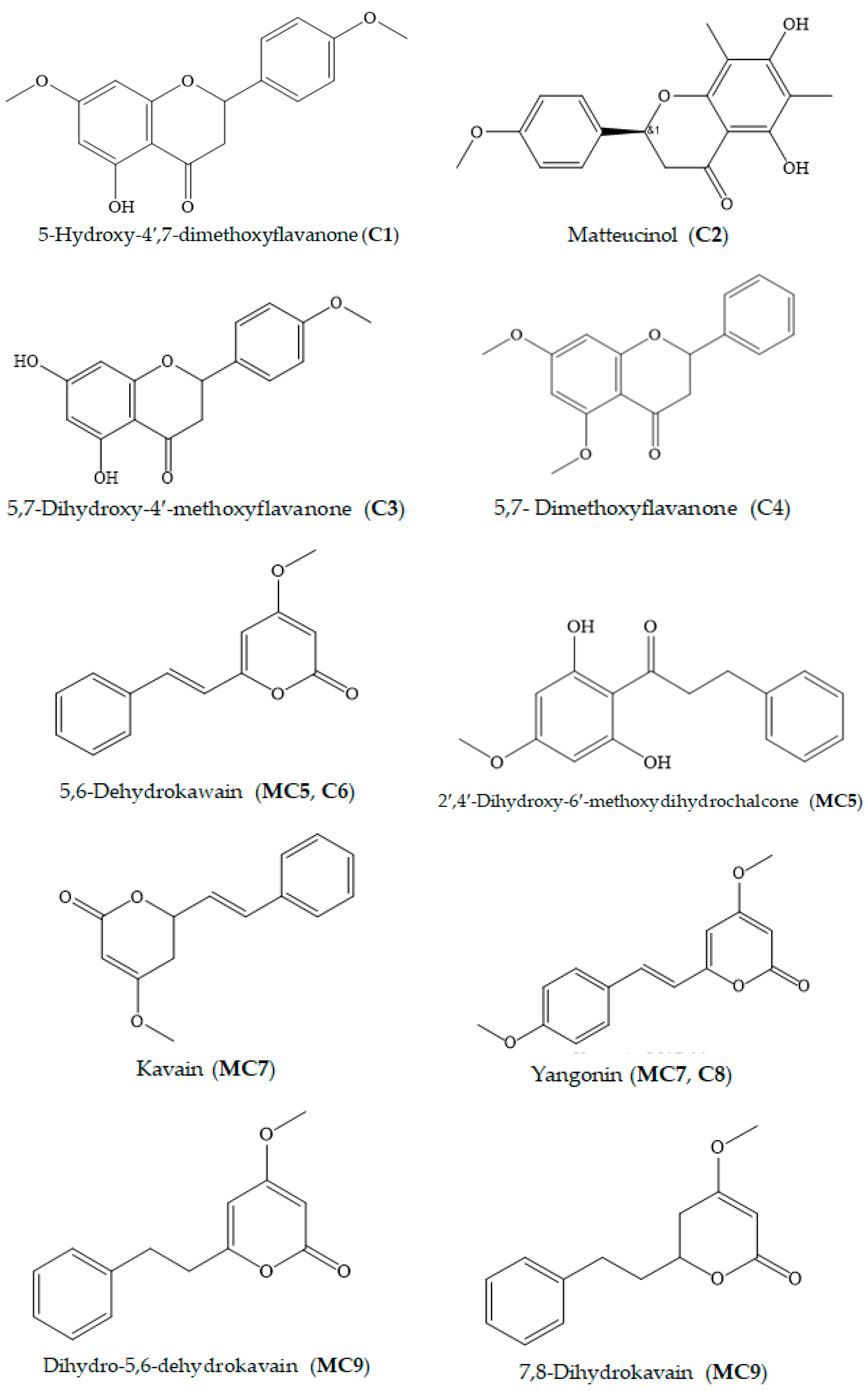
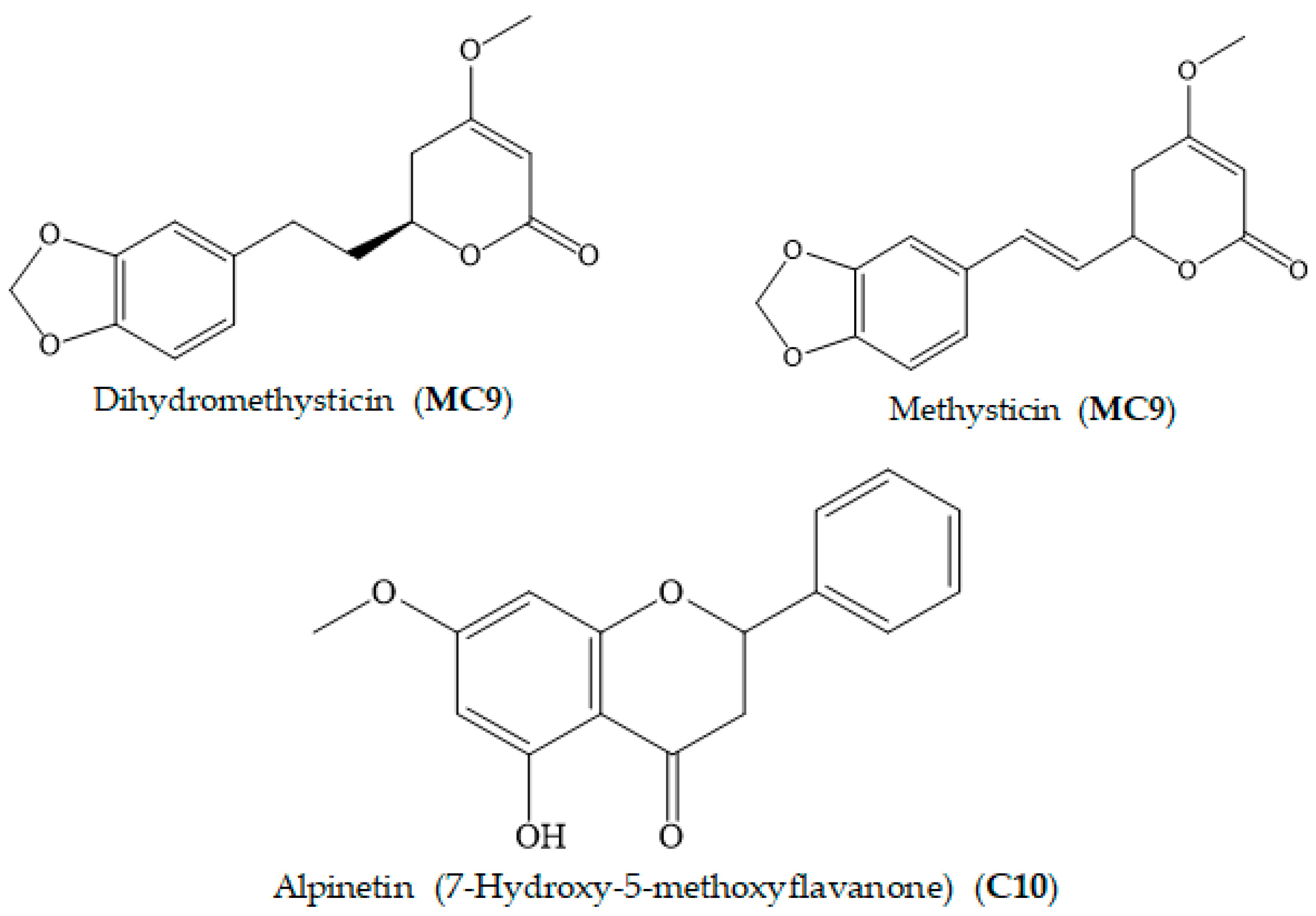
The isolated metabolites were characterized and identified by GC–MS, ESI–MS, 1H and 13C NMR and X-ray analyses. The structure elucidation of compounds C1, C2, C6, MC7, C8 and MC9 was described previously in Van et al. 2018 [26]. The chemical structures of C3 and C4 were determined based on spectroscopic analyses and compared with previous literature data (Supplementary Data Figures S1–S6, Table S1). The X-ray technique was applied to identify the structures of 2′,4′-dihydroxy-6′-methoxydihydrochalcone and alpinetin. The obtained constituents belonged to the following groups. Flavanones: 5-hydroxy-4′,7-dimethoxyflavanone (C1), matteucinol (C2), 5,7-dihydroxy-4′-methoxyflavone (isosakuranetin, C3), 5,7- dimethoxyflavanone (C4) and alpinetin (C10). Kavalactones: DK (MC5 and C6), kavain (MC7), yangonin (MC7, C8), DDK (MC9), 7,8-dihydromethysticin (MC9), dihydromethysticin (MC9) and methysticin (MC9). Chalcones: 2′,4′-dihydroxy-6′-methoxydihydrochalcone (MC5) (Table 1, Figure 2, Supplementary Data Figures S1–S10, CIF1-2). Among the isolated components, the compounds isosakuranetin (C3), isosakuranetin (MC5) and alpinetin (C10) were first identified from kava root.
The spectroscopic results of C3 were interpreted as follows: compound 3, obtained as yellow crystals, has molecular formula C16H15O5, determined by ESI-HRMS at 287.09131 (M+H)+ (calcd. 287.09140). The 1H and 13C NMR spectrum of C3 displayed a typical flavanone-type peak pattern. The signal’s characteristic 1H NMR spectrum of C3, 5,7-dihydroxy-4′-methoxyflavanone, was observed at dH 12.2 (OH chelated), 7.39–7.46 (5H, m) for aromatic protons, and peak combination from dH 2.8, 3.1 and 5.4 could be assigned for the saturated of C-ring of the flavonoid. The B-ring was p-substituted by a phenyl group since both of the signal intensities at dC 128.9 and 126.1 were two-fold higher compared to others based on the signal pattern of the 13C NMR spectrum of C3 (Supplementary Table S1).

3.2. Biological Activity of Crude Extracts

3.2.1. Xanthine Oxidase Inhibitory (XOI) and Antioxidant Activity of Crude Extracts

The various extracts of kava root were investigated for XOI and free radical scavenging capacity using in vitro assays. The results, expressed as IC50 values, are summarized in Table 2. The IC50 was used as a parameter; a lower IC50 value represents a stronger capacity.
Among the studied extracts, the EtOAc extract exhibited the strongest inhibitory effects on both antioxidant and XOI activity. In the radical scavenging assays, the IC50 values of the EtOAc extract were 946 and 1007 µg/mL, as measured by the DPPH and ABTS methods. The antioxidant property of tested extracts decreased in the following order: EtOAc > water > chloroform > hexane. Meanwhile, the order of XOI activity was EtOAc > chloroform > hexane > water. The greatest inhibitory activity of the EtOAc extract revealed that it contained the most abundant active compounds. Therefore, this fraction was chosen for isolation and purification.

3.2.2. Antibacterial Activity of Crude Extracts

The antibacterial activity of the kava root extracts against five human pathogenic bacteria was examined using the agar diffusion method. Ampicillin and streptomycin were used as standards and MeOH was used as a control. The corresponding zones of inhibition diameters are presented in Table 3, in which a higher value shows a stronger growth reduction. In all treatments, ampicillin and streptomycin demonstrated significantly higher suppression against the five studied bacteria compared to tested samples.
The result of a preliminary screening of the extracts indicated that although no extract could inhibit all tested species, each specific extract had a negative influence on the growth of a certain type of bacteria. The polar extract of EtOAc exhibited strong antibacterial activity against the E. coli strain; its zone of growth inhibition was 23 mm. The B. subtilis bacterial strain was observed as the most sensitive bacterium to the chloroform extract, with an inhibition zone of 15 mm. The non-polar hexane extract showed a potential effect against P. mirabilis as the inhibition zone diameter was 10 mm. Among the tested bacteria, K. pneumoniae and L. monocytogenes showed the strongest resistance against all extracts from kava root.

3.3. Biological Activity of Isolated Compounds

3.3.1. Antioxidant Activity of Isolated Compounds

The antioxidant activity of the isolated constituents, estimated by the ABTS and DPPH methods, is presented in Table 4. The results varied among the tested compounds and studied assays. All isolated compounds except MC7 were effective in scavenging ABTS free radicals. The ABTS scavenging activity of four fractions followed the order C3 > C4 > C2 > MC5 > C1 > C6. Meanwhile, the compounds C3, C4 and C6 were able to reduce the stable DPPH radicals and their antioxidant capacity can be ranked as follows: C3 > C4 > C6. Among six fractions isolated from the EtOAc extract, C3 showed the highest inhibitory potential against ABTS and DPPH free radicals, with similar IC50 results (76.5 and 74.8 µg/mL, respectively). By contrast, MC7, C8, MC9 and C10 exhibited negligible antioxidant activity.

3.3.2. XOI Activity of Isolated Compounds

The fractions C4, MC5, C6, MC7, C8, MC9 and C10 of kava root were examined for their inhibitory effects on the uric acid production from xanthine and xanthine oxidase (Table 5). The other fractions, C1C3, with limited quantities, were not tested for this assay. The results showed that most of the tested fractions exhibited an XOI capacity that was comparable to that of allopurinol. The compound C10 was the most promising XO inhibitor (IC50 = 134 µg/mL), followed by MC7 and C4 (IC50 = 242 and 268 µg/mL, respectively). The fractions C6 and MC9 displayed moderate inhibition against the activity of XO, with IC50 values of 452 and 465 µg/mL, respectively. Among the studied fractions, only compound C8 showed trivial inhibition (Table 5).

3.4. Antibacterial Activity

In this study, several compounds isolated from the EtOAc extract were found to be responsible for the antibacterial activity of kava root (Table 6). The antibacterial activity of compounds C1C4 was not examined because their quantity constraints were not sufficient for the antibacterial experiment. Compound MC5 was the most toxic compound against the L. monocytogenes strain, followed by C6. Their zones of inhibition were 13 mm and 9 mm, respectively. Compound MC7 was found to be only potent as an antibacterial agent against K. pneumoniae, as it showed a 9.67 mm zone of inhibition. Two Gram-negative bacteria, i.e., E. coli and P. mirabilis, and one Gram-positive bacterium, B. subtilis, were not susceptible to any of the studied compounds.

4. Discussion

Flavonoids are a large class of natural products characterized by a flavone ring structure [31,32,33]. Flavonoid compounds are well known to possess potential inhibitory action towards free radicals and the XO enzyme, which is responsible for oxidative injury [34]. In this study, the bioassay-guided isolation technique was applied by using in vitro DPPH and ABTS free radical scavenging and XO assays to examine the antioxidant and XOI properties of different polarity extracts and chromatographic fractions. This led to the successful isolation of thirteen components, including six flavonoids (C1, C2, C3, C4, MC5 and C10) and seven kavalactones (MC5, C6, MC7, C8 and MC9). Quantitative analyses revealed various degrees of antioxidant effects in the identified compounds. Among the isolated fractions, flavanones exhibited superior activity as compared with kavalactones (Table 4 and Table 5). The flavonoid C3, with a catechol moiety on the B ring, showed the strongest antioxidant activity in both antioxidant assays. This was followed by C4, C2 and C1, which are structured with more methoxylated groups. In terms of XO-inhibitory activity, the presence of a free hydroxyl group in the flavonoid C10 might be associated with a stronger inhibitory effect compared with the compound C4 (Table 5). The obtained results are consistent with the findings of earlier studies that emphasized the important role of the number and location of OH groups in the radical scavenging and XO activity of flavonoids [35,36,37,38,39,40,41]. Moreover, flavonoids having both XOI and antioxidant activity might be of advantage as potentially applicable compounds to treat gout [34].
Kava lactones have been demonstrated to be involved in the therapeutic value of kava root in the previous literature. In this study, the antioxidant and XOI potential of other constituents besides flavonoids was found in the compound DK (C6) and the mixture of DK and 2′,4′-dihydroxy-6′-methoxydihydrochalcone (MC5). Among them, MC5 exerted a synergistic effect to inhibit ABTS free radicals, which resulted in higher radical scavenging activity compared with other kavalactones (C6, MC7, C8 and MC9) (Supplementary Table S1). However, in comparison with the compound C6, the presence of 2′,4′-dihydroxy-6′-methoxydihydrochalcone reduced the antioxidant activity of the mixture MC5 assayed by the DPPH method. In the present study, yangonin (MC7) did not show an antioxidant capacity. This outcome is contrary to that of a previous study that indicated that yangonin exhibited DPPH radical scavenging ability at 64.7 at 2.5 mg/mL. The discrepancy may be due to its kinetic characteristic. Wu et al. [42] stated that longer reaction times resulted in higher scavenging activity for yangonin. In our experiment, 30 min of reaction might not have been sufficient to cause a reaction, which led to the negligible antioxidant capacity of this compound. In the XOI assay, the compound DK (C6) was also the most effective inhibitor among the isolated kavalactones.
Herein, we also reported the antibacterial properties of different extracts and isolated constituents of kava root against the growth of five Gram-positive and -negative bacteria, namely E. coli, K. pneumoniae, L. monocytogenes, B. subtilis and P. mirabilis (Table 6). These bacterial strains are ubiquitous and rapidly growing pathogens that are responsible for life-threatening infections in humans. When employing the agar diffusion method, the EtOAc extract showed antibacterial activity against the E. coli strain. However, no tested compound (MC5, C6, MC7, C8, MC9 and C10) was found to be involved in the antibacterial activity to constrain this indicator organism. The present result implies that the relatively wide range of antibacterial properties of this extract might be due to the combined modes of active components rather than the individually isolated compounds. Additionally, although the flavanones C1C4 possess inhibitory effects towards these bacteria, they still require further assessment. Several detected components isolated from the EtOAc extract were revealed to be associated with the antibacterial activity of kava root against other tested pathogens. The fractions MC5 and C6 were observed to suppress the growth of L. monocytogenes, with the corresponding zones of inhibition of 13 mm and 9 mm. The results indicated that the combination of kavalactone and chalcone (MC5) produced a stronger inhibitory effect against a wide spectrum of L. monocytogenes as compared to that of single compound DK (C6). Although the EtOAc extract had no effects on K. pneumoniae, MC7 displayed slight antibacterial activity towards this bacterium (inhibition zone = 9.67 mm). The mechanism of action of these compounds in inhibiting bacterial strains should be further studied. This study is the first successful investigation of the antibacterial activity of kava root in vitro. The antibacterial properties of the hexane and chloroform extracts were found to inhibit P. mirabilis and B. subtilis, while those of the other polar extracts were not observed. Elzaawely et al. [30] noted that the growth-inhibitory ability of non-polar extracts (hexane and chloroform) against bacteria might be associated with the presence of several compounds, including oleoresins and sterols. Therefore, the isolation of antibacterial compounds from these extracts should be further investigated.
In this work, we first conducted the isolation, purification and characterization of the components isosakuranetin (C3), 2′,4′-dihydroxy-6′-methoxydihydrochalcone (MC5) and alpinetin (C10) from kava root. The compound 2′,4′-dihydroxy-6′-methoxydihydrochalcone (MC5) was previously identified in Populus candicans [43], but its biological properties have not been documented. Compared to earlier reports concerning the antioxidant activity of constituents isolated from kava root, the compound C3 exhibited the strongest activity. The flavonoid compound alpinetin (C10) has been widely found in many different natural sources, such as honey, Thai ginger (Boesenbergia pandurata), Polygonum limbatum and Propolis [44]. The pharmacological investigations of this compound have reported diverse bioactivity, including anticancer, anti-inflammatory, anti-proliferation and antioxidant effects [45]. In contrast, the radical scavenging activity of pinostrobin was not detected in this work, which might be due to the effects of the different methods, as described earlier. However, in this study, the potent anti-hyperuricemia property of pinostrobin was recorded for the first time.

5. Conclusions

Kava beverages have a long history and play an integral role in the Pacific Island culture. The findings of this study indicate the additional therapeutic value of this herbal plant. Kava root contains bioactive compounds that possess strong antioxidant activity and potent antibacterial properties. By using elution combining hexane and ethyl acetate at ratios of 9:1 and 8:2, the compounds isosakuranetin (C3) and 2′,4′-dihydroxy-6′-methoxydihydrochalcone (MC5) were detected and purified for the first time in P. methysticum root. Moreover, the results of antioxidant assays revealed that they achieved the highest antioxidant capacity compared to other constituents identified in kava root thus far. The antibacterial activity of various extracts and different compounds of this herbal plant was also successfully assayed in in vitro experiments. Moreover, bioassay-guided fractionation was also established to exploit the potential natural therapeutic agents of this medicinal plant, particularly its antioxidant, anti-hyperuricemia and antibacterial properties.

Supplementary Materials

The following supporting information can be downloaded at: https://www.mdpi.com/article/10.3390/foods11233889/s1, Figure S1 (GC-MS chromatography of C3), Figure S2 (ESI-MS spectrum of C3), Figure S3.1 (1H spectrum of C3), Figure S3.2 (13C spectrum of C3), Figure S4.1 (LC-MS of C3), Figure S4.2 (ESI-MS of C3), Figure S5 (GC-MS chromatography of C4), Figure S6 (ESI-MS spectrum of C4), Figure S7 (GC-MS chromatography of C5), Figure S8 (ESI-MS spectrum of C5), Figure S9 (GC-MS chromatography of C10), Figure S10 (ESI-MS spectrum of C10) and Table S1 (X-ray and NMR results of identified compounds from kava root) were provided.

Author Contributions

T.D.X. and T.M.V. conceived the idea of this study. T.M.V. and T.N.M. conducted the experiments. T.M.V. and T.N.M. wrote the manuscript. T.N.M. interpreted the NMR data. T.D.X. and T.D.K. revised the manuscript. All authors have read and agreed to the published version of the manuscript.

Funding

This research is funded by Vietnam National Foundation for Science and Technology Development (NAFOSTED) under grant number 104.01-2019.316.

Institutional Review Board Statement

Not applicable.

Informed Consent Statement

Informed consent was obtained from all subjects involved in the study.

Data Availability Statement

The data presented in this study are available on request from the corresponding author.

Acknowledgments

Yusuf Andriana is appreciated for his support of this research.

Conflicts of Interest

The authors declare no conflict of interest.

References

  1. Lobo, V.; Patil, A.; Phatak, A.; Chandra, N. Free radicals, antioxidants and functional foods: Impact on human health. Pharmacogn. Rev. 2010, 4, 118–126. [Google Scholar] [CrossRef] [PubMed] [Green Version]
  2. Phaniendra, A.; Jestadi, D.B.; Periyasamy, L. Free radicals: Properties, sources, targets, and their implication in various diseases. Indian J. Clin. Biochem. 2015, 30, 11–26. [Google Scholar] [CrossRef] [PubMed] [Green Version]
  3. Valko, M.; Rhodes, C.; Moncol, J.; Izakovic, M.M.; Mazur, M. Free radicals, metals and antioxidants in oxidative stress-induced cancer. Chem. Biol. Interact. 2006, 160, 1–40. [Google Scholar] [CrossRef] [PubMed]
  4. Birben, E.; Sahiner, U.M.; Sackesen, C.; Erzurum, S.; Kalayci, O. Oxidative stress and antioxidant defense. World Allergy. Organ. J. 2012, 5, 9. [Google Scholar] [CrossRef] [PubMed] [Green Version]
  5. Conen, D.; Wietlisbach, V.; Bovet, P.; Shamlaye, C.; Riesen, W.; Paccaud, F.; Burnier, M. Prevalence of hyperuricemia and relation of serum uric acid with cardiovascular risk factors in a developing country. BMC Public Health 2004, 4, 9. [Google Scholar] [CrossRef] [Green Version]
  6. Xuan, T.D.; Gangqiang, G.; Minh, T.N.; Quy, T.N.; Khanh, T.D. An overview of chemical profiles, antioxidant and antimicrobial activities of commercial vegetable edible oils marketed in Japan. Foods 2018, 7, 21. [Google Scholar] [CrossRef] [Green Version]
  7. Patra, J.K.; Das, G.; Baek, K.H. Chemical composition and antioxidant and antibacterial activities of an essential oil extracted from an edible seaweed, Laminaria japonica L. Molecules 2015, 20, 12093–12113. [Google Scholar] [CrossRef] [Green Version]
  8. Rahal, A.; Kumar, A.; Singh, V.; Yadav, B.; Tiwari, R.; Chakraborty, S.; Dhama, K. Oxidative stress, prooxidants, and antioxidants: The interplay. Biomed. Res. Int. 2014, 2014, 761264. [Google Scholar] [CrossRef] [PubMed] [Green Version]
  9. Shaikh, S.M.; Doijad, R.C.; Shete, A.S.; Sankpal, P.S.A. Review on: Preservatives used in pharmaceuticals and impacts on health. Pharma Tutor 2016, 4, 25–34. [Google Scholar]
  10. Kapoor, N.; Saxena, S. Potential xanthine oxidase inhibitory activity of endophytic Lasiodiplodia pseudotheobromae. App. Biochem. Biotechnol. 2014, 173, 1360–1374. [Google Scholar] [CrossRef] [PubMed]
  11. Nguyen, M.T.T.; Awale, S.; Tezuka, Y.; Le Tran, Q.; Watanabe, H.; Kadota, S. Xanthine oxidase inhibitory activity of Vietnamese medicinal plants. Biol. Pharm. Bull. 2004, 27, 1414–1421. [Google Scholar] [CrossRef] [PubMed]
  12. Nguyen, M.T.T.; Awale, S.; Tezuka, Y.; Le Tran, Q.; Kadota, S. Xanthine oxidase inhibitors from the heartwood of Vietnamese Caesalpinia sappan. Chem. Pharm. Bull. 2005, 53, 984–988. [Google Scholar] [CrossRef] [Green Version]
  13. Mohammad, M.K.; Almasri, I.M.; Tawaha, K.; Issa, A.; Al-Nadaf, A.; Hudaib, M.; Alkhatib, H.S.; Abu-Gharbieh, E.; Bustanji, Y. Antioxidant, antihyperuricemic and xanthine oxidase inhibitory activities of Hyoscyamus reticulatus. Pharm. Biol. 2010, 48, 1376–1383. [Google Scholar] [CrossRef] [Green Version]
  14. Neogi, T. Gout. New Engl. J. Med. 2011, 364, 443–452. [Google Scholar] [CrossRef] [PubMed]
  15. Perez-Ruiz, F.; Dalbeth, N.; Bardin, T. A review of uric acid, crystal deposition disease, and gout. Adv. Ther. 2015, 32, 31–41. [Google Scholar] [CrossRef] [PubMed] [Green Version]
  16. Kostić, D.A.; Dimitrijević, D.S.; Stojanović, G.S.; Palić, I.R.; Đorđević, A.S.; Ickovski, J.D. Xanthine oxidase: Isolation, assays of activity, and inhibition. J. Chem. 2015, 2015, 294858. [Google Scholar] [CrossRef] [Green Version]
  17. Davis, R.I.; Brown, J.F. Kava (Piper methysticum) in the South Pacific: Its Importance, Methods of Cultivation, Cultivars, Diseases and Pests; Australian Centre for International Agricultural Research (ACIAR): Canberra, Australia, 1999.
  18. Singh, Y.N. Kava: An overview. J. Ethnopharmacol. 1992, 37, 13–45. [Google Scholar] [CrossRef]
  19. Siméoni, P.; Lebot, V. Identification of factors determining kavalactone content and chemotype in kava (Piper methysticum Forst. f.). Biochem. Syst. Ecol. 2002, 30, 413–424. [Google Scholar] [CrossRef]
  20. Tabudravu, J.N.; Jaspars, M. Anticancer activities of constituents of kava (Piper methysticum). SPJNAS 2005, 23, 26–29. [Google Scholar] [CrossRef]
  21. Meyer, H.J. Pharmacology of kava. In Ethnopharmacologic Search for Psychoactive Drugs, 2nd Ed.; Efron, D.H., Holmstedt, B., Kline, N.S., Eds.; Raven Press: New York, NY, USA, 1979; ISBN 9780300052138. [Google Scholar]
  22. Lebot, V.; Merlin, M.; Lindstrom, L. Kava: The Pacific drug; Yale University Press: New Haven, CT, USA, 1992; ISBN1 10-0300052138. ISBN2 13-978-0300052138. [Google Scholar]
  23. Sarris, J.; LaPorte, E.; Schweitzer, I. Kava: A comprehensive review of efficacy, safety, and psychopharmacology. ANZJP 2011, 45, 27–35. [Google Scholar] [CrossRef]
  24. Lal, B.V.; Fortune, K. (Eds.) The Pacific Islands: An Encyclopedia; University of Hawaii Press: Honolulu, HI, USA, 2000; Volume 1, 619p. [Google Scholar]
  25. Rowe, A.; Narlawar, R.; Groundwater, P.W.; Ramzan, I. Kavalactone pharmacophores for major cellular drug targets. Mini-Rev. Med. Chem. 2011, 11, 79–83. [Google Scholar] [CrossRef] [PubMed]
  26. Van, T.M.; Xuan, T.D.; Minh, T.N.; Quan, N.V. Isolation and purification of potent growth inhibitors from Piper methysticum root. Molecules 2018, 23, 1907. [Google Scholar] [CrossRef] [PubMed] [Green Version]
  27. Minh, T.N.; Xuan, T.D.; Ahmad, A.; Elzaawely, A.A.; Teschke, R.; Van, T.M. Efficacy from different extractions for chemical profile and biological activities of rice husk. Sustainability 2018, 10, 1356. [Google Scholar] [CrossRef] [Green Version]
  28. Tuyen, P.T.; Xuan, T.D.; Khang, D.T.; Ahmad, A.; Quan, N.V.; Tu Anh, T.T.; Anh, L.H.; Minh, T.N. Phenolic compositions and antioxidant properties in bark, flower, inner skin, kernel and leaf extracts of Castanea crenata Sieb. et Zucc. Antioxidants 2017, 6, 31. [Google Scholar] [CrossRef] [Green Version]
  29. Umamaheswari, M.; AsokKumar, K.; Somasundaram, A.; Sivashanmugam, T.; Subhadradevi, V.; Ravi, T.K. Xanthine oxidase inhibitory activity of some Indian medical plants. J. Ethnopharmacol. 2007, 109, 547–551. [Google Scholar] [CrossRef] [PubMed]
  30. Elzaawely, A.A.; Xuan, T.D.; Tawata, S. Antioxidant and antibacterial activities of Rumex japonicus HOUTT. aerial parts. Biol. Pharm. Bull. 2005, 28, 2225–2230. [Google Scholar] [CrossRef] [Green Version]
  31. Kumar, S.; Pandey, A.K. Chemistry and biological activities of flavonoids: An overview. Sci. World J. 2013, 2013, 162750. [Google Scholar] [CrossRef] [PubMed] [Green Version]
  32. Pham-Huy, L.A.; He, H.; Pham-Huy, C. Free radicals, antioxidants in disease and health. IJBS 2008, 4, 89. [Google Scholar]
  33. Srivastava, K.K.; Kumar, R. Stress, oxidative injury and disease. Indian J. Clin. Biochem. 2015, 30, 3–10. [Google Scholar] [CrossRef] [Green Version]
  34. Amic, D.; Davidovic-Amic, D.; Beslo, D.; Rastija, V.; Lucic, B.; Trinajstic, N. SAR and QSAR of the antioxidant activity of flavonoids. Curr. Med. Chem. 2007, 14, 827–845. [Google Scholar] [CrossRef]
  35. Sen, S.; Chakraborty, R. The role of antioxidants in human health. In Oxidative Stress: Diagnostics, Prevention, and Therapy, 1st ed.; Silvana, A., Maria, H., Eds.; State University of New York: Postdam, NY, USA, 2011; pp. 1–37. ISBN 13-9780841226838. [Google Scholar]
  36. Bhouri, W.; Sghaier, M.B.; Kilani, S.; Bouhlel, I.; Dijoux-Franca, M.G.; Ghedira, K.; Ghedira, L.C. Evaluation of antioxidant and antigenotoxic activity of two flavonoids from Rhamnus alaternus L. (Rhamnaceae): Kaempferol 3-O-β-isorhamninoside and rhamnocitrin 3-O-β-isorhamninoside. Food Chem. Toxicol. 2011, 49, 1167–1173. [Google Scholar] [CrossRef]
  37. Teffo, L.S.; Aderogba, M.A.; Eloff, J.N. Antibacterial and antioxidant activities of four kaempferol methyl ethers isolated from Dodonaea viscosa Jacq. var. angustifolia leaf extracts. South Afr. J. Bot. 2010, 76, 25–29. [Google Scholar] [CrossRef]
  38. Wang, T.Y.; Li, Q.; Bi, K.S. Bioactive flavonoids in medicinal plants: Structure, activity and biological fate. Asian J. Pharm. Sci. 2017, 13, 12–13. [Google Scholar] [CrossRef] [PubMed]
  39. Cotelle, N. Role of flavonoids in oxidative stress. Curr. Med. Chem. 2001, 1, 569–590. [Google Scholar] [CrossRef] [PubMed]
  40. Hayashi, T.; Sawa, K.; Kawasaki, M.; Arisawa, M.; Shimizu, M.; Morita, N. Inhibition of cow’s milk xanthine oxidase by flavonoids. J. Nat. Prod. 1988, 51, 345–348. [Google Scholar] [CrossRef] [PubMed]
  41. Van Hoorn, D.E.; Nijveldt, R.J.; Van Leeuwen, P.A.; Hofman, Z.; M’Rabet, L.; De Bont, D.B.; Van Norren, K. Accurate prediction of xanthine oxidase inhibition based on the structure of flavonoids. Eur. J. Pharmacol. 2002, 451, 111–118. [Google Scholar] [CrossRef]
  42. Wu, D.; Yu, L.; Nair, M.G.; DeWitt, D.L.; Ramsewak, R.S. Cyclooxygenase enzyme inhibitory compounds with antioxidant activities from Piper methysticum (kava kava) roots. Phytomedicine 2002, 9, 41–47. [Google Scholar] [CrossRef]
  43. Whatley, F.R.; Greenaway, W.; May, J. Populus candicans and the Balm of Gilead. Z. Nat. C 1989, 44, 353–356. [Google Scholar] [CrossRef] [PubMed]
  44. Shankar, S.; Ravikumar, R.; Palanisamy, A.; Sivasubramanian, A. Development and extraction optimization of baicalein and pinostrobin from Scutellaria violacea through response surface methodology. Pharmacogn. Mag. 2015, 11, 127–138. [Google Scholar]
  45. Fahey, J.W.; Stephenson, K.K. Pinostrobin from honey and Thai ginger (Boesenbergia pandurata): A potent flavonoid inducer of mammalian phase 2 chemoprotective and antioxidant enzymes. J. Agric. Food Chem. 2002, 50, 7472–7476. [Google Scholar] [CrossRef] [PubMed]
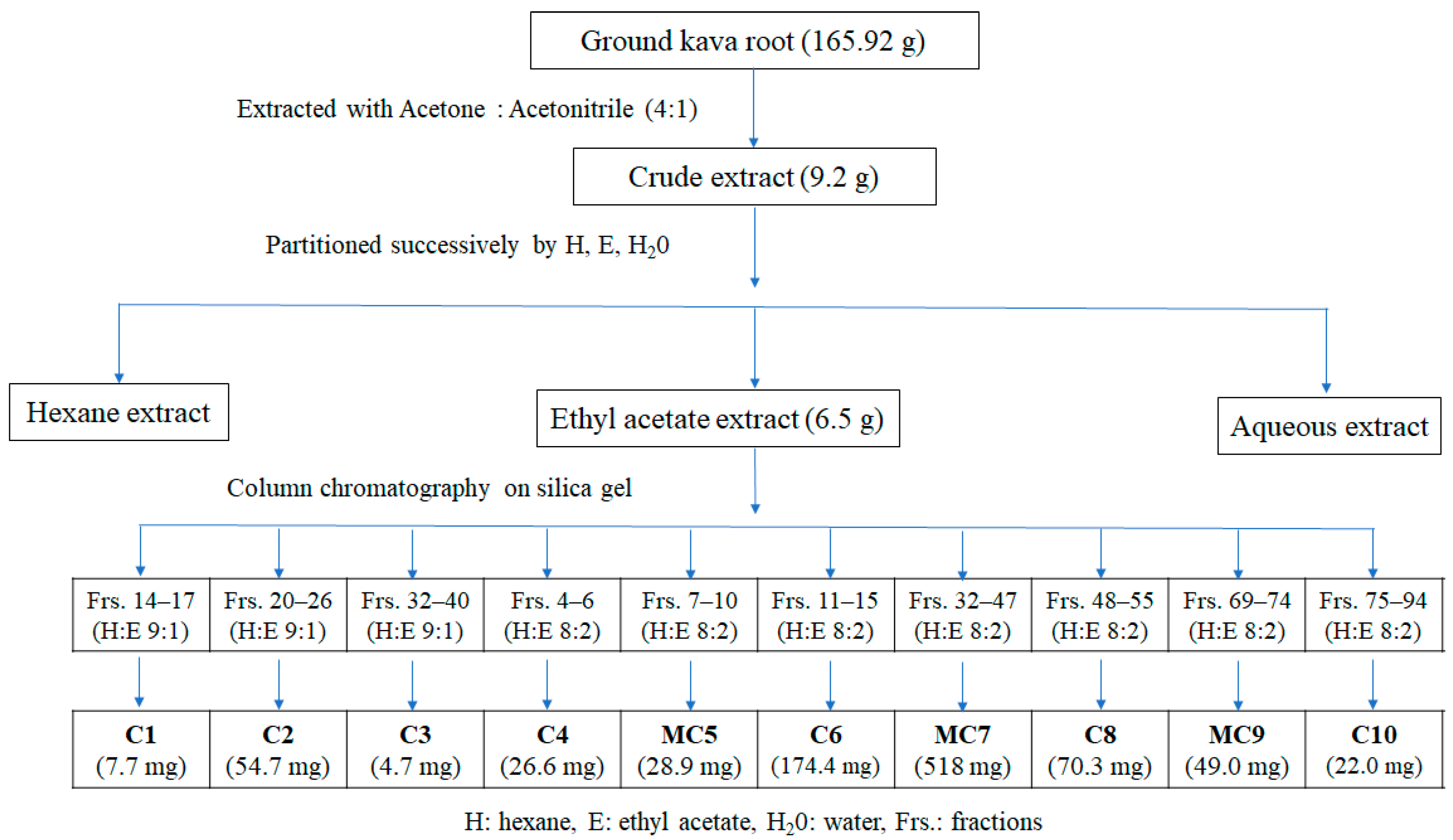
Figure 1. Fractionation and column chromatography separation of kava root.
Foods 11 03889 g001
Figure 2. Chemical structures of components isolated from P. methysticum root.
Foods 11 03889 g002aFoods 11 03889 g002b
Table 1. Identification of isolated compounds from EtOAc extract of kava root by GC–MS, ESI–MS, 1H and 13C NMR.
CodeRetention TimePeak Area (%)CompoundChemical FormulaMolecular WeightChemical Class
C123.493.285-Hydroxy-4′,7-dimethoxyflavanoneC17H16O5300Flavonoids
C225.3790.00MatteucinolC18H18O5314Flavonoids
C321.6797.175,7-Dihydroxy-4′-methoxyflavanone
(Isosakuranetin)
C16H14O5286Flavonoids
C423.0590.005,7- DimethoxyflavanoneC17H16O4284Flavonoids
MC520.8472.055,6-Dehydrokawain (DK)C14H12O3228Kava lactones
22.2627.952′,4′-Dihydroxy-6′-methoxydihydrochalconeC16H16O4272Flavonoids
C620.894.665,6-Dehydrokawain (DK)C14H12O3228Kava lactones
MC720.07
23.34
59.42
40.58
Kavain
Yangonin
C14H14O3
C15H14O4
230
258
Kava lactones
Kava lactones
C823.3493.64YangoninC15H14O4258Kava lactones
MC916.25
18.35
22.45
23.4
12.78
20.44
54.77
8.9
Dihydro-5,6-dehydrokavain (DDK)
7,8-Dihydrokavain
Dihydromethysticin
Methysticin
C14H14O3
C14H16O3
C15H16O5
C15H14O5
230
232
276
274
Kava lactones
Kava lactones
Kava lactones
Kava lactones
C1021.6190.06AlpinetinC16H14O4270Flavonoids
Table 2. Xanthine oxidase inhibitory and antioxidant activity of kava root extracts as IC50 values.
ExtractsRadical Scavenging ActivityXOI Activity
IC50 DPPH (µg/mL)IC50 ABTS (µg/mL)IC50 (µg/mL)
Hexane-1706.0 ± 4.6 a2008.1 ± 6.7 a
Chloroform2392.3 ± 4.5 a1304.0 ± 5.3 b922.7 ± 3.1 b
Ethyl acetate946.3 ± 7.6 b1007.0 ± 6.2 c724.6 ± 2.4 b
Aqueous971.6 ± 9.0 b1017.4 ± 4.7 c-
Data are presented as means ± SD (standard deviation). Mean values with different lowercase letters indicate significant differences in the same column (p < 0.05) (n = 3).
Table 3. Antibacterial activity of the kava root extracts.
Zone of Inhibition (mm)
E. coli(a)K. pneumoniae(a)P. mirabilis(a)B. subtilis(b)L. monocytogenes(b)
Hexane--10.1 ± 2.5--
Chloroform---15.0 ± 2.0-
EtOAc23.0 ± 2.5----
Water-----
Control-----
Standards
(30 µg/disc)
Ampicillin34.0 ± 1.044.1 ± 3.243.0 ± 1.517.1 ± 1.524.0 ± 3.1
Streptomycin19.1 ± 1.015.2 ± 0.629. ± 1.512.0 ± 1.018.1 ± 1.7
-: no inhibition, data are presented as means ± SD (standard deviation) (n = 3). (a): Gram-negative bacteria; (b): Gram-positive bacteria.
Table 4. Antioxidant activity of compounds isolated from kava root.
CompoundsIC50 (µg/mL)
DPPHABTS
C1-205.8 ± 5.7 c
C2-172.8 ± 6.5 d
C374.8 ± 3.5 e76.5 ± 7.1 f
C4477.8 ± 4.7 d143.5 ± 6.5 e
MC51211.5 ± 6.1 a183.4 ± 5.7 d
C6496.6 ± 4.5 c463.5 ± 7.1 b
BHT9.5 ± 0.2 f45.1 ± 3.7 g
Data are presented as means ± SD (standard deviation). Mean values with different lowercase letters indicate significant differences in the same column (p < 0.05) (n = 3). -: no inhibition.
Table 5. Xanthine oxidase inhibitory activity of fractions isolated from kava root.
FractionsIC50 (µg/mL)
C4268.65 ± 2.00 c
MC5452.68 ± 3.43 b
C6242.01 ± 1.12 d
MC9465.27 ± 2.22 a
C10134.52 ± 1.80 e
Allopurinol *21.33 ± 0.19 f
Data are presented as means ± SD (standard deviation). Mean values with different lowercase letters indicate significant differences in the same column (p < 0.05) (n = 3). *: positive control.
Table 6. Antibacterial activity of the isolated compounds extracted from kava root.
Zone of Inhibition (mm)
E. coli(a)K. pneumoniae(a)P. mirabilis(a)B. subtilis(b)L. monocytogenes(b)
MC5----13.0 ± 0.5
C6----9.0 ± 0.5
MC7-9.7 ± 0.5---
Control-----
Standards
(30 µg/disc)
Ampicillin34.0 ± 1.044.1 ± 3.243.0 ± 1.517.1 ± 1.524.0 ± 3.1
Streptomycin19.1 ± 1.015.2 ± 0.629. ± 1.512.0 ± 1.018.1 ± 1.7
Data are presented as means ± SD (standard deviation). -: no inhibition. (a): Gram-negative bacteria; (b): Gram-positive bacteria.
Publisher’s Note: MDPI stays neutral with regard to jurisdictional claims in published maps and institutional affiliations.