Article

Dietary Supplementation with Epicatechin Improves Intestinal Barrier Integrity in Mice

1
International Institute of Food Innovation, Nanchang University, Nanchang 330200, China
2
College of Food Science and Technology, Nanchang University, Nanchang 330047, China
*
Author to whom correspondence should be addressed.
Foods 2022, 11(20), 3301; https://doi.org/10.3390/foods11203301
Submission received: 30 August 2022 / Revised: 10 October 2022 / Accepted: 14 October 2022 / Published: 21 October 2022
(This article belongs to the Section Nutraceuticals, Functional Foods, and Novel Foods)

Abstract

:
Epicatechin (EPI) is a dietary flavonoid that is present in many foods and possesses various bioactivities. We assessed the effects of EPI supplementation on intestinal barrier integrity in mice. Thirty-six mice were assigned to three groups and fed a standard diet or a standard diet supplemented with 50 or 100 mg EPI/kg (n = 12 per group). After 21 days of rearing, blood and intestinal samples were collected from eight randomly selected mice. Supplementation with 50 and 100 mg/kg EPI decreased (p < 0.05) the serum diamine oxidase activity and D-lactic acid concentration and increased (p < 0.05) the duodenal, jejunal, and ileal abundance of tight junction proteins, such as occludin. Moreover, it lowered (p < 0.05) the duodenal, jejunal, and ileal tumor necrosis factor-α contents and enhanced (p < 0.05) the duodenal and jejunal catalase activities and ileal superoxide dismutase activity. Supplementation with a lower dose (50 mg/kg) decreased (p < 0.05) the ileal interleukin-1β content, whereas supplementation with a higher dose (100 mg/kg) increased (p < 0.05) the duodenal and jejunal glutathione peroxidase activities. Furthermore, supplementation with 50 and 100 mg/kg EPI decreased (p < 0.05) cell apoptosis, cleaved cysteinyl aspartate-specific proteinase-3 (caspase-3), and cleaved caspase-9 contents in the duodenum, jejunum, and ileum. In conclusion, EPI could improve intestinal barrier integrity in mice, thereby suppressing intestinal inflammation and oxidative stress and reducing cell apoptosis.

1. Introduction

In addition to serving as the main site for nutrient digestion and absorption, the intestinal epithelium acts as a barrier to inhibit the passage of toxins, allergens, and pathogens from the luminal environment into the circulatory system [1,2]. However, numerous factors, such as inflammation and oxidative stress, can disrupt the intestinal barrier and thereby negatively affect health [3,4]. Therefore, dietary intervention with certain nutrients that can inhibit intestinal inflammation and oxidative stress is likely to attenuate intestinal barrier damage. Flavonoids are polyphenols containing diphenylpropane (C6-C3-C6); they are secondary metabolites that are ubiquitously distributed throughout the plant kingdom [5]. Recently, flavonoids have been a focus of research interest because of their potential modulatory properties with respect to intestinal permeability [6].
Epicatechin (EPI) is a polyphenolic flavonoid that belongs to the flavan-3-ol group; it is present in large amounts in tea, berries, and cocoa [7,8]. Importantly, EPI exhibits a wide array of physiological activities, such as anti-oxidative [9,10], anti-inflammatory [11,12], and anti-apoptotic [13,14] activities. Given these advantages, dietary EPI supplementation may be beneficial for the gastrointestinal tract. For instance, EPI has been reported to protect the gastrointestinal tract from the negative impacts of a high-fat diet [15,16]. However, to date, no study has clearly reported the effects of dietary EPI supplementation on intestinal barrier function in mice. Hence, further research is necessary to elucidate this.
Therefore, this study assesses the effects of EPI supplementation on intestinal barrier integrity in mice and explores its underlying mechanisms. Our findings would provide valuable information on dietary intervention with EPI for improving intestinal barrier function.

2. Materials and Methods

2.1. Animal Management and Diet

A total of 36 healthy mice (4 weeks old, weighing 24–30 g) were bought from Changsha Tianqin Biotechnology Co., Ltd. (Changsha, China) and acclimated for 1 week. Then, the mice were divided into three groups (n = 12 per group). The groups were randomly assigned to one of the three dietary treatments, namely a standard diet (control (CON) group) or a standard diet supplemented with 50 mg/kg EPI (LEPI group) or 100 mg/kg EPI (HEPI group) (purity >90%; Dalian Meilun Biotechnology Co., Ltd., Dalian, China). All the mice were individually caged under controlled conditions (temperature 24 ± 1 °C, humidity 50–60%, 12 h light–dark cycle) and provided with unlimited access to feed and autoclaved water during the 3-week experimental period.

2.2. Assessment of Weight Gain

On days 1 and 22 (08:00 h), all the mice were weighed to calculate their average daily gain (ADG) over the experimental period.

2.3. Sample Collection

After overnight fasting, eight randomly selected mice per group were anesthetized using ether (08:00 h on day 22) and their blood samples were collected via heart puncture. After being placed at room temperature for 30 min, the samples were centrifuged at 3000× g at 4 °C for 10 min to acquire serum and then stored at −20 °C. After blood sampling, the abdominal cavity was opened, and approximately 2 cm segments of the duodenum, jejunum, and ileum were fixed in 4% paraformaldehyde buffer for evaluating intestinal cell apoptosis. The samples were then flushed gently with ice-cold phosphate-buffered saline (PBS). Following this, mucosal samples from the duodenum, jejunum, and ileum were collected by scraping using a scalpel and frozen at −80 °C.

2.4. Measurement of Serum Parameters

Serum diamine oxidase (DAO) activity was measured using kits manufactured by Nanjing Jiancheng Bioengineering Institute (Nanjing, China), and serum D-lactic acid concentration was measured using ELISA kits manufactured by Jiangsu Meimian Industrial Co., Ltd. (Yancheng, China). All assays were performed according to the specific procedures provided in the kits.

2.5. Small Intestine Biochemical Analysis

2.5.1. Preparation of Intestinal Homogenates

Frozen duodenal, jejunal, and ileal mucosal samples were thawed, mixed with ice-cold physiological saline at a ratio of 1:9 (w/v) and centrifuged for 10 min (3000× g at 4 °C) to obtain supernatants. Then, the total protein concentration in the supernatants was estimated using a total protein quantitative assay kit (Nanjing Jiancheng Bioengineering Institute). The antioxidant-related indices and cytokine and cell apoptosis-related protein contents were normalized against the total protein concentration in each sample for intersample comparison.

2.5.2. Determination of Intestinal Antioxidant Capacity

Duodenal, jejunal, and ileal catalase (CAT), superoxide dismutase (SOD), and glutathione peroxidase (GSH-PX) activities and malondialdehyde (MDA) concentration were determined based on the manuals provided by Nanjing Jiancheng Bioengineering Institute.

2.5.3. Assessment of Intestinal Cytokine and Cell Apoptosis-Related Protein Contents

Duodenal, jejunal, and ileal interleukin-1β (IL-1β), IL-10, tumor necrosis factor-α (TNF-α), interferon-γ (IFN-γ), B-cell lymphoma-2-associated X protein (Bax), B-cell lymphoma-2 (Bcl-2), TNF receptor 1 (TNFR1), cleaved cysteinyl aspartate-specific proteinase-3 (caspase-3), cleaved caspase-8, and cleaved caspase-9 contents were assessed using ELISA kits (Jiangsu Meimian Industrial Co., Ltd.). All operations were strictly manipulated according to the manufacturer’s guidelines.

2.6. Detection of Intestinal Cell Apoptosis

Apoptosis was assessed by a terminal deoxynucleotidyl transferase (TdT)-mediated deoxyuridine triphosphate (dUTP) nick-end-labelling (TUNEL) assay using an in situ cell death detection kit (Roche Diagnostics GmbH, Mannheim, Germany). In brief, the fixed duodenal, jejunal, and ileal segments were dehydrated, embedded in paraffin, cut into 3 μm thick sections, dewaxed, rehydrated and then incubated with 20 μg/mL proteinase K at 37 °C for 20 min. Subsequently, the sections were washed thrice with PBS before permeabilizing with 0.5% Triton X-100 at room temperature for 10 min. Next, TdT buffer containing dUTP was used to treat the sections at 37 °C for 2 h, followed by washing thrice with PBS to stop the reaction. Following this, 4′, 6-diamidino-2-phenylindole (DAPI) was used to stain the sections at room temperature for 5 min to detect cell nuclei. Finally, the sections were sealed with antifade mounting medium and imaged under a fluorescent microscope (Nikon Corporation, Tokyo, Japan). A total of six random regions were selected from each section for counting the number of TUNEL-positive cells. The apoptotic index was expressed as the proportion of apoptotic cells to total cells.

2.7. Western Blot Assay

Duodenal, jejunal, and ileal mucosal specimens were homogenized in RIPA buffer containing protease inhibitor cocktail (Beyotime Institute of Biotechnology, Shanghai, China) and then centrifuged at 12,000× g at 4 °C for 15 min to collect lysate supernatants. The total protein concentrations in the lysates were assayed using the bicinchoninic acid method [17]. The proteins were then mixed with 5× sample-loading buffer (Beyotime Institute of Biotechnology) for denaturation at 98 °C for 10 min. Thereafter, equal quantities of proteins per sample were resolved by sodium dodecyl sulphate–polyacrylamide gel electrophoresis and transferred onto polyvinylidene fluoride (PVDF) membranes. The PVDF membranes were then incubated with blocking buffer, i.e., 5% bovine serum albumin in Tris-buffered saline containing 1% Tween-20 (TBS/T), at room temperature for 1 h. After rinsing thrice with TBS/T, the PVDF membranes were probed with primary antibodies against occludin (1:1000 dilution; Abcam plc., Cambridge, UK), zonula occludens-1 (1:1000 dilution; ZO-1; Thermo Fisher Scientific, Inc., Waltham, MA, USA), or glyceraldehyde-3-phosphate dehydrogenase (1:1000 dilution; GAPDH; Cell Signalling Technology, Inc., Danvers, MA, USA) at 4 °C overnight. Following this, the PVDF membranes were rinsed thrice with TBS/T and incubated with a suitable secondary antibody at room temperature for 1 h. Finally, the PVDF membranes were immersed in Clarity™ Western ECL Substrate (Bio-Rad Laboratories, Inc., Hercules, CA, USA), visualized using a ChemiDoc™ XRS+ Imager System (Bio-Rad Laboratories, Inc.), and analyzed using ImageJ software (version 1.8.0; National Institutes of Health, Bethesda, MD, USA).

2.8. Statistical Analysis

All data were assessed by performing one-way analysis of variance followed by Tukey’s multiple-range tests using SAS 9.0 (SAS Inst., Inc., Cary, NC, USA), with each mouse serving as a statistical unit. The results were expressed as the means ± standard errors. p < 0.05 was used to denote statistical significance among means.

3. Results

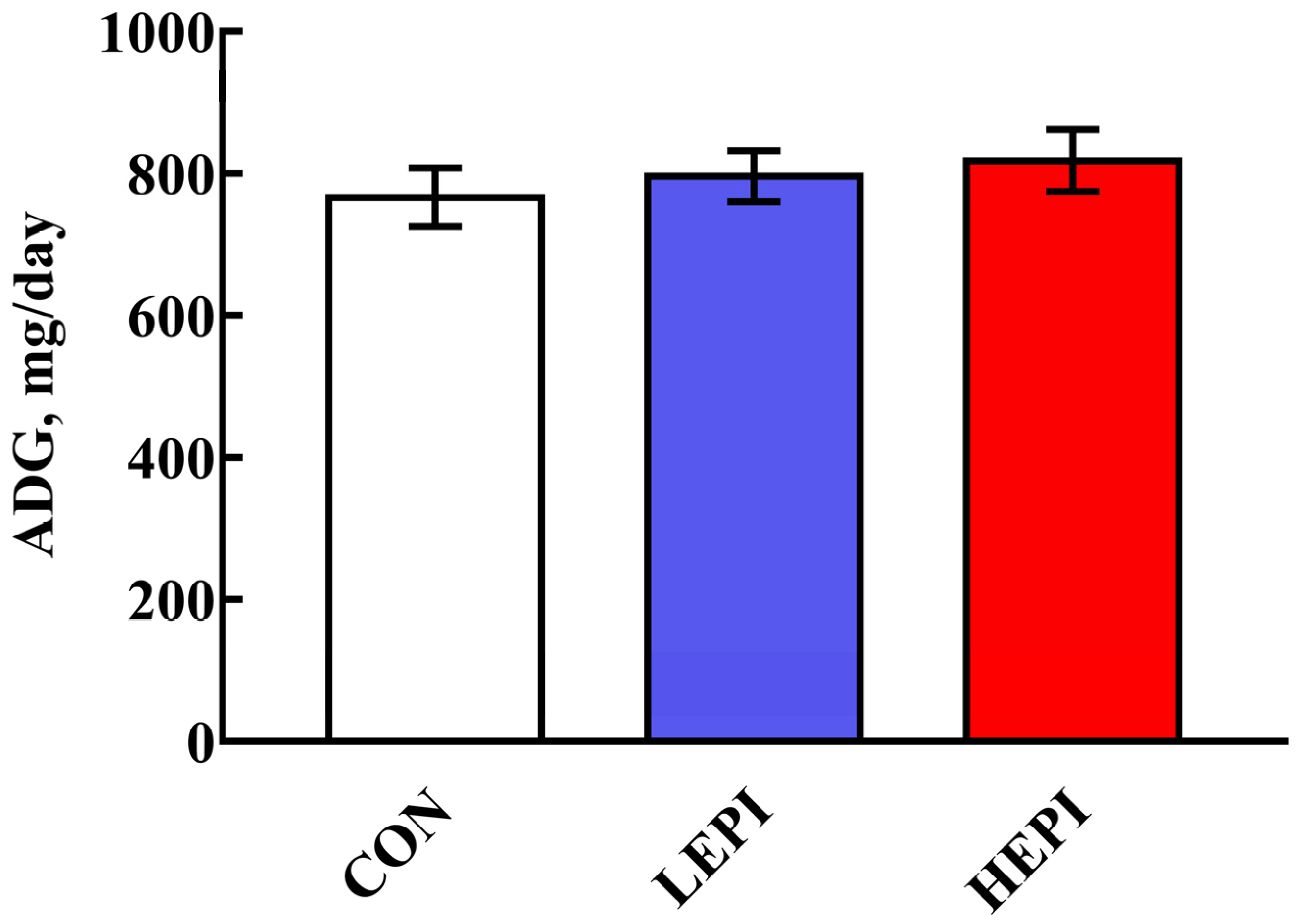
3.1. Growth Performance

The effects of EPI supplementation on the growth performance of the mice are presented in Figure 1. EPI supplementation did not affect (p > 0.05) the ADG of the mice.

3.2. Serum Indices

Table 1 reveals the variations in serum indices of the mice after EPI supplementation. Compared with the CON group, the serum DAO activity and D-lactic acid concentration were notably decreased (p < 0.05) in the LEPI and HEPI groups.

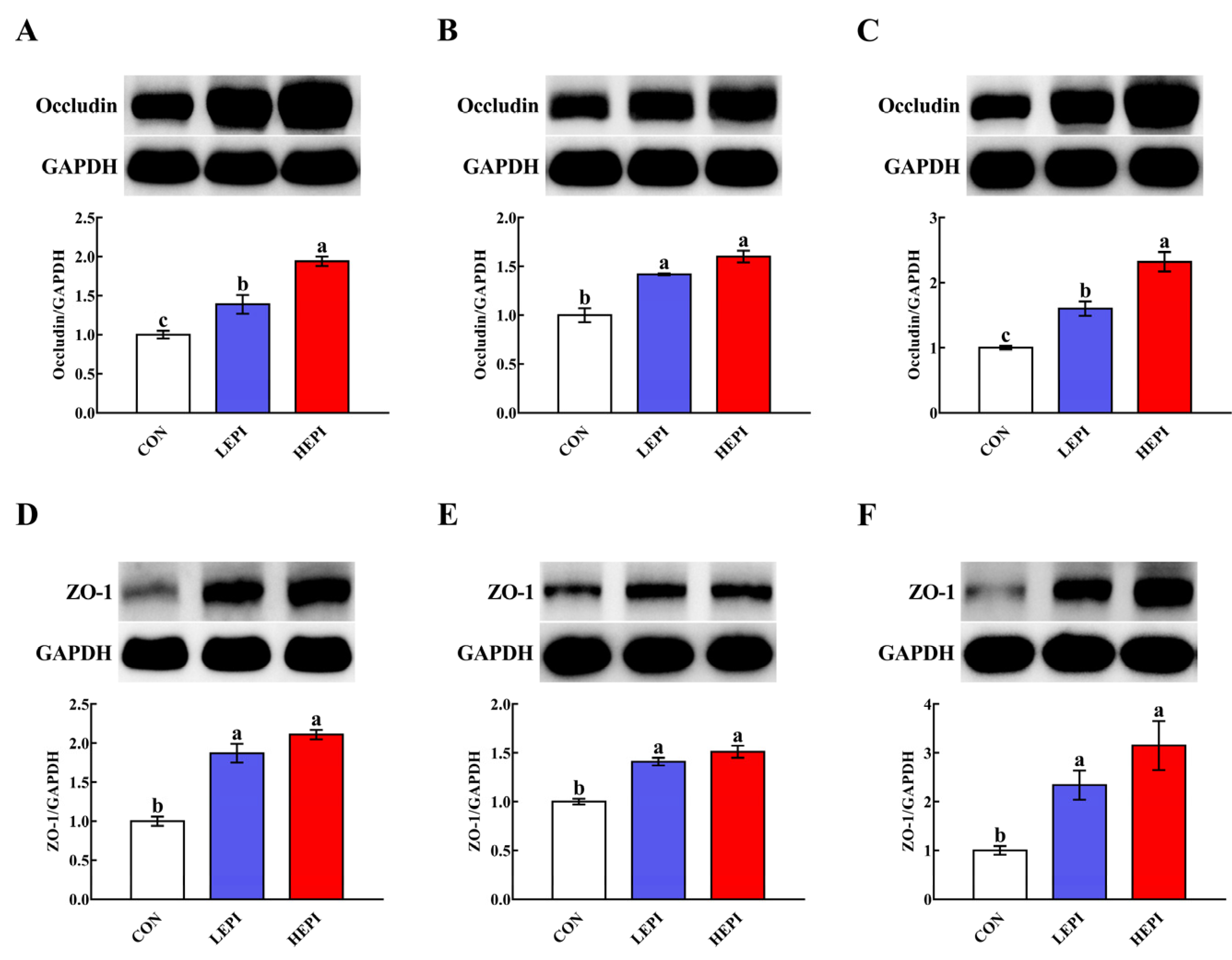
3.3. Abundance of Intestinal Tight Junction Proteins

As shown in Figure 2, supplementation with 50 and 100 mg/kg EPI increased (p < 0.05) the duodenal, jejunal, and ileal abundances of occludin and ZO-1.

3.4. Intestinal Antioxidant Capacity

Dietary supplementation with 50 and 100 mg/kg EPI increased (p < 0.05) the duodenal and jejunal CAT activities and ileal SOD activity (Table 2). Additionally, supplementation with 100 mg/kg EPI increased (p < 0.05) the duodenal and jejunal GSH-PX activities. Moreover, supplementation with either 50 or 100 mg/kg EPI lowered (p < 0.05) the MDA contents in the duodenum, jejunum, and ileum.

3.5. Intestinal Cytokine Contents

Dietary supplementation with 50 mg/kg EPI decreased (p < 0.05) the ileal IL-1β content but increased (p < 0.05) the jejunal IL-10 content (Table 3). Moreover, dietary supplementation with 50 and 100 mg/kg EPI lowered (p < 0.05) the duodenal, jejunal, and ileal TNF-α contents. However, the duodenal, jejunal, and ileal IFN-γ contents remained unchanged (p > 0.05) after supplementation with 50 or 100 mg/kg EPI.

3.6. Intestinal Cell Apoptosis

To assess the impacts of EPI on intestinal cell apoptosis, we observed intestinal cell apoptosis using TUNEL staining. As shown in Figure 3, 50 and 100 mg/kg EPI decreased (p < 0.05) duodenal, jejunal, and ileal cell apoptosis.

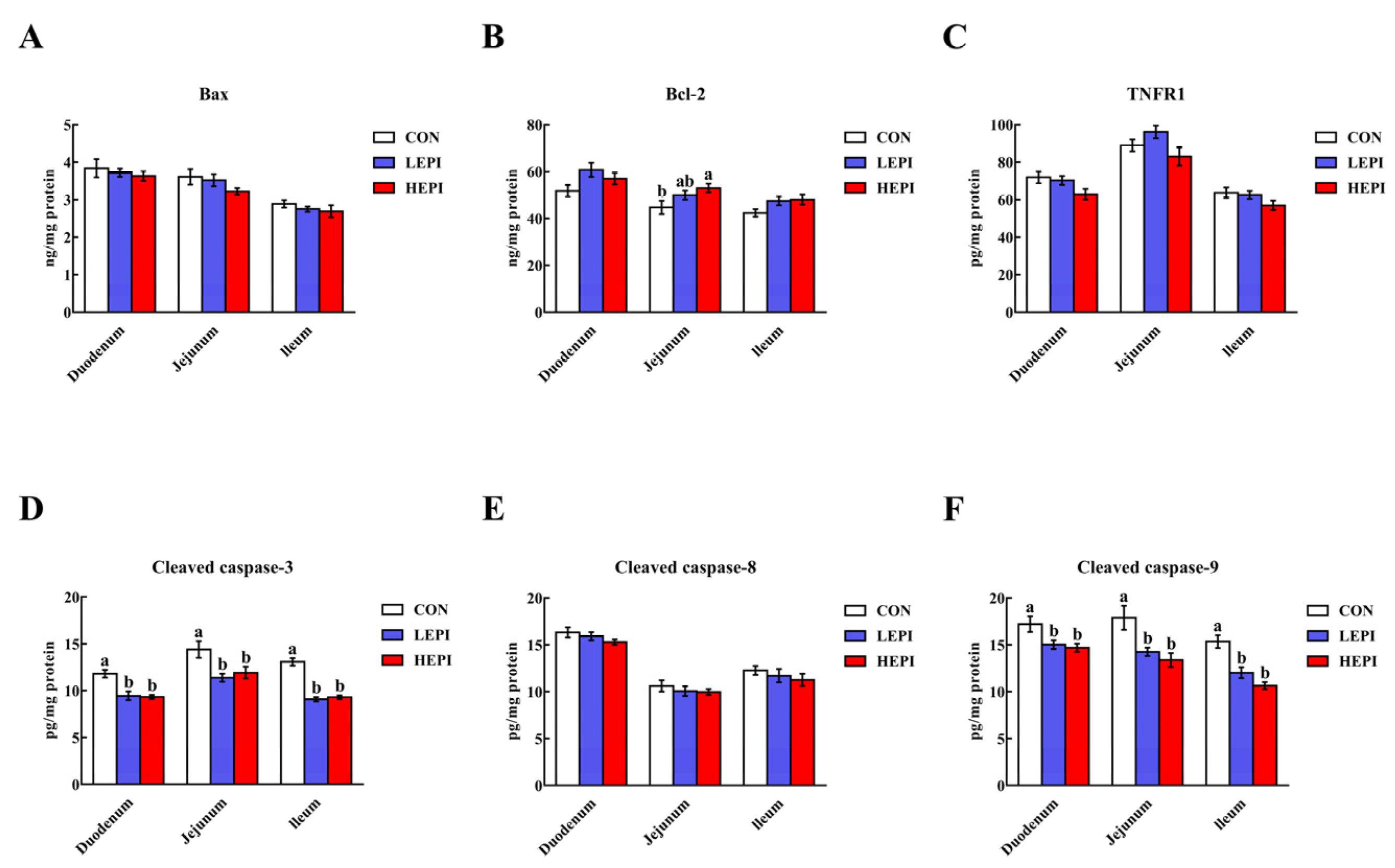
3.7. Intestinal Cell Apoptosis-Related Proteins

Supplementation with 50 and 100 mg/kg EPI decreased (p < 0.05) the cleaved caspase-3 and cleaved caspase-9 contents in the duodenum, jejunum, and ileum (Figure 4). In contrast, supplementation with 100 mg/kg EPI increased (p < 0.05) the jejunal Bcl-2 content. However, no changes (p > 0.05) were noted in the duodenal, jejunal, and ileal Bax, TNFR1, and cleaved caspase-8 contents in the CON, LEPI, and HEPI groups.

4. Discussion

The intestinal barrier is closely related to tight junctions, which are dynamic multifunctional complexes that are located between epithelial cells and function as a critical structure for paracellular permeability [18]. Tight junctions consist of transmembrane proteins, such as occludin, and cytosolic proteins that are recruited to the apicolateral membrane, such as ZO-1 [19,20]. We found that EPI supplementation increased the duodenal, jejunal, and ileal abundances of occludin and ZO-1 (at 50 and 100 mg/kg). These findings indicate that EPI supplementation could improve intestinal barrier integrity in mice. At present, several blood parameters, such as DAO activity and D-lactic acid concentration, are considered circulating markers for evaluating the degree of intestinal barrier damage. Intestinal barrier disruption usually leads to the release of DAO and D-lactic acid into blood circulation [21,22]. Accordingly, mice treated with EPI (50 and 100 mg/kg) exhibited a decrease in serum DAO activity and D-lactic acid concentration, further confirming that EPI supplementation could improve the intestinal barrier integrity in mice.
In addition to playing a crucial role in the modulation of intestinal inflammatory responses, cytokines (both pro-inflammatory and anti-inflammatory cytokines) have an important effect on the regulation of intestinal barrier integrity [23,24]. Most pro-inflammatory cytokines, such as IL-1β and TNF-α, can disrupt the intestinal barrier, thereby increasing intestinal permeability [25,26]. We found that supplementation with 50 mg/kg EPI decreased the ileal IL-1β content, and supplementation with 50 and 100 mg/kg EPI decreased the duodenal, jejunal, and ileal TNF-α contents in mice. These findings indicate that EPI supplementation could improve intestinal barrier integrity by suppressing the release of intestinal pro-inflammatory cytokines. On the other hand, IL-10, an anti-inflammatory cytokine, can attenuate intestinal inflammatory injury [27]. We observed that supplementation with 50 mg/kg EPI increased the jejunal IL-10 content in mice. Intriguingly, EPI supplementation may also improve intestinal barrier integrity by promoting the production of intestinal anti-inflammatory cytokines.
Another pivotal factor contributing to intestinal barrier disruption is oxidative stress, which is widely recognized as a state of imbalance between oxidation and antioxidation [28]. MDA is the primary product of lipid peroxidation and is usually considered an oxidative stress marker [29,30]. We noted a decrease in duodenal, jejunal, and ileal MDA contents in mice treated with EPI (50 and 100 mg/kg), indicating that intestinal oxidative damage was attenuated by EPI supplementation. The activities of antioxidant enzymes, including SOD, CAT, and GSH-PX, are important indicators of antioxidant capacity. SOD catalyzes the conversion of superoxide into hydrogen peroxide, and CAT and GSH-PX convert hydrogen peroxide into water [31,32]. Importantly, we found that supplementation with 50 and 100 mg/kg EPI increased the duodenal and jejunal CAT activities and ileal SOD activity, and supplementation with 100 mg/kg EPI increased the duodenal and jejunal GSH-PX activities in mice. These results suggest that EPI could alleviate intestinal oxidative damage by enhancing antioxidant enzyme activities in the intestines of mice, thereby improving intestinal barrier integrity.
Although cell apoptosis is essential for intestinal epithelial turnover and tissue homeostasis, excessive apoptosis can result in intestinal barrier damage [33]. Therefore, the reduced duodenal, jejunal, and ileal cell apoptosis noted in our study after supplementation with 50 and 100 mg/kg EPI indicates that EPI could improve intestinal barrier integrity in mice through the inhibition of apoptosis. In general, cell apoptosis occurs via the intrinsic pathway (mitochondria-dependent pathway) or the extrinsic pathway (death-receptor-dependent pathway) [34]. The intrinsic pathway is regulated by the Bcl-2 family and is mainly characterized by caspase-9 activation [35,36], while the extrinsic pathway is mediated by membrane death receptors, such as TNFR1, and is mainly characterized by caspase-8 activation [37,38]. Caspase-3 is an executioner caspase that is activated by these two pathways and then initiates apoptosis [39,40]. We found that dietary supplementation with 50 and 100 mg/kg EPI decreased cleaved caspase-3 and cleaved caspase-9 contents in the duodenum, jejunum, and ileum of mice. These results indicate that EPI could repress intestinal cell apoptosis in mice in a mitochondria-dependent manner.

5. Conclusions

In summary, our findings indicate that EPI was beneficial in enhancing intestinal barrier integrity in mice. The underlying mechanisms may be closely related to the suppression of intestinal inflammation and oxidative stress and the reduction in cell apoptosis. These findings provide a foundation for developing EPI as a nutraceutical to improve intestinal health.

Author Contributions

Conceptualization, J.W.; data curation, J.W.; formal analysis, J.W.; funding acquisition, Z.R.; investigation, J.W.; methodology, J.W.; project administration, Z.R.; software, J.W.; supervision, L.Z.; writing—original draft, J.W.; writing—review and editing, J.W. All authors have read and agreed to the published version of the manuscript.

Funding

This study was funded by the National Natural Science Foundation of China (31872897) and the Key Research and Development Projects of Nanchang Metropolis (2019-NC2DSY-002).

Institutional Review Board Statement

All animal study protocols were approved by the Animal Care and Use Committee of Nanchang University (Nanchang, China; permission number: SYXK2015-0001).

Data Availability Statement

All data presented in this study are available through the corresponding author.

Conflicts of Interest

The authors declare that they have no conflict of interest.

References

  1. Turner, J.R. Intestinal mucosal barrier function in health and disease. Nat. Rev. Immunol. 2009, 9, 799–809. [Google Scholar] [CrossRef]
  2. Wijtten, P.J.A.; van der Meulen, J.; Verstegen, M.W.A. Intestinal barrier function and absorption in pigs after weaning: A review. Br. J. Nutr. 2011, 105, 967–981. [Google Scholar] [CrossRef] [PubMed]
  3. Farhadi, A.; Banan, A.; Fields, J.; Keshavarzian, A. Intestinal barrier: An interface between health and disease. J. Gastroenterol. Hepatol. 2003, 18, 479–497. [Google Scholar] [CrossRef]
  4. Blikslager, A.T.; Moeser, A.J.; Gookin, J.L.; Jones, S.L.; Odle, J. Restoration of barrier function in injured intestinal mucosa. Physiol. Rev. 2007, 87, 545–564. [Google Scholar] [CrossRef] [PubMed]
  5. Crozier, A.; Jaganath, I.B.; Clifford, M.N. Dietary phenolics: Chemistry, bioavailability and effects on health. Nat. Prod. Rep. 2009, 26, 1001–1043. [Google Scholar] [CrossRef]
  6. Suzuki, T.; Hara, H. Role of flavonoids in intestinal tight junction regulation. J. Nutr. Biochem. 2011, 22, 401–408. [Google Scholar] [CrossRef]
  7. Vogiatzoglou, A.; Mulligan, A.A.; Luben, R.N.; Lentjes, M.A.H.; Heiss, C.; Kelm, M.; Merx, M.W.; Spencer, J.P.E.; Schroeter, H.; Kuhnle, G.G.C. Assessment of the dietary intake of total flavan-3-ols, monomeric flavan-3-ols, proanthocyanidins and theaflavins in the European union. Br. J. Nutr. 2014, 111, 1463–1473. [Google Scholar] [CrossRef] [PubMed] [Green Version]
  8. Harnly, J.M.; Doherty, R.F.; Beecher, G.R.; Holden, J.M.; Haytowitz, D.B.; Bhagwat, S.; Gebhardt, S. Flavonoid content of U.S. fruits, vegetables, and nuts. J. Agric. Food Chem. 2006, 54, 9966–9977. [Google Scholar] [CrossRef]
  9. Quine, S.D.; Raghu, P.S. Effects of (−)-epicatechin, a flavonoid on lipid peroxidation and antioxidants in streptozotocin-induced diabetic liver, kidney and heart. Pharmacol. Rep. 2005, 57, 610–615. [Google Scholar] [PubMed]
  10. Shariati, S.; Kalantar, H.; Pashmforoosh, M.; Mansouri, E.; Khodayar, M.J. Epicatechin protective effects on bleomycin-induced pulmonary oxidative stress and fibrosis in mice. Biomed. Pharmacother. 2019, 114, 108776. [Google Scholar] [CrossRef] [PubMed]
  11. Vazquez-Prieto, M.A.; Bettaieb, A.; Haj, F.G.; Fraga, C.G.; Oteiza, P.I. (−)-epicatechin prevents TNFα-induced activation of signaling cascades involved in inflammation and insulin sensitivity in 3T3-L1 adipocytes. Arch. Biochem. Biophys. 2012, 527, 113–118. [Google Scholar] [CrossRef] [PubMed] [Green Version]
  12. Xing, J.; Yu, Z.L.; Zhang, X.Y.; Li, W.Y.; Gao, D.N.; Wang, J.; Ma, X.C.; Nie, X.S.; Wang, W. Epicatechin alleviates inflammation in lipopolysaccharide-induced acute lung injury in mice by inhibiting the p38 MAPK signaling pathway. Int. Immunopharmacol. 2019, 66, 146–153. [Google Scholar] [CrossRef] [PubMed]
  13. Wu, H.; Xie, Y.N.; Xu, Y.L.; Hu, Z.H.; Wan, X.; Huang, H.C.; Huang, D.B. Protective effect of epicatechin on apap-induced acute liver injury of mice through anti-inflammation and apoptosis inhibition. Nat. Prod. Res. 2020, 34, 855–858. [Google Scholar] [CrossRef]
  14. Li, Y.; Ma, S.S.; Zhang, Y.T.; Yao, M.H.; Zhu, X.Z.; Guan, F.X. (−)-epicatechin mitigates radiation-induced intestinal injury and promotes intestinal regeneration via suppressing oxidative stress. Free Radic. Res. 2019, 53, 851–864. [Google Scholar] [CrossRef]
  15. Cremonini, E.; Wang, Z.W.; Bettaieb, A.; Adamo, A.M.; Daveri, E.; Mills, D.A.; Kalanetra, K.M.; Haj, F.G.; Karakas, S.; Oteiza, P.I. (−)-epicatechin protects the intestinal barrier from high fat diet-induced permeabilization: Implications for steatosis and insulin resistance. Redox Biol. 2018, 14, 588–599. [Google Scholar] [CrossRef] [PubMed]
  16. Wang, Z.W.; Litterio, M.C.; Müller, M.; Vauzour, D.; Oteiza, P.I. (−)-epicatechin and NADPH oxidase inhibitors prevent bile acid-induced Caco-2 monolayer permeabilization through ERK1/2 modulation. Redox Biol. 2020, 28, 101360. [Google Scholar] [CrossRef] [PubMed]
  17. Smith, P.K.; Krohn, R.I.; Hermanson, G.T.; Mallia, A.K.; Gartner, F.H.; Provenzano, M.D.; Fujimoto, E.K.; Goeke, N.M.; Olson, B.J.; Klenk, D.C. Measurement of protein using bicinchoninic acid. Anal. Biochem. 1985, 150, 76–85. [Google Scholar] [CrossRef]
  18. Yap, A.S.; Mullin, J.M.; Stevenson, B.R. Molecular analyses of tight junction physiology: Insights and paradoxes. J. Membr. Biol. 1998, 163, 159–167. [Google Scholar] [CrossRef] [PubMed]
  19. Gonzalez-Mariscal, L.; Betanzos, A.; Nava, P.; Jaramillo, B.E. Tight junction proteins. Prog. Biophys. Mol. Biol. 2003, 81, 1–44. [Google Scholar] [CrossRef]
  20. Suzuki, T. Regulation of intestinal epithelial permeability by tight junctions. Cell. Mol. Life Sci. 2013, 70, 631–659. [Google Scholar] [CrossRef] [PubMed]
  21. Fukudome, I.; Kobayashi, M.; Dabanaka, K.; Maeda, H.; Okamoto, K.; Okabayashi, T.; Baba, R.; Kumagai, N.; Oba, K.; Fujita, M.; et al. Diamine oxidase as a marker of intestinal mucosal injury and the effect of soluble dietary fiber on gastrointestinal tract toxicity after intravenous 5-fluorouracil treatment in rats. Med. Mol. Morphol. 2014, 47, 100–107. [Google Scholar] [CrossRef] [PubMed]
  22. Nielsen, C.; Lindholt, J.S.; Erlandsen, E.J.; Mortensen, F.V. D-lactate as a marker of venous-induced intestinal ischemia: An experimental study in pigs. Int. J. Surg. 2011, 9, 428–432. [Google Scholar] [CrossRef] [PubMed] [Green Version]
  23. Al-Sadi, R.; Boivin, M.; Ma, T. Mechanism of cytokine modulation of epithelial tight junction barrier. Front. Biosci. 2009, 14, 2765–2778. [Google Scholar] [CrossRef] [Green Version]
  24. Capaldo, C.T.; Nusrat, A. Cytokine regulation of tight junctions. Biochim. Biophys. Acta Biomembr. 2009, 1788, 864–871. [Google Scholar] [CrossRef] [PubMed] [Green Version]
  25. Ye, D.M.; Ma, I.; Ma, T.Y. Molecular mechanism of tumor necrosis factor-α modulation of intestinal epithelial tight junction barrier. Am. J. Physiol. Gastroint. Liver Physiol. 2006, 290, G496–G504. [Google Scholar] [CrossRef] [Green Version]
  26. Al-Sadi, R.; Ye, D.M.; Said, H.M.; Ma, T.Y. IL-1β-induced increase in intestinal epithelial tight junction permeability is mediated by MEKK-1 activation of canonical NF-κB pathway. Am. J. Pathol. 2010, 177, 2310–2322. [Google Scholar] [CrossRef]
  27. Madsen, K.L.; Lewis, S.A.; Tavernini, M.M.; Hibbard, J.; Fedorak, R.N. Interleukin 10 prevents cytokine-induced disruption of T84 monolayer barrier integrity and limits chloride secretion. Gastroenterology 1997, 113, 151–159. [Google Scholar] [CrossRef]
  28. Circu, M.L.; Aw, T.Y. Intestinal redox biology and oxidative stress. Semin. Cell Dev. Biol. 2012, 23, 729–737. [Google Scholar] [CrossRef] [PubMed] [Green Version]
  29. Kadiiska, M.B.; Gladen, B.C.; Baird, D.D.; Germolec, D.; Graham, L.B.; Parker, C.E.; Nyska, A.; Wachsman, J.T.; Ames, B.N.; Basu, S.; et al. Biomarkers of oxidative stress study II: Are oxidation products of lipids, proteins, and DNA markers of CCl4 poisoning? Free Radic. Biol. Med. 2005, 38, 698–710. [Google Scholar] [CrossRef]
  30. Pirinccioglu, A.G.; Gökalp, D.; Pirinccioglu, M.; Kizil, G.; Kizil, M. Malondialdehyde (MDA) and protein carbonyl (PCO) levels as biomarkers of oxidative stress in subjects with familial hypercholesterolemia. Clin. Biochem. 2010, 43, 1220–1224. [Google Scholar] [CrossRef]
  31. Finkel, T.; Holbrook, N.J. Oxidants, oxidative stress and the biology of ageing. Nature 2000, 408, 239–247. [Google Scholar] [CrossRef]
  32. Bhattacharyya, A.; Chattopadhyay, R.; Mitra, S.; Crowe, S.E. Oxidative stress: An essential factor in the pathogenesis of gastrointestinal mucosal diseases. Physiol. Rev. 2014, 94, 329–354. [Google Scholar] [CrossRef] [PubMed] [Green Version]
  33. Günther, C.; Neumann, H.; Neurath, M.F.; Becker, C. Apoptosis, necrosis and necroptosis: Cell death regulation in the intestinal epithelium. Gut 2013, 62, 1062–1071. [Google Scholar] [CrossRef] [Green Version]
  34. Ghobrial, I.M.; Witzig, T.E.; Adjei, A.A. Targeting apoptosis pathways in cancer therapy. CA-Cancer J. Clin. 2005, 55, 178–194. [Google Scholar] [CrossRef] [PubMed] [Green Version]
  35. Cory, S.; Adams, J.M. The Bcl2 family: Regulators of the cellular life-or-death switch. Nat. Rev. Cancer 2002, 2, 647–656. [Google Scholar] [CrossRef]
  36. Youle, R.J.; Strasser, A. The Bcl-2 protein family: Opposing activities that mediate cell death. Nat. Rev. Mol. Cell Biol. 2008, 9, 47–59. [Google Scholar] [CrossRef]
  37. Duprez, L.; Wirawan, E.; Berghe, T.V.; Vandenabeele, P. Major cell death pathways at a glance. Microbes Infect. 2009, 11, 1050–1062. [Google Scholar] [CrossRef] [PubMed]
  38. Ashkenazi, A. Targeting death and decoy receptors of the tumour-necrosis factor superfamily. Nat. Rev. Cancer 2002, 2, 420–430. [Google Scholar] [CrossRef]
  39. Elmore, S. Apoptosis: A review of programmed cell death. Toxicol. Pathol. 2007, 35, 495–516. [Google Scholar] [CrossRef]
  40. Riedl, S.J.; Shi, Y.G. Molecular mechanisms of caspase regulation during apoptosis. Nat. Rev. Mol. Cell Biol. 2004, 5, 897–907. [Google Scholar] [CrossRef]
Figure 1. Effects of epicatechin on the growth performance of mice. Data are expressed as the means (12 mice/treatment), with standard errors represented by vertical bars. CON group: control group—mice received a standard diet; LEPI group—mice received a standard diet supplemented with 50 mg/kg epicatechin; HEPI group—mice received a standard diet supplemented with 100 mg/kg epicatechin; ADG—average daily gain.
Foods 11 03301 g001
Figure 2. Effects of epicatechin on the abundance of intestinal tight junction proteins in mice. (AC) Duodenal, jejunal, and ileal abundances of occludin, respectively. (DF) Duodenal, jejunal, and ileal abundances of ZO-1, respectively. Data are expressed as means (eight mice/treatment), with standard errors represented by vertical bars. a–c Mean values with different superscripts indicate a significant difference (p < 0.05). CON group: control group—mice received a standard diet; LEPI group—mice received a standard diet supplemented with 50 mg/kg epicatechin; HEPI group—mice received a standard diet supplemented with 100 mg/kg epicatechin; ZO-1—zonula occludens-1; GAPDH—glyceraldehyde-3-phosphate dehydrogenase.
Foods 11 03301 g002
Figure 3. Effects of epicatechin on intestinal cell apoptosis in mice. (A) Representative small intestinal cross-sections of mice in the CON, LEPI, and HEPI groups after TUNEL staining (200×). Nuclei were stained with DAPI (blue) and TUNEL (green) staining. (BD) Quantification of apoptotic cells in the duodenum, jejunum, and ileum. Data are expressed as means (eight mice/treatment), with standard errors represented by vertical bars. a,b Mean values with different superscripts indicate a significant difference (p < 0.05). CON group: control group—mice received a standard diet; LEPI group—mice received a standard diet supplemented with 50 mg/kg epicatechin; HEPI group—mice received a standard diet supplemented with 100 mg/kg epicatechin; DAPI—4′, 6-diamidino-2-phenylindole; TUNEL—terminal deoxynucleotidyl transferase-mediated deoxyuridine triphosphate nick-end labelling.
Foods 11 03301 g003
Figure 4. Effects of epicatechin on intestinal Bax (A), Bcl-2 (B), TNFR1 (C), cleaved caspase-3 (D), cleaved caspase-8 (E), and cleaved caspase-9 (F) contents in mice. Data are expressed as means (eight mice/treatment), with standard errors represented by vertical bars. a,b Mean values with different superscripts indicate a significant difference (p < 0.05). CON group: control group—mice received a standard diet; LEPI group—mice received a standard diet supplemented with 50 mg/kg epicatechin; HEPI group—mice received a standard diet supplemented with 100 mg/kg epicatechin; Bax—B-cell lymphoma-2-associated X protein; Bcl-2—B-cell lymphoma-2; TNFR1—tumor necrosis factor receptor 1; cleaved caspase-3—cleaved cysteinyl aspartate-specific proteinase-3; cleaved caspase-8—cleaved cysteinyl aspartate-specific proteinase-8; cleaved caspase-9—cleaved cysteinyl aspartate-specific proteinase-9.
Foods 11 03301 g004
Table 1. Effects of epicatechin on the serum indices of mice .
Items §Treatment Groups p-Value
CONLEPIHEPI
DAO, U/L9.04 ± 0.25 a7.48 ± 0.14 b7.27 ± 0.16 b<0.001
D-Lactic acid, ng/mL563.64 ± 8.17 a488.49 ± 5.73 b507.46 ± 4.32 b<0.001
a,b Mean values with different superscripts in the same row indicate a significant difference (p < 0.05). Values are the means of eight replicates per treatment. CON group: control group—mice received a standard diet; LEPI group—mice received a standard diet supplemented with 50 mg/kg epicatechin; HEPI group—mice received a standard diet supplemented with 100 mg/kg epicatechin. § DAO—diamine oxidase.
Table 2. Effects of epicatechin on the intestinal antioxidant capacity of mice .
Items §Treatment Groups p-Value
CONLEPIHEPI
Duodenum
SOD, U/mg protein70.90 ± 2.89 81.74 ± 3.5079.20 ± 4.660.127
CAT, U/mg protein46.56 ± 2.48 c66.33 ± 2.88 b78.54 ± 2.79 a<0.001
GSH-PX, U/mg protein184.44 ± 4.55 b201.16 ± 4.75 b241.52 ± 7.83 a<0.001
MDA, nmol/mg protein2.97 ± 0.18 a1.66 ± 0.09 b1.47 ± 0.10 b<0.001
Jejunum
SOD, U/mg protein50.14 ± 1.8853.99 ± 2.6251.10 ± 2.150.461
CAT, U/mg protein23.26 ± 0.91 b29.07 ± 1.85 a30.65 ± 1.58 a0.006
GSH-PX, U/mg protein170.46 ± 4.85 b180.83 ± 3.37 a, b185.56 ± 3.79 a0.044
MDA, nmol/mg protein1.97 ± 0.08 a1.53 ± 0.07 b1.38 ± 0.04 b<0.001
Ileum
SOD, U/mg protein54.23 ± 2.76 b71.78 ± 2.24 a80.04 ± 2.60 a<0.001
CAT, U/mg protein29.24 ± 1.2930.53 ± 1.5033.89 ± 1.880.123
GSH-PX, U/mg protein127.92 ± 3.85138.81 ± 5.06141.77 ± 4.470.095
MDA, nmol/mg protein2.69 ± 0.09 a2.11 ± 0.10 b2.01 ± 0.07 b<0.001
a–c Mean values with different superscripts in the same row indicate a significant difference (p < 0.05). Values are the means of eight replicates per treatment. CON group: control group—mice received a standard diet; LEPI group—mice received a standard diet supplemented with 50 mg/kg epicatechin; HEPI group—mice received a standard diet supplemented with 100 mg/kg epicatechin. § SOD—superoxide dismutase; CAT—catalase; GSH-PX—glutathione peroxidase; MDA—malondialdehyde.
Table 3. Effects of epicatechin on the intestinal cytokine contents of mice .
Items §Treatment Groups p-Value
CONLEPIHEPI
Duodenum, pg/mg protein
IL-1β30.85 ± 1.3832.75 ± 1.2328.47 ± 1.130.075
IL-10486.92 ± 12.63517.91 ± 16.42497.73 ± 12.890.308
TNF-α309.87 ±7.39 a230.81 ± 5.28 b212.71 ± 9.55 b<0.001
IFN-γ338.01 ± 9.43324.94 ± 7.32319.80 ± 8.590.315
Jejunum, pg/mg protein
IL-1β32.68 ± 1.1830.68 ± 0.8030.31 ± 0.850.193
IL-10440.39 ± 11.10 b535.18 ± 8.24 a466.93 ± 14.38 b<0.001
TNF-α325.84 ± 7.47 a301.07 ± 6.86 b297.19 ± 6.17 b0.015
IFN-γ310.94 ± 6.12301.32 ± 7.30294.26 ± 6.780.238
Ileum, pg/mg protein
IL-1β27.69 ± 1.32 a22.74 ± 0.57 b25.46 ± 1.12 a,b0.011
IL-10351.85 ± 12.71353.51 ± 10.48380.03 ± 9.190.147
TNF-α231.54 ± 6.88 a203.52 ± 5.97 b194.74 ± 9.31 b0.006
IFN-γ240.08 ± 11.15225.44 ± 8.35234.89 ± 5.880.497
a,b Mean values with different superscripts in the same row indicate a significant difference (p < 0.05). Values are the means of eight replicates per treatment. CON group: control group—mice received a standard diet; LEPI group—mice received a standard diet supplemented with 50 mg/kg epicatechin; HEPI group—mice received a standard diet supplemented with 100 mg/kg epicatechin. § IL-1β—interleukin-1β; IL-10—interleukin-10; TNF-α—tumor necrosis factor-α; IFN-γ—interferon-γ.
Publisher’s Note: MDPI stays neutral with regard to jurisdictional claims in published maps and institutional affiliations.

