Article

Genetic Diversity and Potential Virulence of Listeria monocytogenes Isolates Originating from Polish Artisanal Cheeses

by
Renata Pyz-Łukasik
1,*,
Waldemar Paszkiewicz
1,
Michał Kiełbus
2,
Monika Ziomek
1,
Michał Gondek
1,
Piotr Domaradzki
3,
Katarzyna Michalak
4 and
Dorota Pietras-Ożga
4
1
Department of Food Hygiene of Animal Origin, Faculty of Veterinary Medicine, University of Life Sciences in Lublin, Akademicka 12, 20-033 Lublin, Poland
2
Department of Experimental Hematooncology, Medical University of Lublin, Chodźki 1, 20-093 Lublin, Poland
3
Department of Quality Assessment and Processing of Animal Products, University of Life Sciences in Lublin, Akademicka 13, 20-950 Lublin, Poland
4
Department of Epizootiology and Clinic of Infectious Diseases, Faculty of Veterinary Medicine, University of Life Sciences in Lublin, Głęboka 30, 20-612 Lublin, Poland
*
Author to whom correspondence should be addressed.
Foods 2022, 11(18), 2805; https://doi.org/10.3390/foods11182805
Submission received: 20 July 2022 / Revised: 7 September 2022 / Accepted: 8 September 2022 / Published: 11 September 2022
(This article belongs to the Section Food Microbiology)

Abstract

:
Artisanal cheeses can be sources of Listeria monocytogenes and cause disease in humans. This bacterial pathogen is a species of diverse genotypic and phenotypic characteristics. The aim of the study was to characterize 32 isolates of L. monocytogenes isolated in 2014–2018 from artisanal cheeses. The isolates were characterized using whole genome sequencing and bioinformatics analysis. The artisanal cheese isolates resolved to four molecular groups: 46.9% of them to IIa (1/2a-3a), 31.2% to IVb (4ab-4b-4d-4e), 12.5% to IIc (1/2c-3c), and 9.4% to IIb (1/2b-3b-7). Two evolutionary lineages emerged: lineage II having 59.4% of the isolates and lineage I having 40.6%. The sequence types (ST) totaled 18: ST6 (15.6% of the isolates), ST2, ST20, ST26, and ST199 (each 9.4%), ST7 and ST9 (each 6.3%), and ST1, ST3, ST8, ST16, ST87, ST91, ST121, ST122, ST195, ST217, and ST580 (each 3.1%). There were 15 detected clonal complexes (CC): CC6 (15.6% of isolates), CC9 (12.5%), CC2, CC20, CC26, and CC199 (each 9.4%), CC7 and CC8 (each 6.3%), and CC1, CC3, CC14, CC87, CC121, CC195, and CC217 (each 3.1%). The isolates were varied in their virulence genes and the differences concerned: inl, actA, LIPI-3, ami, gtcA, aut, vip, and lntA.

1. Introduction

Listeria monocytogenes is a foodborne pathogen that is a serious and constant threat to public health worldwide, as documented in annual official reports from many countries and in other publications [1,2]. This pathogen has been associated with both sporadic episodes and large outbreaks of human listeriosis. Although the incidence rate is low (0.42 per 100,000 population), the disease commonly results in severe clinical outcomes, with high hospitalization (97.1%) and mortality rates (13%) [1]. According to the last European Food Safety Authority (EFSA) and European Centre for Disease Prevention and Control (ECDC) report, the highest notification rates were observed in Finland, Slovenia, Malta, and Sweden (1.7, 1.2, 0.97, and 0.85 cases per 100,000 population, respectively) and the lowest notification rates were reported by Romania, Bulgaria, Croatia, Ireland, Slovakia, Czechia, Poland, and Greece (≤0.19 per 100,000 population). In 2020, data from the 27 European Union (EU) member states gave the number of confirmed cases of invasive listeriosis in humans as 1876, including 62 cases in Poland. In that year, the numbers of hospitalizations and deaths were 780 and 167, respectively [1]. Listeriosis may occur in a non-invasive form and an invasive form. The non-invasive form of the disease is found in adults with competent immune systems and takes a mild course with flu-like symptoms and/or inflammation of the stomach and intestines. In contrast, the invasive form of the disease affects the young, old, pregnant, and immunocompromised segments of the population (the YOPIS risk group) and takes a grave, life-threatening course. The most common clinical forms of listeriosis include sepsis, meningitis, encephalitis (or other infections of the central nervous system), and in pregnant women, miscarriage, premature birth, stillbirth, or death of the fetus [3,4].
This pathogen is characterized by great genetic diversity and variability in virulence potential [5]. Consequently, L. monocytogenes isolates originating from food belong to different molecular groups, evolutionary lineages, sequence types (ST), and clonal complexes (CC) [6,7,8,9]. Due to differences in the virulence potential of L. monocytogenes, not all strains are equally capable of causing human infection. Many virulence factors participate in the pathogenicity of L. monocytogenes and allow it to infect, survive in, and replicate in a variety of host cell types [5]. Subgroups of this pathogen have different virulence phenotypes that may be associated with niche specificity [9]. Assessing the genetic diversity of L. monocytogenes is critical to understanding the epidemiology, ecology, and pathogenicity of this bacterium [10].
Artisanal cheeses have been produced continuously in many countries of the world. This type of foodstuff is produced in some regions of those countries by traditional methods (no advanced technological solutions are used nor production standards established for the process) and according to long-standing recipes indigenous to each region. Artisanal cheeses are appreciated by consumers around the world [11] and have cultural, social, and economic importance [12]. According to the literature, artisanal cheeses were vehicles of L. monocytogenes [13,14,15,16] and the cause of listeriosis outbreaks [17,18,19]. Reporting foodborne outbreaks is mandatory under Zoonoses Directive 2003/99/EC. The data of EFSA represent the most comprehensive set of data available at the EU level for assessing their public health burden, including those caused by L. monocytogenes [1]. In 2020, 16 listeriosis outbreaks were noted, and in one case, cheese was the vehicle for the outbreak [1]. The same report revealed that the prevalence rates of L. monocytogenes in various types of cheeses were in the range of 0.29–1.4% [1]. In contrast, artisanal cheeses from some countries were markedly more frequently contaminated, and L. monocytogenes was present in 4.1–30% of samples [13,15,16,20].
Addressing the pathogen’s complexity and variability, the aim of the study was to characterize L. monocytogenes isolates originating from Polish artisanal cheeses in terms of the molecular groups (including serogroups), evolutionary lineages, sequence types, clonal complexes, and the presence of virulence genes from among a set of 40.

2. Materials and Methods

2.1. Sampling

Artisanal cheeses contaminated with the pathogen came from 16 cheese dairies located in four administrative divisions in southern Poland (Table 1). Listeria monocytogenes were isolated from artisanal cheeses (hard cheeses n = 29; cottage cheeses n = 3) in the years 2014–2018. A total of 32 isolates were investigated. The presence of the pathogen was determined according to the methodology of the International Organization for Standardization (ISO) standards [21,22].

2.2. Whole-Genome Sequencing

2.2.1. DNA Extraction

A single colony of bacteria was suspended in 100 µL of Tris-HCl (1 M, pH 8.0, RNase/DNase-free; EURX). To cell suspension, 20 µL of Lysozyme (concentration 10 mg/mL; A&A Biotechnology) and 10 µL of Lysostaphin from Staphylococcus staphylolyticus (concentration 2 mg/mL; Sigma-Aldrich, St. Louis, MO, USA) was added, then incubated for 30 min at 37 °C. DNA extraction was performed using an automated Maxwell RSC system (Promega) and Cell Culture Maxwell kit (Promega) according to the manufacturer’s instructions. Elution of DNA was performed in 100 µL of Tris-HCl (1 M, pH 8.0, RNase/DNase-free; EURX). The quantity and quality of DNA were evaluated by fluorometric (Qubit 3.0, Thermo Fisher Scientific, Waltham, MA, USA) and spectrophotometric (NanodropOne, Thermo Fisher Scientific) methods. Additionally, the integrity of isolated DNA was confirmed by capillary electrophoresis (DNF–488 High Sensitivity Genomic DNA Analysis Kit, Fragment Analyzer, Agilent, Santa Clara, CA, USA).

2.2.2. Library Preparation

Libraries were prepared with the use of an automated pipetting station (Biomek i5; Beckman Culter, Indianapolis, IN, USA). For library construction, 100 ng of genomic DNA was enzymatically fragmented with the use of a KAPA Frag Kit (Roche) for 15 min at 37 °C. Then, the procedure was continued with a KAPA HypPlus kit (Roche, Basel, Switzerland) and KAPA DI Adapter Kit (Roche) according to the manufacturer’s recommendation. The quality and quantity of libraries were confirmed by capillary gel electrophoresis (DNF-473 Standard Sensitivity NGS Fragment Analysis Kit, Fragment Analyzer, Agilent, Santa Clara, CA, USA) and fluorimeter (BR/HS assay kit, Qubit 3.0, Thermo Fisher Scientific).

2.2.3. Sequencing

The Paired-end (2 × 300 bp) sequencing was performed using the V3 kit (Illumina, San Diego, CA, USA) and a Miseq platform (Illumina).

2.2.4. Data Analysis

The molecular group described by Doumith et al. [23] was extracted from whole genome sequencing (WGS) data using the scheme described by Moura et al. [24] (https://bigsdb.pasteur.fr/listeria/listeria.html (accessed on 8 September 2021)).
The seven-gene multilocus sequence typing (MLST) scheme, evolutionary lineages, ST, and CC were deducted using the BIGSdb-Lm database (http://bigsdb.pasteur.fr/listeria (accessed on 8 September 2021)). A minimum spanning tree was constructed based on MLST data to show the phylogenetic relationship between the 32 L. monocytogenes isolates. A core genome MLST (cgMLST) tree was built using ape and cluster R package. CgMSLT allele matrix for analysis was performed using chewBBACA (https://github.com/B-UMMI/chewBBACA (accessed on 8 September 2021)) using Pasteur Institut cgMLST schema. The software Dendroscope 3 and iTOL 5.5 were used for visualizing the phylogenetic tree.
The whole-genome alignment and clustering were performed using parsnp 1.2-01 software (https://www.ncbi.nlm.nih.gov/pmc/articles/PMC4262987/ (accessed on 1 June 2022). We used L. monocytogenes (GeneBank GCA_000196035.1) as a reference genome. For dendrogram visualization, the ggtree R package was applied [25].
Analyzing the position of premature stop codons in obtained sequences facilitated the estimation of the loss of function genes among the analyzed isolates. The total coding sequence (CDS) lengths were shown, starting from transcription initiation to the stop codon (Figure S1). The relative CDS lengths were also calculated as percentage values in the range of 0% to 100% for each sequence. Relative CDS percentages that were set at 0 indicated no detection of the gene at the level of sequencing data, whereas percentages of 100 were considered the full-length CDS based on the UniProt database. Hierarchical clustering and heatmap visualization were performed with the use of the complexheatmap package [26] running in the R environment in version 4.1.

3. Results

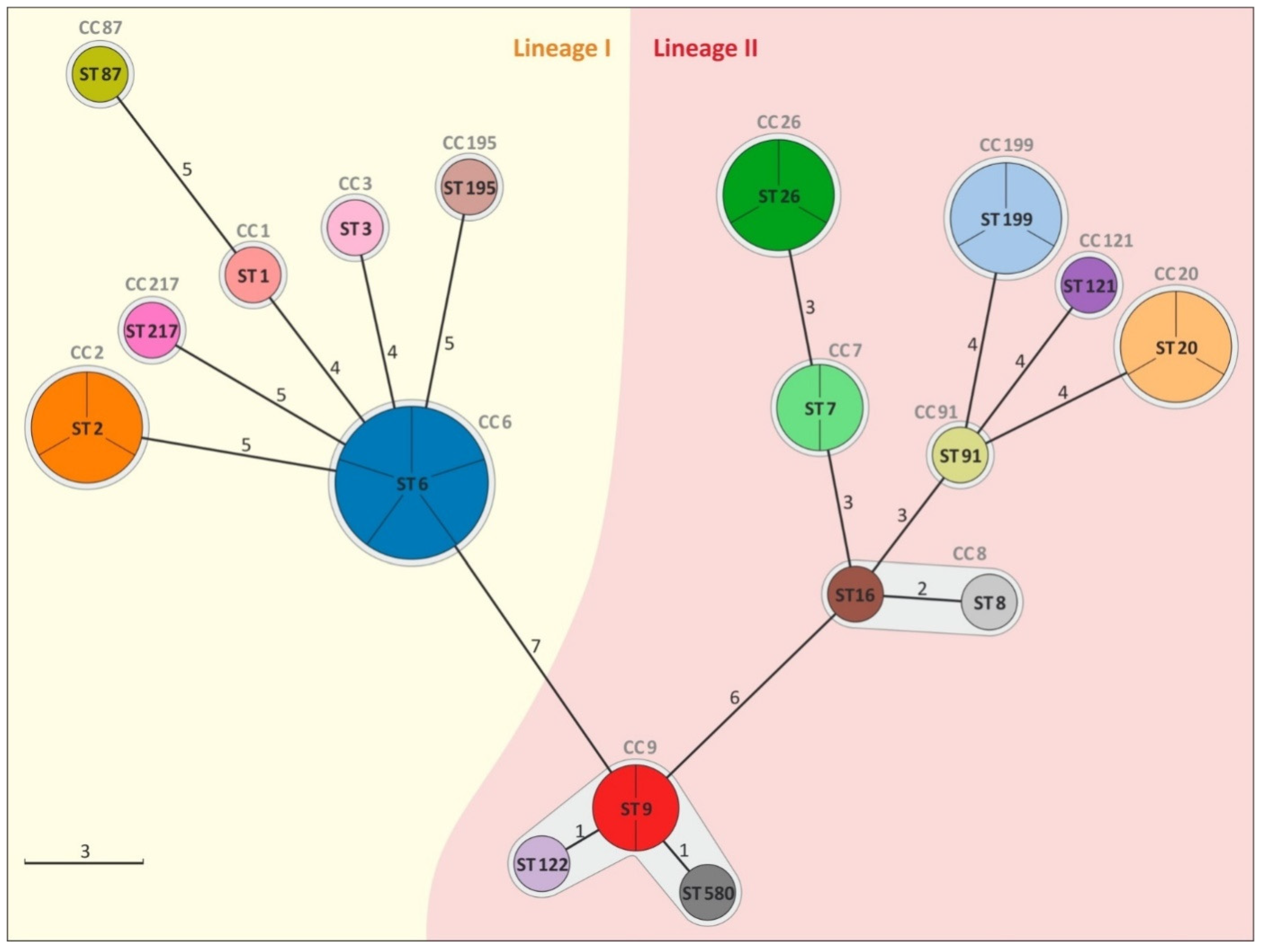
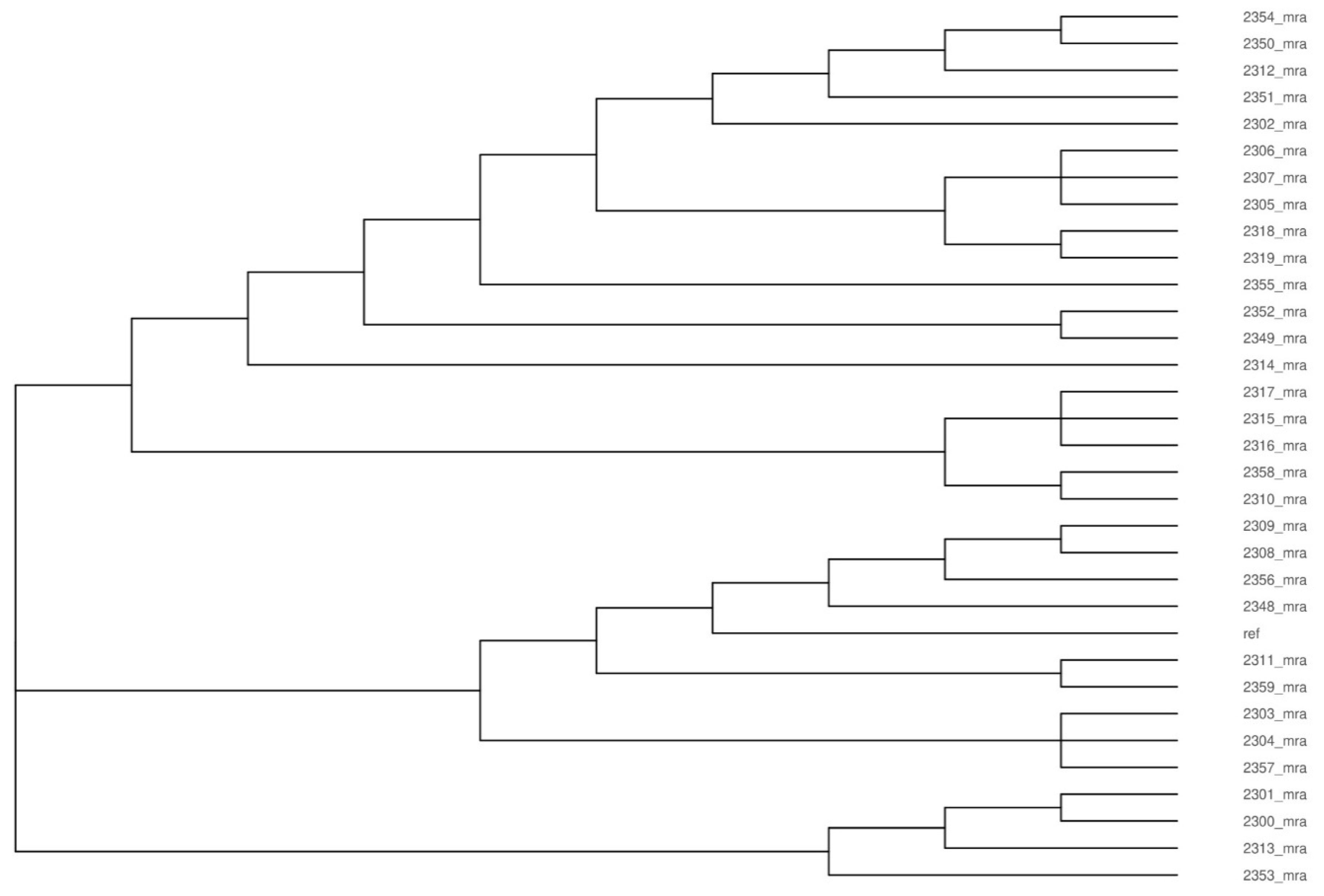
3.1. Analysis of Genetic Diversity of L. monocytogenes Isolates

The L. monocytogenes isolates (n = 32) were classified to four molecular groups (including serogroups): 15 isolates belonged to group IIa (1/2a-3a) (46.9%), 10 isolates belonged to group IVb (4ab-4b-4d-4e) (31.2%), 4 isolates belonged to group IIc (1/2c-3c) (12.5%), and 3 isolates belonged to group IIb (1/2b-3b-7) (9.4%) (Table 1).
The isolates were classified into lineage I or lineage II, respectively, containing 40.6% and 59.4% of isolates. A total of 18 different MLST sequence types were identified in the 32 isolates derived from the cheeses: ST 6 (n = 5, 15.6% of isolates), ST 2 and ST 20 (each n = 3, 9.4%), ST 26 and ST 199 (each n = 3, 9.4%), ST 7 and ST 9 (each n = 2, 6.3%), and ST 1, ST 3, ST 8, ST 16, ST 87, ST 91, ST 121, ST 122, ST 195, ST 217, and ST 580 (each n = 1, 3.1%). Sequence type 7, ST 8, ST 16, ST 20, ST 26, ST 91, ST 121, and ST 199 belonged to serogroup IIa and serovar 1/2a-3a; ST 3, ST 87, and ST 195 belonged to serogroup IIb and serovar 1/2b-3b-7; ST 9, ST 122, and ST 580 belonged to serogroup IIc and serovar 1/2c-3c; and ST 1, ST 2, and ST 6 belonged to serogroup IVb and serovar 4ab-4b-4d-4e. The L. monocytogenes isolates were grouped into 15 clonal complexes: CC 6 (n = 5), CC 9 (n = 4), CC 2, CC 20, CC 26, and CC 199 (each n = 3), CC 7 and CC 8 (each n = 2), CC 1, CC 3, CC 14, CC 87, CC 121, CC 195, and CC 217 (each n = 1) (Table 1). The minimum spanning tree shows that the number of allelic differences between neighboring STs ranged from 1 to 7 (Figure 1).
The parsnp dendrogram of the whole-genome alignment shows the similarity of sequenced Listeria isolates on the genomic scale. The total coverage among all the sequences reached 82.2%. The dendrogram shows that the analyzed isolates formed three main clusters that covered 19, 9, and 4 isolates. Isolates belonging to cluster 2 showed the greatest similarity to the reference strain of L. monocytogenes (Figure 2).

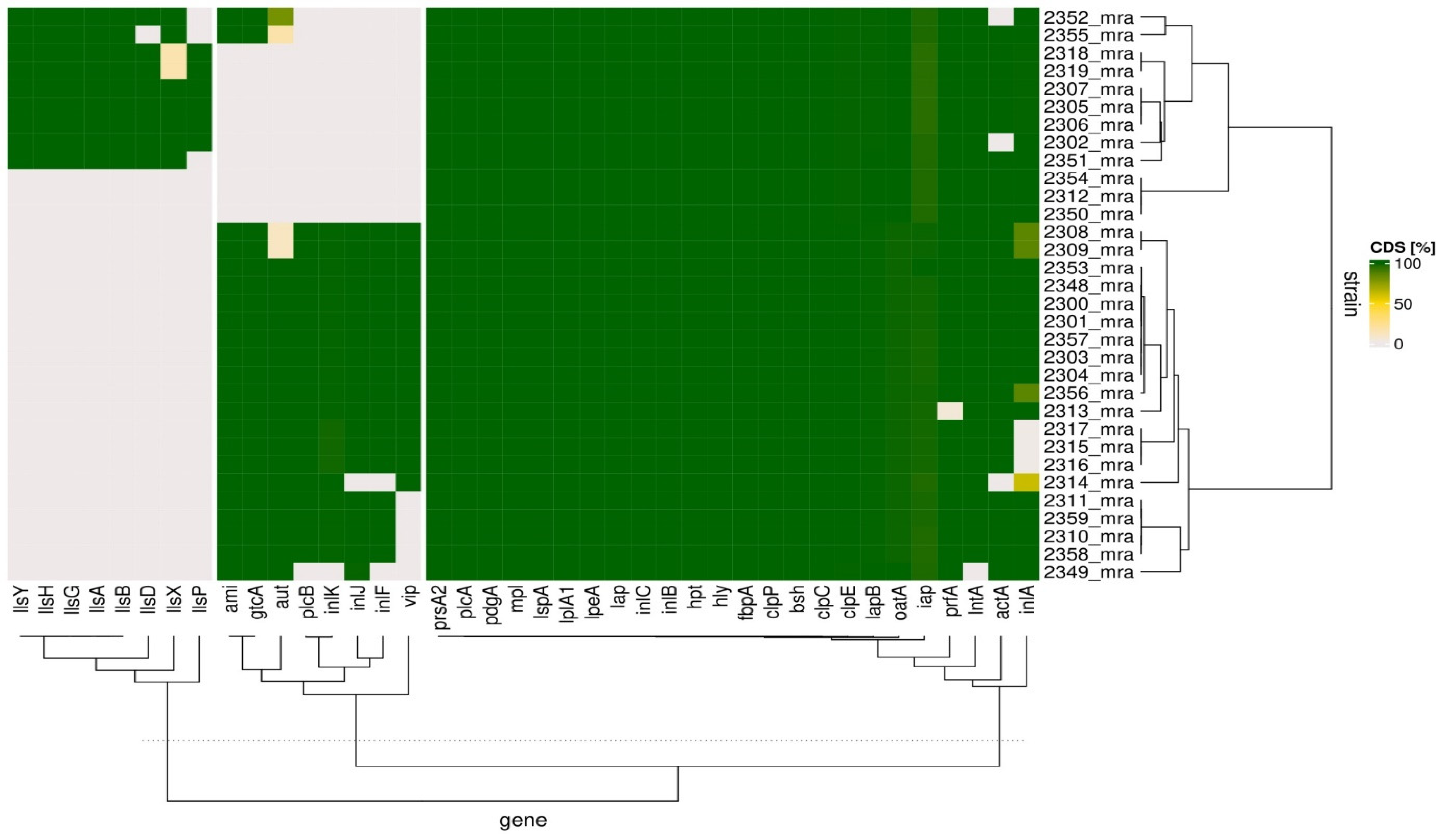
3.2. Analysis of Potential Virulence of L. monocytogenes Isolates

Based on the BIGSdb-Lm database, we selected 40 genes reported previously to be important for the virulence of Listeria to elucidate the potential virulence of the isolates (Figure 3). The heatmap shows that the analyzed isolates clustered hierarchically into two groups according to the coding sequence (CDS) length of the virulence genes. One of these groups (cluster 1) contained twelve isolates, and the other group (cluster 2) contained twenty isolates. With only a few exceptions, the Listeria isolates showed the presence of rather uniform and full CDS-length prsA2, prfA, plcA, pdgA, oatA, mpl, lspA, lplA1, lpeA, lntA, lapB, lap, inlC, inlB, inlA, iap, hpt, hly, fbpA, clpP, clpE, clpC, bsh, and actA genes. These exceptions were that some isolates’ inlA copies were judged to be non-functional because of premature stop codon insertion; this applied to more than a fifth (7 out of 32) of the isolates (2308_mra, 2309_mra, 2314_mra, 2315_mra, 1216_mra, 2317_mra, and 2356_mra); actA was detected as truncated in three isolates (2302_mra, 2314_mra, and 2352_mra); and prfA and lntA were truncated in the 2313_mra and 2349_mra isolates, respectively. The bacterial isolates that had been grouped as cluster 1 almost ubiquitously lacked functional sequences in their aut, inlF, inlJ, inlK, plcB, and vip genes. In this cluster, only ami and gtcA were found with full CDS in two isolates, which were 2352_mra and 2355_mra. Among the isolates that had been grouped in cluster 2, a truncated aut gene was present in the 2308_mra and 2309_mra isolates. A set of truncated inlF, inlK, plcB, and vip genes was specific only to the 2349_mra isolate in cluster 2. The isolate labeled as 2314_mra in this cluster was negative for inlF and inlJ. Additionally, five isolates of the same cluster were also negative for vip CDS, and these isolates were 2310_mra, 2311_mra, 2349_mra, 2358_mra, and 2359_mra; cluster 2 isolates were also negative for genes that code listeriolysins which had been reported as specific for Listeria innocua, namely llsA, llsB, llsD, llsG, llsH, llsP, llsX, and llsY, whereas the complete CDS of most of the studied listeriolysin genes were present in cluster 1 isolates. In cluster 1, only three isolates (2312_mra, 2350_mra, and 2354_mra) were negative for all of these listeriolysin-coding genes. The absence of llsP characterized three other isolates (2351_mra, 2352_mra, and 2355_mra), and llsX was not present in 2318_mra or 2319_mra. Additionally, the 2355_mra isolate was negative for the llsD gene.

4. Discussion

4.1. Genetic Diversity of L. monocytogenes Isolates

A total of 32 L. monocytogenes isolates originating from artisanal cheeses were typed and characterized for the presence of selected virulence genes. The majority of isolates were classified into group IIa (1/2a-3a), the next group by size was IVb (4ab-4b-4d-4e), then came IIc (1/2c-3c), and the smallest was IIb (1/2b-3b-7). These results are consistent with an analysis of 1698 food-derived isolates, which showed the dominance of the IIa and high representation of the IVb molecular groups among various food matrices, including milk and milk products [6]. In turn, Espinosa–Mata et al. (2022) and Barría et al. (2020) reported that group IVb dominated among L. monocytogenes isolates originating from artisanal cheeses [13,15]. Therefore, it seems reasonable to conclude that the potential for L. monocytogenes to contaminate food is not limited to one molecular group [6]. Furthermore, analysis of L. monocytogenes from artisanal cheeses revealed the presence of isolates from different molecular groups (IIa and IVb) in the same batch of cheese, as well as the presence of isolates from different molecular groups (IIb and IIc) in different products from the same production site, which could suggest coexisting sources of pathogen contamination in these cases (data not shown). Additionally, it should be noted that L. monocytogenes belonging to groups IIa, IVb, and IIb were responsible for human infections [7], which, to relate this to the present research, indicated a potential risk associated with the consumption of artisanal cheeses.
Listeria monocytogenes strains form a structured population and are differentiated into distinct evolutionary lineages, i.e., I, II, III, and IV, which represent different ecological, genetic, and phenotypic characteristics [27]. Most human listeriosis outbreaks are associated with lineage I isolates, while lineage II strains are common in foods. Lineage II strains are widespread in natural and farm environments and are also commonly isolated from animal listeriosis cases and sporadically isolated from human clinical cases. In contrast, lineage III and IV strains are rare and are predominantly isolated from animal sources [27]. Isolates from artisanal cheeses belonged to lineages I and II in 40.6% and 59.4% proportions, respectively, indicating a significant number of them from lineage I in the pool of isolates. Therefore, a perception of overrepresentation of lineage I strains in sporadic listeriosis cases in proportion to their prevalence in food appears inconsistent with the evidence from these products [27].
Artisanal cheese isolates were revealed to harbor a wide range of sequence types (18 ST having been detected), and those types were shown to have dissimilar prevalences (ST 6 being dominant), geographical distributions (ST 2 occurring in two administrative regions), and frequent co-occurrence in products from the same cheese dairy (ST 6, ST 26, and ST 122 being detected in dairy I; ST 7 and ST 16 in dairy F; ST 195 and ST 580 in dairy C; ST 1 and ST 20 in dairy G1; and ST 3 and ST 9 in dairy G2) (Table 1). The existence of an MLST database enables data comparison at the international level [28]. The comparison showed that all STs found in artisanal cheeses were known and had previously been reported in certain countries (http://bigsdb.pasteur.fr/listeria (accessed on 8 September 2021)). According to the available literature, 16 ST were found in various foodstuffs, and they were ST1, ST2, ST3, ST6, ST7, ST8, ST9, ST16, ST20, ST26, ST87, ST91, ST121, ST122, ST217, and ST 580 [8,29,30,31,32]. It is significant that some of these STs have been associated with listeriosis (ST 1, ST 2, ST 3, ST 6, ST 7, ST 8, ST 9, ST 20, ST 26, ST 87, and ST 121) [8,33,34,35] and epidemics (ST 6 and ST 87) [36,37]. It is also on record that ST 195 was isolated from clinical cases [6]. Artisanal cheese isolates were also classified into 15 CC (Table 1; Figure 1). In the presented studies, the dominant CC was CC 6, which, according to the results of studies by other authors, was more often attributed to clinical cases than detected in food [38]. This CC was regarded as rare until 2000, while since that year, its relative frequency has increased [7]. In the same time interval, a similar trend was observed for CC 8, CC 9, and CC 121, while the relative frequency of CC 1, CC 2, CC 3, CC 7, CC 14, CC 20, and CC 199 was lower [7]. The frequency distribution of L. monocytogenes CC in food and clinical sources is highly uneven, and three categories of highly prevalent complexes can be distinguished: infection-associated (CC 1, CC 2, CC 4, and CC 6), food-associated (CC 9 and CC 121), and intermediate (others) [38]. The results of the presented studies showed that there were CCs from these three categories in the artisanal cheeses. It should be noted that, with the exception of CC 199, the isolates identified in these studies (totaling 14: CC 1, CC 2, CC 3, CC 6, CC 7, CC 8, CC 9, CC 14, CC 20, CC 26, CC 87, CC 121, CC 195, and CC 217) have had clinical cases attributed to them in many countries around the world [8,38,39]. In addition, the artisanal cheeses studied contained complexes from among the most common CCs in Europe (CC 2, CC 6, CC 7, and CC 9) [8].
Multiple alignments of analyzed genomes indicated the close evolutionary relationship of isolated L. monocytogenes. Neither the administrative division, the cheese dairy, nor the year of isolation had relevance to the extent to which Listeria monocytogenes isolates were related (Figure 2).

4.2. Virulence Genes of L. monocytogenes Isolates

According to the microbiological criterion for L. monocytogenes in food [40], all strains of this pathogen are considered equally virulent. However, differences in pathogen virulence have been described in the subject literature [38,41]. The isolates from artisanal cheeses were compared for selected 40 virulence genes (Figure 3 and Figure S1) that affect the intracellular life cycle of L. monocytogenes, i.e., host cell adhesion and invasion, intracellular multiplication and motility, and intercellular spread [42].
The L. monocytogenes genome includes a large family of genes expressing proteins known as internalins (Inl) [43], which are important in the early stages of infection. Literature data showed that beyond the extensively-studied invasins inlA and inlB (considered species-specific), other internalins such as inlF, inlJ, inlC, and inlK also fulfill important functions in the infectious process. The internalin genes are involved in adherence (inlF and inlJ), cell-to-cell spread (inlC), and autophagy evasion (inlK) [43,44,45,46,47]. The inlA, inlB, and inlC genes were found in all isolates of lineages I and II (32/32), but some isolates of lineage II had premature stop codons (PMSC) in the inlA gene (7/19), which suggests a reduced ability to invade human intestinal epithelial cells in vitro [48]. In contrast, the inlF and inlK genes were found only in isolates from lineage II (19/19 and 18/19, respectively), whereas the inlJ gene was present in isolates from lineage II (18/19) and only 1/13 from lineage I.
LIPI-1 (Listeria pathogenicity island 1) and LIPI-3 are considered to be responsible for the increased virulence in some strains of the pathogen [41]. It is pertinent that all invasive strains but one possessed LIPI-1 [41]. Intracellular pathogenesis heavily relies on factors transcribed by prfA, hly, plcA, mpl, actA, and plcB genes located in the LIPI-1 (Listeria pathogenicity island 1) [49], which are all absent from L. innocua, the closest non-pathogenic Listeria species to L. monocytogenes [50]. The prfA gene is the major regulator of the virulence gene expression [49]; the hly gene encodes listeriolysin O (LLO), which plays a central role in the cell-to-cell spread process of L. monocytogenes [51] as well as being involved in several stages of the intracellular lifecycle of the pathogen [52]; the plcA and plcB genes encode phosphatidylinositol-specific phospholipase C and broad-range phospholipase C, respectively [53], which are necessary at the stage of phagosome lysis and pathogen release into the cytoplasm of host cells; the mpl gene encoding metalloprotease is required for actA processing and protrusion resolution, besides being involved in plcB processing and vacuole escape [54]; and the actA gene is an essential virulence factor of L. monocytogenes and its extracellular action enhances virulence in contributing to aggregation and biofilm formation to mediate colonization of the gut lumen, promoting and enhancing bacterial host cell entry, facilitating evasion of autophagy and vacuolar exit, as well as activating nuclear factor of kappa light polypeptide gene enhancer in B-cells (NF-κB) [55]. The results of the presented studies show that the prfA, plcA, plcB, mpl, and hly genes were present in 32/32 and the actA gene in 29/32 of isolates. Premature stop codons in the plcB and prfA genes occurred in all lineage I isolates (13/13) and 1 lineage II isolate (1/19).
Listeria monocytogenes strains may carry the llsA, llsB, llsD, llsG, llsH, llsP, llsX, and llsY genes, together designated LIPI-3 [5]. Pathogenicity studies in murine models indicated that LIPI-3 was responsible for the increased virulence of some strains of the pathogen [41]. LIPI-3 encodes an additional hemolysin, listeriolysin S (LLS), which is a hemolytic/cytolytic factor impacting potential virulence [56]. LIPI-3 genes were only found in isolates from lineage I (9/13). These genes were found in a set of 8 in 6/13 isolates, a set of 7 in 2/13 (with the llsP gene absent), and a set of 6 in 1/13 (with the llsP and llsD genes absent), which implied these isolates higher potential virulence compared to the other isolates of both lineages at the stage of pathogen release from the phagosome. Furthermore, Vilchis–Rangel et al. [41] pointed to a strong association between llsX and the invasiveness of L. monocytogenes. The llsX gene was found in all nine isolates of this line; however, two isolates had premature stop codons.
The ami gene mediates the adhesion of L. monocytogenes to eukaryotic cells via its cell wall–anchoring domain, and its inactivation leads to a severe loss of adhesion [57]. The gtcA protein is necessary for lipoteichoic acid glycosylation in L. monocytogenes [58]. A mutation in the gtcA gene reduced the ability of strains carrying it to invade intestinal epithelial cells under experimental conditions, which may be associated with reduced virulence of those strains [59]. All isolates from lineage II (19/19) and three from lineage I (3/13) contained the ami and gtcA genes.
The aut gene encodes the auto surface protein with autolytic activity, which is required for the entry of L. monocytogenes into eukaryotic cells and contributes to virulence in vivo [60]. Studies in mice and guinea pigs showed significantly less virulence of L. monocytogenes with an inactivated aut gene [60]. Among the researched L. monocytogenes, all isolates of lineage II (19/19) and only three isolates of lineage I (3/13) had the aut gene, while two isolates of lineage I and lineage II (2/13 and 2/19) were detected to have premature stop codons in this gene.
The vip gene is a virulence factor only present in pathogenic Listeria species, which is required for entry into some mammalian cells and for virulence (for efficient entry into Caco-2 human intestinal epithelial cells and L2071 mouse fibroblast cells, but not into GPC16 guinea pig colon adenocarcinoma or Vero African green monkey kidney cells) [61]. This virulence factor gene is present in all L. monocytogenes lineage I and II serovars that include serovars implicated in human disease (1/2a, 1/2c, 1/2b, and 4b) and absent from non-pathogenic species of Listeria [44]. Among artisanal cheese isolates, the vip gene was found in only 15 isolates from lineage II (15/19).
The prsA2 gene plays a unique and important role in L. monocytogenes pathogenesis by promoting the activity and stability of at least two critical secreted virulence factors: LLO and a broad-specificity phospholipase. Loss of prsA2 activity severely attenuated virulence in mice and impaired bacterial cell-to-cell spread in host cells [62]. All artisanal cheese-derived isolates from both lineages had the prsA2 gene (32/32).
The lntA gene encodes a bacterial nucleomodulin which acts directly in the nucleus to manipulate a chromatin regulatory protein. The secreted virulence factor allows a pathogen to control host chromatin composition, transcription, and gene expression [63]. The lntA gene was found in isolates of lineage II (19/19) and lineage I (12/13).
The iap gene of L. monocytogenes encodes the extracellular protein p60 (a murein hydrolase), which is necessary for septum separation and for the successful invasion of host cells [42]. The iap gene was found in lineage I and lineage II isolates (32/32).
The bsh gene determines the production of bile salt hydrolase, which is a protein conferring on L. monocytogenes resistance to the action of bile salts, and its production increases the chance of survival of the pathogen in the gastrointestinal tract [64]. The bsh gene was present in all isolates from artisanal cheeses from lineages I and II (32/32).
The genes encoding stress proteins, i.e., clpP, clpC, or clpE, are crucial to L. monocytogenes at the stage of intracellular growth. The clpP gene encodes a protein that is involved in the rapid adaptive response of intracellular pathogens during the infectious process [65]. The clpC gene encodes a virulence factor promoting the intracellular survival of L. monocytogenes by facilitating its early bacterial escape from the phagosomal compartment of a macrophage [66]. It is also required for adhesion and invasion, and it governs InlA, InlB, and ActA expression [67]. The ClpE protein acts synergistically with ClpC in cell septation [68]. Active clpP, clpC, and clpE genes were found in all isolates from lineages I and II (32/32).
The pdgA and oatA genes encode peptidoglycan N-deacetylase (PdgA) and peptidoglycan O-acetyltransferase (OatA), respectively, by which L. monocytogenes acquires resistance to antibacterial compounds of which the action is directed at the cell wall of the pathogen, including lysozyme. The resistance is gained by modifying the structure of peptidoglycans, and these peptidoglycan modifications are essential for the survival of the pathogen in macrophages. In addition, PdgA and OatA enable L. monocytogenes to suppress the cytokine response of the infected organism. The pdgA and oatA genes are important factors used by L. monocytogenes for the effective colonization of host cells [69]. The pdgA and oatA genes were found in all isolates from lineages I and II (32/32).
The hpt gene encodes the transporter protein (Hpt) that L. monocytogenes exploits to be able to use glucose-1-phosphate from the host cell as a source of carbon and energy at the stage of intracellular multiplication. In in vivo experiments in mice, the absence of the hpt gene in L. monocytogenes impaired the bacterium’s proliferation within the host cell and decreased its virulence [3,42]. The hpt gene was found in all isolates from lineages I and II (32/32).
The lap gene encodes the Lap adhesion protein, which, when bound to the host cell receptor, enhances the adhesion of L. monocytogenes to intestinal epithelial cells [42]. All isolates of L. monocytogenes from artisanal cheeses (32/32) carried the lap gene.
The lapB gene encodes the LapB adhesion protein characteristic only of pathogenic species of Listeria. This protein is essential for adhesion to and penetration into host intestinal epithelial cells by L. monocytogenes [42]. All isolates of L. monocytogenes from artisanal cheeses (32/32) also had the lapB gene.
The fbpA gene encodes the production of fibronectin-binding protein (FbpA), which increases the adhesion of L. monocytogenes to intestinal epithelial cells and acts as a chaperone protein to stabilize and/or ensure proper secretion of LLO and InlB [42]. All isolates of L. monocytogenes from artisanal cheeses (32/32) were found to have the fbpA gene.
The lspA gene encodes a signal peptidase in class II (SPase II), which is a co-actor in the maturation of lipoproteins in L. monocytogenes infection. The maturation of lipoproteins (from a precursor form to mature protein) is crucial for the surface protein anchoring and effective phagosome escape and intracellular survival of L. monocytogenes [70]. All isolates of L. monocytogenes from artisanal cheeses (32/32) carried the lspA gene.
The lpeA gene is responsible for the synthesis of the lipoprotein promoting entry A (LpeA) necessary for L. monocytogenes to penetrate eukaryotic cells and acts to facilitate intracellular survival of the pathogen in infected macrophages. Mutated L. monocytogenes cells with lpeA deletion failed to infect human Caco-2 cells or mouse hepatocytes (tib73) in cell cultures [42,71]. The lpeA gene was found in all isolates (32/32).
The lplA1 gene encodes lipoate protein ligase (LplA1) necessary for intracellular replication of L. monocytogenes. LplA1 allows the pathogen to use lipoic peptides derived from the cytosol of host cells as a source of lipoate, which promotes the adaptation of L. monocytogenes to living in infected mammalian cells [72]. The lplA1 gene was present in all isolates of L. monocytogenes from artisanal cheeses (32/32).
In general, the comparison of 32 artisanal cheese isolates with respect to 40 selected virulence genes showed a greater diversity of isolates between lineages than within the same lineage. In contrast to lineage I isolates, those of lineage II did not have LIPI-3 genes, which confirms that lineage II strains may be more adapted to an environmental lifestyle than lineage I strains [27]. An additional difference between lineage I and lineage II involved the vip, inlF, and inlK genes, which were only present in lineage II isolates. This may be related to the diverging evolutionary pathways followed by virulence genes, which are affected by a strain’s origin and serotype [73]. A common feature of isolates from both lineages was the presence of most LIPI-1 genes (prfA, plcA, mpl, hly, plcB, and actA) and the presence of the inlA, inlC, inlB, prsA2, iap, bsh, clpP, clpE, clpC, pdgA, oatA, hpt, lap, lapB, fbpA, lspA, lpeA, and lplA1 genes. Bechtel and Gobbons [74] reported that some L. monocytogenes populations may associate with particular foods, including cheese, and that gene content may contribute to this pattern, which may, to some extent, explain the observed similarities and differences in genes among artisanal cheese isolates.

5. Conclusions

The studies provided information on the genetic diversity and selected virulence genes of the isolates of L. monocytogenes derived from specific dairy products, i.e., artisanal cheeses. The populations of L. monocytogenes isolated from the cheeses were differentiated in terms of molecular groups (including serogroups), evolutionary lineages, sequence types, clonal complexes, and potential virulence. The small number of isolates obtained from artisanal cheeses limits the representativeness of the results; however, the presence in this foodstuff of isolates from lineages I and II in comparable numbers suggests that attributing dominance to lineage II in this type of food is unjustified. The complete absence of lineage III and IV isolates is consistent with the observation that these two lineages are rarely isolated from food-related environments. The sequence types and clonal complexes to which the isolates from the cheeses belonged were distributed locally and globally. The study also confirmed that premature stop codon mutations in inlA and the absence of LIPI-3 are characteristic of lineage II isolates. In turn, the presence of premature stop codon mutations in the plcB gene and the absence of the vip gene in all isolates from lineage I appear to be characteristic of isolates of this line. The administrative division, cheese dairy, or year of isolation had no relevance to the extent to which L. monocytogenes isolates were related.
Artisanal cheeses can carry virulent strains of L. monocytogenes. Products originating in the same batch (produced in the same cheese dairy, under the same conditions, and at the same time) can simultaneously harbor isolates from different molecular groups, including serovars, different evolutionary lineages, different sequence types, different clonal complexes, and of different virulences, and that dairy can be a place where strains classified into different ST and CC are in circulation.
Relating the presented studies to food safety, the effective treatment of listeriosis, and potential new scientific research, they indicate an area for further research on the sensitivity to disinfectants and antibiotics of isolates from artisanal cheeses.

Supplementary Materials

The following supporting information can be downloaded at: https://www.mdpi.com/article/10.3390/foods11182805/s1, Figure S1: Diverging graphs of coding sequence length among analyzed Listeria isolates. The absolute length in base pairs (bp) of the gene is shown for all of the sequenced Listeria isolates. The CDS length range for a selected gene is color-coded dark blue (minimum length) to light blue (maximum length).

Author Contributions

Conceptualization, W.P., R.P.-Ł., M.Z. and M.G.; methodology, W.P. and R.P.-Ł.; software, M.K.; validation, M.K.; formal analysis, M.K.; investigation, W.P. and R.P.-Ł.; resources, W.P. and R.P.-Ł.; data curation, W.P., R.P.-Ł. and M.K.; writing—original draft preparation, R.P.-Ł., W.P., M.K., M.Z., M.G., P.D., K.M. and D.P.-O.; writing—review and editing, R.P.-Ł., W.P., M.K., M.Z., M.G., P.D., K.M. and D.P.-O.; visualization, R.P.-Ł., W.P. and M.K.; supervision, W.P. and R.P.-Ł.; project administration, W.P. and R.P.-Ł.; funding acquisition, R.P.-Ł. and W.P. All authors have read and agreed to the published version of the manuscript.

Funding

This research received no external funding.

Data Availability Statement

The data of the present study are available from the corresponding author upon request.

Acknowledgments

The authors thank the staff of the Department of Omics Analyses, National Veterinary Research Institute, Puławy, Poland, for sample sequencing.

Conflicts of Interest

The authors declare no conflict of interest.

References

  1. EFSA and ECDC (European Food Safety Authority and European Centre for Disease Prevention and Control). The European Union One Health 2020 Zoonoses Report. EFSA J. 2021, 19, 6971. [Google Scholar] [CrossRef]
  2. Desai, A.N.; Anyoha, A.; Madoff, L.C.; Lassmann, B. Changing epidemiology of Listeria monocytogenes outbreaks, sporadic cases, and recalls globally: A review of ProMED reports from 1996 to 2018. Int. J. Infect. Dis. 2019, 84, 48–53. [Google Scholar] [CrossRef] [PubMed]
  3. Allerberger, F.; Wagner, M. Listeriosis: A resurgent foodborne infection. Clin. Microbiol. Infect. 2010, 16, 16–23. [Google Scholar] [CrossRef] [PubMed]
  4. Lewańska, M.; Godela, A.; Myga-Nowak, M. Listeriosis. Modern perception of epidemiological threat. Post. Mikrobiol. 2018, 57, 106–116. [Google Scholar] [CrossRef]
  5. Schiavano, G.F.; Ateba, C.N.; Petruzzelli, A.; Mele, V.; Amagliani, G.; Guidi, F.; De Santi, M.; Pomilio, F.; Blasi, G.; Gattuso, A.; et al. Whole-Genome Sequencing characterization of virulence profiles of Listeria monocytogenes food and human isolates and in vitro adhesion/invasion assessment. Microorganisms 2022, 10, 62. [Google Scholar] [CrossRef] [PubMed]
  6. Henri, C.; Félix, B.; Guillier, L.; Leekitcharoenphon, P.; Michelon, D.; Mariet, J.F.; Aarestrup, F.M.; Mistou, M.Y.; Hendriksen, R.S.; Roussel, S. Population genetic structure of Listeria monocytogenes strains as determined by pulsed-field gel electrophoresis and multilocus sequence typing. Appl. Environ. Microbiol. 2016, 82, 5720–5728. [Google Scholar] [CrossRef]
  7. Bergholz, T.M.; Shah, M.K.; Burall, L.S.; Rakic-Martinez, M.; Datta, A.R. Genomic and phenotypic diversity of Listeria monocytogenes clonal complexes associated with human listeriosis. Appl. Microbiol. Biotechnol. 2018, 102, 3475–3485. [Google Scholar] [CrossRef]
  8. Zhang, Y.; Dong, S.; Chen, H.; Chen, J.; Zhang, J.; Zhang, Z.; Yang, Y.; Xu, Z.; Zhan, L.; Mei, L. Prevalence, genotypic characteristics and antibiotic resistance of Listeria monocytogenes from retail foods in bulk in Zhejiang Province, China. Front. Microbiol. 2019, 10, 1710. [Google Scholar] [CrossRef]
  9. Cardenas-Alvarez, M.X.; Townsend Ramsett, M.K.; Malekmohammadi, S.; Bergholz, T.M. Evidence of hypervirulence in Listeria monocytogenes clonal complex 14. J. Med. Microbiol. 2019, 68, 1677–1685. [Google Scholar] [CrossRef]
  10. Chen, Y.; Chen, Y.; Pouillot, R.; Dennis, S.; Xian, Z.; Luchansky, J.B.; Porto-Fett, A.C.S.; Lindsay, J.A.; Hammack, T.S.; Allard, M.; et al. Genetic diversity and profiles of genes associated with virulence and stress resistance among isolates from the 2010–2013 interagency Listeria monocytogenes market basket survey. PLoS ONE 2020, 15, e0231393. [Google Scholar] [CrossRef]
  11. Meng, Z.; Zhang, L.; Xin, L.; Lin, K.; Yi, H.; Han, X. Technological characterization of Lactobacillus in semihard artisanal goat cheeses from different Mediterranean areas for potential use as nonstarter lactic acid bacteria. J. Dairy Sci. 2018, 101, 2887–2896. [Google Scholar] [CrossRef] [PubMed]
  12. Mata, G.M.; Martins, E.; Machado, S.G.; Pinto, M.S.; de Carvalho, A.F.; Vanetti, M.C. Performance of two alternative methods for Listeria detection throughout Serro Minas cheese ripening. Braz. J. Microbiol. 2016, 47, 749–756. [Google Scholar] [CrossRef] [PubMed]
  13. Barría, C.; Singer, R.S.; Bueno, I.; Estrada, E.; Rivera, D.; Ulloa, S.; Fernández, J.; Mardones, F.O.; Moreno-Switt, A.I. Tracing Listeria monocytogenes contamination in artisanal cheese to the processing environments in cheese producers in southern Chile. Food Microbiol. 2020, 90, 103499. [Google Scholar] [CrossRef] [PubMed]
  14. Pyz-Łukasik, R.; Knysz, P.; Gondek, M. Hygiene quality and consumer safety of traditional short- and long-ripened cheeses from Poland. J. Food Qual. 2018, 2018, 8732412. [Google Scholar] [CrossRef]
  15. Espinosa-Mata, E.; Mejía, L.; Villacís, J.E.; Alban, V.; Zapata, S. Detection and genotyping of Listeria monocytogenes in artisanal soft cheeses from Ecuador. Rev. Argent. Microbiol. 2022, 54, 53–56. [Google Scholar] [CrossRef] [PubMed]
  16. Kevenk, T.O.; Gulel, G.T. Prevalence, antimicrobial resistance and serotype distribution of Listeria monocytogenes isolated from raw milk and dairy products. J. Food Saf. 2016, 36, 11–18. [Google Scholar] [CrossRef]
  17. Jackson, K.A.; Gould, L.H.; Hunter, J.C.; Kucerova, Z.; Jackson, B. Listeriosis outbreaks associated with soft cheeses, United States, 1998–2014. Emerg. Infect. Dis. 2018, 24, 1116–1118. [Google Scholar] [CrossRef]
  18. Centers for Disease Control and Prevention (CDC). Outbreak of listeriosis associated with homemade Mexican-style cheese--North Carolina. Morb. Mortal. Wkly Rep. 2001, 50, 560–562. [Google Scholar]
  19. Acciari, V.A.; Iannetti, L.; Gattuso, A.; Sonnessa, M.; Scavia, G.; Montagna, C.; Addante, N.; Torresi, M.; Zocchi, L.; Scattolini, S.; et al. Tracing sources of Listeria contamination in traditional Italian cheese associated with a US outbreak: Investigations in Italy. Epidemiol. Infect. 2016, 144, 2719–2727. [Google Scholar] [CrossRef]
  20. Pyz-Łukasik, R.; Gondek, M.; Winiarczyk, D.; Michalak, K.; Paszkiewicz, W.; Piróg-Komorowska, A.; Policht, A.; Ziomek, M. Occurrence of Listeria monocytogenes in artisanal cheeses from Poland and its identification by MALDI-TOF MS. Pathogens 2021, 10, 632. [Google Scholar] [CrossRef]
  21. PN-EN ISO 11290-1:1999; Microbiology of Food and Animal Feeding Stuffs—Horizontal Method for the Detection and Enumeration of Listeria monocytogenes—Part 1: Detection Method. PKN: Warszawa, Poland, 1999.
  22. PN-EN ISO 11290-1: 2017-07; Microbiology of the Food Chain—Horizontal Method for the Detection and Enumeration of Listeria monocytogenes and of Listeria spp.—Part 1: Detection Method. PKN: Warszawa, Poland, 2017.
  23. Doumith, M.; Buchrieser, C.; Glaser, P.; Jacquet, C.; Martin, P. Differentiation of the major Listeria monocytogenes serovars by multiplex PCR. J. Clin. Microbiol. 2004, 42, 3819–3822. [Google Scholar] [CrossRef] [PubMed] [Green Version]
  24. Moura, A.; Criscuolo, A.; Pouseele, H.; Maury, M.M.; Leclercq, A.; Tarr, C.; Björkman, J.T.; Dallman, T.; Reimer, A.; Enouf, V.; et al. Whole genome-based population biology and epidemiological surveillance of Listeria monocytogenes. Nat. Microbiol. 2016, 2, 16185. [Google Scholar] [CrossRef] [PubMed]
  25. Yu, G.; Lam, T.T.; Zhu, H.; Guan, Y. Two Methods for Mapping and Visualizing Associated Data on Phylogeny Using Ggtree. Mol. Biol. Evol. 2018, 35, 3041–3043. [Google Scholar] [CrossRef] [PubMed]
  26. Gu, Z.; Eils, R.; Schlesner, M. Complex heatmaps reveal patterns and correlations in multidimensional genomic data. Bioinformatics 2016, 32, 2847–2849. [Google Scholar] [CrossRef] [PubMed]
  27. Orsi, R.H.; den Bakker, H.C.; Wiedmann, M. Listeria monocytogenes lineages: Genomics, evolution, ecology, and phenotypic characteristics. Int. J. Med. Microbiol. 2011, 301, 79–96. [Google Scholar] [CrossRef]
  28. Salcedo, C.; Arreaza, L.; Alcalá, B.; de la Fuente, L.; Vázquez, J.A. Development of a multilocus sequence typing method for analysis of Listeria monocytogenes clones. J. Clin. Microbiol. 2003, 41, 757–762. [Google Scholar] [CrossRef]
  29. Wang, Y.; Zhao, A.; Zhu, R.; Lan, R.; Jin, D.; Cui, Z.; Wang, Y.; Li, Z.; Wang, Y.; Xu, J.; et al. Genetic diversity and molecular typing of Listeria monocytogenes in China. BMC Microbiol. 2012, 12, 119. [Google Scholar] [CrossRef]
  30. Yin, Y.; Tan, W.; Wang, G.; Kong, S.; Zhou, X.; Zhao, D.; Jia, Y.; Pan, Z.; Jiao, X. Geographical and longitudinal analysis of Listeria monocytogenes genetic diversity reveals its correlation with virulence and unique evolution. Microbiol. Res. 2015, 175, 84–92. [Google Scholar] [CrossRef]
  31. Kim, S.W.; Haendiges, J.; Keller, E.N.; Myers, R.; Kim, A.; Lombard, J.E.; Karns, J.S.; Van Kessel, J.A.S.; Haley, B.J. Genetic diversity and virulence profiles of Listeria monocytogenes recovered from bulk tank milk, milk filters, and milking equipment from dairies in the United States (2002 to 2014). PLoS ONE 2018, 13, e0197053. [Google Scholar] [CrossRef]
  32. Caruso, M.; Fraccalvieri, R.; Pasquali, F.; Santagada, G.; Latorre, L.M.; Difato, L.M.; Miccolupo, A.; Normanno, G.; Parisi, A. Antimicrobial susceptibility and multilocus sequence typing of Listeria monocytogenes isolated over 11 years from food, humans, and the environment in Italy. Foodborne Pathog. Dis. 2020, 17, 284–294. [Google Scholar] [CrossRef]
  33. Ragon, M.; Wirth, T.; Hollandt, F.; Lavenir, R.; Lecuit, M.; Le Monnier, A.; Brisse, S. A new perspective on Listeria monocytogenes evolution. PLoS Pathog. 2008, 4, e1000146. [Google Scholar] [CrossRef] [PubMed] [Green Version]
  34. Wang, Y.; Jiao, Y.; Lan, R.; Xu, X.; Liu, G.; Wang, X.; Zhang, L.; Pang, H.; Jin, D.; Dai, H.; et al. Characterization of Listeria monocytogenes isolated from human listeriosis cases in China. Emerg. Microbes. Infect. 2015, 4, e50. [Google Scholar] [CrossRef] [PubMed]
  35. Huang, Y.T.; Ko, W.C.; Chan, Y.J.; Lu, J.J.; Tsai, H.Y.; Liao, C.H.; Sheng, W.H.; Teng, L.J.; Hsueh, P.R. Disease burden of invasive listeriosis and molecular characterization of clinical isolates in Taiwan, 2000–2013. PLoS ONE 2015, 10, e0141241. [Google Scholar] [CrossRef]
  36. Pérez-Trallero, E.; Zigorraga, C.; Artieda, J.; Alkorta, M.; Marimón, J.M. Two outbreaks of Listeria monocytogenes infection, Northern Spain. Emerg. Infect. Dis. 2014, 20, 2155–2157. [Google Scholar] [CrossRef]
  37. Thomas, J.; Govender, N.; McCarthy, K.M.; Erasmus, L.K.; Doyle, T.J.; Allam, M.; Ismail, A.; Ramalwa, N.; Sekwadi, P.; Ntshoe, G.; et al. Outbreak of listeriosis in South Africa associated with processed meat. N. Engl. J. Med. 2020, 382, 632–643. [Google Scholar] [CrossRef]
  38. Maury, M.M.; Tsai, Y.H.; Charlier, C.; Touchon, M.; Chenal-Francisque, V.; Leclercq, A.; Criscuolo, A.; Gaultier, C.; Roussel, S.; Brisabois, A.; et al. Uncovering Listeria monocytogenes hypervirulence by harnessing its biodiversity. Nat Genet. 2016, 48, 308–313, Erratum in: Nat. Genet. 2017, 49, 651. [Google Scholar] [CrossRef]
  39. Knabel, S.J.; Reimer, A.; Verghese, B.; Lok, M.; Ziegler, J.; Farber, J.; Pagotto, F.; Graham, M.; Nadon, C.A.; Canadian Public Health Laboratory Network (CPHLN). Sequence typing confirms that a predominant Listeria monocytogenes clone caused human listeriosis cases and outbreaks in Canada from 1988 to 2010. J. Clin. Microbiol. 2012, 50, 1748–1751. [Google Scholar] [CrossRef]
  40. European Commission. Commission Regulation (EC) No 2073/2005 of 15 November 2005 on Microbiological Criteria for Foodstuffs. Off. J. Eur. Union 2005, 338, 1–32. [Google Scholar]
  41. Vilchis-Rangel, R.E.; Espinoza-Mellado, M.D.R.; Salinas-Jaramillo, I.J.; Martinez-Peña, M.D.; Rodas-Suárez, O.R. Association of Listeria monocytogenes LIPI-1 and LIPI-3 marker llsX with invasiveness. Curr. Microbiol. 2019, 76, 637–643. [Google Scholar] [CrossRef]
  42. Camejo, A.; Carvalho, F.; Reis, O.; Leitão, E.; Sousa, S.; Cabanes, D. The arsenal of virulence factors deployed by Listeria monocytogenes to promote its cell infection cycle. Virulence 2011, 2, 379–394. [Google Scholar] [CrossRef]
  43. Bierne, H.; Sabet, C.; Personnic, N.; Cossart, P. Internalins: A complex family of leucine-rich repeat-containing proteins in Listeria monocytogenes. Microbes Infect. 2007, 9, 1156–1166. [Google Scholar] [CrossRef] [PubMed]
  44. Doumith, M.; Cazalet, C.; Simoes, N.; Frangeul, L.; Jacquet, C.; Kunst, F.; Martin, P.; Cossart, P.; Glaser, P.; Buchrieser, C. New aspects regarding evolution and virulence of Listeria monocytogenes revealed by comparative genomics and DNA arrays. Infect. Immun. 2004, 72, 1072–1083. [Google Scholar] [CrossRef] [Green Version]
  45. Dortet, L.; Mostowy, S.; Cossart, P. Listeria and autophagy escape: Involvement of InlK, an internalin-like protein. Autophagy 2012, 8, 132–134. [Google Scholar] [CrossRef] [PubMed]
  46. Ghosh, P.; Halvorsen, E.M.; Ammendolia, D.A.; Mor-Vaknin, N.; O’Riordan, M.X.D.; Brumell, J.H.; Markovitz, D.M.; Higgins, D.E. Invasion of the brain by Listeria monocytogenes is mediated by InlF and host cell vimentin. mBio 2018, 9, e00160-18. [Google Scholar] [CrossRef] [PubMed]
  47. Leung, N.; Gianfelice, A.; Gray-Owen, S.D.; Ireton, K. Impact of the Listeria monocytogenes protein InlC on infection in mice. Infect. Immun. 2013, 81, 1334–1340. [Google Scholar] [CrossRef] [PubMed]
  48. Su, X.; Cao, G.; Zhang, J.; Pan, H.; Zhang, D.; Kuang, D.; Yang, X.; Xu, X.; Shi, X.; Meng, J. Characterization of internalin genes in Listeria monocytogenes from food and humans, and their association with the invasion of Caco-2 cells. Gut Pathog. 2019, 11, 30. [Google Scholar] [CrossRef]
  49. Vázquez-Boland, J.A.; Kuhn, M.; Berche, P.; Chakraborty, T.; Domínguez-Bernal, G.; Goebel, W.; González-Zorn, B.; Wehland, J.; Kreft, J. Listeria pathogenesis and molecular virulence determinants. Clin. Microbiol. Rev. 2001, 14, 584–640. [Google Scholar] [CrossRef] [PubMed]
  50. Milohanic, E.; Glaser, P.; Coppée, J.Y.; Frangeul, L.; Vega, Y.; Vázquez-Boland, J.A.; Kunst, F.; Cossart, P.; Buchrieser, C. Transcriptome analysis of Listeria monocytogenes identifies three groups of genes differently regulated by PrfA. Mol. Microbiol. 2003, 47, 1613–1625. [Google Scholar] [CrossRef] [PubMed]
  51. Osborne, S.E.; Brumell, J.H. Listeriolysin O: From bazooka to Swiss army knife. Philos. Trans. R. Soc. Lond. B Biol. Sci. 2017, 372, 20160222. [Google Scholar] [CrossRef] [PubMed]
  52. Cheng, C.; Sun, J.; Yu, H.; Ma, T.; Guan, C.; Zeng, H.; Zhang, X.; Chen, Z.; Song, H. Listeriolysin O pore-forming activity is required for ERK1/2 phosphorylation during Listeria monocytogenes infection. Front. Immunol. 2020, 11, 1146. [Google Scholar] [CrossRef] [PubMed]
  53. Quereda, J.J.; Andersson, C.; Cossart, P.; Johansson, J.; Pizarro-Cerdá, J. Role in virulence of phospholipases, listeriolysin O and listeriolysin S from epidemic Listeria monocytogenes using the chicken embryo infection model. Vet. Res. 2018, 49, 13. [Google Scholar] [CrossRef] [PubMed]
  54. Alvarez, D.E.; Agaisse, H. The metalloprotease Mpl supports Listeria monocytogenes dissemination through resolution of membrane protrusions into vacuoles. Infect. Immun. 2016, 84, 1806–1814. [Google Scholar] [CrossRef] [PubMed] [Green Version]
  55. Pillich, H.; Puri, M.; Chakraborty, T. ActA of Listeria monocytogenes and its manifold activities as an important listerial virulence factor. Curr. Top. Microbiol. Immunol. 2017, 399, 113–132. [Google Scholar] [CrossRef]
  56. Cotter, P.D.; Draper, L.A.; Lawton, E.M.; Daly, K.M.; Groeger, D.S.; Casey, P.G.; Ross, R.P.; Hill, C. Listeriolysin S, a novel peptide haemolysin associated with a subset of lineage I Listeria monocytogenes. PLoS Pathog. 2008, 4, e1000144. [Google Scholar] [CrossRef] [PubMed]
  57. Milohanic, E.; Pron, B.; The European Listeria Genome Consortium; Berche, P.; Gaillard, J.L. Identification of new loci involved in adhesion of Listeria monocytogenes to eukaryotic cells. European Listeria Genome Consortium. Microbiology 2000, 146, 731–739. [Google Scholar] [CrossRef] [PubMed]
  58. Rismondo, J.; Haddad, T.F.M.; Shen, Y.; Loessner, M.J.; Gründling, A. GtcA is required for LTA glycosylation in Listeria monocytogenes serovar 1/2a and Bacillus subtilis. Cell Surf. 2020, 6, 100038. [Google Scholar] [CrossRef]
  59. Faith, N.; Kathariou, S.; Cheng, Y.; Promadej, N.; Neudeck, B.L.; Zhang, Q.; Luchansky, J.; Czuprynski, C. The role of L. monocytogenes serotype 4b gtcA in gastrointestinal listeriosis in A/J mice. Foodborne Pathog. Dis. 2009, 6, 39–48. [Google Scholar] [CrossRef]
  60. Cabanes, D.; Dussurget, O.; Dehoux, P.; Cossart, P. Auto, a surface associated autolysin of Listeria monocytogenes required for entry into eukaryotic cells and virulence. Mol. Microbiol. 2004, 51, 1601–1614. [Google Scholar] [CrossRef]
  61. Cabanes, D.; Sousa, S.; Cebriá, A.; Lecuit, M.; García-del Portillo, F.; Cossart, P. Gp96 is a receptor for a novel Listeria monocytogenes virulence factor, Vip, a surface protein. EMBO J. 2005, 24, 2827–2838. [Google Scholar] [CrossRef]
  62. Alonzo, F., 3rd; Port, G.C.; Cao, M.; Freitag, N.E. The posttranslocation chaperone PrsA2 contributes to multiple facets of Listeria monocytogenes pathogenesis. Infect. Immun. 2009, 77, 2612–2623. [Google Scholar] [CrossRef]
  63. Lebreton, A.; Job, V.; Ragon, M.; Le Monnier, A.; Dessen, A.; Cossart, P.; Bierne, H. Structural basis for the inhibition of the chromatin repressor BAHD1 by the bacterial nucleomodulin LntA. mBio 2014, 5, e00775-13. [Google Scholar] [CrossRef] [PubMed]
  64. de las Heras, A.; Cain, R.J.; Bielecka, M.K.; Vázquez-Boland, J.A. Regulation of Listeria virulence: PrfA master and commander. Curr. Opin. Microbiol. 2011, 14, 118–127. [Google Scholar] [CrossRef] [PubMed]
  65. Gaillot, O.; Pellegrini, E.; Bregenholt, S.; Nair, S.; Berche, P. The ClpP serine protease is essential for the intracellular parasitism and virulence of Listeria monocytogenes. Mol. Microbiol. 2000, 35, 1286–1294. [Google Scholar] [CrossRef] [PubMed]
  66. Rouquette, C.; de Chastellier, C.; Nair, S.; Berche, P. The ClpC ATPase of Listeria monocytogenes is a general stress protein required for virulence and promoting early bacterial escape from the phagosome of macrophages. Mol. Microbiol. 1998, 27, 1235–1245. [Google Scholar] [CrossRef] [PubMed]
  67. Nair, S.; Derré, I.; Msadek, T.; Gaillot, O.; Berche, P. CtsR controls class III heat shock gene expression in the human pathogen Listeria monocytogenes. Mol. Microbiol. 2000, 35, 800–811. [Google Scholar] [CrossRef]
  68. Nair, S.; Frehel, C.; Nguyen, L.; Escuyer, V.; Berche, P. ClpE, a novel member of the HSP100 family, is involved in cell division and virulence of Listeria monocytogenes. Mol. Microbiol. 1999, 31, 185–196. [Google Scholar] [CrossRef]
  69. Aubry, C.; Goulard, C.; Nahori, M.A.; Cayet, N.; Decalf, J.; Sachse, M.; Boneca, I.G.; Cossart, P.; Dussurget, O. OatA, a peptidoglycan O-acetyltransferase involved in Listeria monocytogenes immune escape, is critical for virulence. J. Infect. Dis. 2011, 204, 731–740. [Google Scholar] [CrossRef]
  70. Réglier-Poupet, H.; Frehel, C.; Dubail, I.; Beretti, J.L.; Berche, P.; Charbit, A.; Raynaud, C. Maturation of lipoproteins by type II signal peptidase is required for phagosomal escape of Listeria monocytogenes. J. Biol. Chem. 2003, 278, 49469–49477. [Google Scholar] [CrossRef]
  71. Réglier-Poupet, H.; Pellegrini, E.; Charbit, A.; Berche, P. Identification of LpeA, a PsaA-like membrane protein that promotes cell entry by Listeria monocytogenes. Infect. Immun. 2003, 71, 474–482. [Google Scholar] [CrossRef]
  72. Keeney, K.M.; Stuckey, J.A.; O’Riordan, M.X. LplA1-dependent utilization of host lipoyl peptides enables Listeria cytosolic growth and virulence. Mol. Microbiol. 2007, 66, 758–770. [Google Scholar] [CrossRef]
  73. Poimenidou, S.V.; Dalmasso, M.; Papadimitriou, K.; Fox, E.M.; Skandamis, P.N.; Jordan, K. Virulence gene sequencing highlights similarities and differences in sequences in Listeria monocytogenes Serotype 1/2a and 4b strains of clinical and food origin from 3 different geographic locations. Front. Microbiol. 2018, 9, 1103. [Google Scholar] [CrossRef] [PubMed]
  74. Bechtel, T.D.; Gibbons, J.G. Population genomic analysis of Listeria monocytogenes from food reveals substrate-specific genome variation. Front. Microbiol. 2021, 12, 620033. [Google Scholar] [CrossRef] [PubMed]
Figure 1. Minimum spanning tree (MST) illustrating the phylogenetic relationship based on sequence types (STs) allelic profiles of L. monocytogenes isolates. Each circle represents one ST. The size of the circle is proportional to the number of isolates. The fragment of the circle corresponds to single isolates. Links between the circles are represented according to the number of allelic mismatch between STs.
Foods 11 02805 g001
Figure 2. Parsnp dendrogram of the whole-genome alignment showing the similarity of L. monocytogenes isolates. All the Listeria genomic sequences were aligned to the reference genome, which is labeled as ‘ref’ on the plot.
Foods 11 02805 g002
Figure 3. Clustering analysis of virulence genes in L. monocytogenes isolates. The heatmap shows the presence of the selected virulence gene set among the analyzed isolates. Normalized coding sequence (CDS) lengths are shown in colors.
Foods 11 02805 g003
Table 1. Distribution (administrative divisions and cheese dairies), numbers, molecular groups, lineages, allelic profiles (ST), clonal complexes (CC) of the L. monocytogenes isolates.
Administrative DivisionsCheese DairyNumber of Isolates
n = 32
Year of IsolationMolecular GroupLineageSTCC
2D1, D2, I52016, 2018IV b 4ab-4b-4d-4eI66
2D3, G132014, 2018II a 1/2a-3aII2020
2, 3G3, J32015, 2017, 2018IV b 4ab-4b-4d-4eI22
2I32015II a 1/2a-3aII2626
2H32018II a 1/2a-3aII199199
2G222016II c 1/2c-3cII99
2F22017II a 1/2a-3aII77
2G112014IV b 4ab-4b-4d-4eI11
2F12018II a 1/2a-3aII168
4K12018II a 1/2a-3aII121121
2I12018II c 1/2c-3cII1229
2D412014IIb 1/2b-3b-7I8787
1A12016IV b 4ab-4b-4d-4eI217217
1C12016II b 1/2b-3b-7I195195
2E12018II a 1/2a-3aII9114
2G212017II b 1/2b-3b-7I33
1C12017II c 1/2c-3cII5809
1B12018II a 1/2a-3aII88
1–4 administrative divisions/A–K cheese dairy: 1/A, B, C; 2/D, E, F, G, H, I; 3/J; 4/K. Different capital letters (from A to K) indicate the production sites in different towns, while the same capital letters with different subscripts (D1–D4, G1–G3) show different production sites in one town. ST—sequence type; CC—clonal complex.
Publisher’s Note: MDPI stays neutral with regard to jurisdictional claims in published maps and institutional affiliations.

